BIO 305 MOLECULAR BIOLOGY NOUN - nouedu.net 305 MOLECULAR... · 3.3.1 Expression of Cloned Genes...
77
NATIONAL OPEN UNIVERSITY OF NIGERIA SCHOOL OF SCIENCE AND TECHNOLOGY COURSE CODE: BIO 305 COURSE TITLE: MOLECULAR BIOLOGY
Transcript of BIO 305 MOLECULAR BIOLOGY NOUN - nouedu.net 305 MOLECULAR... · 3.3.1 Expression of Cloned Genes...
NATIONAL OPEN UNIVERSITY OF NIGERIA
SCHOOL OF SCIENCE AND TECHNOLOGY
COURSE CODE: BIO 305
COURSE TITLE: MOLECULAR BIOLOGY
1
BIO 305 MOLECULAR BIOLOGY
TABLE OF CONTENTS PAGE
Module 1………………………………………………………
Unit 1 Molecular Biology – An Overview…………....
Unit 2 Microbial Genetics…………………………….
Module 2……………………………………………………..
Unit 1 Genes and Chromosomes…………………...
Unit 2 NucleicAcids…………………………………..
Module 3…………………………………………………….
Unit 1 DNA Replication……………………………....
Unit 2 DNA Transcription……………………………
Module 4…………………………………………………….
Unit 1 Genetic Code…………………………….
Unit 2 Gene Expression…………………………
Unit 3 Protein Synthesis……………………………
Module 5 …………………………………………………
Unit 1 Metabolic Pathways…………………….
Unit 2 DNA Sequencing……………………………
2
MOLECULAR BIOLOGY
INTRODUCTION
Molecular biology is concerned with understanding the
mechanisms responsible for the transmission and expression of
the genetic information that ultimately governs cell structure and
function. Understanding the molecular biology of cells is
fundamental to all biological sciences. Because of its growing
number of practical applications in Agriculture, Biotechnology, and
Medicine.
All cells share a number of basic properties (a kind of
underlying unity of cell biology) that are particularly apparent at the
molecular level. Such unity has allowed scientists to choose simple
organisms like bacteria as models for many fundamental
experiments, with the expectation that similar molecular
mechanisms are operative in organisms as diverse as E. coli and
humans.
3
UNIT 1: MOLECULAR BIOLOGY – AN OVERVIEW
CONTENTS
1.0 Introduction
2.0 Objectives
3.0 Main Body
3.1 Meaning of Molecular Biology
3.2 Relationship to Other Biological Sciences
3.3 Techniques of Molecular Biology
3.3.1 Expression of Cloned Genes
3.3.2 Polymerase Chain Reaction
3.3.3 Gel Electrophoresis
3.3.4 Nucleic Acid Hybridisation
3.3.5 Restriction Fragment Length Polymorphism
Analysis
4.0 Conclusion
5.0 Summary
6.0 Tutor-Marked Assignments
7.0 References/Further Readings
4
1.0 INTRODUCTION
Molecular biology is concerned with understanding the
mechanisms responsible for the transmission and expression of
the genetic information that ultimately governs cell structure and
function. Understanding the molecular biology of cells is
fundamental to all biological sciences, because of its growing
number of practical applications in agriculture, biotechnology and
medicine.
2.0 OBJECTIVES
At the end of studying this unit, you should be able to:
1. Define molecular biology.
2. Outline its relationship with other biological sciences.
3. Describe the techniques of molecular biology.
4. Identify the significance of molecular biology.
3.0 MAIN BODY
3.1 Meaning of Molecular Biology
The term molecular biology was first used in 1945 by William
Astbury to refer to the study of the chemical and physical structure
of biological molecules. Contemporary molecular biology is the
study of life at the molecular level; and it is principally concerned
with the understanding and interactions between the various
systems of a cell, including the interactions between the different
types of DNA, RNA and protein biosynthesis and how these
reactions are regulated – the mechanisms responsible for the
transmission and expression of the genetic information that
ultimately governs cell structure and function i.e. the study of the
process of replication, transcription and translation of genetic
5
material. The central dogma of molecular biology where the
genetic material (DNA) is transcribed into RNA and then translated
into protein provides the starting point for understanding the field.
3.2 Relationship With Other Biological Sciences
Molecular signs of evolution show that evolutionary
relationships among species are reflected in their DNA and
proteins – in their genes and gene products. Researchers in
molecular biology use techniques and ideas from other areas of
biology and chemistry particularly genetics – the transfer of
biological information from cell to cell, from parents to offsprings
and thus from generation to generation and to the effects on
organisms – and biochemistry – the study of the chemical
substances and vital processes occurring in living organisms
particularly the role, the function and the structure of biomolecules.
Much of the work in molecular biology is quantitative and
require ideas from computer science in bioinformatics and
computational biology. Molecular genetics – the study of gene
structure and function is a prominent sub-field of molecular
biology.
Many other areas of biology focus on molecules, either
directly studying their interactions in their own right such as in cell
biology and developmental biology or indirectly where the
techniques of molecular biology are used to infer historical
attributes of populations or species as in fields of evolutionary
biology such as population genetics and phylogenetics.
3.3 Techniques of Molecular Biology
6
To understand different biochemical events of prokaryotic
and eukaryotic cells at molecular level and be able to characterise,
isolate and manipulate molecular components of cells and
organisms, a wide array of bio-physico-chemical techniques are
used in molecular biology. These include:
3.3.1 Expression of Cloned Genes
Molecular cloning enables the determination of the
nucleotide sequences of genes and also provide new approaches
to obtaining large amounts of proteins for structural and functional
characterisation. In expression colony, DNA coding for a protein of
interest is cloned using polymerase chain reaction or restriction
enzymes into a plasmid or phase vector known as expression
vector.
The plasmid can be inserted into either bacterial or animal
cells; DNA coding for a protein of interest is now inside a cell, and
the protein can now be expressed. A variety of systems, such as
inducible promoters and specific cell-signalling factors, are
available to help express the protein of interest at high levels.
Large quantities of a protein can then be extracted from the
bacterial or eukaryotic cell. The protein can be tested for
enzymatic activity under a variety of situations, the protein may be
crystallised so its tertiary structure can be studied, or in the
pharmaceutical industry, the activity of new drugs against the
protein can be studied.
3.3.2 Polymerase Chain Reaction
Molecular cloning allows individual DNA fragments to be
propagated in bacteria and isolated in large amounts. The
7
polymerase chain reaction is a versatile technique for copying
DNA, and allows a single DNA sequence to be copied repeatedly,
or altered in predetermined ways; essentially it is used for
repeated replication of a defined segment of DNA. Single DNA
molecules can thus be amplified to yield readily detectable
quantities of DNA that can be isolated and quantitatively
measured.
3.3.3 Gel Electrophoresis
Gel electrophoresis is one of the principal tools of molecular
biology. It is a common method in which molecules (DNA, RNA
and proteins) are separated based on the rates of their migration in
an electric field. A gel, usually formed from agarose or
polyacrylamide, is placed between two buffer compartments
containing electrodes. The sample is then pipetted into preformed
slots in the gel, and the electric field is turned on; the gel acts like a
sieve, selectively retarding the movement of larger molecules.
Smaller molecules therefore move through the gel more rapidly,
allowing a mixture of nucleic acids to be separated on the basis of
size.
3.3.4 Nucleic Acid Hybridisation
Cloning enabled the isolation and characterisation of
individual genes. However, understanding the role of genes within
cells, requires analysis of the intracellular organisation and
expression of individual genes and their encoded proteins. Nucleic
acid hybridisation is a method for detecting and analysing
sequences of homologous DNA. This enables the mapping of
genes, to chromosomes, the analysis of gene expression, and the
8
localisation of proteins to subcellular organelles. In this way, it is
possible to study genetic differences between organisms or
individuals.
Hybridisation can be achieved by southern or northern
blotting.
Southern blotting is a method for probing for the presence of
a specific DNA sequence within a DNA sample and it enables a
researcher to determine not only whether a particular sequence is
present within a sample of DNA, but how many such sequences
there are; and the size of the restriction fragments that contain
these sequences.
Messenger RNA can also be subjected to hybridisation
analysis, in an analogous process known as Northern blotting used
to study the expression patterns of a specific type of RNA
molecule and is essentially a combination of denaturing RNA gel
electrophoresis and a blot. In this process RNA is separated based
an size and is their transferred to a membrane that is then probed
with a labelled complement of a sequence of interest. It is used to
determine whether a particular gene is made into MRNA, how
much of that MRNA is present, and whether the abundance of that
specific MRNA changes at different stages of development or in
response to certain regulatory signals; that control gene
expression.
3.3.5 Restriction Fragment Length Polymorphism Anal ysis
DNA fragments that result from cutting a particular piece of
DNA wit a specific restriction enzyme give a characteristic pattern
of bands upon gel electrophoresis. Fact band corresponds to a
DNA restriction fragment of a certain length. Such difference are
9
called restriction fragment length polymorphisms (RLFPs) serving
as genetic marker for a particular location the genome. A given
FLP marker frequently occurs in numerous variants in a population
and is inherited in a mendelian fashion. Genetic markers are used
for making linkage maps. RLFP analysis is important in the
diagnosis of genetic disorders and in forensic applications.
4.0 CONCLUSION
Molecular biology seeks to understand the molecular basis of
life. Relating the structure of specific molecules of biological
importance – such as proteins, enzymes and nucleic acids – to
their functional roles in cells and organisms.
5.0 SUMMARY
Researchers in molecule biology use specific techniques
native to molecule biology, but combine these with techniques and
ideas from genetics and biochemistry to characterise, isolate and
manipulate the molecule components of cells and organisms.
6.0 TUTOR-MARKED ASSIGNMENTS
1. Briefly define molecular biology
2. Outline the relationship of molecular biology to other
biological sciences.
3. What is nucleic acid hybridisation.
7.0 REFERENCES/FURTHER READINGS
Robert, K.M.; Daryl, K.G.; Peter, A.M. and Victor, W.R.
(2003). Harper’s Illustrated Biochemistry, Twenty-Sixth
Edition. McGraw Hill Publishers India.
10
Raven, P. H. and Johnson, G. B. (1985). Biology. Second
Edition. Times Mirror/Mosby College Publishing.
Campbell, N. A. (1996). Biology. Fourth Edition. The
Benjamin/Cummings Publishing Company Inc.
California.
Bruce, A., Dennis, B., Lewis,J., Martin, R., Keith, R. and
James, D.W. (1994). Molecular Biology of the Cell. Third
Edition. Garland Publishing , Inc. New York

11
12
MICROBIAL GENETICS
CONTENTS
1.0 Introduction
2.0 Objectives
3.0 Main Body
3.1 Escherichia coli as Experimental Models
3.2 Yeast as Experimental Models
4.0 Conclusion
5.0 Summary
6.0 Tutor-Marked Assignments
7.0 References/Further Readings
13
1.0 INTRODUCTION
It studies the genetics of micro-organisms, which involves
the study of the genotype of microbial species and also the
expression system in the form of phenotypes. It also involves the
study of genetic processes taking place in these micro organisms.
2.0 OBJECTIVES
At the end of this unit you should be able to:
1. Recognise the importance of E. coli as an experimental
model.
2. Recognise yeasts as models for studies of eukaryotic
cells.
3.0 MAIN BODY
3.1 Escherichia coli as Experimental Model
Because of their comparative simplicity, prokaryotic cells
(bacteria) are ideal models for studying many fundamental aspects
of biochemistry and molecular biology. The most thoroughly
studied species of bacteria is E. coli which is the most favoured
organism for investigation of the basic mechanisms of molecular
genetics. Most of the present concepts of molecular biology – DNA
replication, the genetic code, gene expression and protein
synthesis – are derived from studies of E. coli.
E. coli is useful to molecular biologists because of its relative
simplicity and the ease of its propagation and study in the
laboratory. For example the genome of E. coli consists of
approximately 4.6 million base pairs and encodes about 4000
different proteins. While the human genome is more complex with
approximately 3 billion base pairs and encodes about 100,000
14
different proteins. The small size of E.coli genome provides
advantages for genetic analysis and the sequence of the entire
E.coli genome has been determined.
Molecular genetic experiments are further facilitated by the
rapid growth of E.coli under well defined laboratory conditions. E.
coli can divide every 20-60 minutes, depending on culture
conditions and a clonal population of E.coli all cells derived by
division of a single cell of origin – can be isolated as a colony
grown on agar – containing medium. Bacterial colonies contain
many cells, and selecting and analysing genetic variants of E.coli
strain is easy and rapid. This generally contributes to the success
of experiments in molecular genetics.
E.coli can divide rapidly in nutrient mixtures like glucose,
salts, amino acids, vitamins and nucleic acid precursors. However,
E. coli can also grow in much simpler media consisting of only
salts as source of nitrogen (such as ammonia) and a source of
carbon and energy (such as glucose). But in such simple medium,
the bacteria grow a little slowly (a division time of about 40
minutes) because they must synthesise all their own amino acids,
nucleotides and other organic compounds.
The ability of E. coli to carry out these biosynthetic reactions
in simple defined media has made them extremely useful in
elucidating the biochemical pathways involved. Thus, the rapid
growth and simple nutritional requirements of E. coli have greatly
facilitated fundamental experiments in both molecular biology and
biochemistry.
Although bacteria are models for studies of cell properties,
they cannot be used to study aspects of cell structure and function
that are unique to eukaryotes. Yeasts, the simplest eukaryotes,
15
have a number of experimental advantages similar to those of E.
coli and have provided a model for studies of many aspects of
eukaryotic cell biology.
The genome of the most studied yeasts, Saccharomyces
cervisae, consists of 12 million base pairs of DNA and contains
about 6000 genes; and is about 3 times larger than that of E. coli it
is much more manageable than the genomes of more complex
eukaryotes, such as humans. Yeasts can be readily grown in the
laboratory and can be studied by many of the same molecular
genetic approaches that have proved successful with E. coli.
Although yeasts do not replicate as rapidly as bacteria, they still
divide as frequently as every 2 hours and can easily be grown as
colonies from a single cell. Yeasts can be used for a variety of
genetic manipulations similar to those that can be performed using
bacteria. Yeast mutants have been important in understanding
many fundamental processes in eukaryotes, including DNA
replication, transcription, RNA processing, protein sorting and
regulation of cell division.
4.0 CONCLUSION
The evolution of present day cells from a common ancestor
has important implications for cell and molecular biology as an
experimental science. Because the fundamental properties of all
cells have been conserved during evolution, the basic principles
learned from experiments performed with one type of cell are
generally applicable to other cells.
16
5.0 SUMMARY
Because of their genetic simplicity and ease of study,
bacteria such as E. coli are particularly useful for investigation of
fundamental aspects of biochemistry and molecular biology.
Yeasts, as the simplest eukaryotic cells, yeasts are an important
model for studying various aspects of eukaryotic cell biology.
6.0 TUTOR-MARKED ASSIGNMENTS
1. Give two reasons why E. coli is useful in molecular
biology.
2. Yeasts have been used as models for the study of many
aspects of the biology of eukaryotic cells. Why are they
not suitable for analysis of animal cell movements?
8.0 REFERENCES/FURTHER READINGS
Robert, K.M.; Daryl, K.G.; Peter, A.M. and Victor, W.R.
(2003). Harper’s Illustrated Biochemistry, Twenty-Sixth
Edition. McGraw Hill Publishers India.
Raven, P. H. and Johnson, G. B. (1985). Biology. Second
Edition. Times Mirror/Mosby College Publishing.
Campbell, N. A. (1996). Biology. Fourth Edition. The
Benjamin/Cummings Publishing Company Inc.
California.
Bruce, A., Dennis, B., Lewis,J., Martin, R., Keith, R. and
James, D.W. (1994). Molecular Biology of the Cell.
Third Edition. Garland Publishing , Inc. New York
17
GENES AND CHROMOSOMES
CONTENTS
1.0 Introduction
2.0 Objectives
3.0 Main Body
3.1 Genes
3.1.1 Cistron
3.1.2 Muton
3.1.3 Recon
3.2 Chromosomes
3.2.1 Chromosomes Structure
3.2.2 Chromosomes Number
3.2.3 Sex Chromosomes
3.2.4 Human Chromosomes and Genetic Disorders
4.0 Conclusion
5.0 Summary
6.0 Tutor-Marked Assignments
7.0 References/Further Readings
18
1.0 INTRODUCTION
Order implies information, instructions are required to
arrange parts or processes in a non-random way. Biological
instructions are encoded in the DNA which is a substance of
genes; the units of inheritance that transmit information from
parents to offspring to ensure continuity of life.
2.0 OBJECTIVES
At the end of this unit you should be able to:
1. Define the gene.
2. Define chromosomes.
3. Describe the structure of chromosomes.
4. Recognise the importance of genes and chromosomes in
heredity.
3.0 MAIN BODY
3.1 Genes
The classical principles of genetics were deduced by Gregor
Mendel in 1865 on the basis of the results of breeding experiments
with peas. Mendel studied the inheritance of a number of well-
defined traits such as plant height and was able to deduce general
rules for their transmission. In all cases, Mendel could correctly
interpret the observed patterns of inheritance by assuming that
each trait is determined by a pair of inherited “factors”, which are
now called genes.
The term gene (Gr genos descent) was coined by W.
Johannsen in 1909 to refer to the hereditary “factors” of Mendel.
The gene can be defined as a functional unit of heredity which
occupies a specific place (locus) on a chromosome, is capable of
19
reproducing itself exactly at each cell division and directs the
formation of an enzyme or other protein.
Cytologic and genetic studies show that genes are the
fundamental units of inheritance regarded as indivisible units of the
chromosomes on which they are located like “beads on a string”.
Genes as the chief functional genetic unit, determine the
basic architecture of every cell, the nature and life of the cell, the
specific protein synthesis, the enzyme formation and the self-
reproduction of the cell.
Genes are molecular patterns that can maintain their
identities for many generations, can be self-duplicated in each
generation and can control cell processes by allowing their
specificities to be occupied. Genes can mutate, can be assorted,
can be shuffled in different combinations, therefore, genes are
regarded as the basis for modern interpretation of evolution.
On operational or functional basis as a cistron, a muton or a
recon as determined or encountered by different approaches.
3.1.1 A Cistron: A gene can be referred to as a unit of function
called a cistron which is the smallest functional region on a
chromosome. This idea replaced the unitary concept of the
physical gene with the concept of an operational gene
composed of one or more functional components (mutant
alleles) that embraces an array of mutant sites, that behaves
in a Mendelian fashion. The term cistron was coined by
Seymour Benzer and is regarded as a portion of DNA
specifying a single polypeptide chain. Each cistron is
responsible for coding one messenger RNA molecule, which
in turn participate in the formation of a polypeptide chain. It
20
has been discovered that hundreds of units of mutation
(mutons) and recombination (recons) exist within each
cistron. Cistrons, therefore, occupy a much greater
chromosomal length than mutons or recons.
3.1.2 Muton: There are many positions or sites within a cistron
where mutations can occur. A muton is the smallest genetic
unit or length of DNA that can mutate i.e. a change in muton
could result in mutation to produce a phenotypic effect. A
muton may consist of a single nucleotide or many
nucleotides.
3.1.3 Recon: Sometimes crossing over or recombination occur in
a cistron to provide another sub-divisional concept of the
cistron, the recon. The recon is the smallest genetic unit that
can undergo crossing over (exchange of genetic material), or
recombination i.e. the smallest unit within the DNA capable
of being independently involved in recombination.
3.2 CHROMOSOMES
Thomas Morgan after looking at Hugo De Vries theories of
mutations, bred the fruit fly Drosophila melanogaster for a year
with no mutations, but a fly appeared with white eye instead of red
eyes, within another year. 40 different kinds of mutations had been
noticed. In trying to explain how organisms inherit characteristics
Morgan discovered that the various mutations of the flies were
associated with 4 pairs of chromosome possessed by Drosophila.
Thomas Morgan’s work proved the chromosomal theory of
inheritance which states that chromosome are the elements that
21
transmits inheritable characteristics, also discovered that
chromosome are the carriers of genes which cause the expression
of individual characteristics.
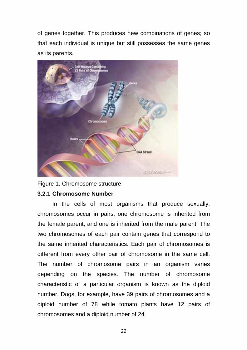
Chromosomes are filamentous rod-like or thread-like gene
bearing bodies found in the nucleus during cell division. Each
nucleus contain information coded in the form of DNA and
organised into groups called genes. Genes are arranged on the
chromosome and each gene contain enough information for the
production of one protein which can have some effects on the
individual chromosome vary widely between different organisms.
The chromosome molecule may be circular or linear, typically
eukaryotic cells (cells with nuclei) have large linear chromosomes
and prokaryotic cells (cells without defined nuclei) have circular
chromosomes. Chromosomes are the essential unit for cellular
division and must be replicated, divided and passed successfully
to their daughter cells so as to ensure the genetic diversity and
survival of their progeny.
3.1.2 Chromosome Structure
Chromosome within a cell occur in matched pairs called
homologous xomes, joined at the centre by a centromse. Each
chromosome contains many genes, and each gene is located at a
particular site on the chromosome, known as the locus. Like
chromosome, genes typically occur in pairs. A gene found on one
chromosome in a pair usually has the same locus as another gene
in the other chromosome of the pair, and these two genes are
called alleles. Alleles are alternate forms of the same gene.
In organisms that use sexual reproduction, offspring inherit
one-half of their genes from each parent and then mix the two sets
22
of genes together. This produces new combinations of genes; so
that each individual is unique but still possesses the same genes
as its parents.
Figure 1. Chromosome structure
3.2.1 Chromosome Number
In the cells of most organisms that produce sexually,
chromosomes occur in pairs; one chromosome is inherited from
the female parent; and one is inherited from the male parent. The
two chromosomes of each pair contain genes that correspond to
the same inherited characteristics. Each pair of chromosomes is
different from every other pair of chromosome in the same cell.
The number of chromosome pairs in an organism varies
depending on the species. The number of chromosome
characteristic of a particular organism is known as the diploid
number. Dogs, for example, have 39 pairs of chromosomes and a
diploid number of 78 while tomato plants have 12 pairs of
chromosomes and a diploid number of 24.
23
Gametes or sex cells (eggs and sperm) contain only half the
number of chromosomes found in the other cells of an organism.
This reduced number of chromosomes in the gametes is known as
the haploid number. During fertilisation the gametes unite to form a
cell known as a zygote containing the diploid number of
chromosomes characteristics of the species.
3.2.3 Sex Chromosomes
Most organisms have complete sets of matching
chromosomal pairs, known as autosomes. In mammals, birds and
some other organisms, one pair of chromosomes is not identical.
Known as the sex chromosomes, this pair plays a dominant role in
determining the sex of an organism. Females have two copies of
the X chromosome while males have one Y chromosome and one
X chromosome. Both males and females inherit one sex
chromosome from the mother (always an X chromosome) and one
sex chromosome from the father (an X chromosome in female
offspring and a Y chromosome in male offspring). The presence of
the Y chromosome determines that a zygote will develop into a
male.
3.2.4 Human Chromosomes and Genetic Disorders
Humans have 23 pairs of chromosomes, with a diploid
number of 46 numbered according to their size. The largest is
chromosome 1 and the smallest is chromosome 23. Physical and
chemical meiosis (formation of gametes) can damage
chromosomes or alter their number in a cell, to give rise to
embryos with more of less genetic material, sometimes resulting in
developmental disabilities or health problems. In a process called
24
non disjunction, paired members of chromosomes fail to separate
from one another during meiosis. Non disjunction can lead to a
condition known as Down Syndrome, in which a person inherits
three copies of chromosome 21. Another condition that may result
from non disjunction is Turner Syndrome, a disorder in which a
female inherits only a single X chromosome.
Breakage of a chromosome can lead to four types of
changes in chromosome structure. A deletion occurs when a
chromosome fragment lacking a centromere is lost during cell
division. In some cases the fragment may join to the homologous
chromosome to produce a duplication. It may reattach to the
original chromosome but in a reverse orientation, producing an
inversion; the fragment can join a non homologous chromosome a
rearrangement called translocation.
4.0 CONCLUSION
Every chromosome in a cell contains many genes and each
gene is located at a particular site or locus, on the chromosome.
Chromosomes vary in size and shape and usually occur in
matched pairs called homologoues. The number of homologous
chromosomes in a cell depends upon the organism.
5.0 SUMMARY
Genes are present in all living cells. They are copied in their
entirety every time a cell divides so that each new cell gets a
complete set. They determine virtually all traits that living
organisms possess. Chromosome within a cell occurs in matched
pairs. Each chromosome contains many genes. Like
25
chromosomes, genes typically occur in pairs, the two genes are
called alleles. Alleles are alternate forms of the same gene.
6.0 TUTOR MARKED ASSIGNMENTS
1. What is a gene’s locus?
2. Mendel assumed that each trait is determined by?
3. Briefly define (a) Gene (b) Cistron (c) Recon.
4. Briefly state the chromosomal theory of inheritance.
5. What are chromosomes?
6. What is chromosomal non disjunction?
7. Chromosomes within a cell occur in matched pair called ?
7.0 REFERENCES/FURTHER READINGS
Geoffrey, M. C. (2000). The Cell. A Molecular Approach, Second
Edition. ASM Press Washington D.C.
Nancy, L. P.; Larry, S. U. and William, S. (2003). Bioinquiry:
making Connections in Biology. John Wiley & Sons Inc.
26
NUCLEIC ACIDS
CONTENTS
1.0 Introduction
2.0 Objectives
3.0 Main Body
3.1 Nucleic Acids
3.2 Types of Nucleic Acids
3.3 DNA
3.4 DNA Structure
3.5 RNA
3.6 RNA Structure
3.7 Types of RNA
4.0 Conclusion
5.0 Summary
6.0 Tutor-Marked Assignments
7.0 References/Further Readings
27
1.0 INTRODUCTION
The principal genetic informational materials or molecules of
living organisms are chemically called nucleic acids. Both DNA
and RNA are polymers of complex molecules containing carbon,
oxygen, hydrogen, nitrogen and phosphorus. The molecules of
nucleic acids (polymers) are composed of monomers called
nucleotides joined by covalent bonds.
2.0 OBJECTIVES
At the end of this unit you should be able to:
1. Describe the chemical composition of nucleic acids.
2. Identify the types of nucleic acids.
3. Describe the structure of DNA and RNA.
4. Highlight the roles of DNA and RNA.
5. Differentiate DNA from RNA.
3.0 MAIN BODY
3.1 Nucleic Acids
Nucleic acids are extremely complex molecules produced by
living cells and viruses, to pass on hereditary characteristics from
one generation to the next, and to trigger the manufacture of
specific proteins. The name nucleic acids comes from their initial
isolation from the nucleic of living cells. However, certain nucleic
acids are found not in the cell nucleus but in cell cytoplasm.
Nucleic acid molecules are very large chains of repeating
nucleotide units linked in many sequences. Thus, nucleic acids are
high polymers with very high molecular weights. A nucleotide is a
molecular unit or a nucleic acid molecule that consists of 3
subunits:
28
• a phosphate group.
• a pentose sugar (ribose or deoxyribose).
• a nitrogen base (purine or pyrimidine).
A nucleoside is a compound consisting of 2 subunits – a
pentose sugar and a nitrogen base. It is a precursor of a
nucleotide. A summary of nucleic acid formation is:
• Pentose sugar + Nitrogen base = Nucleoside
• Nucleoside + Phosphate group = Nucleotide
• Nucleotide + Nucleotide + Nucleotide = Nucleic acid.
3.2 TYPE OF NUCLEIC ACIDS
There are two types of nucleic acids – DNA (Dexyribonucleic
acid) and RNA (Ribonucleic acid); both are chemical relatives that
are universally present in all living cells and they form the chemical
basis of life. Both DNA and RNA contain the purines – Adenine (A)
and Guanine (G) and the pyrimidine cytosine (C). The second kind
of pyrimidine in DNA is Thymine (T) where as it is Uracil (U) in
RNA. Therefore a unique pyrimidine distinguishes DNA from RNA.
3.3 DNA
The DNA is a polymer made up of repeating units of
mononucleotides carrying the genetic material of all cellular
organisms and most viruses. DNA carries the information needed
to direct protein synthesis ad replication. Protein synthesis is the
production of the proteins needed by the cell or virus for its
activities and development. Replication is the process of which
DNA copies itself for each descendant cell or virus, passing on the
information needed for protein synthesis. In most cellular
29
organisms, DNA is organised on chromosomes located in the
nucleus of the cell.
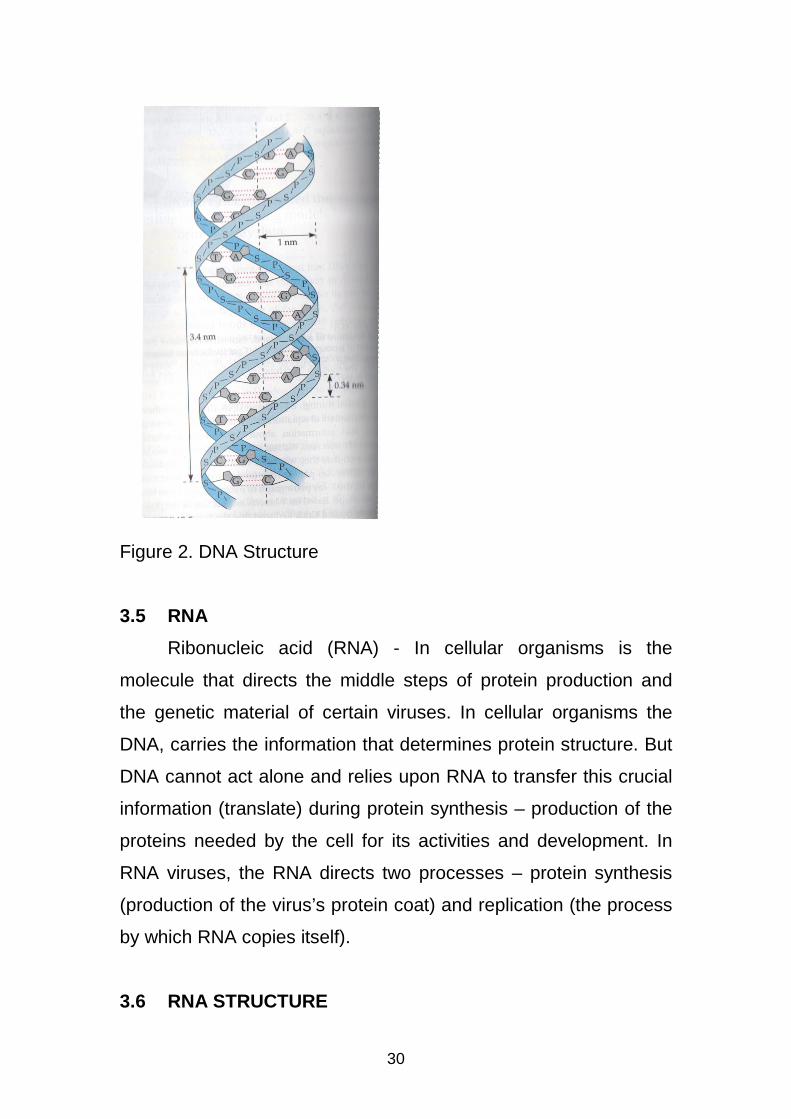
3.4 DNA STRUCTURE
The DNA is a spiral ladder with the nucleotides forming the
side pieces and the steps composed of a combination of purine
and pyrimidine which join the deoxyribose sugars in the side
pieces to hold them together. The purines are of two types
Adenine (A) and Guanine (G), the pyrimidines too are of two types
Cytosine (C) and Thymine (T). In forming the steps of the ladder G
may join C, or C may join G, and T may join A, or A may join T,
usually by hydrogen bond. With the pairings G–C, C–G, A–T or T–
A occurring throughout the length of the DNA molecule. The
combination of the sugar molecule, phosphate group and a
nitrogenous base completes the basic structure of a nucleotide.
With purine linked to a pyrimidine precisely adenine (A) always
pairing with thymine (T) and guanine (G) pairing with cytosine (C).
A mirror image of the nucleotide is added to produce a double
nucleotide chain which will twist to produce the α – helix.
30
Figure 2. DNA Structure
3.5 RNA
Ribonucleic acid (RNA) - In cellular organisms is the
molecule that directs the middle steps of protein production and
the genetic material of certain viruses. In cellular organisms the
DNA, carries the information that determines protein structure. But
DNA cannot act alone and relies upon RNA to transfer this crucial
information (translate) during protein synthesis – production of the
proteins needed by the cell for its activities and development. In
RNA viruses, the RNA directs two processes – protein synthesis
(production of the virus’s protein coat) and replication (the process
by which RNA copies itself).
3.6 RNA STRUCTURE
31
The structure of RNA is similar to that of DNA and it is
composed of a single string of ribonucleotides, each of which is
composed of
• a pentose sugar (ribose sugar)
• a phosphate group
• a nitrogenous base (one of the two bases – adenine,
guanine, uracil and cytosine)
These components are joined together in the same manner
as in DNA molecule. But RNA differs chemically from DNA by
being single stranded, having a D-ribose sugar instead of
Deoxyribose sugar and having uracil as nitrogenous base instead
of thymine. These nitrogenous bases can occur in any sequence.
3.7 TYPES OF RNA
There are three types of RNA classified based on their
molecular size. The smallest type of RNA is called transfer – RNA
(tRNA) which carries amino acids to the ribosomes for
incorporation into a protein. Each amino acid has different classes
of tRNA that read the codes of mRNA, therefore involved in protein
synthesis. The tRNA receives information from mRNA, through
pairing of their bases and accordingly selects particular amino
acids and pass to the ribosome.
The second type of RNA is the ribosomal – RNA (rRNA), this
is larger than tRNA and composes the ribosomes in the cytoplasm,
the specialised structures that are the sites of protein synthesis.
Transfer – RNA are the most abundant type of RNA and they
coordinate the sequential coupling of tRNA molecules to the series
of mRNA codons.
32
The largest type of RNA is the messenger – RNA (mRNA).
Messenger – RNA is a strand of RNA that is complementary to the
DNA sequence for a gene and carries the genetic blueprint copied
from the sequence of bases in a cell’s DNA. This blue print
specifies the sequence of amino acids in a protein. All the types of
RNA are formed as needed, using specific sections of the cell’s
DNA as template.
4.0 CONCLUSION
Two classes of nucleic acids are the deoxyribonucleic acids
(DNA) and ribonucleic acids (RNA). All living cells contain the
genetic material DNA, that determines the shape, the form and the
function of the offspring. While the RNA takes part in the actual
synthesis of the proteins a cell produces, the structure of which is
specified by the DNA.
5.0 SUMMARY
The DNA occurs almost exclusively in the chromosomes and
to a small extent in the mitochondria and chloroplasts. RNA occurs
mostly in the cytoplasm, nucleolus, ribosomes and to some extent
in the chromosomes. DNA is the sole genetic material that
migrates intact from generation to generation; through the
reproductive units (gametes). The DNA is responsible for the
development of specific characters in the successive generations.
It is also the controlling agent of all the vital activities of the cell
and is responsible for all biosynthetic processes including protein
synthesis. Therefore the DNA holds and controls all the secrets of
life of the cell. The RNA is under the instructions of DNA and acts
33
as a messenger carrying information from the DNA to the
ribosomes for synthesis of proteins.
6.0 TUTOR-MARKED ASSIGNMENTS
1. What are the 3 major components of a nucleotide?
2. What are the major differences between DNA and RNA?
3. Briefly outline the major functions of the 3 types of RNA.
7.0 REFERENCES/FURTHER READINGS
Geoffrey, M. C. (2000). The Cell. A Molecular Approach, Second
Edition. ASM Press Washington D.C.
Nancy, L. P.; Larry, S. U. and William, S. (2003). Bioinquiry:
making Connections in Biology. John Wiley & Sons Inc.
Verma, P. S. and Agarwal, V. K. (2008). Cell Biology, Genetics,
Molecular Biology, Evolution and Ecology. S. Chand and
Company Limited, New Delhi.
34
DNA REPLICATION
CONTENTS
1.0 Introduction
2.0 Objectives
3.0 Main Body
4.0 Conclusion
5.0 Summary
6.0 Tutor-Marked Assignments
7.0 References/Further Readings
35
1.0 INTRODUCTION
DNA is the genetic material that makes up the genes; it
contains all the information needed for the cell’s growth, operation,
and division into two similar cells. During replication, an exact copy
of the DNA is made; with the existing DNA being used as a
template for the synthesis of new DNA strands in the cell nucleus.
2.0 OBJECTIVES
At the end of this unit you should be able to:
1. Show how the Watson-Crick Model accounts for
replication.
2. Know the semi-conservative nature of DNA replication.
3.0 MAIN BODY
DNA as the sole genetic material of living organisms must be
able to replicate itself exactly if information is to be transferred
from parents to offspring and from generation to generation. In
most cellular organisms, replication of a DNA molecule takes place
in the cell nucleus and occurs just before the cell divides.
During replication the parent double helix DNA molecule
uncoils when the hydrogen bonds between the nitrogenous bases
are broken, and as a result the double helix DNA begins to unzip
and unwind. The unzipping creates two separate parent strands of
DNA. Each parent strand becomes the template (pattern) for the
creation of a daughter strand.
The unzipping exposes chemical bonds on the purines and
pyrimidines, the nucleoplasm is a reservoir of free nucleotides from
which each A on the parent strand attracts a T nucleotide, each C
attracts a G nucleotide and so on. When the nucleotides are lined
36
up they join together to form a polynucleolide chain, the DNA
polymerase helps the nucleotides link up; by bonding the
phosphate group of nucleotide to the sugar molecule of the
adjacent nucleotide during the side rail of the new DNA molecule.
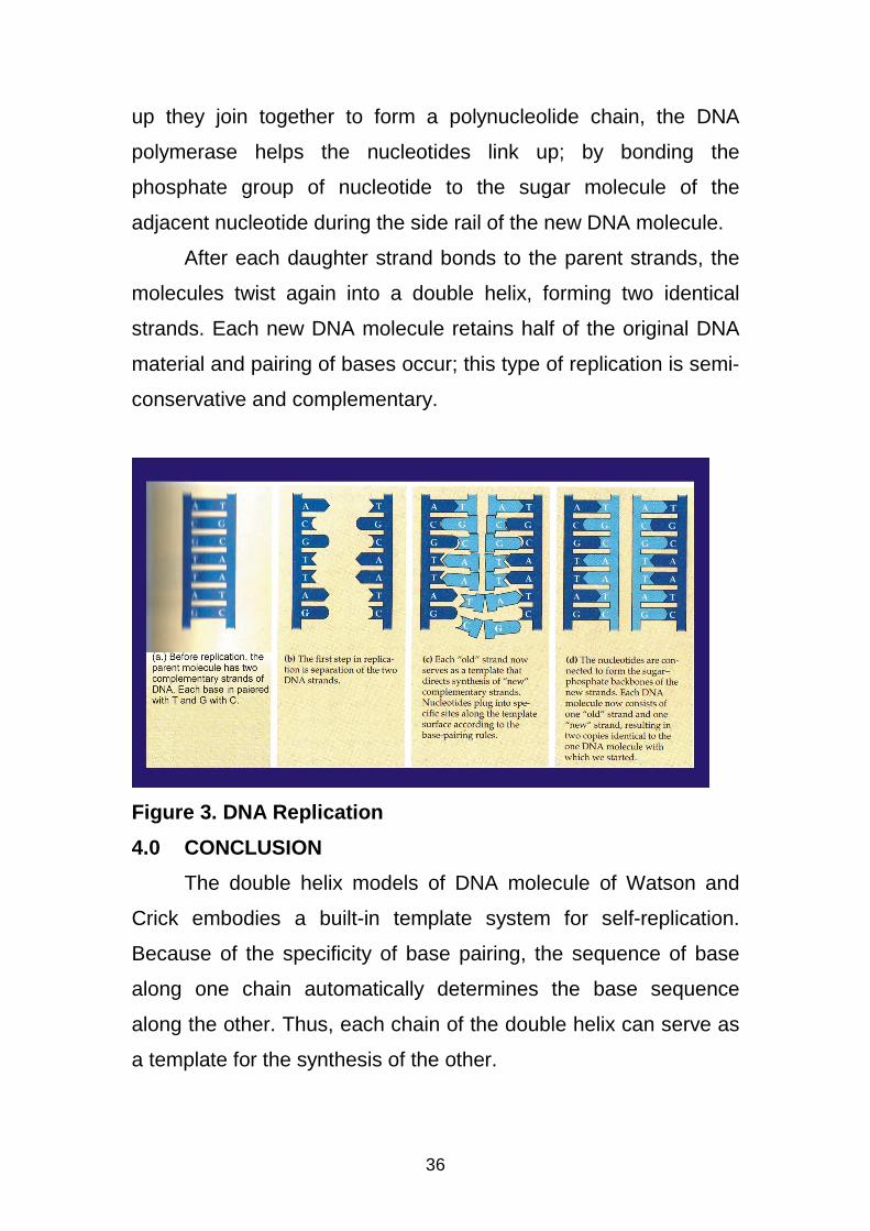
After each daughter strand bonds to the parent strands, the
molecules twist again into a double helix, forming two identical
strands. Each new DNA molecule retains half of the original DNA
material and pairing of bases occur; this type of replication is semi-
conservative and complementary.
Figure 3. DNA Replication
4.0 CONCLUSION
The double helix models of DNA molecule of Watson and
Crick embodies a built-in template system for self-replication.
Because of the specificity of base pairing, the sequence of base
along one chain automatically determines the base sequence
along the other. Thus, each chain of the double helix can serve as
a template for the synthesis of the other.
37
5.0 SUMMARY
The Watson-Crick Model of DNA explains how genetic
replication occurs. The process produces two complete double-
chained molecules, each identical in base sequence to the original
double-chained molecule.
6.0 TUTOR-MARKED ASSIGNMENT
1. Define DNA replication.
2. What is a template?
8.0 REFERENCES/FURTHER READINGS
Robert, K.M.; Daryl, K.G.; Peter, A.M. and Victor, W.R.
(2003). Harper’s Illustrated Biochemistry, Twenty-Sixth
Edition. McGraw Hill Publishers India.
Raven, P. H. and Johnson, G. B. (1985). Biology. Second
Edition. Times Mirror/Mosby College Publishing.
Campbell, N. A. (1996). Biology. Fourth Edition. The
Benjamin/Cummings Publishing Company Inc.
California.
Bruce, A., Dennis, B., Lewis,J., Martin, R., Keith, R. and
James, D.W. (1994). Molecular Biology of the Cell.
Third Edition. Garland Publishing , Inc. New York
38
DNA TRANSCRIPTION
CONTENTS
1.0 Introduction
2.0 Objectives
3.0 Main Body
3.1 Transcription
3.2 Pre-Initiation
3.3 Initiation
3.4 Promoter Clearance
3.5 Elongation
3.6 Termination of Transcription
4.0 Conclusion
5.0 Summary
6.0 Tutor-Marked Assignments
7.0 References/Further Readings
39
1.0 INTRODUCTION
Genes are the instructions for making specific proteins. But a
gene does not build a protein directly. The bridge between genetic
information and protein synthesis is ribonucleic acid (RNA). The
synthesis (creation) of RNA from a DNA template occurs during
transcription.
2.0 OBJECTIVES
At the end of this unit you should be able to:
1. Explain the process of transcription.
2. Recognise the importance of transcription in protein
synthesis.
3.0 MAIN BODY
3.1 Transcription
The DNA molecule represents information required for the
building of a phenotype structure i.e. the DNA is a molecule
carrying a message that when passed in some way to a
‘manufacturing site’ and translated controls the formation of the
individual structural units necessary to the complete individual.
The DNA does not play any part in the manufacturing
process but merely give instructions as to what shall be processed
and how this should be done. The DNA carries its message in
some form of code, this being represented by the sequence of the
bases in the poly-nucleotide chain. Each message or sequence
being equivalent to the gene.
This DNA with its genetic code is located in the nucleus of
cells and within chromatin and chromosomes. While the protein
synthesising machinery (ribosomes) of the cell is located in the
40
cytoplasm, so the information must therefore be transported from
the nucleus to the cytoplasm. This usually happens was the code
along the DNA molecule is copied by a strand of mRNA. The
copying of codon sequences from DNA to mRNA is called
transcription. Transcription results in an RNA complement that
includes uracil (U) in all instances where thymine (T) would have
occurred in a DNA complement.
If one codon on the DNA molecule is AAA the
complementary condon on a strand of mRNA would be UUU, TAT
would transcribe as AUA. Then the mRNA with a faithful reverse
copy of the genetic code, separates from the DNA template. The
mRNA then passes through minute pores in the nuclear
membrane and into the cytoplasm.
Unlike replication, transcription does not progress along the
entire length of a chromosome. Instead, certain parts of the
chromosome are transcribed. The whole process is divided into
the following stages: Pre-initiation, initiation, promoter clearance,
elongation and termination.
3.2 Pre-Initiation
The first step in transcription is binding of RNA polymerase
to a DNA molecule. Binding occurs at particular sites, the
promotes, which are specific sequences of 20 to 200 bases at
which several interactions occur or regions of DNA which promote
transcription. A special promoter region has been identified in
eukaryotic organisms. It is a short DNA sequence known as a
TATA box, because it is enriched with the nitrogenous bases
thymine (T) and adenine (A) found 25-30 base pairs upstream
from the short site of transcription. TAT box orient the RNA
41
polymerase enzyme, so that synthesis proceeds from left to right.
It is also the region at which the double helix opens to form the
open promoter complex which is the binding site for a transcription
factor known as TATA bining protein (TBP) constituting the pre-
initiation complex which is a highly stable complex and an active
intermediate in chain initiation. It is in this complex a local
unwinding or melting of the DNA helix occurs, which is necessary
for pairing of the incoming ribonucleotides.
3.3 Initiation
Once an open-promoter complex has been formed, RNA
polymerase is ready to initiate RNA synthesis. RNA polymerase
contains two nucleotides binding sites, called the initiation site and
the elongation.
In eukaryotes, RNA polymerase does not directly recognise
the core promoter sequences. Instead, a collection of proteins
called transcription which are proteins needed to initiate
transcription but are not part of the RNA polymerase mediate the
binding of RNA polymerase and the initiation of transcription. Only
after certain transcription factors are attached to the promoter does
the RNA polymerase bind to it. The completed assembly of
transcription factors and RNA polymerase bind to the promoter,
forming a transcription initiation complex. Once active RNA
polymerase is bound to a promoter region, the enzyme begins to
separate the two DNA strands at the initiation site, and
transcription is underway.
42
3.4 Promoter Clearance
After the first bond is synthesised, the RNA polymerase must
clear the promoter. During promoter clearance there is tendency
for the RNA transcript to be released to produce truncated
transcripts in a process known as abortive initiation. Abortive
initiation continues to occur resulting in the transcription elongation
complex.
3.5 Elongation
One strand of the DNA, the template strand (non coding
strand), is used as a template for RNA synthesis. As transcription
proceeds, RNA polymerase transverses the template strand from
3′→5′ direction and uses base pairing complementarity with the
DNA template to create an RNA copy. Transcription proceeds in
the 5′→3′ direction. This produces an RNA molecule from 5′→3′,
an exact copy of the coding strand (with thymines replacing uracils
and ribose sugar replacing deoxyribose in the sugar phosphate
backbone). During transcription multiple RNA polymerase can be
involved on a single DNA template and multiple rounds of
transcriptions, so many mRNA molecules can be rapidly produced
from a single copy of a gene.
Elongation also involves a proof reading mechanism that can
replace incorrectly incorporated bases. This may correspond with
short pauses during transcription that allow appropriate RNA
editing factors to bind.
3.6 Termination of Transcription
Transcription proceeds until the RNA polymerase reaches a
termination site on the DNA. The sequence of nitrogenous bases
43
that marks this site signals RNA polymerase to stop adding
nucleotides to the RNA strand and release the RNA molecule.
4.0 CONCLUSION
Transcription is a process of creating an equivalent RNA
copy of a sequence of DNA. During transcription a DNA sequence
is read by RNA polymerase, which produces a complementary
RNA strand.
5.0 SUMMARY
Transcription involves the production of a special kind of
RNA known as messenger RNA (mRNA). The process can be
summarised in these simple steps. DNA unwinds or unzips as the
hydrogen bonds break, the free nucleotides of the RNA pair with
complementary DNA bases, RNA sugar-phosphate backbone
forms aided by RNA polymerase, the hydrogen bonds of the
untwisted RNA and DNA ladder break, freeing the new RNA.
6.0 TUTOR-MARKED ASSIGNMENTS
1. What is transcription?
2. What is transcription unit?
3. What are transcription factors?
8.0 REFERENCES/FURTHER READINGS
Robert, K.M.; Daryl, K.G.; Peter, A.M. and Victor, W.R.
(2003). Harper’s Illustrated Biochemistry, Twenty-Sixth
Edition. McGraw Hill Publishers India.
Raven, P. H. and Johnson, G. B. (1985). Biology. Second
Edition. Times Mirror/Mosby College Publishing.
44
Campbell, N. A. (1996). Biology. Fourth Edition. The
Benjamin/Cummings Publishing Company Inc.
California.
45
GENE EXPRESSION
CONTENTS
1.0 Introduction
2.0 Objectives
3.0 Main Body
3.1 Control of Gene Expression
3.2 Gene Expression in Bacteria
4.0 Conclusion
5.0 Summary
6.0 Tutor-Marked Assignments
7.0 References/Further Readings
46
1.0 INTRODUCTION
In multicellular organism every cell in the body has identical
genetic information, individual cells have different structural and
functional characteristics. Gene expression is the most
fundamental level at which genotype gives rise to the phenotype.
The genetic code stored in DNA in form of nucleotide sequence is
interpreted by gene expression, and the properties of the
expression products give rise to the organism’s phenotype.
2.0 OBJECTIVES
At the end of this unit you should be able to:
1. Explain how gene expression controls the process of
development in multicellular organisms.
3.0 MAIN BODY
3.1 Control of Gene Expression
Gene expression is a process by which genes coded
information is converted into the structures operating in a cell. The
process is used by all known life – eukaryotes, prokaryotes and
viruses – to generate the macromolecular machinery for life. Gene
expression gives the cell control over structure and function and is
the basis for cellular differentiation, morphogenesis and the
versatility and adaptability of any organism.
3.2 Gene Expression in Bacteria
Bacterial cells are genetically simpler than eukaryotic cells,
with just one chromosome and only about 3,000 genes. Bacteria
can be grown rapidly in large numbers under controlled conditions
in the laboratory, they have been especially useful for studying the
47
regulation of gene expression. Francois Jacob and Jacques
Monod in 1960, formulated a powerful model of the control of gene
expression in bacterial cells, based on their investigation of
enzyme synthesis in E. coli. The Jacob-Monod model proposes
that three parts of the chromosome are involved in controlling
transcription of the structural genes. The regulator gene, the
operator region and the promoter region.
The regulator gene, controls indirectly the activity of the
structural genes. The regulator gene is located near the structural
genes and encodes the information for the synthesis of a repressor
protein. When the repressor binds to the operator, it blocks the
promoter’s binding sites for RNA polymerase and thus prevents
transcription of the structural genes. In this system, the genes
specifying particular enzymes are inactive until turned on by an
inducer substance. In a negative control system such as the lac
operon, a repressor protein binds to the operator and turns off
transcription. When the repressor protein is inactive, the operator
is turned on and transcription and translation automatically occur.
In a positive control system, proteins called transcription factors
bind to the promoter and activate transcription.
3.3 Hormonal Control of Gene Expression
In higher plants and animals signals in various glands and/ or
secretory cells somehow stimulate target tissue or target cells to
undergo dramatic changes in their metabolic patterns. These
changes frequently include altered pattern of differentiation that
are generally dependent on altered patterns of gene expression.
Peptide hormones such as insulin and steroid hormones such as
estrogen, progestrone, testosterone (in animals like mammals) and
48
ecdysone (in insects). In higher animals, hormones are
synthesized in specialized secretory cells called endocrine cells
and are released into the blood stream. The peptide hormones
do not normally enter cells because of their relative large size.
Their effects are mediated by receptor proteins located in target-
cell membranes and by the intracellular levels of secondary
messenger called cyclic AMP (cAMP). The cAMP activates a
protein kinase which activates many specific enzymes. The
steroid hormones on the other hand, are small molecules that
readily enter cells through the plasma membrane. Once inside the
appropriate target cells, the steroid hormones become attached to
specific receptor proteins which are present only in the cytoplasm
of target cells. The hormone-receptor protein complexes activate
the transcription of specific and correct genes by binding to
specific DNA sequences present in the cis-acting regulatory
regions of the genes.
4.0 Conclusion
In genetics, gene expression is the most fundamental level at
which genotype gives rise to the phenotype. The genetic code
stored in the DNA in form of nucleotide sequence is interpreted by
gene expression , and the properties of the expression products
give rise to the organism’s phenotype.
5.0 Summary
Gene expression is the process by which coded information in
from a gene is used in the synthesis of functional gene products,
which are often proteins. The process of gene expression is used
49
by all known life- eukaryotes, prokaryotes and viruses – to
generate the macromolecular machinery for life.
6.0 Tutor-Marked Assignments
1. Write short notes on (i) gene expression and (ii) steroid
hormones.
7.0 REFERENCES/FURTHER READINGS
Robert, K.M.; Daryl, K.G.; Peter, A.M. and Victor, W.R. (2003).
Harper’s Illustrated Biochemistry, Twenty-Sixth Edition.
McGraw Hill Publishers India.
http://en.wikipedia.org/wiki/geneexpression.
Raven, P. H. and Johnson, G. B. (1985). Biology. Second Edition.
Times Mirror/Mosby College Publishing.
Campbell, N. A. (1996). Biology. Fourth Edition. The
Benjamin/Cummings Publishing Company Inc. California.
50
GENETIC CODE
CONTENTS
1.0 Introduction
2.0 Objectives
3.0 Main Body
3.1 Nature of the Genetic Code
3.2 Characteristics of the Genetic Code
4.0 Conclusion
5.0 Summary
6.0 Tutor-Marked Assignments
7.0 References/Further Readings
51
1.0 INTRODUCTION
The essential question of gene expression is how does the
order of nucleotides in a DNA molecule encode the information
that specifies the order of amino acids in a protein, i.e. the
correspondence between nucleotide triplets and amino acids in
proteins. The letters A,G,T, and C correspond to the nucleotides
found in DNA. They are organised into three-letter code words
called codons, and the collection of these codons makes up the
genetic code.
2.0 OBJECTIVES
At the end of this unit you should be able to:
1. Know the nature of the genetic code.
2. Outline the characteristics of the genetic code.
3. Know the condon, anti-condon and nonse condon.
3.0 MAIN BODY
3.1 The Nature of the Genetic Code
The genetic code is the set of rules by which information
encoded in genetic material (DNA or mRNA sequences) is
translated into proteins (amino acids) by living cells.
In 1961 Francis Crick and his colleagues reasoned that the
genetic must likely consist of a series of blocks of information,
each block corresponding to an amino acid in the encoded protein.
They further hypothesised that the information in one block was
probably a sequence of three nucleotides specifying a particular
amino acid, they arrived at the number three because a two
nucleotide block will not yield enough different combinations to
52
code for the 20 different kinds of amino acids that commonly occur
in proteins.
Within genes that encode proteins the nucleotide sequences
of DNA is usually in increment of three (3) consecutive nucleotide
without penetration between the increment. Each block of three (3)
nucleotide code of one amino acid. These 3 nucleotide blocks are
called codons. Translation occurs on the ribosome, first the initial
portion of mRNA transcribed in a gene binds to an rNRA molecule
interwoven in the ribosome, the mRNA lies on the ribosomes in
such a way that only the 3 nucleotide portion of the mRNA
molecule – the codon is exposed at the polypeptide making site as
each bit of the mRNA message is exposed in turn. A molecule of
tRNA in the complementary 3 nucleotide sequences or anticodon
binds to the mRNA, because the tRNA molecule carries a
particular amino acid, that amino acid and no other is added to the
polypeptide chain in that position.
Protein synthesis occurs as a series of tRNA molecules bond
one after another to the exposed portion of mRNA molecule as it
moves through the ribosomes, each of this tRNA molecule has
attached to it an amino aid and the amino acid it brings to the
ribosome is added one after another to the end of a growing
polypeptide chain. The anticodon of a tRNA is 3 nucleotide long;
the base sequences of the tRNA anticodons are complementary to
the associated sequences of mRNA. Since there are 4 different
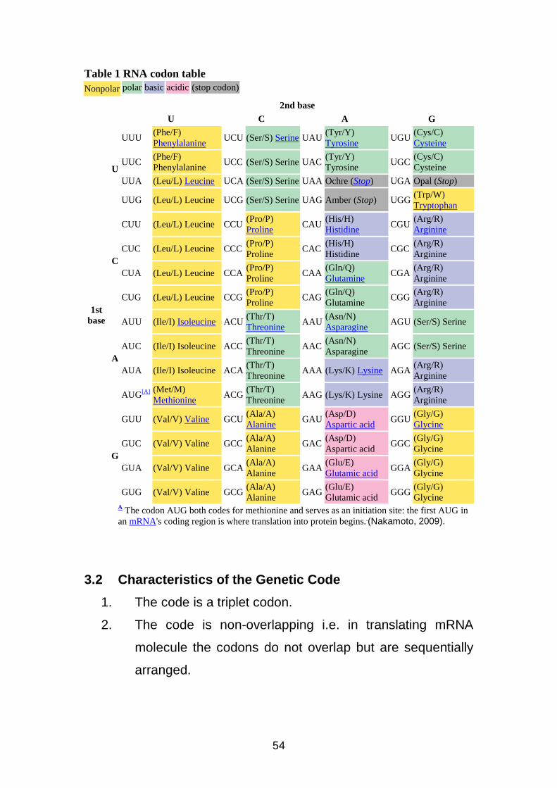
kinds of nucleotides in mRNA (C, G, A, U) there are 43 or 64
different 3 letter code words or codons possible. The list of
different mRNA codons specific for each of the 20 amino acids is
called the Genetic code. The genetic code is the same in all
organisms with only a few exceptions. A particular codon such as
53
AGA corresponds to the same amino acid (Arginine) in bacteria as
in humans.
Note: That 3 out of the 64 codons – UAA, UAG and UGA do not
correspond to triplets that are recognised by any activating
enzyme. These 3 codons, called nonsense codons they serve as
chain terminators or as stop signals in the mRNA message,
marking the end of a polypeptide i.e. they specify where the
polymerisation of amino acids into a protein is to stop.
The codon AUG both codes for methionine and serves as an
initiation site. The first AUG in an mRNA’s coding region is where
translation into protein begins, marking the beginning of a
polypeptide amino acid sequence. The ribosome uses the first
AUG that it encounters in the mRNA message to signal the start of
its translation.
54
Table 1 RNA codon table
Nonpolar polar basic acidic (stop codon)
2nd base
U C A G
1st base
U
UUU (Phe/F) Phenylalanine
UCU (Ser/S) Serine UAU (Tyr/Y) Tyrosine
UGU (Cys/C) Cysteine
UUC (Phe/F) Phenylalanine
UCC (Ser/S) Serine UAC (Tyr/Y) Tyrosine
UGC (Cys/C) Cysteine
UUA (Leu/L) Leucine UCA (Ser/S) Serine UAA Ochre (Stop) UGA Opal (Stop)
UUG (Leu/L) Leucine UCG (Ser/S) Serine UAG Amber (Stop) UGG (Trp/W) Tryptophan
C
CUU (Leu/L) Leucine CCU (Pro/P) Proline
CAU (His/H) Histidine
CGU (Arg/R) Arginine
CUC (Leu/L) Leucine CCC (Pro/P) Proline
CAC (His/H) Histidine
CGC (Arg/R) Arginine
CUA (Leu/L) Leucine CCA (Pro/P) Proline
CAA (Gln/Q) Glutamine
CGA (Arg/R) Arginine
CUG (Leu/L) Leucine CCG (Pro/P) Proline
CAG (Gln/Q) Glutamine
CGG (Arg/R) Arginine
A
AUU (Ile/I) Isoleucine ACU (Thr/T) Threonine
AAU (Asn/N) Asparagine
AGU (Ser/S) Serine
AUC (Ile/I) Isoleucine ACC (Thr/T) Threonine
AAC (Asn/N) Asparagine
AGC (Ser/S) Serine
AUA (Ile/I) Isoleucine ACA (Thr/T) Threonine
AAA (Lys/K) Lysine AGA (Arg/R) Arginine
AUG[A] (Met/M) Methionine
ACG (Thr/T) Threonine
AAG (Lys/K) Lysine AGG (Arg/R) Arginine
G
GUU (Val/V) Valine GCU (Ala/A) Alanine
GAU (Asp/D) Aspartic acid
GGU (Gly/G) Glycine
GUC (Val/V) Valine GCC (Ala/A) Alanine
GAC (Asp/D) Aspartic acid
GGC (Gly/G) Glycine
GUA (Val/V) Valine GCA (Ala/A) Alanine
GAA (Glu/E) Glutamic acid
GGA (Gly/G) Glycine
GUG (Val/V) Valine GCG (Ala/A) Alanine
GAG (Glu/E) Glutamic acid
GGG (Gly/G) Glycine
A The codon AUG both codes for methionine and serves as an initiation site: the first AUG in an mRNA's coding region is where translation into protein begins. (Nakamoto, 2009).
3.2 Characteristics of the Genetic Code
1. The code is a triplet codon.
2. The code is non-overlapping i.e. in translating mRNA
molecule the codons do not overlap but are sequentially
arranged.
55
3. The code is commaless i.e. no punctuation and once the
reading is commenced at a specific codon, there is no
punctuation between codons, and the message is read in
a continuing sequence of nucleotide triplets until a
translation stop codon is reached.
4. The genetic code is unambiguous, with a particular codon
always coding for the same amino acid.
5. The code is universal ranging from bacteria to man.
6. Some codes act as start codons (AUG).
7. Some act as stop codons (UAA, UAG, UGA).
8. The code has polarity i.e. it is always read in a fixed
direction the 5′ 3′ direction.
9. Degenerate: The code is degenerate i.e. more than one
condon may specify the same amino acid.
4.0 CONCLUSION
The (genome) full complement of genetic information that an
organism inherits from its parents is inscribed in DNA. The portion
of the genome that codes for a protein or RNA is referred to as a
gene. Those genes that code for proteins are composed of tri-
nucleotide units called cocoons each coding to a single amino
acid. The genetic code represents the order of the nucleotide
sequences in DNA or RNA that form the basis of heredity through
their role in protein synthesis.
5.0 SUMMARY
Genetic instructions from DNA are written in three
nucleotide units called codons. There are 64 codons in the genetic
56
code, 61 of the codons code for amino acids and 3 of the 64
cocons function as stop signals.
6.0 TUTOR-MARKED ASSIGNMENTS
1. What do you understand by ‘genetic code’?
2. Outline any four characteristics of the genetic code.
3. Briefly define the following: codon, anticodon and
nonsense codon.
7.0 REFERENCES/FURTHER READINGS
Robert, K.M.; Daryl, K.G.; Peter, A.M. and Victor, W.R. (2003).
Harper’s Illustrated Biochemistry, Twenty-Sixth Edition.
McGraw Hill Publishers India.
http://en.wikipedia.org/wiki/geneticcode.
Raven, P. H. and Johnson, G. B. (1985). Biology. Second Edition.
Times Mirror/Mosby College Publishing.
Campbell, N. A. (1996). Biology. Fourth Edition. The
Benjamin/Cummings Publishing Company Inc. California.
Nakamoto T ( 2009). "Evolution and the universality of the mechanism of initiation of protein synthesis". Gene 432 (1-2): 1–6. doi:10.1016/j.gene.2008.11.001. PMID 19056476.
Pamela K. Mulligan; King, Robert C.; Stansfield, William D. (2006). A dictionary of genetics. Oxford [Oxfordshire ]: Oxford University Press. pp. 608. ISBN 0-19-530761-5.
57
PROTEIN SYNTHESIS
CONTENTS
1.0 Introduction
2.0 Objectives
3.0 Main Body
3.1 The Central Dogma
3.2 Mechanism of Protein Synthesis
3.2.1 Chain Initiation
3.2.2 Chain Elongation
3.2.2.1 Codon Recognition
3.2.2.2 Peptide Bond Formation
3.2.2.3 Translocation
3.2.3 Chain Termination
3.3 From Polypeptide to Functional Protein
4.0 Conclusion
5.0 Summary
6.0 Tutor-Marked Assignments
7.0 References/Further Readings
58
1.0 INTRODUCTION
DNA with its correct mechanism of replication, serves to
carry genetic information from cell to cell and from generation to
generation. The information is translated into proteins that
determine the phenotype. Protein synthesis involves how the
information present in the sequences of bases (triplet codons) of
the mRNA is translated into a sequence of amino acids in proteins.
2.0 OBJECTIVES
At the end of this unit you should be able to:
1. Describe the mechanism of protein synthesis.
2. Describe the central dogma.
3. Identify the roles of transcription and translation in the flow
of genetic information.
3.0 MAIN BODY
3.1 The Central Dogma
Genes are the instructions for making specific proteins. But a
gene does not build a protein directly. The bridge between genetic
information and protein synthesis is RNA. The process of
synthesis of protein involves one of the central dogma of molecular
biology; which postulates that genetic information flows from
nucleic acids to protein. The first step of the central dogma is
known as transcription and does not involve a change of code
since DNA and mRNA are complementary. The second step
involves a change of code from nucleotide sequences to amino
acid sequences and is called translation illustrated as follows:
Duplication DNA RNA Protein
Transcription Translation
59
3.2 Mechanism of Protein Synthesis
Protein synthesis is a very complex biochemical
transformation performed by cells resulting in the formation of a
polypeptide chain. The mechanism of protein synthesis can be
divided into the following 3 main steps: chain initiation, chain
elongation and chain termination. All three steps require protein
factors (about 200 different proteins) mostly enzymes that aid in
mRNA, tRNA and ribosomes in the translation process. Chain
initiation and elongation require energy usually provided by GTP
(guanosine triphosphate), a molecule closely related to ATP.
3.2.1 Chain Initiation
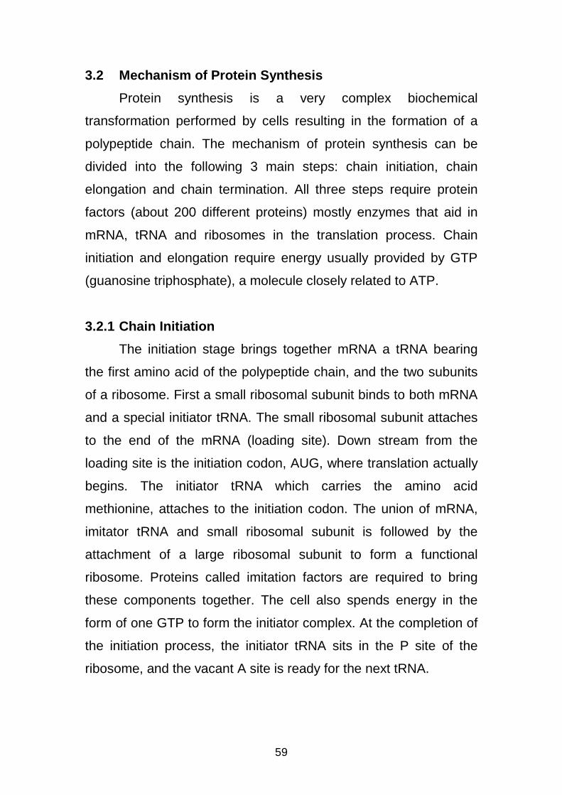
The initiation stage brings together mRNA a tRNA bearing
the first amino acid of the polypeptide chain, and the two subunits
of a ribosome. First a small ribosomal subunit binds to both mRNA
and a special initiator tRNA. The small ribosomal subunit attaches
to the end of the mRNA (loading site). Down stream from the
loading site is the initiation codon, AUG, where translation actually
begins. The initiator tRNA which carries the amino acid
methionine, attaches to the initiation codon. The union of mRNA,
imitator tRNA and small ribosomal subunit is followed by the
attachment of a large ribosomal subunit to form a functional
ribosome. Proteins called imitation factors are required to bring
these components together. The cell also spends energy in the
form of one GTP to form the initiator complex. At the completion of
the initiation process, the initiator tRNA sits in the P site of the
ribosome, and the vacant A site is ready for the next tRNA.
60
Figure 4. Chain Initiation
61
3.2.2 Chain Elongation
The elongation stage amino acids are added one by one to
the initial amino acid joined by peptide bond. Each addition
involves the participation of several proteins called elongation
factors. The whole process occurs in a thee-step cycle.
3.2.2.1 Codon Recognition
The mRNA codon in the A site of the ribosome forms
hydrogen bonds with the anticodon of an incoming molecule of
tRNA carrying its approrpite amino acid. An elongation factor
ushers the tRNA into the A site. This step requires the hydrolysis
of a phosphate bond from GTP.
3.2.2.2 Peptide Bond Formation
A component of the large ribosomal subunit catolyzes the
formation of a peptide bond between the polypeptide extending
from the P site and the newly arrived amino acid in the A site. In
this step, the polypeptide separates from the tRNA to which it was
bound and is transferred to the amino acid carried by the tRNA in
the A site.
3.2.2.3 Translation
The tRNA in the P site dissociates from the ribosome. The
tRNA in the A site, now attached to the growing polypeptide, is
translocated to the P site. As the tRNA changes sites, its anticodon
remains hydrogen-bonded to the mRNA codon, allowing the
mRNA and tRNA molecules to move as a unit. This movement
brings the next codon to be translated into the A site. The
62
translocation step requires energy, which is provided by hydrolysis
of a GTP molecule.
Figure 5. The elongation cycle translation
3.2.3 Chain Termination
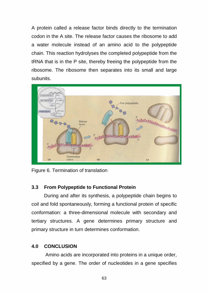
The final stage of translation is termination. Elongation
continues until a termination codon reaches the A site of the
ribosome. Nonsense base triplets – UAA, UAG and UGA – do not
code for amino acids but instead act as signals to stop translation.
63
A protein called a release factor binds directly to the termination
codon in the A site. The release factor causes the ribosome to add
a water molecule instead of an amino acid to the polypeptide
chain. This reaction hydrolyses the completed polypeptide from the
tRNA that is in the P site, thereby freeing the polypeptide from the
ribosome. The ribosome then separates into its small and large
subunits.
Figure 6. Termination of translation
3.3 From Polypeptide to Functional Protein
During and after its synthesis, a polypeptide chain begins to
coil and fold spontaneously, forming a functional protein of specific
conformation: a three-dimensional molecule with secondary and
tertiary structures. A gene determines primary structure and
primary structure in turn determines conformation.
4.0 CONCLUSION
Amino acids are incorporated into proteins in a unique order,
specified by a gene. The order of nucleotides in a gene specifies
64
the amino acid sequence of a protein through translation in which
messenger RNA acts as a template for protein synthesis.
5.0 SUMMARY
Cells are governed by a molecular chain of commands that
flows from DNA → RNA → protein. DNA transcribes RNA of the
copying of the information contained in DNA by the RNA. The RNA
the translates the information and assemble protein on the
ribosomes.
6.0 TUTOR-MARKED ASSIGNMENTS
1. Write short notes on (i) Central Dogman (ii) Transcription
(iii) Translation.
2. What is initiation complex?
3. Which process in protein require hydrolysis of GTP.
7.0 REFERENCES/FURTHER READINGS
Raven, P. H. and Johnson, G. B. (1985). Biology. Second Edition.
Times Mirror/Mosby College Publishing.
Campbell, N. A. (1996). Biology. Fourth Edition. The
Benjamin/Cummings Publishing Company Inc. California.
Robert, K.M.; Daryl, K.G.; Peter, A.M. and Victor, W.R. (2003).
Harper’s Illustrated Biochemistry, Twenty-Sixth Edition.
McGraw Hill Publishers India.
65
DNA SEQUENCING
CONTENTS
1.0 Introduction
2.0 Objectives
3.0 Main Body
4.0 Conclusion
5.0 Summary
6.0 Tutor-Marked Assignments
7.0 References/Further Studies
66
1.0 INTRODUCTION
Once an interesting piece of DNA has been isolated or
identified, there is need to determine if the sequence of
nucleotides in the fragment is related to known genes and to
determine what kind of protein it might produce. DNA sequencing
refers to sequencing methods for determining the order of the
nucleotides bases – adenine, guanine, cytosine and thymine – in a
molecule of DNA.
2.0 OBJECTIVES
At the end of this unit you should be able to know:
1. What DNA sequencing is.
2. The methods of DNA sequencing.
3.0 MAIN BODY
3.1 DNA SEQUENCING
DNA sequencing makes it possible to determine the precise
order, or sequence of nucleotide bases within a fragment of DNA.
In DNA sequencing, many copies of a single-stranded DNA
fragment that will be used to synthesise a new DNA strand, are
created. Through DNA sequencing a gene can be characterised in
terms of a linear sequence of AGCT bases that in turn, can be
used to predict the amino acid sequence of the corresponding
protein using the genetic code. There are three methods for
determining DNA sequences.
3.2 Chain-Termination Method (Sanger Method)
This method requires a single-stranded DNA template, a
DNA primer, a DNA polymerase, radioactively labelled nucleotides
67
and modified nucleotides that terminate DNA strand elongation.
The DNA sample is divided into four separate sequencing
reactions, containing all four of the stranded deoxynucleotides and
the DNA polymerase. To each reaction is added only one of the
four dideoxynucleotides which are the chain-terminating
nucleotides, that terminate DNA strand extension and result in
DNA fragments of varying lengths.
The newly synthesised and labelled DNA fragments are heat
denatured, and separated by size, by gel electrophoresis on a
denaturing polyacrylamide-urea gel with each of the four reactions
run in one of four individual lanes (lanes A, T, G, C) the DNA
bands are then visualised by autoradiography or UV light and the
DNA sequence can be directly read off the x-ray film.
3.3 Maxam and Gilbert’s Chemical (Degradation Metho d)
The method is based on chemical modification of DNA and
subsequent clearage at specific bases; it requires potassium
labelling at one end and purification of the RNA fragment to be
sequenced. The chemical treatment with **** RNAs generates
breaks at every nucleotide base. Thus a series of labelled
fragments is generated from the potassium labelled end to the first
cut site in each molecule. The fragments in the four reactions are
arranged side by side in gel electrophoresis for size separation.
The fragments are visualised when the gel is exposed to
hydrolysis enzymes for autoradiography, yielding a series of cubes
each corresponding to a RNA fragment from which the sequence
may be determined.
68
4.0 CONCLUSION
DNA sequencing allows the isolation of individual fragments
of DNA in quantities suitable for detailed characterisation and the
determination of nucleotide sequence.
69
METABOLIC PATHWAYS
CONTENTS
1.0 Introduction
2.0 Objectives
3.0 Main Body
3.1 Meaning of Metabolism
3.2 Glycolysis
3.3 Fermentation
3.4 The Krebs Critic Acid Cycle
3.5 Oxidative Phosphorylation
4.0 Conclusion
5.0 Summary
6.0 Tutor-Marked Assignments
7.0 References/Further Readings
70
1.0 INTRODUCTION
Metabolic pathways are a series of chemical reactions
occurring within a cell. In each pathway or chemical reaction, a
principal chemical is modified by a series of chemical reactions.
Enzymes catalyse these reactions, and often require dietary
minerals, vitamins and other cofactors in order to function properly.
Because of the many chemicals (metabolites) that may be involved
metabolic pathways can be elaborate. In addition, numerous
distinct pathways co-exist within a cell; collectively called the
metabolic network. Metabolic pathways are important to the
maintenance of homeostatis within an organism.
2.0 OBJECTIVES
At the end of this unit you should be able to:
1. Define metabolism, catabolism and anabolism.
2. Describe the stages involved in the break down of
glucose.
3. Outline the processes involved in glycolysis.
3.0 MAIN BODY
3.1 Meaning of Metabolism
Metabolism: The sum of the enzyme-mediated chemical reactions
occurring within a cell or a whole organism. These chemical
reactions are divided into two – catabolism and anabolism.
Catabolism (catabolic pathway) is a metabolic pathway that
releases energy by breaking down complex molecules into simpler
compounds.
71
Anabolism (anabolic pathway) is a metabolic pathway that
involves the synthesis or assembling of organic molecules and
new protoplasm.
Before the energy stored in lipids, proteins and
carbohydrates can be used by the cell to do work, the molecules
must be broken down in a series of chemical reactions and the
energy produced used to synthesise adenosine triphosphate (ATP)
– an adenine – containing nucleoside triphosphate that releases
free energy when its phosphate bonds are hydrolyzed. ATP is
regarded as the universal energy currency of living organisms. The
energy is used to drive endergonic reactions in cells.
The complete degradation of an energy-rich compound such
as glucose to carbondioxide and water involves many
enzymatically controlled reactions. The complete breakdown of
glucose involves four stages:
• Glycolysis (Stage I)
• Fermentation or oxidation of pyruvic acid to acetyl-coenzyme
A (Stage II).
• The Kreb’s Critic Acid Cycle (Stage III).
• Oxidative phosphorylation (Stage IV).
3.2 Glycolysis
The first series of reactions in the degradation of glucose is
termed glycolysis and the most important features of glycolysis
are:
• Each molecule of glucose (C6 H12 O6) is broken down to two
molecules of pyruvic acid (C3 H4 O3).
72
• Two molecules of ATP are used to initiate the process, later
four new ATP molecules are synthesised with a net gain of
two molecules of ATP and two molecules of NADH (an
energy or electron carrier molecule).
• ATP molecules are produced by substrate level
phosphorylation and NAD+ (nicotinamide adenine
dinucleotide) is reduced to NADH by oxidation of the food.
• The net energy yield from glycolysis, per glucose molecule is
2 ATP and 2 NADH (Stage I).
• No molecular oxygen is used, glycolysis can occur with
(aerobic metabolism) or without oxygen (anerobic
metabolism).
• The reactions of glycolysis occur within the cytosol of the cell
in the cytoplasm outside the mitochondria. While the krebs
cycle enzymes and intermediates are dissolved in the fluid
within the mitochondria.
3.3 Fermentation
The fate the pyruvic acid produced during glycolysis depends
on oxygen supply. Fermentation enables a cell to continue the
reactions of glycolysis in the absence of oxygen.
• In the absence of O2, the pyruvic acid may be reduced by
NADH to CO2 and ethyl alcohol or lactic acid, in a process
called fermentation. In the process the glycolytic pathway
leads to the production of alcohol or lactic acid and it enables
the cell to continue synthesising ATP molecules by the
breakdown of nutrients under anerobic conditions.
73
• Under aerobic conditions the pyruvic acid can be further
oxidised, with the accompanying synthesis of ATP. The
process begins when pyruvic acid moves from the cytoplasm
into the inner compartment of the mitochondrion to form two
each of acetyl-COA, CO2 and NADH (Stage II).
• The acetyl-CoA formed is fed into a complex circular series
of reactions.
3.4 The Krebs Citric Acid Cycle
The cycle functions as a metabolic “furnace” that oxidises
organic fuel derived from pyruvate, the product of glycolysis.
• In the course of the cycle, two carbon atoms are lost as CO2,
a molecule of ATP is synthesised, and eight electrons and
eight hydrogens are picked up by carrier compounds,
forming three molecules of NADH and one of FADH2.
• The cycle generates 1 ATP per turn by substrate
phosphorylation.
• Since one molecule of glucose gives rise to two molecules of
acetyl-CoA, two turns of the cycle occur for each molecule of
glucose oxidised.
3.5 Oxidative Phosphorylation (Electron Transport C hain)
A series of oxidation – reduction reactions that passes
electrons from higher energy levels to lower energy levels. As a
result, ADP is phosphorylated to form ATP. The exergonic reaction
of hydrogen with oxygen to form water releases a large amount of
energy in the form of heat and light. In cellular respiration, an
electron transport chain breaks the “fall” of electrons in this
74
reaction into a series of small steps and stores some of the
released energy in a form that can be used to make ATP.
The electron transport chain molecules are found in the inner
membrane of the mitochondria. The transfer of electrons along the
electron-transport chain results in the pumping of H+ ions from the
inner compartment of the mitochondria to the outer compartment.
As a result, the H+ concentration increases in the outer
compartment and an electrostatic and osmotic concentration
gradient is built up across the inner membrane. Special enzyme
complexes, called ATP synthetases act as H+ ion channels in the
inner mitochondrial membrane. As the H+ ions move down the
electrochemical gradient through the complex, energy is released
and can be used to synthesise ATP. The total number of new ATP
molecules produced by the complete metabolic breakdown of
glucose is usually 36, two from glycolysis, two from krebs cycle
and 32 from electron-transport phosphorylation in the
mitochondria. This yield represent about 39% of the energy of
glucose, the rest of the energy is released as heat.
4.0 CONCLUSION
Cellular metabolism is characterised by metabolic pathways
or sequences of enzyme-catalysed reactions in which the product
of one reaction serves as the reactant of the next. Sometimes
pathways are linear, but can be circular, or interconnect with each
other, they can even be branched, in which the product of one
reaction can go in either directions depending on the needs of the
cell at any particular time.
75
5.0 SUMMARY
A metabolic pathway involves step-by-step modification of an
initial molecule to form another product. The resulting product can
be used-either immediately, as the end product of a metabolic
pathway, to initiate another metabolic pathway or be stored by the
cell. Metabolic pathways usually provide precursors for cell
components and provide energy for synthetic and other energy
requiring processes.
6.0 TUTOR MARKED ASSIGNMENT
1. What are the two major functions of metabolic pathways?
2. What are the four stages involved in the breakdown of
glucose?
3. What is the function of the krebs citric acid cycle?
7.0 REFERENCES/FURTHER READINGS
McFadden, C. H. and Keeton, T. W. (1995). Biology: An
Exploration of Life. W. W. Norton and Company Inc., New
York.
Verma, P. S. and Agarwal, V. K. (2008). Cell Biology, Genetics,
Molecular Biology, Evolution and Ecology. S. Chand and
Company Limited, New Delhi.
Hans, G. S. (1995). General Microbiology. Cambridge University
Press.

76