BIO 206 / 308 – Unit 2 Ch 12 & 13, NeuroPhysiology Denver School of Nursing – ADN & BSN Programs...
100
BIO 206 / 308 – Unit 2 Ch 12 & 13, NeuroPhysiology Denver School of Nursing – ADN & BSN Programs No Laboratory component for this class
-
Upload
frank-sutton -
Category
Documents
-
view
219 -
download
3
Transcript of BIO 206 / 308 – Unit 2 Ch 12 & 13, NeuroPhysiology Denver School of Nursing – ADN & BSN Programs...

BIO 206 / 308 – Unit 2 Ch 12 & 13, NeuroPhysiology
Denver School of Nursing – ADN & BSN ProgramsNo Laboratory component for this class

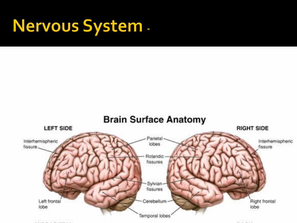
Lets start at the basics and pull this brain system apart…
What is the purpose of the Nervous system?
Just as always make it simple…
The crazy complexity of the Nervous system can be be broken down to THREE words… What are the basic 3 Functions of the NS?
▪ 1) ?▪ 2) ?▪ 3) ?
The nervous system is organized to:
1) Detect
2) Evaluate
3) Respond

The nervous system is organized to:
1) DETECT changes (from stimuli) in the internal and external environment,
2) EVALUATE the level and type of stimuli, &
3) RESPOND by initiating changes in muscles or glands or not initiating change

CNS vs PNS
Autonomic vs Somatic
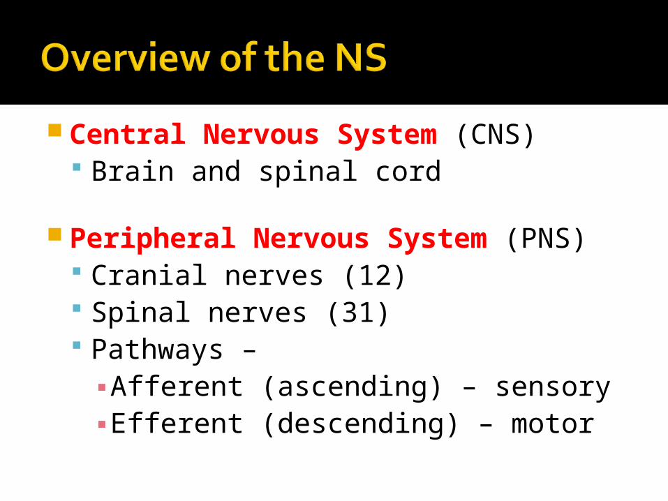
Central Nervous System (CNS) Brain and spinal cord
Peripheral Nervous System (PNS) Cranial nerves (12) Spinal nerves (31) Pathways –
▪ Afferent (ascending) – sensory▪ Efferent (descending) – motor

Neuron Glia (glial
cells)
DendritesAxon

Neuron – Single unit of the NSGlia (glial cells) – supportive cells to
neuronsDendrites – Receptor arms of the neuronAxon – delivery arm of the neuron

• Three components– Cell body (soma)
• Mainly in the CNS• Densely packed in CNS → NUCLEI• Densely packed in PNS → GANGLIA
– Dendrite• Receptive portion of neuron
– Axon• Carries nerve impulse away from the cell
body• One per neuron

• Axon– Hillock
• Leaves the cell body, Nissl free• Lowest threshold for stimulation so → AP begins here
– Myelin• Insulating layer of lipid material segmented: ↑ speed of
conduction• Multiple sclerosis, Guillain-Barre’
– Endoneurium• Delicate layer of connective tissue around each axon
– Neurilemma• Thin membrane between the myelin sheath and the
endoneurium

Functional classification– Sensory
• Receptors to CNS• Afferent
– Associational• Interneurons• Sensory to motor
– Motor • Form CNS to effector organ*• Efferent
*(skeletal muscle or organs)

Support cells: 50% brain and spinal column volume: 5 – 10 x more numerous than neurons

Oligodendrocytes
Schwann cells
Ependymal cells
Astrocytes
Microglia


Neurons generate and conduct electrical and chemical impulses by selectively changing the electrical portion of their
plasma membranes and
influencing other nearby neurons by the release of neurotransmitters.


RESTING Electric Potential: 1) K+ > INside 2) Na+ > OUTside 3) Ca2+ > OUTside

Electrical Action Potential: Initially, the inside of the cell is negative. Chemical / Charge imbalance creates the
Resting Potential ▪ INSIDE of the CELL - High K, Low Na / Ca▪ OUTSIDE of the Cell - High Na / Ca, Low K
“Excitable cells” have the capability of changing their membrane charge, when the NET charge changes other transporters (Voltage gated channels) open causing a chain reaction of increasing positive and then negative charge.
The Action Potential – is the name of the rapid change and propagation of a chemical membrane potential.

Net membrane electrical potential is determined by: 1) The concentration gradients for K+, Na+, and Ca2+
across the membrane. 2) The relative permeability (electrical conductance)
of the membrane to each of these ions)
▪ Therefore the NET electrical potential is determined by multiplying each individual ion equilibrium potentials multiplied by their membrane permeability and added together.▪ (DON’T Panic this pink bullet point will not be on any exam)

Movement of these ions according to concentration gradient

Summary of Ion pumpsThese are ACTIVE Transport Pumps to
set the cells back up to “Resting Potential” 1) Na/K ATPase (3Na+ OUT (/2K+ IN) 2) Ca ATPase (Ca2+ OUT) 3) Na/Ca Exchanger (3Na+ IN /1Ca2+ OUT)

Summary of Cardiac Ion pumps: 1) Na/K ATPase
(3Na+/2K+) 2) Ca ATPase
(Ca2+ OUT) 3) Na/Ca
Exchanger (3Na+/1Ca2)

Action Potential of a nerve cell: Here the Resting Potential would be -60
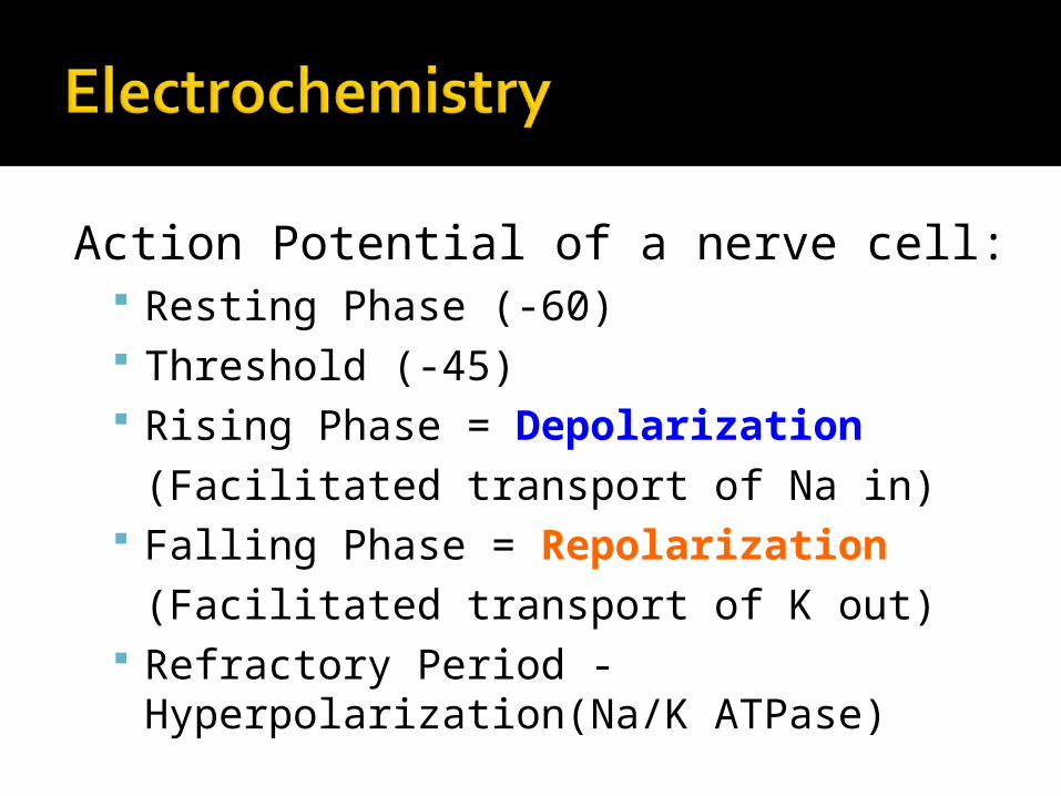
Action Potential of a nerve cell: Resting Phase (-60) Threshold (-45) Rising Phase = Depolarization
(Facilitated transport of Na in) Falling Phase = Repolarization
(Facilitated transport of K out) Refractory Period - Hyperpolarization(Na/K
ATPase)


So how do these nerves actually talk to each other??
The answer is in the… “Synaptic Cleft”
Electrochemical regulation of neurotransmitter release…

Electrochemical regulation of neurotransmitter release…
LETS Break it down: 1) “Electricity” (change in charge of the nerve) 2) Open or closed gate 3) Release of messenger 4) Messenger received 5) New message delivered

Synaptic Cleft – Intersection for delivery and acceptance of Info… what are the messengers called?
Do NOT panic, you do NOT need to memorize detailed names… this is just a pictures to help visualize the “chemical intersection”
Which is called the Synaptic Cleft

Neurons are not physically continuous with one another
Region between adjacent neurons – synapse
Impulses are transmitted across the synapse by neurotransmitters.

Presynaptic neurons and postsynaptic neurons – “to and from the synapse”
Synaptic bouton – vesicles containing neurotransmitters More than 30 substances Excitatory (excitatory postsynaptic
potential) Inhibitory (inhibitory postsynaptic
potential)
Synaptic cleft Space between neurons

Principles of Human Anatomy and Physiology, 11e 38
Astrocytes
Oligodendrocytes
Schwann cells

Astrocytes (only in CNS) – surround and deliver nutrients and blood supply to neurons.
Oligodendrocytes (CNS) – structural support &produce myelin sheath to multiple CNS nerve fibers (Stroke recovery)
Schwann cells (PNS) – Mylinate single axon regions of the same neuron. (lack of healing)

Image from Iowa State University: http://www.public.iastate.edu

Image from Iowa State University: http://www.public.iastate.edu
A. Afferent neuron
A. Efferent neuron
A. Interneuron

Image from BIOPRO Baden-Württemberg Institute of Physiology: http://www.biovalley.com

Does anyone know what the Blood Brain Barrier is??
Any guesses?


Blood Brain Barrier- is a tight web of astrocytes around the brain capillaries that form the BBB
The concept of the blood brain barrier was first introduced by Paul Ehrlich. He found that intravenous injection of dyes into the bloodstream stained all the tissues in most organs except the brain. Using electron microscopy and electron- dense tracers such as horseradish peroxidase (HRP) a group of scientists demonstrated that the blood- brain barrier is located in endothelial cells of capillaries of the brain.

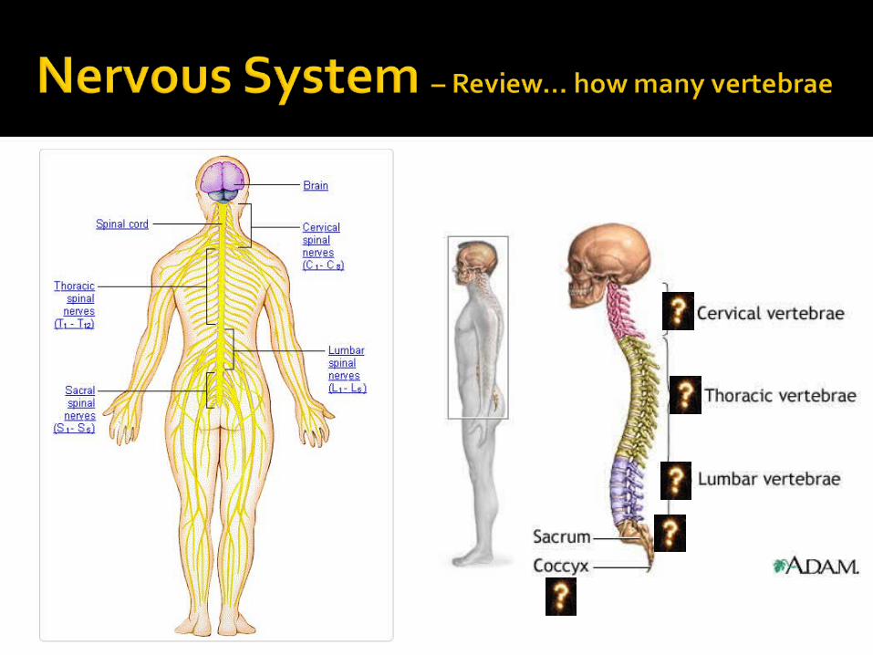


Image from: http://kalibneil.tripod.com
Spinal Nerves:~ 8 Cervical nerves~ 12 Thoracic nerves~ 5 Lumbar nerves~ 5 Sacral nerves~ ONE Coccygeal nerve
Total of 31 Paired Spinal Nerves

Neuromuscular JunctionMotor unit – neuron and
skeletal muscleJunction – axon and plasma
membrane of muscle


Grey vs White Matter


Protective Structure Meninges
Protective membranes surrounding the brain and spinal cord
▪ Dura mater ▪ subdural space – veins
▪ Arachnoid ▪ CSF
▪ Pia mater


Cerebrospinal fluid (CSF) and the ventricular system
CSF – clear, colorless similar to blood plasma and interstitial fluid 125 to 150 ml Produced by choroid plexus (ependymal
cells) within the ventricles (lateral, 3rd & 4th)
Reabsorbed through the arachnoid villi


Cranial Nerves

Cranial Nerves (PNS) Olfactory (I) Optic (II) Oculomotor (III) Trochlear (IV) Trigeminal (V) Abducens (VI)
Facial (VII) Vestibulocochlear
(VIII)(acoustic)
Glossopharyngeal (IX)
Vagus (X) Accessory (XI) Hypoglossal (XII)


How to memorize the cranial nerves…


Image from: http://efildenimaxenu.blogspot.com
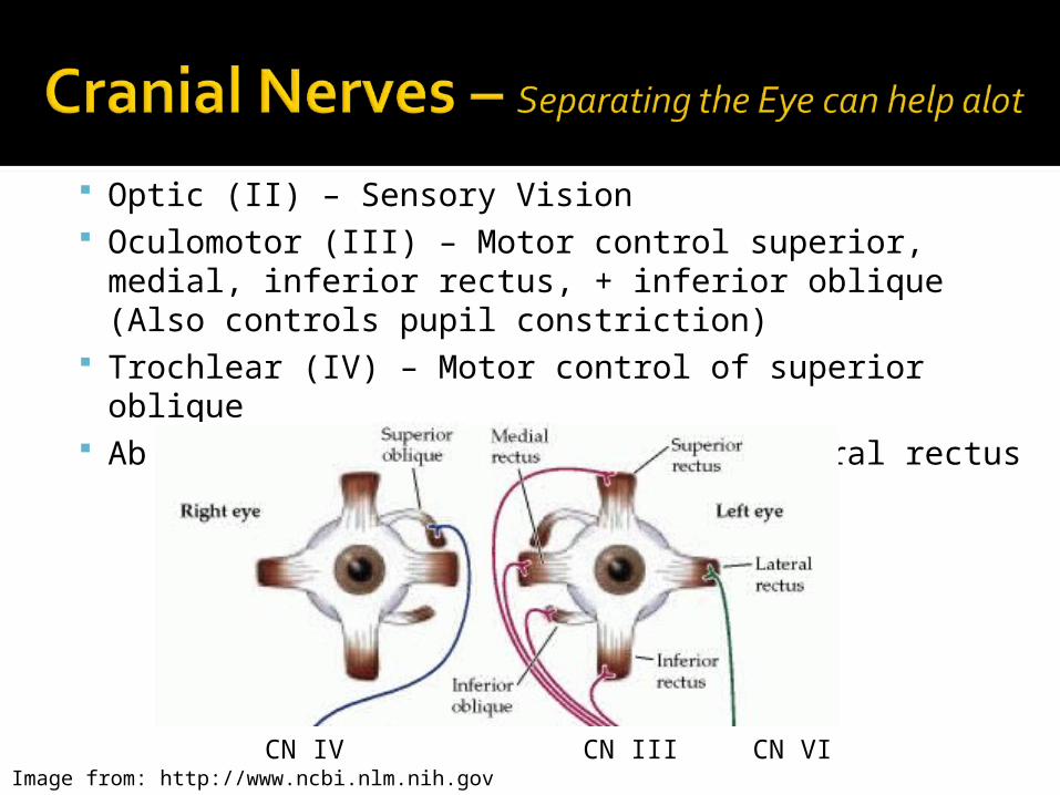
Optic (II) – Sensory Vision Oculomotor (III) – Motor control superior, medial,
inferior rectus, + inferior oblique (Also controls pupil constriction)
Trochlear (IV) – Motor control of superior oblique Abducens (VI) – Motor control of lateral rectus
CN IV CN III CN VIImage from: http://www.ncbi.nlm.nih.gov


Reflex ArcReceptorAfferent (sensory) neuronEfferent (motor) neuronEffector – muscle/gland


Spinal Cord nerve roots Dorsal nerve root localized / specific cord
level afferent sensation (dermatomes)
Ventral nerve root localized / specific cord level efferent motor control (myotomes)

Image from: http://www.whiplash101.com / Apparelyzed.com

Figure 8-17
AnteriorWith SC comparison

Posterior

List of Myotomes of Commonly Injured Nerve RootsC5 – The deltoid muscle (abduction of the arm at the shoulder).C6 – The biceps (flexion of the arm at the elbow).C7 – The triceps (extension of the arm at the elbow).C8 – The small muscles of the hand. L4 – The quadriceps (extension of the leg at the knee).L5 – The tibialis anterior (Dorsiflexion).S1 – The gastrocnemius muscle (Plantarflexion).
Image from: http://neurotalk.psychcentral.com

Image from: http://neurotalk.psychcentral.com

Divisions of the Nervous System Central Nervous System (2 main components) Peripheral Nervous System (All the REST)
▪ Autonomic (What is an example of this function?)▪ Somatic (What is an example of this function?)▪ Sensory nerves (What is an example of their
function?)

A) Autonomic = Involuntary control of nerves (ANS) THE ANS is further divided into: (p.212)
▪ i) Sympathetic NS▪ ii) Parasympathetic NS
Autonomic NS controls everything we take for granted…

A) Autonomic = Involuntary control of nerves (ANS) THE ANS is further divided into: (p.212)
▪ i) Sympathetic NS – “FIGHT or FLIGHT”▪ ii) Parasympathetic NS - “REST and DIGEST”
Autonomic NS controls everything we take for granted… if it was up to us we would stop breathing as soon as we fall asleep in class, but the autonomic NS keeps you alive!!

“Fight or Flight” Accelerates heart rate Constricts blood vessels to smooth muscle Dilates blood vessels to skeletal muscle Decreases GI movement Dilation of pupil Effects on glands: Increases epinephrine,
sweat secretion and decreases digestive secretion

“Fight or Flight” HR Vasoconstriction to Smooth Ms Vasodilatation to Skeletal Ms peristalsis Mydriasis Effects on glands: Increases epinephrine,
sweat secretion and decreases digestive secretion

“Rest and Digest” Slows heartbeat NO effect on blood vessels to smooth muscle NO effect on blood vessels to skeletal
muscle Increases peristalsis Contraction of pupil Effects on glands: No effect on adrenal
medulla or sweat glands, but increases secretion of digestive enzymes

“Rest and Digest” HR NO Δ smooth muscle vasculature NO Δ skeletal muscle vasculature peristalsis Miosis Effects on glands: No effect on adrenal
medulla or sweat glands, but increases secretion of digestive enzymes

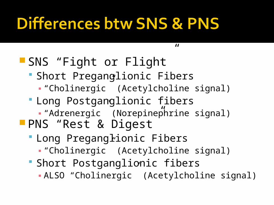
SNS “Fight or Flight” Short Preganglionic Fibers
▪ “Cholinergic” (Acetylcholine signal) Long Postganglionic fibers
▪ “Adrenergic” (Norepinephrine signal)PNS “Rest & Digest”
Long Preganglionic Fibers ▪ “Cholinergic” (Acetylcholine signal)
Short Postganglionic fibers ▪ ALSO “Cholinergic” (Acetylcholine signal)

SNS “Fight or Flight” Primary chemical signal
▪ Epinephrine / Norepinephrine
PNS “Rest & Digest” Primary chemical signal
▪ Acetylcholine


B) Somatic= Voluntary control of motor nerves (SNS) This is the division that provides nervous
control of the musculoskeletal system

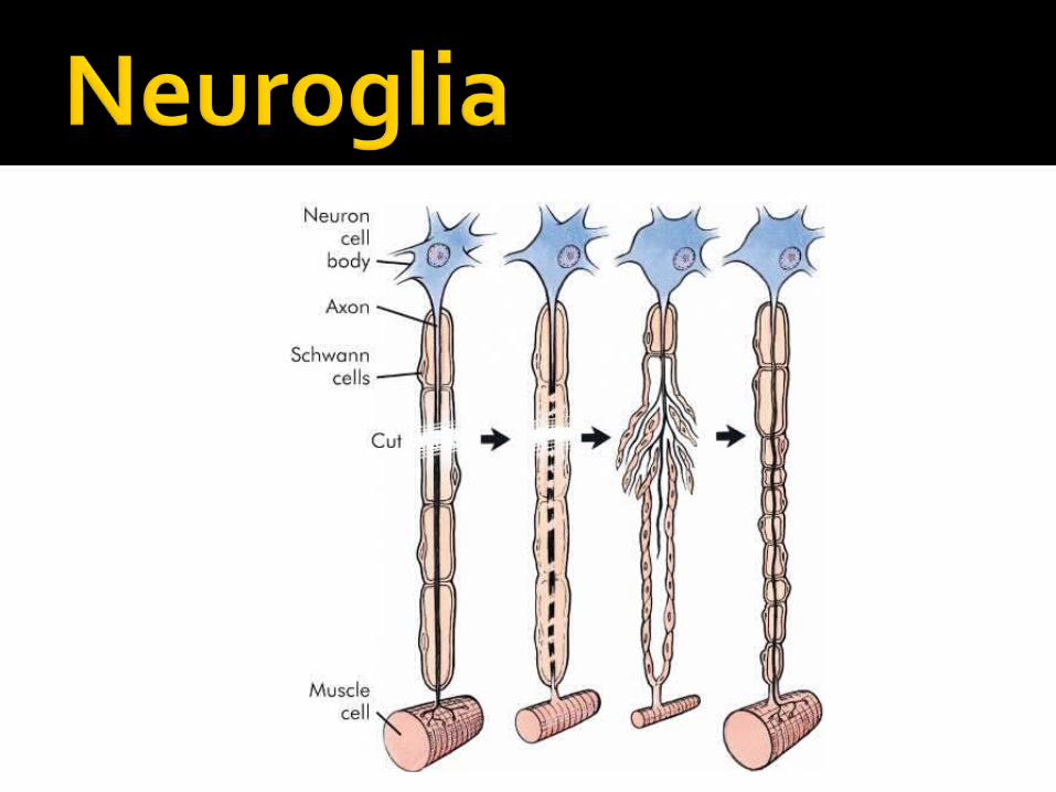
Distal to the injury Wallerian degeneration
▪ Swelling▪ Neurofilaments hypertrophy▪ Myelin sheath shrinks and disintegrates
▪ Axon portion degenerates▪ Myelin sheath ⇨ Schwann cell pathway

Limited to myelinated axons Generally only in the PNS (Schwann cells) CNS limited by ↑ scar formation and
different type of myelin(oligodendrocytes)
Depends upon location, type of injury (crush vs. cut), inflammatory response and scar tissue formation
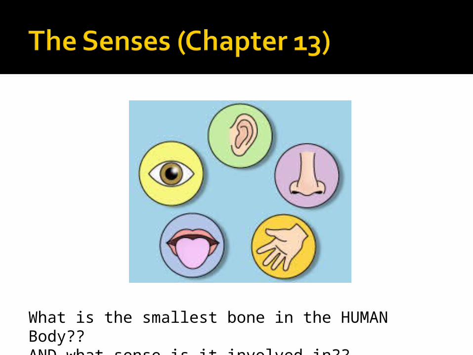
What is the smallest bone in the HUMAN Body?? AND what sense is it involved in??
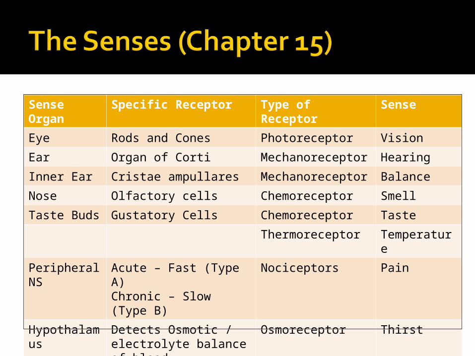
Sense Organ
Specific Receptor Type of Receptor
Sense
Eye Rods and Cones Photoreceptor Vision
Ear Organ of Corti Mechanoreceptor Hearing
Inner Ear Cristae ampullares Mechanoreceptor Balance
Nose Olfactory cells Chemoreceptor Smell
Taste Buds Gustatory Cells Chemoreceptor Taste
Thermoreceptor Temperature
Peripheral NS
Acute – Fast (Type A)Chronic – Slow (Type B)
Nociceptors Pain
Hypothalamus
Detects Osmotic / electrolyte balance of blood.
Osmoreceptor Thirst
a) Muscles b) Tendons
a) Muscle spindlesb) Golgi tendon
receptors
a + b = Proprioceptors(stretch+mechano)
Proprioception

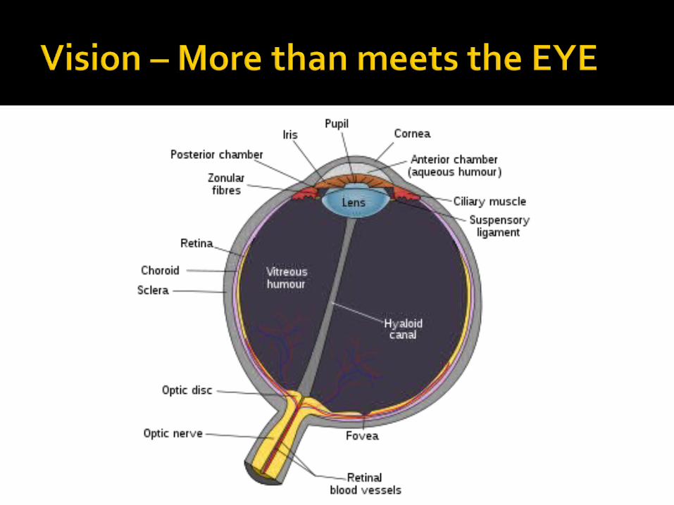

Retina is made of two specialized photoreceptors: RODS and CONES
Do you know the specific function of these two receptors??

The Average human eye has 125 Million Rods and Cones
Rods are the photoreceptors that detect monochromatic image and image quality (black and white)
Cones are the photoreceptors that detect detailed images and have three subgroups to detect the array of color images.
Humans have 18 times as many rods as cones, however the cones are not evenly distributed, instead they are concentrated in the Fovea centralis and center of the retina