
Bio 134 Pathology: Cell injury
37
CELL INJURY Prof. Mark Anthony I. Jose, MSc
-
Upload
shaina-mavreen-villaroza -
Category
Education
-
view
256 -
download
3
Transcript of Bio 134 Pathology: Cell injury

CELL INJURY
Prof. Mark Anthony I. Jose, MSc

Reversible Injury
and
Cell Death

Reversible Injury
– The two main morphologic correlates of reversible cell injury are
cellular swelling and fatty change.
– Cellular swelling is the result of failure of energy-dependent ion
pumps in the plasma membrane, leading to an inability to
maintain ionic and fluid homeostasis.
– Fatty change occurs in hypoxic injury and various forms of toxic or
metabolic injury, and is manifested by the appearance of small or
large lipid vacuoles in the cytoplasm.
– It occurs mainly in cells involved in and dependent on fat
metabolism, such as hepatocytes and myocardial cells.

Cell Death
– With continuing damage, the injury becomes irreversible, at which
time the cell cannot recover and it dies.
– There are two types of cell death— necrosis and apoptosis—which
differ in their morphology, mechanisms, and roles in disease and
physiology.


Causes of Cell Injury
– The causes of cell injury range from the
gross physical trauma of a motor vehicle
accident to the single gene defect that
results in a defective enzyme underlying a
specific metabolic disease.

HYPOXIA
– Also known as OxygenDeprivation or OxygenDeficiency.
– interferes with aerobicoxidative respiration and isan extremely important andcommon cause of cell injuryand death.
– Can be caused by Ischemia,Anemia, Carbon monoxidepoisoning, and pooroxygenation of blood due topulmonary disease

CAUSES OF HYPOXIA
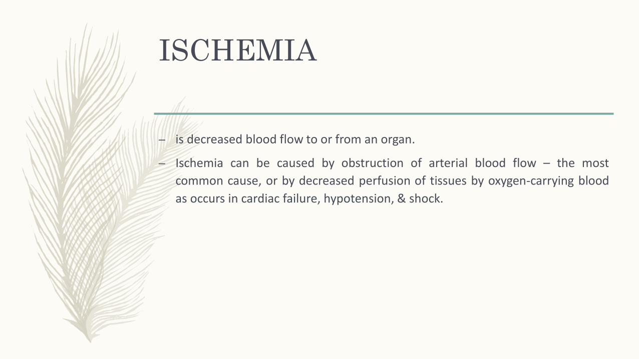
ISCHEMIA
– is decreased blood flow to or from an organ.
– Ischemia can be caused by obstruction of arterial blood flow – the most
common cause, or by decreased perfusion of tissues by oxygen-carrying blood
as occurs in cardiac failure, hypotension, & shock.

ISCHEMIA
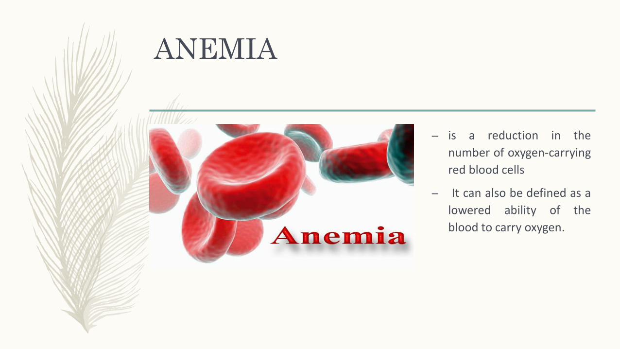
ANEMIA
– is a reduction in the
number of oxygen-carrying
red blood cells
– It can also be defined as a
lowered ability of the
blood to carry oxygen.

CARBON MONOXIDE
POISONING
– CO decreases the oxygen-
capacity of red blood cells
by chemical alteration of
hemoglobin.

Poor oxygenation of blood
due to pulmonary disease
– cause increased carbon dioxide retention,
which may cause drowsiness, headaches,
and in severe cases lack of respiration,
which may lead to death.
– People with lung ailments or with central
respiratory depression, who receive
supplemental oxygen, require careful
monitoring.
CAUSES OF CELL
INJURY
CONTINUATION

Chemical Agents
– An enormous number of chemical substances can injure cells; even innocuous
substances such as glucose or salt, if sufficiently concentrated, can so derange
the osmotic environment that cell injury or death results.
– Agents commonly known as poisons cause severe damage at the cellular level
by altering membrane permeability, osmotic homeostasis, or the integrity of an
enzyme or cofactor, and exposure to these poisons can culminate in the death
of the whole organism.
– Other potentially toxic agents are encountered daily in our environment; these
include air pollutants, insecticides, CO, asbestos, and social “stimuli” such as
ethanol. Even therapeutic drugs can cause cell or tissue injury in a susceptible
patient or if used excessively or inappropriately.

Infectious Agents
– These range from submicroscopic
viruses to meter-long tapeworms;
in between are the rickettsiae,
bacteria, fungi, and protozoans.
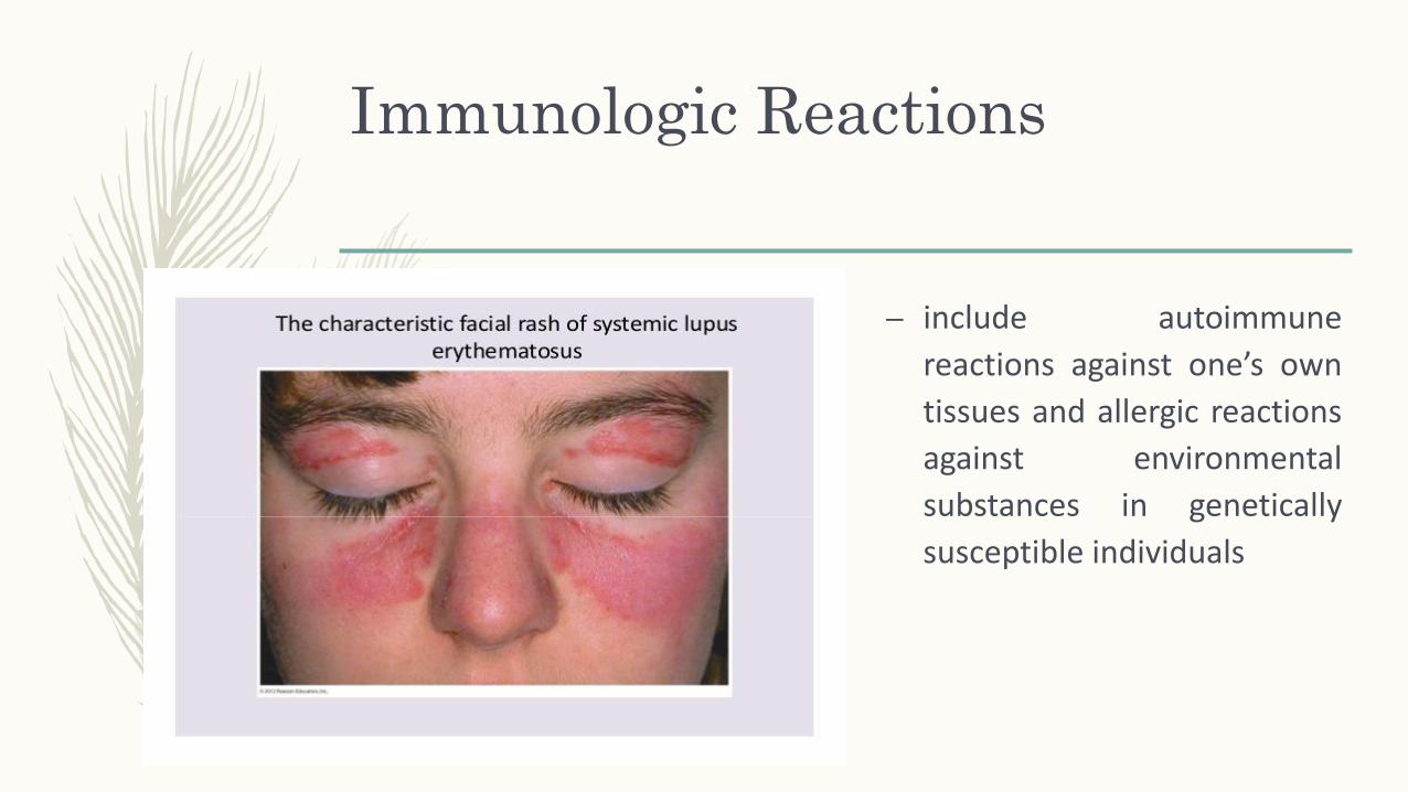
Immunologic Reactions
– include autoimmune
reactions against one’s own
tissues and allergic reactions
against environmental
substances in genetically
susceptible individuals

Genetic Defects
– can result in pathologic changes as conspicuous as the congenital
malformations associated with Down syndrome or as subtle as the
single amino acid substitution in hemoglobin S giving rise to sickle
cell anemia.
– Genetic defects may cause cell injury because of deficiency of
functional proteins, such as enzymes in inborn errors of
metabolism, or accumulation of damaged DNA or misfolded
proteins, both of which trigger cell death when they are beyond
repair.

Nutritional Imbalances
– Protein-calorie insufficiency among underprivileged populations is
only the most obvious example; specific vitamin deficiencies are
not uncommon even in developed countries with high standards
of living.
– Ironically, excesses of nutrition are also important causes of
morbidity and mortality; for example, obesity markedly increases
the risk for type 2 diabetes mellitus.
– Moreover, diets rich in animal fat are strongly implicated in the
development of atherosclerosis as well as in increased
vulnerability to many disorders, including cancer.

Physical Agents
– Trauma, extremes of
temperatures, radiation, electric
shock, and sudden changes in
atmospheric pressure all have
wide-ranging effects on cells

Aging
– Cellular senescence leads to
alterations in replicative and repair
abilities of individual cells and
tissues.
– All of these changes result in a
diminished ability to respond to
damage and, eventually, the death
of cells and of the organism.


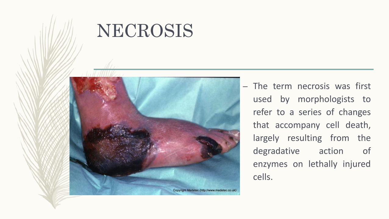
NECROSIS
– The term necrosis was first
used by morphologists to
refer to a series of changes
that accompany cell death,
largely resulting from the
degradative action of
enzymes on lethally injured
cells.

PATTERNS OF
NECROSIS
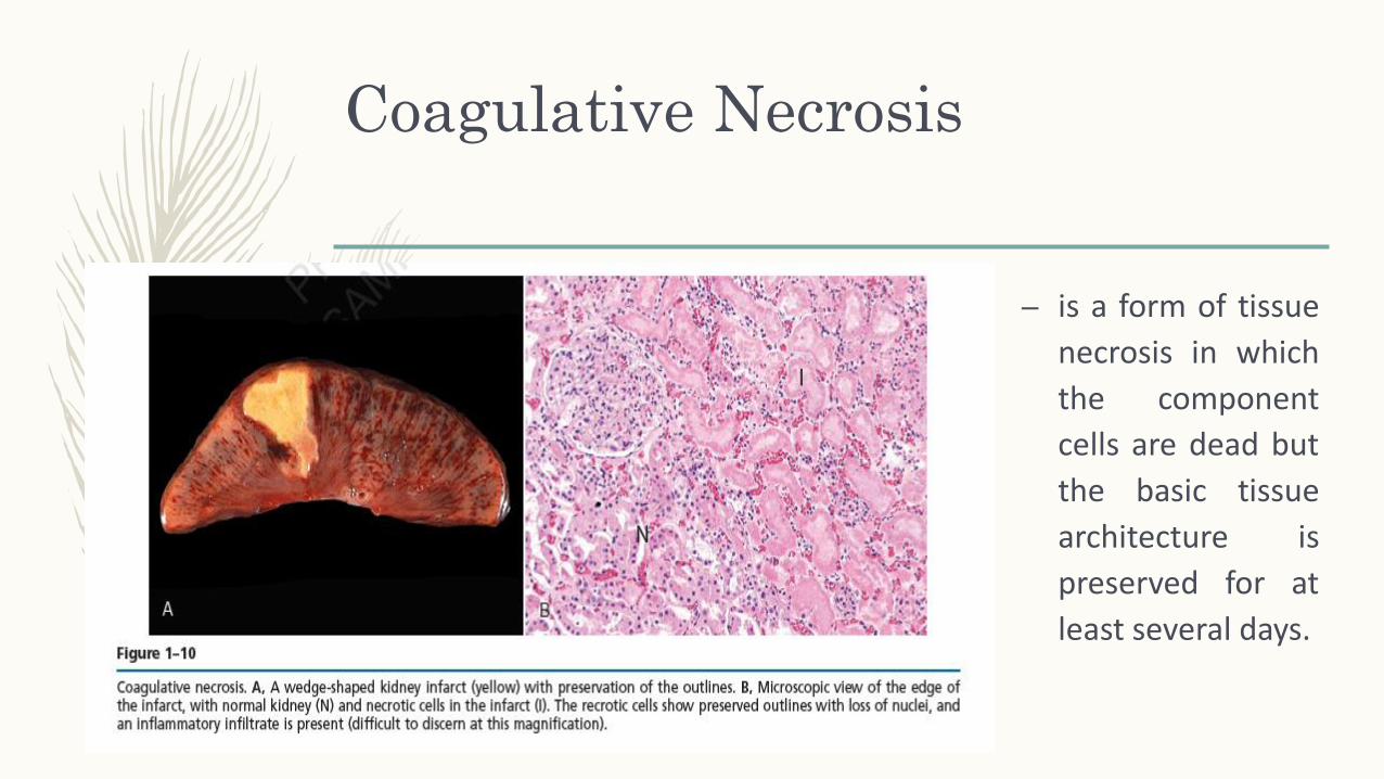
Coagulative Necrosis
– is a form of tissue
necrosis in which
the component
cells are dead but
the basic tissue
architecture is
preserved for at
least several days.

Liquefactive Necrosis
– is seen in focal bacterial or,
occasionally, fungal infections,
because microbes stimulate
the accumulation of
inflammatory cells and the
enzymes of leukocytes digest
(“liquefy”) the tissue.

Gangrenous Necrosis
– is not a distinctive pattern of cell death, theterm is still commonly used in clinicalpractice.
– It is usually applied to a limb, generally thelower leg, that has lost its blood supply andhas undergone coagulative necrosisinvolving multiple tissue layers.
– When bacterial infection is superimposed,coagulative necrosis is modified by theliquefactive action of the bacteria and theattracted leukocytes (so called wetgangrene).

Caseous Necrosis
– is encountered most often in
foci of tuberculous infection.
The term “caseous”
(cheeselike) is derived from
the friable yellow-white
appearance of the area of
necrosis

APOPTOSIS
– is a pathway of cell death
that is induced by a tightly
regulated suicide program in
which cells destined to die
activate enzymes capable of
degrading the cells’ own
nuclear DNA and nuclear and
cytoplasmic proteins.

Causes of Apoptosis


![Pathology Lecture 3, Cell Injury (Continued) [Lecture Notes]](https://static.fdocuments.in/doc/165x107/5525f9b64a7959c2488b4e6a/pathology-lecture-3-cell-injury-continued-lecture-notes.jpg)


