BIO 121 – Molecular Cell Biology Lecture Section IV A. Cells in the Context of Tissue, Organ and...
75
BIO 121 – Molecular Cell Biology Lecture Section IV A. Cells in the Context of Tissue, Organ and Organismal Architecture B. Wound Healing
-
Upload
gary-harrell -
Category
Documents
-
view
214 -
download
1
Transcript of BIO 121 – Molecular Cell Biology Lecture Section IV A. Cells in the Context of Tissue, Organ and...

BIO 121 – Molecular Cell Biology
Lecture Section IV
A. Cells in the Context of Tissue, Organ and Organismal Architecture
B. Wound Healing

Four Types of Vertebrate Tissue
1.Epithelium
2.Connective Tissue
3.Muscle
4.Nervous Tissue

1. Architecture of Epithelium• Simple, Stratified, Pseudostratified, Transitional
• Squamous, Cuboidal, Columnar
• Ciliated or not
• Examples:– Small Intestine = Simple Columnar Epithelium– Trachea = Ciliated Pseudostratified Columnar Epithelium– Blood Vessel = Simple Squamous Epithelium– Skin = Stratified Squamous Epithelium

Structure equals Function
– Small Intestine: Simple Columnar Epithelium = absorption
– Trachea: Ciliated Pseudostratified Columnar Epithelium = filtering debris
– Blood Vessel: Simple Squamous Epithelium = gas exchange
– Skin: Stratified Squamous Epithelium = protective physical barrier

Function: 1. absorption of nutrients2. enzymatic digestion at neutral pH3. multiple defensive mechanisms
Simple,ColumnarEpithelium

4 Cell types in Small Intestine

Small Intestine

Cellular Adhesion in Small Intestine
DesmosomesHemidesmosomesAdherens JunctionsOccluding Junctions

Tracheal Epithelium
Ciliated Pseudostratified Columnar Epithelium
with Goblet Cells
1. Mucus traps dust and air-borne microorganisms
2. Ciliar waving gets rid of unwanted material

The Vasculature: Simple, Squamous Epithelium
Gas Exchange Fluid Exchenge

Epidermis of Skin
Stratified Squamous Epithelium
Creates tough, waterproof
barrier

Differentiation and Direction of Movement in Epidermis
Cornification is the over-
production of cytokeratins, ECM and the adhesions to a degree that stops cellular metabolism.


2. Mesenchymal Cell Types and Connective Tissues

Figure 23-52 Molecular Biology of the Cell (© Garland Science 2008)

The Fibroblast

Loose Connective Tissue
Dense Regular CT
Dense Irregular CT
Elastic Connective Tissue

The dermis is as complex as the epidermis and contributes greatly to skin function
Cartilage and the Chondrocyte

Lacunar Structure of the Hyaline Cartilage
Extremely low blood flow

Lacunar structure of the long bones
Osteoblasts

Cortical Bone vs. Spongy Bone

Marrow of Long Bones has Stem Cells
Cell Types of the Bone

Start out as cartilage models built by chondrocytes
Chondrocytes hypertrophy, calcify and die
Osteoblasts and osteoclasts finish up

The Adipocyte
MesenchymalStem Cells are a
continuous source of adipocytes

Figure 23-47a Molecular Biology of the Cell (© Garland Science 2008)
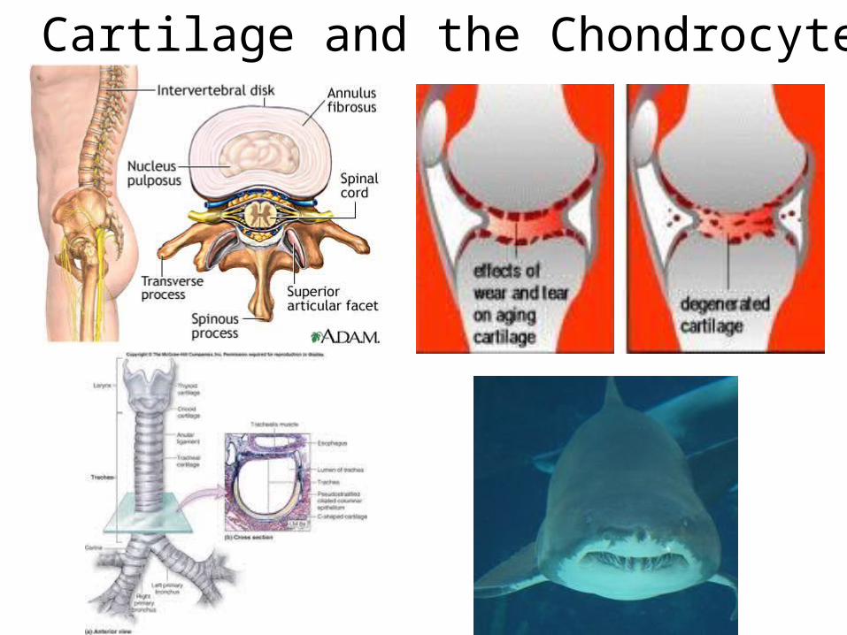
3. Contractile Tissue


Arteries, veinsLymphatic vesselsGastrointestinal tractRespiratory tractUrinary bladderReproductive tractUrinary tractIris of the eyeErector pili of skin

4. Nervous Tissue

Nerve Bundles

Cutaneous Wound Healing
The skin is a complex organ...

Many cells and activities involved

Many cells and activities involved in Healing
Clotting
Scarring
Re-establishingFunction

• Four overlapping stages to wound healing
– Hemostasis
– Inflammation
– Proliferation
– Maturation

Blood flows into the exposed ECM of the injured tissue.

RBC and Platelets Trapped in Fibrin Clot

Clotting factor VII from the
blood contacts tissue factor on
cells in the damaged tissues to
activate clotting

.
Camacho A , Dimsdale J E Psychosom Med 2000;62:326-336©2000 by Lippincott Williams & Wilkins
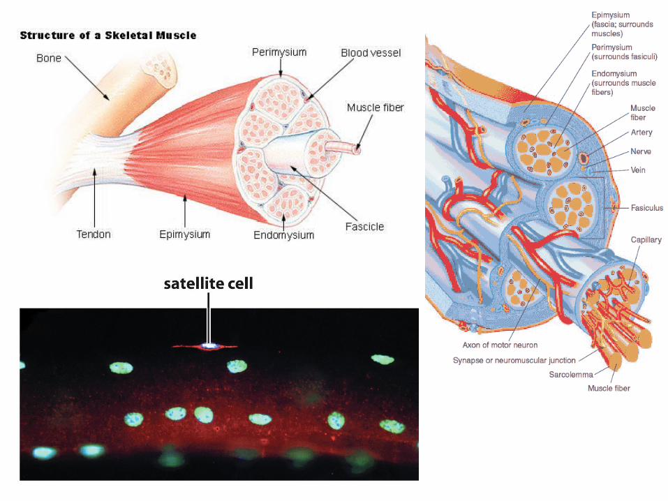
Platelet activation in the clot makes them sticky and releases their signal storage vesicles

Positive feedbackactivates even more
Platelet activation releases growth
factors by regulated secretion


Inflammation is a process mediated primarily by WBC as part of our innate immunity
- Resident mast cells and macrophages
- Recruited monocytes and neutrophils

Resident mast cells also degranulate
rubor = rednesscalor = heattumor = swellingdolor = pain

Activated mast cell activities

Figure 1 Development and differentiation of macrophages.
Rickard A J , Young M J J Mol Endocrinol 2009;42:449-459
© 2011 Society for Endocrinology

Activated macrophage activities

Neutrophil Diapedesis


Activated neutrophils are phagocytic

Proliferation re-establishes tissue function
• Reconnection of the dermal connective tissue
• Integrity of the epidermal layers
• Re-establishment of blood flow

Reconnection of the dermal CT

Cell Migration or “Crawling”• The Basic Mechanism
– Triggered by signals from outside the cell– Actin-myosin based movement– Requires attachments to outside to pull against– Gotta’ drag all of the cell contents along for the ride

Chemotaxis
Circumferential receptors
Rho-family GTPases (monomeric)
Rho-dependent kinases
1. Actin monomer nucleotide exchange 2. Actin fiber polymerization and disassembly 3. Myosin motor ATPase activity

Figure 17-62 (part 1 of 3) Molecular Biology of the Cell (© Garland Science 2008)

Formation of the scar matrix1. glycosaminoglycans2. proteoglycans 3. fibrous proteins 4. elastic proteins

Re-establishment of the epidermal epithelium involves both mitosis
and epithelial migration

Also must reform the basal lamina

Re-epithelialization below the scab
scar
Fi

Model depicting α3β1-integrin-mediated functions of epidermis that contribute to wound healing.
Mitchell K et al. J Cell Sci 2009;122:1778-1787
©2009 by The Company of Biologists Ltd


Figure 23-34 Molecular Biology of the Cell (© Garland Science 2008)

Maturation Phase

Wound contraction by myofibroblasts

Stitches Perform Wound Contracture

Collagen Remodeling

A scar never reaches the strength of undamaged tissue

Healing Abnormalities
• Failure to heal: Excessive Inflammation
• Excessive scarring: Wound Fibrosis– Hypertrophic Scarring– Keloid Scarring


Biofilms May Block Healing


Hypertrophic scars result from failed fibroblast contracture
Don’t extend beyond the original wound edge

Keloid scars result from excessive TGF-b receptors on fibroblasts
Extend to fibroblasts outside the wound

People have exploited these conditions to create the ‘keloid tattoo’