binds with high affinity to isolated alveolar type II cells
5
Proc. Natl. Acad. Sci. USA Vol. 86, pp. 5410-5414, July 1989 Cell Biology Lung surfactant apoprotein SP-A (26-36 kDa) binds with high affinity to isolated alveolar type II cells (receptors/epithelial celis/lipoproteins) Jo RAE WRIGHT*t, JENNY D. BORCHELT*, AND SAMUEL HAWGOOD*t *Cardiovascular Research Institute and the Departments of tPhysiology and SPediatrics, University of California, San Francisco, CA 94143 Communicated by John A. Clements, March 24, 1989 (received for review May 24, 1988) ABSTRACT Pulmonary surfactant is synthesized and se- creted by alveolar type H cells. These cells recycle surfactant lipids by an internalization process that is enhanced in vitro by the surfactant proteins with molecular masses of 26-36 kDa (SP-A). SP-A also inhibits the secretion of lipid by type II cells. These results suggest that SP-A may play a role in feedback regulation of surfactant pool size and are consistent with the hypothesis that the type II cell surface has receptors for SP-A. The goal of this study is to characterize the binding of radio- iodinated SP-A to isolated rat type II cells. Binding of SP-A to type II cells at 40C has a K112 of approximately 5 X 10⁻¹⁰ M, is saturable, and is inhibited by excess unlabeled SP-A. Binding is dependent on calcium and is reduced by heat treatment of SP-A. The binding of a proteolytic fragment of SP-A that is produced by collagenase treatment is reduced by excess unla- beled SP-A. The binding of the fragment to macrophages and lung fibroblasts is not inhibited by excess unlabeled SP-A. Trypsinization of the type II cell surface reduces the binding of both intact SP-A and the collagenase-resistant fragment. These results show that SP-A binds to type II cells with high affinity and suggest that these cells have receptors that recognize the carboxyl-terminal domain of SP-A. Lung surfactant is synthesized by the alveolar type II cell and stored intracellularly in lamellar bodies until it is secreted into the alveolar air space. Once in the alveoli, the secreted lamellar body contents can form tubular myelin, which is thought to be a precursor to the monomolecular surface film (for review, see ref. 1). Several studies suggest that alveolar surfactant can be internalized by type II cells, incorporated into lamellar bodies, and eventually resecreted (2-5). It has been calculated that between 25 and 95% of the alveolar surfactant enters this "recycling" pathway (6, 7). Results from recent studies suggest that the major surfac- tant protein group of 26- to 36-kDa SP-A (previously referred to as SP 26-36) can enhance the uptake of lipids by type II cells (5, 8, 9). SP-A has also been shown to inhibit the secretion of phosphatidylcholine by type II cells in primary culture (10-12). These results suggest that SP-A may play an important role in regulating alveolar surfactant pool size by balancing secretion and reuptake. These results also suggest that SP-A may interact with receptors on type II cells. The goal of the work reported here was to characterize the interaction of SP-A with isolated alveolar type II cells. A preliminary report of this work has appeared in abstract form (13). While this manuscript was in preparation, Kuroki et al. (14) reported that isolated type II cells that have been maintained in primary culture bind SP-A with high affinity. MATERIALS AND METHODS Materials. Agarose, Bio-Gel P-6DG, and Enzymobeads were obtained from Bio-Rad. Sephacryl S-200 and heparin- Sepharose CL-6B were obtained from Pharmacia. Elastase was obtained from Cooper Biomedical. Collagenase was purchased from Worthington. Media and serum were ob- tained from the University of California Cell Culture Facility. Na1251 was obtained from Amersham. All other enzymes and chemicals were obtained from Sigma. Isolation, lodination, and Analysis of SP-A and Its Collage- nase-Resistant Fragment (CRF). SP-A was isolated from rat lung lavage and the lavage fluid of a patient with alveolar proteinosis as described in detail (9). CRF was isolated from human SP-A because only small amounts of rat SP-A can be obtained by lung lavage (8-10 pug per lung). We found no qualitative differences between binding of rat SP-A and human SP-A (i.e., binding of both was saturable, required calcium, and was inhibited by excess unlabeled SP-A). CRF was obtained by digesting human SP-A with collagenase from Clostridium histolyticum. Non-specific protease contami- nants were removed from the collagenase by chromatogra- phy over heparin-Sepharose CL-6B and Sephacryl S-200 as described (15). SP-A was incubated with collagenase in 5 mM Tris-HCl/5 mM CaCl2, pH 7.40 at 370C for 16 hr. After digestion the sample was centrifuged (100,000 X g, 60 min, SW 40 rotor), the supernatant was applied to a Sephacryl S-200 column, and material was eluted in 5 mM Tris-HCl/100 mM NaCl, pH 7.40 to separate CRF from intact SP-A. Protein was measured by the method of Lowry et al. (16). SP-A and CRF were iodinated using Bio-Rad Enzymo- beads according to the manufacturer's directions. Rat or human SP-A (75-300 Ag) in 5 mM Tris HCl (pH 7.40) or human CRF in 5 mM Tris HCI/154 mM NaCl, pH 7.40 was incubated at room temperature with 0.5-1 mCi Na1251 for 15-20 min (1 Ci = 37 GBq). Free Na125I was separated from 125I-labeled protein on 8-ml columns of Bio-Gel P-6DG that had been pre-eluted with 20-50 pug of the appropriate protein. Fractions that contained radioactivity greater than 88% pre- cipitable by trichloroacetic acid were combined and then stored at 40C. Specific activities ranged from 0.2 to 2 ACi/ttg. Radiolabeled SP-A was used within 2-3 weeks after the iodination; storage for longer periods of time reduced binding of protein to cells. Radiolabeled CRF was used within 1-2 weeks. NaDodSO4/PAGE was performed on slab gels as de- scribed (8). All samples were reduced with 50 mM dithio- threitol. Gels were stained with Coomassie blue, dried, and exposed to Kodak diagnostic film X-OMAT AR in a Kodak X-OMATIC cassette with intensifying screens at -70'C. Abbreviations: SP-A, surfactant proteins of 26-36 kDa; CRF, col- lagenase-resistant fragment of SP-A. 5410 The publication costs of this article were defrayed in part by page charge payment. This article must therefore be hereby marked "advertisement" in accordance with 18 U.S.C. §1734 solely to indicate this fact.
Transcript of binds with high affinity to isolated alveolar type II cells

Proc. Natl. Acad. Sci. USAVol. 86, pp. 5410-5414, July 1989Cell Biology
Lung surfactant apoprotein SP-A (26-36 kDa) binds with highaffinity to isolated alveolar type II cells
(receptors/epithelial celis/lipoproteins)
Jo RAE WRIGHT*t, JENNY D. BORCHELT*, AND SAMUEL HAWGOOD*t*Cardiovascular Research Institute and the Departments of tPhysiology and SPediatrics, University of California, San Francisco, CA 94143
Communicated by John A. Clements, March 24, 1989 (receivedfor review May 24, 1988)
ABSTRACT Pulmonary surfactant is synthesized and se-creted by alveolar type H cells. These cells recycle surfactantlipids by an internalization process that is enhanced in vitro bythe surfactant proteins with molecular masses of 26-36 kDa(SP-A). SP-A also inhibits the secretion of lipid by type II cells.These results suggest that SP-A may play a role in feedbackregulation of surfactant pool size and are consistent with thehypothesis that the type II cell surface has receptors for SP-A.The goal of this study is to characterize the binding of radio-iodinated SP-A to isolated rat type II cells. Binding of SP-A totype II cells at 40C has a K112 of approximately 5 X 10⁻¹⁰ M,is saturable, and is inhibited by excess unlabeled SP-A. Bindingis dependent on calcium and is reduced by heat treatment ofSP-A. The binding of a proteolytic fragment of SP-A that isproduced by collagenase treatment is reduced by excess unla-beled SP-A. The binding of the fragment to macrophages andlung fibroblasts is not inhibited by excess unlabeled SP-A.Trypsinization of the type II cell surface reduces the binding ofboth intact SP-A and the collagenase-resistant fragment. Theseresults show that SP-A binds to type II cells with high affinityand suggest that these cells have receptors that recognize thecarboxyl-terminal domain of SP-A.
Lung surfactant is synthesized by the alveolar type II cell andstored intracellularly in lamellar bodies until it is secreted intothe alveolar air space. Once in the alveoli, the secretedlamellar body contents can form tubular myelin, which isthought to be a precursor to the monomolecular surface film(for review, see ref. 1). Several studies suggest that alveolarsurfactant can be internalized by type II cells, incorporatedinto lamellar bodies, and eventually resecreted (2-5). It hasbeen calculated that between 25 and 95% of the alveolarsurfactant enters this "recycling" pathway (6, 7).
Results from recent studies suggest that the major surfac-tant protein group of 26- to 36-kDa SP-A (previously referredto as SP 26-36) can enhance the uptake of lipids by type IIcells (5, 8, 9). SP-A has also been shown to inhibit thesecretion of phosphatidylcholine by type II cells in primaryculture (10-12). These results suggest that SP-A may play animportant role in regulating alveolar surfactant pool size bybalancing secretion and reuptake. These results also suggestthat SP-A may interact with receptors on type II cells.The goal of the work reported here was to characterize the
interaction of SP-A with isolated alveolar type II cells. Apreliminary report of this work has appeared in abstract form(13). While this manuscript was in preparation, Kuroki et al.(14) reported that isolated type II cells that have beenmaintained in primary culture bind SP-A with high affinity.
MATERIALS AND METHODSMaterials. Agarose, Bio-Gel P-6DG, and Enzymobeads
were obtained from Bio-Rad. Sephacryl S-200 and heparin-Sepharose CL-6B were obtained from Pharmacia. Elastasewas obtained from Cooper Biomedical. Collagenase waspurchased from Worthington. Media and serum were ob-tained from the University of California Cell Culture Facility.Na1251 was obtained from Amersham. All other enzymes andchemicals were obtained from Sigma.
Isolation, lodination, and Analysis of SP-A and Its Collage-nase-Resistant Fragment (CRF). SP-A was isolated from ratlung lavage and the lavage fluid of a patient with alveolarproteinosis as described in detail (9). CRF was isolated fromhuman SP-A because only small amounts of rat SP-A can beobtained by lung lavage (8-10 pug per lung). We found noqualitative differences between binding of rat SP-A andhuman SP-A (i.e., binding of both was saturable, requiredcalcium, and was inhibited by excess unlabeled SP-A). CRFwas obtained by digesting human SP-A with collagenase fromClostridium histolyticum. Non-specific protease contami-nants were removed from the collagenase by chromatogra-phy over heparin-Sepharose CL-6B and Sephacryl S-200 asdescribed (15). SP-A was incubated with collagenase in 5 mMTris-HCl/5 mM CaCl2, pH 7.40 at 370C for 16 hr. Afterdigestion the sample was centrifuged (100,000 X g, 60 min,SW 40 rotor), the supernatant was applied to a SephacrylS-200 column, and material was eluted in 5 mM Tris-HCl/100mM NaCl, pH 7.40 to separate CRF from intact SP-A.Protein was measured by the method of Lowry et al. (16).SP-A and CRF were iodinated using Bio-Rad Enzymo-
beads according to the manufacturer's directions. Rat orhuman SP-A (75-300 Ag) in 5 mM Tris HCl (pH 7.40) orhuman CRF in 5 mM Tris HCI/154 mM NaCl, pH 7.40 wasincubated at room temperature with 0.5-1 mCi Na1251 for15-20 min (1 Ci = 37 GBq). Free Na125I was separated from125I-labeled protein on 8-ml columns of Bio-Gel P-6DG thathad been pre-eluted with 20-50 pug ofthe appropriate protein.Fractions that contained radioactivity greater than 88% pre-cipitable by trichloroacetic acid were combined and thenstored at 40C. Specific activities ranged from 0.2 to 2 ACi/ttg.Radiolabeled SP-A was used within 2-3 weeks after theiodination; storage for longer periods of time reduced bindingof protein to cells. Radiolabeled CRF was used within 1-2weeks.NaDodSO4/PAGE was performed on slab gels as de-
scribed (8). All samples were reduced with 50 mM dithio-threitol. Gels were stained with Coomassie blue, dried, andexposed to Kodak diagnostic film X-OMAT AR in a KodakX-OMATIC cassette with intensifying screens at -70'C.
Abbreviations: SP-A, surfactant proteins of 26-36 kDa; CRF, col-lagenase-resistant fragment of SP-A.
5410
The publication costs of this article were defrayed in part by page chargepayment. This article must therefore be hereby marked "advertisement"in accordance with 18 U.S.C. §1734 solely to indicate this fact.
Proc. Natl. Acad. Sci. USA 86 (1989) 5411
The molecular size ofSP-A was determined by gel filtrationchromatography. Samples (0.1 ml) containing 1-10 jug wereapplied to a 1.9 x 90 cm, 4% agarose column and eluted with5 mM Tris HCl/154 mM NaCl, pH 7.40 at room temperatureat a flow rate of 8 ml/hr. Fractions of 2.5 ml were collectedand analyzed for radioactivity. The elution profiles of thestandards blue dextran, thyroglobulin, apoferritin, catalase,and albumin were monitored by measuring absorbance at 280nm.
Isolation and Primary Culture of Lung Cells. Type II cells(17) and macrophages (9) were isolated by described meth-ods. Type II cells and macrophages were either used on theday of isolation (freshly isolated cells) or they were placed inprimary culture. For primary culture, the cells were resus-pended in Dulbecco's modified Eagle's medium (DMEM)containing 10o (vol/vol) fetal bovine serum and gentamicin(50 pg/ml). Cells were plated in either 96-well plates at 5 x10⁵ cells per 0.25 ml per well or in 24-well plates at 106 cellsper 0.5 ml per well. The cells were incubated for 16-18 hr inan atmosphere of 10% CO2/90M air.
Fibroblasts were prepared from rat lung tissue. Lungs fromtwo rats were minced finely and incubated with occasionalagitation in 20 ml of Dulbecco's phosphate-buffered salinecontaining 0.2% trypsin (type III, from bovine pancreas) and0.006% DNase I at 370C for 40 min, after which time 25 ml ofphosphate-buffered saline (40C, pH 7.40) containing 0.1%trypsin inhibitor (type II-S from soybean) was added to theminced lung tissue. The digest was centrifuged at 140 x g for10 min and the pellet was resuspended in 20 ml of DMEMcontaining 4.5 g of glucose/liter, 1o fetal bovine serum, andgentamicin (50 Ag/ml). The cell suspension was filteredthrough two layers of surgical gauze and the cells were thencultured at 1-2 x 10⁶ cells per 100-mm tissue culture dish.Cells were allowed to grow to confluence and passaged onto75-cm2 tissue culture plates. Cells were routinely used be-tween passages 5 and 15.On the day before the binding assay was performed,
fibroblasts were removed from flasks by trypsin treatmentand were resuspended in plating medium and plated in 96- or24-well plates as described above.
Trypsin Treatment of Type II Cells. For some studies,freshly isolated type II cells were suspended in Dulbecco'sphosphate-buffered saline containing 0.025% trypsin (frombovine pancreas, type III) and incubated with gentle agitationat room temperature for 10 min, after which time fetal calfserum or trypsin inhibitor (from soybean, type II-S) wasadded to a final concentration of 20%o or 0.05%, respectively.Cells and medium were separated by centrifugation and cellswere resuspended in 1 ml of the appropriate medium con-taining radiolabeled protein. Control cells were treated in anidentical fashion except that trypsin was not included in theincubation medium.
Binding Assays with Freshly Isolated Cells. From 2 to 2.5 X10⁶ cells were resuspended at 4°C in a custom-formulatedmedium containing modified Krebs improved salts (18) (1.6mM K2HPO4/5.4 mM KCI/1.29 mM MgC12-6H20/112 mMNaCl/4 mM NaHCO3/15.7 mM sodium phosphate, pH 7.40),13 mM glucose, 25 mM Hepes, 2 mM sodium pyruvate, andEagle's minimal essential medium (binding medium). Thismedium contains a low concentration ofadded calcium (2AMas calcium pantothenate). Bovine serum albumin (0.1%,fraction V) was included in the binding medium becausealbumin reduced adherence of SP-A to microcentrifuge tubesand tissue culture plastic ware but did not alter the binding ofSP-A to lung cells. Cells were incubated in a microcentrifugetube and mixed every 7-10 min. At the end of the incubationperiod, cells and medium were separated by centrifugation at140 x g for 10 min in a Beckman model 12 Microfuge inhorizontal tube holders in a cold room. The medium wasremoved and the cells were gently resuspended in a small
volume of medium, transferred to a fresh tube, and thenwashed and collected by centrifugation twice. The final cellpellet was resuspended in 0.5 ml of medium; an aliquot wasremoved for determination of cell number (in duplicate) in aNeubauer counting chamber.Samples without cells were carried through the procedure
concurrently and the amount of radioactivity that sedimentedor remained associated with the centrifuge tubes in theabsence of cells was determined and was subtracted from thesamples with cells. This correction increased with increasingprotein concentration and was generally 5-20%.
Binding Assays with Adherent Cells. Because only smallamounts of rat SP-A can be obtained by lung lavage, amicroassay with adherent cells in primary culture was de-veloped to assess the potential inhibitory role of excessunlabeled SP-A. (The advantage of this assay is that fewercells and smaller volumes of medium can be used than arerequired for the binding assays using cells in suspension. Itwas necessary to use a minimum of2 x 106 cells in the bindingassays with freshly isolated cells to minimize losses duringwashing and centrifugation.) After the cells had been allowedto adhere for 16-18 hr, they were removed from the CO2incubator, placed in room air on ice, and washed four timeswith binding medium. Fresh binding medium containing 0.1%bovine serum albumin was added to the wells (0.2 ml per wellof a 24-well plate or 0.1 ml per well of a 96-well plate). At theend ofthe incubation with radiolabeled protein, the cells werewashed four times with binding medium and the cells weredissolved in 0.1 M NaOH and analyzed for radioactivity.Binding ofSP-A to both freshly isolated type II cells and cellsin primary culture required calcium, was inhibited by excessunlabeled SP-A, and was saturable.
RESULTSCharacterization of Radiolabeled SP-A and CRF. A repre-
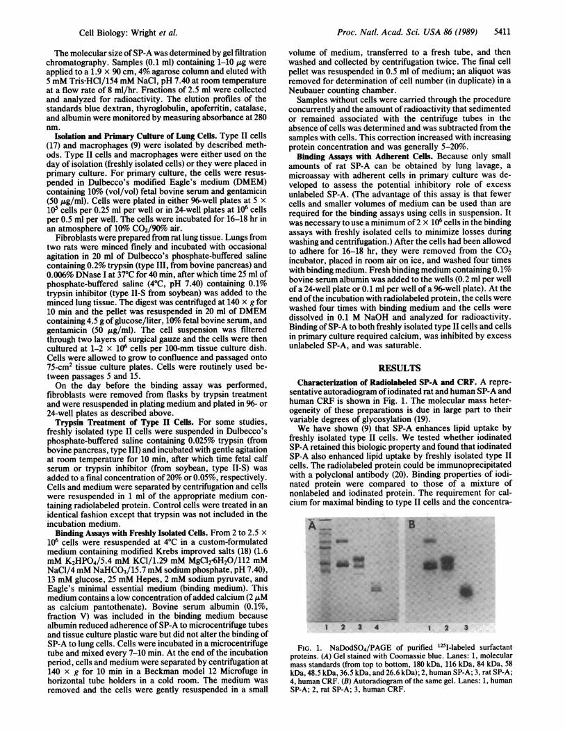
sentative autoradiogram ofiodinated rat and human SP-A andhuman CRF is shown in Fig. 1. The molecular mass heter-ogeneity of these preparations is due in large part to theirvariable degrees of glycosylation (19).We have shown (9) that SP-A enhances lipid uptake by
freshly isolated type II cells. We tested whether iodinatedSP-A retained this biologic property and found that iodinatedSP-A also enhanced lipid uptake by freshly isolated type IIcells. The radiolabeled protein could be immunoprecipitatedwith a polyclonal antibody (20). Binding properties of iodi-nated protein were compared to those of a mixture ofnonlabeled and iodinated protein. The requirement for cal-cium for maximal binding to type II cells and the concentra-
A B~~As......
3 1 2 3
FIG. 1. NaDodSO4/PAGE of purified 'l25-labeled surfactantproteins. (A) Gel stained with Coomassie blue. Lanes: 1, molecularmass standards (from top to bottom, 180 kDa, 116 kDa, 84 kDa, 58kDa, 48.5 kDa, 36.5 kDa, and 26.6 kDa); 2, human SP-A; 3, rat SP-A;4, human CRF. (B) Autoradiogram of the same gel. Lanes: 1, humanSP-A; 2, rat SP-A; 3, human CRF.
Cell Biology: Wright et A

Proc. Natl. Acad. Sci. USA 86 (1989)
-650,000
120-co
0
X 80-E-o
N9 40-
0
Vo
40
Fraction
VI
75
Un
Co00
0)C
-6C
.0l 25a-C,
tUr60 80
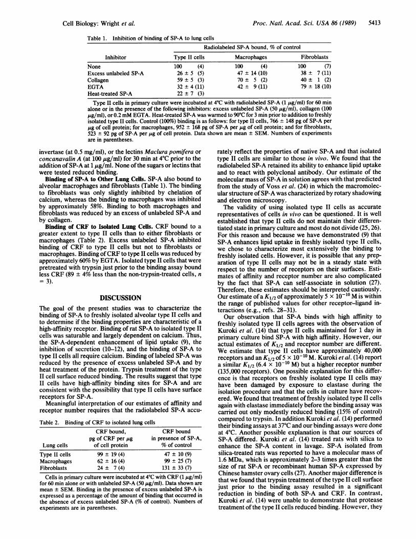
FIG. 2. Analysis of 1251-labeled rat SP-A by gel filtration chro-matography. The elution position is indicated for blue dextran by VO(void volume), for thyroglobulin by -650,000, and for the includedvolume by V1. Recovery of radioactivity was approximately 70%o.This profile is representative of those obtained for four preparations.
tion at which binding reached a plateau with rat SP-A weresimilar with either preparation (data not shown).
lodinated rat SP-A was sized by gel filtration chromatog-raphy (Fig. 2). Ninety percent of the recovered material waseluted with an approximate molecular mass of 650 kDa.Approximately 10% of the material was eluted with theincluded volume, was not precipitable with trichloroaceticacid, and was probably mostly free 1251. Occasionally a verysmall amount (less than 1% of the recovered material) waseluted in the void volume. Similar results were obtained whenthe elution was performed at 40C.
Binding of SP-A to Freshly Isolated Type II Cells. Bindingof rat SP-A was rapid. We estimated that approximately 2 minelapsed between the addition of SP-A and the beginning ofcentrifugation, and thus our first time point was obtained atapproximately 2 min. Binding reached maximal levels within20 min (Fig. 3). The binding of SP-A increased rapidly at lowconcentrations and less rapidly at higher concentrations (Fig.4). Specific high-affinity binding was calculated using theslope-peeling method described by Goldstein and Brown(21). An analysis of the binding as described by Klotz (22)(Fig. 4 Inset) reveals that half-maximal binding occurs at 5 x10⁻¹⁰ M and that there are approximately 40,000 SP-Abinding sites per cell. Estimates of specific binding of SP-A
0 20
Time, min40 60
FIG. 3. Time course of binding of rat SP-A (1 ug/ml) to freshlyisolated type II cells. Cells (2-2.5 x 10⁶ cells per ml of bindingmedium) were incubated at 40C. Cells were separated from themedium and washed by centrifugation. Data shown are mean ±SEM, n = four to six experiments.
to type II cells in primary culture were similar when calcu-lated either by slope peeling or by measuring binding in thepresence of excess unlabeled SP-A (data not shown).
Inhibitors of Binding to Type II Cells. Heating SP-A at 90'Cfor 3 min reduced binding by 75-80%. Binding was largelydependent on calcium since inclusion of EGTA in the me-dium reduced binding by approximately 70% (Table 1).Excess unlabeled SP-A reduced the binding of iodinated ratSP-A both to freshly isolated type II cells (data not shown)and to isolated type II cells in primary culture by approxi-mately 74%. Collagen (type IV, from human placenta) re-duced binding to a lesser extent. Freshly isolated type II cellsthat were pretreated with trypsin just prior to the bindingassay bound less SP-A than control cells (50 + 14% less thanthe non-trypsin-treated cells, n = 5).
Since SP-A is a lectin (23), the effects of carbohydrates andlectins that could potentially compete for binding sites weretested. Cells were preincubated with mannose, galactose,glucose, or N-acetylglucosamine (at 60 mM), mannan or
FIG. 4. Concentration-dependent binding ofrat SP-A to freshly isolated type II cells. Incu-bation condition and analyses were as describedin Fig. 3. Specific binding (o) was calculated bysubtracting nonspecific binding (as estimated bycurve peeling) from total binding (0). Datashown are mean + SEM, n = 3. (Inset) Klotzplot of the binding data. The x axis is the log ofthe concentration free SP-A (ng/ml), and the yaxis is the amount of specifically bound SP-A(ng per 106 cells).
751
50C
25C.
axCD0
0 200CL~0)aC
0.0
C/)
2 3log free
4
I
5412 Cell Biology: Wright et al.
SP-A Aiglml
Proc. Natl. Acad. Sci. USA 86 (1989) 5413
Table 1. Inhibition of binding of SP-A to lung cells
Radiolabeled SP-A bound, % of control
Inhibitor Type II cells Macrophages Fibroblasts
None 100 (4) 100 (4) 100 (7)Excess unlabeled SP-A 26 ± 5 (5) 47 ± 14 (10) 38 ± 7 (11)Collagen 59 ± 5 (3) 70 ± 5 (2) 40 ± 1 (2)EGTA 32 ± 4 (11) 42 ± 9 (11) 79 ± 18 (10)Heat-treated SP-A 22 ± 7 (3)Type II cells in primary culture were incubated at 40C with radiolabeled SP-A (1 j.g/ml) for 60 min
alone or in the presence of the following inhibitors: excess unlabeled SP-A (50 jzg/ml), collagen (100jig/ml), or 0.2mM EGTA. Heat-treated SP-A was warmed to 900C for 3 min prior to addition to freshlyisolated type II cells. Control (100%o) binding is as follows: for type II cells, 766 ± 148 pg of SP-A perjig of cell protein; for macrophages, 952 ± 168 pg of SP-A per jLg of cell protein; and for fibroblasts,523 ± 92 pg of SP-A per jIg of cell protein. Data shown are mean ± SEM. Numbers of experimentsare in parentheses.
invertase (at 0.5 mg/ml), or the lectins Maclura pomifera or
concanavalin A (at 100 jig/ml) for 30 min at 4°C prior to theaddition ofSP-A at 1 jg/ml. None ofthe sugars or lectins thatwere tested reduced binding.
Binding of SP-A to Other Lung Cells. SP-A also bound toalveolar macrophages and fibroblasts (Table 1). The bindingto fibroblasts was only slightly inhibited by chelation ofcalcium, whereas the binding to macrophages was inhibitedby approximately 58%. Binding to both macrophages andfibroblasts was reduced by an excess of unlabeled SP-A andby collagen.
Binding of CRF to Isolated Lung Cells. CRF bound to a
greater extent to type II cells than to either fibroblasts or
macrophages (Table 2). Excess unlabeled SP-A inhibitedbinding of CRF to type II cells but not to fibroblasts ormacrophages. Binding ofCRF to type II cells was reduced byapproximately 60% by EGTA. Isolated type II cells that werepretreated with trypsin just prior to the binding assay boundless CRF (89 ± 4% less than the non-trypsin-treated cells, n
= 3).
DISCUSSIONThe goal of the present studies was to characterize thebinding of SP-A to freshly isolated alveolar type II cells andto determine if the binding properties are characteristic of a
high-affinity receptor. Binding of rat SP-A to isolated type IIcells was saturable and largely dependent on calcium. Thus,the SP-A-dependent enhancement of lipid uptake (9), theinhibition of secretion (10-12), and the binding of SP-A totype II cells all require calcium. Binding of labeled SP-A wasreduced by the presence of excess unlabeled SP-A and byheat treatment of the protein. Trypsin treatment of the typeII cell surface reduced binding. The results suggest that typeII cells have high-afflinity binding sites for SP-A and are
consistent with the possibility that type II cells have surfacereceptors for SP-A.
Meaningful interpretation of our estimates of affinity andreceptor number requires that the radiolabeled SP-A accu-
Table 2. Binding of CRF to isolated lung cellsCRF bound,
pg of CRF per jugCRF bound
in presence of SP-A,Lung cells of cell protein % of control
Type II cells 99 ± 19 (4) 47 ± 10 (9)Macrophages 62 ± 16 (4) 99 ± 25 (7)Fibroblasts 24 ± 7 (4) 131 ± 33 (7)
Cells in primary culture were incubated at 4°C with CRF (1 jig/ml)for 60 min alone or with unlabeled SP-A (50 jig/ml). Data shown are
mean SEM. Binding in the presence of excess unlabeled SP-A isexpressed as a percentage of the amount of binding that occurred inthe absence of excess unlabeled SP-A (% of control). Numbers ofexperiments are in parentheses.
rately reflect the properties of native SP-A and that isolatedtype II cells are similar to those in vivo. We found that theradiolabeled SP-A retained its ability to enhance lipid uptakeand to react with polyclonal antibody. Our estimate of themolecular mass of SP-A in solution agrees with that predictedfrom the study of Voss et al. (24) in which the macromolec-ular structure ofSP-A was characterized by rotary shadowingand electron microscopy.The validity of using isolated type II cells as accurate
representatives of cells in vivo can be questioned. It is wellestablished that type II cells do not maintain their differen-tiated state in primary culture and most do not divide (25, 26).For this reason and because we have demonstrated (9) thatSP-A enhances lipid uptake in freshly isolated type II cells,we chose to characterize most extensively the binding tofreshly isolated cells. However, it is possible that any prep-aration of type II cells may not be in a steady state withrespect to the number of receptors on their surfaces. Esti-mates of affinity and receptor number are also complicatedby the fact that SP-A can self-associate in solution (27).Therefore, these estimates should be interpreted cautiously.Our estimate of a K112 of approximately 5 x 10⁻¹⁰ M is withinthe range of published values for other receptor-ligand in-teractions (e.g., refs. 28-31).Our observation that SP-A binds with high affinity to
freshly isolated type II cells agrees with the observation ofKuroki et al. (14) that type II cells maintained for 1 day inprimary culture bind SP-A with high affinity. However, ouractual estimates of K112 and receptor number are different.We estimate that type II cells have approximately 40,000receptors and an K112 of 5 x 10⁻¹⁰ M. Kuroki et al. (14) reporta similar K1/2 (6.4 x 10⁻¹⁰ M) but a higher receptor number(135,000 receptors). One possible explanation for this differ-ence is that receptors on freshly isolated type II cells mayhave been damaged by exposure to elastase during theisolation procedure and that the cells in culture have recov-ered. We found that treatment of freshly isolated type II cellsagain with elastase immediately before the binding assay wascarried out only modestly reduced binding (15% of control)compared to trypsin. In addition Kuroki et al. (14) performedtheir binding assays at 37°C and our binding assays were doneat 4°C. Another possible explanation is that our sources ofSP-A differed. Kuroki et al. (14) treated rats with silica toenhance the SP-A content in lavage. SP-A isolated fromsilica-treated rats was reported to have a molecular mass of1.6 MDa, which is approximately 2-3 times greater than thesize of rat SP-A or recombinant human SP-A expressed byChinese hamster ovary cells (27). Another major difference isthat we found that trypsin treatment of the type II cell surfacejust prior to the binding assay resulted in a significantreduction in binding of both SP-A and CRF. In contrast,Kuroki et al. (14) were unable to demonstrate that proteasetreatment of the type II cells reduced binding. However, they
Cell Biology: Wright et al.

Proc. Natl. Acad. Sci. USA 86 (1989)
used 20 times less trypsin than we did, presumably becausehigher concentrations may have resulted in detachment ofcells from the tissue culture plates.Because SP-A has sequence homology with several man-
nose-binding proteins (32) and was shown to bind severalsugars (23), we evaluated the effects of various potentiallycompeting sugars and lectins on binding of SP-A. Since noneof the substances reduced binding, it is possible that SP-Ainteracts with the type II cells independent of binding tocarbohydrates. However, it is also possible that the carbo-hydrates we tested do not have high enough affinities toeffectively compete for binding sites.We observed that intact SP-A bound specifically (i.e.,
binding could be reduced by excess unlabeled SP-A) to typeII cells, macrophages, and fibroblasts. Because SP-A con-tains a collagen-like domain (33) and because we have not yetbeen able to isolate a purified preparation of the collagen-likedomain of SP-A, we tested the effects of commerciallyprepared collagen on binding of intact SP-A. Collagen re-duced the binding of radiolabeled SP-A to all three cell types.Voss et al. (24) concluded that SP-A has a similar structureto that of complement component C1q, which also containsa collagen-like domain. Tenner et al. (34) observed that bothClq and SP-A enhance the phagocytosis by macrophages ofsheep erythrocytes opsonized with IgG or with IgM andcomplement. Tenner and Cooper (31) have demonstrated thatClq binds to peripheral blood mononuclear cells by a recep-tor-mediated, ionic strength-dependent process and havesuggested that it is the collagen-like portion of the Clqmolecule that contains the receptor-binding site. These ob-servations suggest that SP-A and Clq may interact withseveral types of cells through their collagen-like domains.(However, we have not excluded the possibility that a directinteraction between SP-A and collagen reduced SP-A bindingto the cells.)Human CRF (i.e., the non-collagen-like domain) bound to
type II cells to a greater extent than to macrophages orfibroblasts. The binding of CRF to only type II cells wasinhibited by excess unlabeled SP-A. We speculate that SP-Amay interact with cells through a variety of mechanisms,including association with cell surface lipids. It has beenshown that SP-A binds to lipids (35-37) and that lipid bindingdoes not require calcium. The CRF prepared in our labora-tory by collagenase treatment of human SP-A binds verypoorly to lipid mixtures or to solvent-extracted surfactantlipids (S.H., unpublished observations). Ross et al. (19) alsoreported that a canine CRF bound weakly to lipid. Therefore,the CRF binding may reflect better the association of SP-Awith nonlipid components of the cell surface. Our data areconsistent with the possibility that type II cells may havereceptors that recognize the carboxyl-terminal portion ofSP-A. However, SP-A binds to several types ofcells, perhapsthrough a variety of mechanisms, and possibly to more thanone type of receptor. Additional studies are required toelucidate the detailed nature of the interaction of SP-A withcell surface receptors.
We thank Dr. John Clements for valuable discussions and encour-agement, Drs. Leland Dobbs and Henk Haagsman for their criticalreading of the manuscript, Ms. May Huang and Ms. Tania Sargeantfor their technical assistance, and Ms. Noelle Gielen for preparationof the manuscript and computer graphics. We are grateful to Dr.Jeffrey Golden for providing lavage fluid from an alveolar proteinosispatient. This work was supported by National Institutes of Healthgrants [HL-30923 (J.R.W.) and HL-24075 (J.R.W. and S.H.)].
1. Wright, J. R. & Clements, J. A. (1987) Am. Rev. Respir. Dis.135, 426-444.
2. Hallman, M., Epstein, B. L. & Gluck, L. (1981) Am. Soc. Clin.Invest. 68, 742-751.
3. Morimoto, Y. & Adachi, Y. (1982) Chem. Pharm. Bull. 30,2248-2251.
4. Jacobs, H., Jobe, A., Ikegami, M. & Conaway, D. (1983) J.Biol. Chem. 258, 4156-4165.
5. Wright, J. R., Wager, R. E., Hamilton, R. L., Huang, M. &Clements, J. A. (1986) J. Appl. Physiol. 60, 817-825.
6. Magoon, M. W., Wright, J. R., Baritussio, A., Williams,M. C., Goerke, J., Benson, B. J., Hamilton, R. L. & Clements,J. A. (1983) Biochim. Biophys. Acta 750, 18-31.
7. Jacobs, H. C., Ikegami, M., Jobe, A. H., Berry, D. D. &Jones, S. (1985) Biochim. Biophys. Acta 837, 77-84.
8. Wright, J. R., Benson, B. J., Williams, M. C., Goerke, J. &Clements, J. A. (1984) Biochim. Biophys. Acta 791, 320-332.
9. Wright, J. R., Wager, R. E., Hawgood, S., Dobbs, L. &Clements, J. A. (1987) J. Biol. Chem. 262, 2888-2894.
10. Dobbs, L. G., Wright, J. R., Hawgood, S., Gonzalez, R.,Venstrom, K. & Nellenbogen, J. (1987) Proc. Natl. Acad. Sci.USA 84, 1010-1014.
11. Rice, W. R., Ross, G. F., Singleton, F. M., Dingle, S. &Whitsett, J. A. (1987) J. Appl. Physiol. 63, 692-698.
12. Kuroki, Y., Mason, R. J. & Voelker, D. R. (1988) J. Biol.Chem. 263, 3388-3394.
13. Wright, J. R., Borchelt, J. D. & Hawgood, S. (1988) FASEB J.2, 959 (abstr.).
14. Kuroki, K., Mason, R. J. & Voelker, D. R. (1988) Proc. Nail.Acad. Sci. USA 85, 5566-5570.
15. Bicsak, T. A. & Harper, E. (1985) Anal. Biochem. 145, 286-291.
16. Lowry, 0. H., Rosebrough, N. J., Farr, A. L. & Randall, R. J.(1951) J. Biol. Chem. 193, 265-275.
17. Dobbs, L. D., Gonzalez, R. & Williams, M. C. (1986)Am. Rev.Respir. Dis. 134, 141-145.
18. Dawson, R. M. C., Elliot, D. C., Elliot, W. H. & Jones, K. M.(1969) DataforBiochemical Research (Clarendon, Oxford), pp.507.
19. Ross, G. F., Notter, R. H., Meuth, J. & Whitsett, J. A. (1986)J. Biol. Chem. 261, 14283-14291.
20. Walker, S. R., Williams, M. C. & Benson, B. (1986) J. His-tochem. Cytochem. 34, 1137-1148.
21. Goldstein, J. L. & Brown, M. S. (1974) J. Biol. Chem. 249,5153-5162.
22. Klotz, I. M. (1982) Science 217, 1247-1249.23. Haagsman, H. P., Hawgood, S., Sargeant, T., Buckley, D.,
White, R. T., Drickamer, K. & Benson, B. J. (1987) J. Biol.Chem. 262, 13877-13880.
24. Voss, T., Eistetter, H., Schafer, K. P. & Engel, J. (1988) J.Mol. Biol. 201, 219-227.
25. Mason, R. J., Dobbs, L. G., Gonzalez, R. D. & Williams,M. C. (1977) Fed. Proc. Fed. Am. Soc. Exp. Biol. 36, 2697-2702.
26. Diglio, C. A. & Kikkawa, Y. (1977) Lab. Invest. 37, 622-631.27. Haagsman, H. P., Hawgood, S. & Benson, B. J. (1988) FASEB
J. 2, 1044 (abstr.).28. Stahl, P., Schlesinger, P. H., Sigardson, E., Rodman, J. S. &
Lee, Y. C. (1980) Cell 19, 207-215.29. Goldstein, J. L. & Brown, M. S. (1977) Annu. Rev. Biochem.
46, 897-930.30. Sawyer, S. T. & Krantz, S. B. (1986) J. Biol. Chem. 261,
9187-9195.31. Tenner, A. J. & Cooper, N. R. (1980) J. Immunol. 125, 1658-
1664.32. Drickamer, K., Dordal, M. S. & Reynolds, L. (1986) J. Biol.
Chem. 791, 226-238.33. White, R. T., Damm, D., Miller, J., Spratt, K., Schilling, J.,
Hawgood, S., Benson, B. & Cordell, B. (1985) Nature (London)317, 361-363.
34. Tenner, A. J., Robinson, S., Borchelt, J. D. & Wright, J. R.(1988) J. Cell Biol. 107, 115 (abstr.).
35. King, R. J. & MacBeth, M. C. (1981) Biochim. Biophys. Acta647, 159-168.
36. King, R. J., Carmichael, M. C. & Horowitz, P. M. (1983) J.Biol. Chem. 258, 10672-10680.
37. Hawgood, S., Benson, B. J. & Hamilton, R. L. (1985) Bio-chemistry 24, 184-190.
5414 Cell Biology: Wright et al.