Binding of Tissue Plasminogen Activator Cultured...
8
Binding of Tissue Plasminogen Activator to Cultured Human Endothelial Cells Katherine A. Hajjar,** Nancy M. Hamel, Peter C. Harpel,$ and Ralph L. Nachmant Divisions of Hematology-Oncology, Departments of *Pediatrics and tMedicine, and the Specialized Center ofResearch in Thrombosis, Cornell University Medical College, New York 10021 Abstract Tissue plasminogen activator (t-PA) and urokinase (u-PA), the major activators of plasminogen, are synthesized and released from endothelial cells. We previously demonstrated specific and functional binding of plasminogen to cultured human um- bilical vein endothelial cells (HUVEC). In the present study we found that t-PA could bind to HUVEC. Binding of t-PA to HUVEC was specific, saturable, plasminogen-independent, and did not require lysine binding sites. The t-PA bound in a rapid and reversible manner, involving binding sites of both high (Kd, 28.7±10.8 pM; B.x, 3,700±300) and low (Kd, 18.1±3.8 nM; B.m 815,000±146,000) affinity. t-PA binding was 70% inhibited by a 100-fold molar excess of u-PA. When t-PA was bound to HUVEC, its apparent catalytic efficiency increased by three- or fourfold as measured by plasminogen activation. HUVEC-bound t-PA was active site-protected from its rapidly acting inhibitor: plasminogen activator inhibi- tor. These results demonstrate that t-PA specifically binds to HUVEC and that such binding preserves catalytic efficiency with respect to plasminogen activation. Therefore, endothelial cells can modulate hemostatic and thrombotic events at the cell surface by providing specific binding sites for activation of plasminogen. Introduction Human tissue plasminogen activator (t-PA)' is a single chain seine protease of M, 68,000. This enzyme is synthesized and secreted by endothelial cells and is one of the two major circu- lating plasminogen activators (1). After cleavage with plasmin (2, 3), two distinct portions of the molecule have been defined, Address correspondence to Katherine A. Hajjar, M.D., Division of Hematology-Oncology, Department of Medicine, Cornell University Medical College, 1300 York Ave., New York, NY 10021. Receivedfor publication 22 December 1986 and in revisedform 29 April 1987. 1. Abbreviations used in this paper: AFC, 7-amino-4-trifluoromethyl- coumarin; a2PI, a2 antiplasmin; Bm., number of binding sites per cell; EACA, e-aminocaproic acid; HUVEC, human umbilical vein endothe- lial cells; IB(0), II mM n-2-hydroxyethylpiperazine-n'-2-ethanesul- fonic acid, 137 mM NaCl, 4 mM KCl, 3 mM CaCl2, 1 mM MgCl2, li mM glucose; IB(2), IB(0) containing 2 mg/ml BSA; IB(5), IB(0) con- taining 5 mg/ml BSA; K123, elastase-derived "kringle" 1,2,3 fragment of plasminogen; k,.t, catalytic rate constant; LDH, lactate dehydroge- nase; PAI; plasminogen activator inhibitor; PCM, postculture me- dium; tAMCHA, tranexamic acid; t-PA, tissue plasminogen activator; u-PA, urokinase. an 'A' or heavy chain (Mr 36,000) possessing two plasmino- gen-like "kringle" structures, an epidermal growth factor-like region, and a fibronectin-like "finger" structure, plus a 'B' or light chain (M, 32,000) which contains the active site (4, 5). The light chain of t-PA shows considerable homology with other serine proteases (6, 7). For optimal activity, t-PA re- quires fibrin as a cofactor that augments its catalytic efficiency by two to three orders of magnitude, possibly via formation of a cyclic ternary complex involving t-PA, plasminogen, and fibrin (8). Other proteins such as thrombospondin and histi- dine-rich glycoprotein may also enhance the enzymatic activ- ity of t-PA in vitro (9). In addition to t-PA, cultured endothelial cells synthesize and secrete urokinase-like plasminogen activators (u-PA) (10-12), and a plasminogen activator inhibitor (PAI) (10, 13). In the resting state, both forms of plasminogen activator are measurable in plasma (50-100 pM and - 100 pM, respec- tively), and additional t-PA is released from the vascular wall after various stimuli (14). Cultured bovine capillary endothe- lial cells secrete t-PA into the medium, and binding to the surface is minimal (15). However, it is not clear whether large vessel endothelial cells behave similarly. In contrast, u-PA binds to human monocytes and a monocytic cell line (U937) (16, 17), normal and virus-transformed fibroblasts (3T3) (18, 19), a human epidermoid carcinoma (A43 1) cell line (20-22), and possibly bovine corneal (23) and capillary (15) endothelial cells. We recently reported binding and activation of human glu-plasminogen on the surface of cultured human umbilical vein endothelial cells (HUVEC) (24). Binding was specific, of high affinity, and associated with a log-order increase in cata- lytic activity when bound zymogen was activated by t-PA. We now demonstrate plasminogen-independent, specific, and sat- urable binding of t-PA to both high- and low-affinity sites on the HUVEC surface. Binding was independent of the lysine- binding sites of t-PA, and was 70% inhibitable by a 100-fold molar excess of urokinase. t-PA binding to HUVEC was asso- ciated with preservation of enzyme activity as determined by plasminogen activation, and binding protected t-PA from its fast-acting inhibitor, PAL. Methods Materials. 96 (Falcon Labware, Oxnard, CA) and 24 (Costar, Cam- bridge, MA)-well tissue culture plates were employed. L-Glutamine, penicillin-streptomycin, fungizone, porcine intestinal mucosal hepa- rin, BSA (essentially fatty acid- and globulin-free), lactoperoxidase, tissue culture grade EDTA, p-nitrophenylphosphate, I-arginine, and l-lysine were from Sigma Chemical Co., St. Louis, MO. Collagenase (type I) was obtained from Worthington Diagnostics, Bedford, MA, lysine-Sepharose and Concanavalin A-Sepharose from Pharmacia Fine Chemicals, Piscataway, NJ, diethanolamine from Fisher Scien- tific, Springfield, NJ, e-aminocaproic acid from Aldrich Chemical Co., Milwaukee, WI, and tranexamic acid from Kabi, Stockholm, Sweden. Serum-free medium (PC- 1) was purchased from Ventrex Laboratories 1712 K. A. Hajjar, N. M. Hamel, P. C. Harpel, and R. L. Nachman J. Clin. Invest. © The American Society for Clinical Investigation, Inc. 0021-9738/87/12/1712/08 $2.00 Volume 80, December 1987, 1712-1719
Transcript of Binding of Tissue Plasminogen Activator Cultured...
Binding of Tissue Plasminogen Activator to Cultured Human Endothelial CellsKatherine A. Hajjar,** Nancy M. Hamel, Peter C. Harpel,$ and Ralph L. NachmantDivisions of Hematology-Oncology, Departments of *Pediatrics and tMedicine, and the Specialized Center of Research in Thrombosis,Cornell University Medical College, New York 10021
Abstract
Tissue plasminogen activator (t-PA) and urokinase (u-PA), themajor activators of plasminogen, are synthesized and releasedfrom endothelial cells. Wepreviously demonstrated specificand functional binding of plasminogen to cultured human um-bilical vein endothelial cells (HUVEC). In the present study wefound that t-PA could bind to HUVEC. Binding of t-PA toHUVECwas specific, saturable, plasminogen-independent,and did not require lysine binding sites. The t-PA bound in arapid and reversible manner, involving binding sites of bothhigh (Kd, 28.7±10.8 pM; B.x, 3,700±300) and low (Kd,18.1±3.8 nM; B.m 815,000±146,000) affinity. t-PA bindingwas 70% inhibited by a 100-fold molar excess of u-PA. Whent-PA was bound to HUVEC, its apparent catalytic efficiencyincreased by three- or fourfold as measured by plasminogenactivation. HUVEC-bound t-PA was active site-protectedfrom its rapidly acting inhibitor: plasminogen activator inhibi-tor. These results demonstrate that t-PA specifically binds toHUVECand that such binding preserves catalytic efficiencywith respect to plasminogen activation. Therefore, endothelialcells can modulate hemostatic and thrombotic events at the cellsurface by providing specific binding sites for activation ofplasminogen.
Introduction
Human tissue plasminogen activator (t-PA)' is a single chainseine protease of M, 68,000. This enzyme is synthesized andsecreted by endothelial cells and is one of the two major circu-lating plasminogen activators (1). After cleavage with plasmin(2, 3), two distinct portions of the molecule have been defined,
Address correspondence to Katherine A. Hajjar, M.D., Division ofHematology-Oncology, Department of Medicine, Cornell UniversityMedical College, 1300 York Ave., NewYork, NY 10021.
Receivedfor publication 22 December 1986 and in revisedform 29April 1987.
1. Abbreviations used in this paper: AFC, 7-amino-4-trifluoromethyl-coumarin; a2PI, a2 antiplasmin; Bm., number of binding sites per cell;EACA, e-aminocaproic acid; HUVEC,human umbilical vein endothe-lial cells; IB(0), II mMn-2-hydroxyethylpiperazine-n'-2-ethanesul-fonic acid, 137 mMNaCl, 4 mMKCl, 3 mMCaCl2, 1 mMMgCl2, limMglucose; IB(2), IB(0) containing 2 mg/ml BSA; IB(5), IB(0) con-taining 5 mg/ml BSA; K123, elastase-derived "kringle" 1,2,3 fragmentof plasminogen; k,.t, catalytic rate constant; LDH, lactate dehydroge-nase; PAI; plasminogen activator inhibitor; PCM, postculture me-dium; tAMCHA, tranexamic acid; t-PA, tissue plasminogen activator;u-PA, urokinase.
an 'A' or heavy chain (Mr 36,000) possessing two plasmino-gen-like "kringle" structures, an epidermal growth factor-likeregion, and a fibronectin-like "finger" structure, plus a 'B' orlight chain (M, 32,000) which contains the active site (4, 5).The light chain of t-PA shows considerable homology withother serine proteases (6, 7). For optimal activity, t-PA re-quires fibrin as a cofactor that augments its catalytic efficiencyby two to three orders of magnitude, possibly via formation ofa cyclic ternary complex involving t-PA, plasminogen, andfibrin (8). Other proteins such as thrombospondin and histi-dine-rich glycoprotein may also enhance the enzymatic activ-ity of t-PA in vitro (9).
In addition to t-PA, cultured endothelial cells synthesizeand secrete urokinase-like plasminogen activators (u-PA)(10-12), and a plasminogen activator inhibitor (PAI) (10, 13).In the resting state, both forms of plasminogen activator aremeasurable in plasma (50-100 pM and - 100 pM, respec-tively), and additional t-PA is released from the vascular wallafter various stimuli (14). Cultured bovine capillary endothe-lial cells secrete t-PA into the medium, and binding to thesurface is minimal (15). However, it is not clear whether largevessel endothelial cells behave similarly. In contrast, u-PAbinds to human monocytes and a monocytic cell line (U937)(16, 17), normal and virus-transformed fibroblasts (3T3) (18,19), a human epidermoid carcinoma (A43 1) cell line (20-22),and possibly bovine corneal (23) and capillary (15) endothelialcells.
We recently reported binding and activation of humanglu-plasminogen on the surface of cultured human umbilicalvein endothelial cells (HUVEC) (24). Binding was specific, ofhigh affinity, and associated with a log-order increase in cata-lytic activity when bound zymogen was activated by t-PA. Wenow demonstrate plasminogen-independent, specific, and sat-urable binding of t-PA to both high- and low-affinity sites onthe HUVECsurface. Binding was independent of the lysine-binding sites of t-PA, and was 70% inhibitable by a 100-foldmolar excess of urokinase. t-PA binding to HUVECwas asso-ciated with preservation of enzyme activity as determined byplasminogen activation, and binding protected t-PA from itsfast-acting inhibitor, PAL.
Methods
Materials. 96 (Falcon Labware, Oxnard, CA) and 24 (Costar, Cam-bridge, MA)-well tissue culture plates were employed. L-Glutamine,penicillin-streptomycin, fungizone, porcine intestinal mucosal hepa-rin, BSA (essentially fatty acid- and globulin-free), lactoperoxidase,tissue culture grade EDTA, p-nitrophenylphosphate, I-arginine, andl-lysine were from Sigma Chemical Co., St. Louis, MO. Collagenase(type I) was obtained from Worthington Diagnostics, Bedford, MA,lysine-Sepharose and Concanavalin A-Sepharose from PharmaciaFine Chemicals, Piscataway, NJ, diethanolamine from Fisher Scien-tific, Springfield, NJ, e-aminocaproic acid from Aldrich Chemical Co.,Milwaukee, WI, and tranexamic acid from Kabi, Stockholm, Sweden.Serum-free medium (PC- 1) was purchased from Ventrex Laboratories
1712 K. A. Hajjar, N. M. Hamel, P. C. Harpel, and R. L. Nachman
J. Clin. Invest.©The American Society for Clinical Investigation, Inc.0021-9738/87/12/1712/08 $2.00Volume 80, December 1987, 1712-1719
Inc., Portland, ME. Pooled human serum was obtained from the NewYork Blood Center.
Purified proteins. Human recombinant tissue plasminogen activa-tor (t-PA) was provided by Genentech Inc., South San Francisco, CA.Samples were assessed by SDS-PAGE, and only "single-chain" t-PAwas used. Activity of t-PA was assessed in a fluid phase fluorogenicplasminogen activation assay with urokinase as standard. Urokinase(winkinase) was obtained from Sterling-Winthrop Research Institute,Rensselaer, NY. Human glu-plasminogen and fibrinogen were pur-chased from IMCO, Stockholm, Sweden. Murine epidermal growthfactor (receptor grade) and human prothrombin were from Calbio-chem-Behring Corp., La Jolla, CA; human fibronectin was kindlyprovided by Dr. Domenick Falcone, Cornell University Medical Col-lege. The elastase-derived "kringle 1,2,3" fragment (K123) of humanplasminogen was prepared by the method of Sottrup-Jenson et al. (25).Recombinant human interleukin- I (IL- 1) was provided by Dr. CharlesDinarello, Tufts University School of Medicine, Boston, MA. Rabbitanti-human t-PA was kindly supplied by Dr. Nils Bang, Eli Lilly,Indianapolis, IN, rabbit anti-human plasminogen activator-I by Dr.E. K. 0. Kruithof, Centre Hospitalier Universitaire Vaudois, Lau-sanne, Switzerland, mouse anti-F, receptor by DamonBiotech, Need-ham Heights, MA. Rabbit anti-human prothrombin was purchasedfrom Calbiochem-Behring Corp., and alkaline phosphatase-conju-gated goat anti-rabbit was from CooperBiomedical, Inc., Malvern, PA.
Cell culture. Early passage (P2-5) human umbilical vein endothelialcells (HUVEC) were cultured in plasminogen-depleted human serum(< 1%) (24). For binding or activation studies the cells were used atconfluency (20,000-30,000 cells/well). These cells did not react inELISAs with antibodies to human albumin, aI-antitrypsin, a2-plasmininhibitor, a2-macroglobulin, or histidine-rich glycoprotein. The cellsurface was specifically unreactive toward fibrinogen at an antibodydilution of 1:500. At this level, the antibody could detect human fi-brinogen applied to polystyrene wells in coating concentrations as lowas 0. 147 fmol/well. Cell monolayers reacted strongly with rabbit anti-serum to human lung angiotensin converting enzyme, rabbit anti-serum to whole HUVEC, and with rabbit IgG directed against humanfibronectin, each at dilutions of up to 1:50,000.
Cell quantitation. Each well to be counted was washed once withHepes-buffered saline. Cells were detached by incubation (20 min,37°C) with Hepes-buffered saline containing 0.05% type I collagenase,0.0 1% EDTA, and 0.25% BSA. After gentle trituration, the single-cellsuspensions were enumerated in a hemocytometer. Counts were veri-fied in an electronic apparatus (Model ZBI, Coulter Electronics Inc.,Hialeah, FL). Trypan blue exclusion by the cells was 98%.
Cell monolayer ELISAs. Cell monolayer ELISAs were carried outessentially as previously described (24). For t-PA binding studies,monolayers were equilibrated to 4°C (5 min), washed once with incu-bation buffer (IB[2]), once with IB(2) containing 10 mMEACA, andthree times again with IB(2). Various concentrations of t-PA or othercontrol proteins (100 ,l/well) were then added and the plates incubatedat 4°C for various time periods. The monolayers were then washedthree times with cold IB(2), lightly fixed with 0.02% glutaraldehyde,washed again three times, and exposed to rabbit anti-human t-PA (3 h,37°C). After three additional washes, the cells were exposed to alkalinephosphatase-conjugated goat anti-rabbit IgG (3 h, 37°C or 18 h, 4°C).After three final washes, the alkaline phosphatase substrate p-nitro-phenylphosphate was added and substrate hydrolysis monitored at 405nm as a reflection of protein binding.
Radioisotope labeling. Human recombinant t-PA was radio-iodin-ated by the lactoperoxidase method (26). Labeling was carried out for 3min at 20°C in a total volume of 390 gl at pH 7.4 using a reactionmixture consisting of 4.4 MMt-PA, 13 mMbenzamidine, 1.3 IMEDTA, 22 U/ml lactoperoxidase, 70 MMH202 in PBS containing0.0 1%Tween 80. The reaction was stopped with excess KI and bound125i separated from unbound on a Sephadex G-25 column. t-PA activ-ity in radio-iodinated samples was comparable to starting materialas assessed by a fluid phase fluorometric assay of plasminogen activa-tion. Structural integrity of all radiolabeled samples was monitored by
SDS-polyacrylamide gel autoradiography. Labeled t-PA was stored at-70'C in the presence of 0.1% gelatin.
SDS-PAGE. Samples were dissolved in 5% SDS, 50 mMTris, 5mMEDTA, 25% sucrose (wt/vol), and 50 Mg/ml bromphenol blue.Reduced samples were heated to 100IC for 10 min in the presence of200 mMdithiothreitol. Samples were applied to 9%Laemmli (27) slabgels (3.9% stacking gels), and electrophoresed overnight at constantpower. The gels were fixed in 10% methanol/7% acetic acid, dried, andautoradiographed.
'251-t-PA binding studies. Measurement of '251I-t-PA binding toHUVECwas carried out as described previously for '251-plasminogen(24). Cells were grown to confluency in gelatin or fibronectin-coated24-well tissue culture plates, washed once with IB(5), once with IB(5)/10 mMEACA, and three times with IB(5). Cell counts were thenperformed. Various concentrations of trace-labeled '251-t-PA wereadded to duplicate wells (450 Ml/well), and the plates incubated at 40Cfor 30 min. Aliquots (100 Ml) of free '25I-t-PA were counted in a 1 185gammacounter (Searle Radiographics, Des Plaines, IL). HUVECwerethen washed rapidly, solubilized in 1% SDS, 0.5 MNaOH, 0.01 MEDTA, and I00-l aliquots counted as described above. The data wereanalyzed using the "Ligand" program for fitting multiple binding sites(Biomedical Computing Technology Information Center, VanderbiltUniversity, Nashville, TN) (28). The Ligand program was used toestimate Kd and Bmax using both one- and two-site models.
Fluorometric assay for t-PA activation of plasminogen. Plasmino-gen activation by both fluid phase and HUVEC-bound t-PA was car-ried out as previously described (24). Confluent HUVECwere washedfive times and incubated with various concentrations of t-PA at 4°C.The monolayers were then rapidly washed three times (wash time,< 35 s), and immediately thereafter a plasmin substrate (D-val-leu-lys-7-amino-4-trifluoromethyl-coumarin; 165 MMfinal concentration),human glu-plasminogen (75-328 nM final), and IB(2) were added togive a total volume of 300 MI. In some experiments, t-PA activity wasmeasured in the presence of Con A eluates of post-culture mediumfrom interleukin 1-stimulated EC; under these circumstances, t-PAactivity was measured in the presence of 31 mMa-methylmannoside.
Plasminogen activator inhibitor (PAI). Confluent HUVECin T25tissue culture flasks were washed twice with serum-free medium(PC- 1), and then treated with recombinant human interleukin 1 (IL- 1)at 0, 10.6, or 53.2 ng/ml in PC-l for 18 h (29). Postculture medium(PCM) was harvested and passed twice over a Con A column (0.3 ml)(13, 30) previously equilibrated with PBS, 0.0 1% Tween 80, 0.02%NaN3 (PBS-Tw8O-N3). After washes with 10 column volumes of PBS-Tw8O-N3 containing 1 MNaCl and 10 column volumes of PBS-Tw8O-N3 alone, the column was eluted with 0.5 Ma-methylmanno-side in the same buffer. Fractions (100 Ml) were collected and urokinaseinhibitory activity tested as follows. Equal volumes of urokinase(u-PA) (2.5 CTAunits/ml) and HUVECconditioned medium or ConA eluates of the culture fluid were incubated for 30 min at 37°. Resid-ual enzyme activity was measured by incubating 12.5 Ml of these mix-tures containing 0.015 U urokinase for 2 h at 37°C, with 1 Mgglu-plas-minogen, in a total volume of 0.2 ml in 0.01 MNa2HPO4, pH 7.4,containing 0.14 MNaCl, 0.01 MEACA, 0.25% gelatin, 0.006% Tween20, 0.1% NaN3. 50 Ml of substrate solution containing a mixture ofthiobenzylbenzyloxycarbonyl-L-lysinate (0.68 mg/ml H20) and 5,5'-dithiobis (2-nitrobenzoic acid) (10 mg/ml dimethylformamide) in theratio of 1:1 1 (vol:vol) was added and incubated at 37°C (31). Colordevelopment was followed at 405 nm in a Multitek ELISA platereader. The concentration of plasminogen activator inhibitor per mil-liliter of HUVECculture fluid or of chromatographic fraction was de-termined using a linear urokinase standard curve produced with en-zyme concentrations of 0.002 to 0.015 U per incubation mixture.
Results
Characteristics of HUVECbinding of t-PA. Upon exposure ofHUVECto t-PA, binding took place in a concentration-de-
Tissue Plasminogen Activator Binding to Endothelial Cells 1713
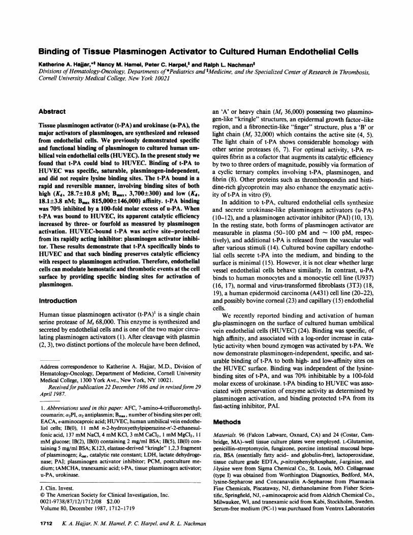
pendent and saturable manner (Fig. 1). At a t-PA level of 100IU/ml (14.6 nM), binding approached saturation. This bind-ing isotherm displayed an apparent Kd of 5.4± 1.1 nM(SE, n= 5). The binding isotherm for t-PA suggested the possibility ofa two-component system with a rapid increase in binding atlow t-PA concentrations and a slower accumulation of bindingat higher concentrations. HUVECalso bound u-PA, with satu-ration being approached at a u-PA concentration of - 4 nM.Half-maximal u-PA binding was observed at a level of -1 nMu-PA. Prothrombin, a protein that physically resembles t-PAand u-PA in that it possesses a double "kringle" structure,served as control. Prothrombin was not bound by HUVECunder identical incubation conditions and concentrationranges. Binding of DFP-t-PA to HUVECwas essentially indis-tinguishable from that of untreated t-PA.
All antigen-antibody systems employed in the experimentswere evaluated in serial plate dilutions and found to be sensi-tive at coating concentrations of < 1.0 ng/well and antibodydilutions of up to 1:50,000. Furthermore, t-PA and plasmino-gen binding to HUVECwere mutually independent, sinceprior addition of either protein had no effect on binding of theother (data not shown).
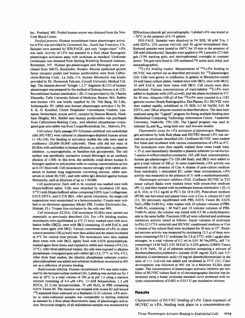
As demonstrated in Fig. 2, HUVECbound t-PA rapidly,reaching a steady state at - 10 min. Reversibility of bindingwas shown by "infinite dilution" of unbound ligand at 10 min.At this time point, the t-PA dissociation curve was biphasicwith a rapid initial dissociation phase during the first 2.5 min(T1/2, 9 min) followed by a slower second phase over the next24 min (T1/2, 54 min). Under conditions utilized, 77% of totalbinding was dissociable.
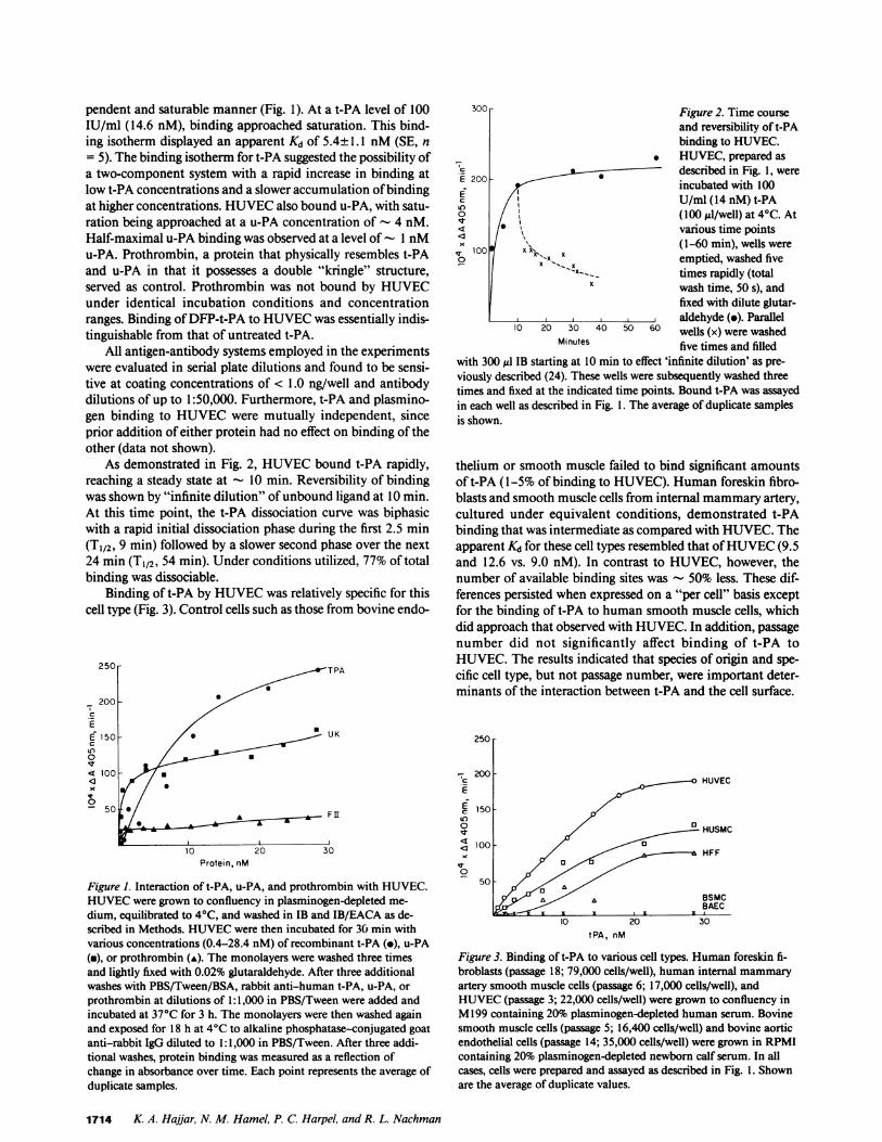
Binding of t-PA by HUVECwas relatively specific for thiscell type (Fig. 3). Control cells such as those from bovine endo-
Protein, nM
Figure 1. Interaction of t-PA, u-PA, and prothrombin with HUVEC.HUVECwere grown to confluency in plasminogen-depleted me-dium, equilibrated to 4°C, and washed in IB and IB/EACA as de-scribed in Methods. HUVECwere then incubated for 30 min withvarious concentrations (0.4-28.4 nM) of recombinant t-PA (-), u-PA(a), or prothrombin (A). The monolayers were washed three timesand lightly fixed with 0.02% glutaraldehyde. After three additionalwashes with PBS/Tween/BSA, rabbit anti-human t-PA, u-PA, orprothrombin at dilutions of 1:1,000 in PBS/Tween were added andincubated at 37°C for 3 h. The monolayers were then washed againand exposed for 18 h at 4°C to alkaline phosphatase-conjugated goatanti-rabbit IgG diluted to 1:1,000 in PBS/Tween. After three addi-tional washes, protein binding was measured as a reflection ofchange in absorbance over time. Each point represents the average ofduplicate samples.
Figure 2. Time courseand reversibility of t-PAbinding to HUVEC.
. HUVEC, prepared asE 200 described in Fig. 1, wereE 200 incubated with 100
U/ml (14 nM) t-PAo / \ (100 til/well) at 4°C. At< /\0various time pointsXbx1OO#x< ~~~~(1-60 min), wells were
° X emptied, washed five'--X__ times rapidly (total
X wash time, 50 s), andfixed with dilute glutar-aldehyde (o). Parallel
10 20 30 40 50 60 wells (x) were washedMinutes five times and filled
with 300 ul IB starting at 10 min to effect 'infinite dilution' as pre-viously described (24). These wells were subsequently washed threetimes and fixed at the indicated time points. Bound t-PA was assayedin each well as described in Fig. 1. The average of duplicate samplesis shown.
thelium or smooth muscle failed to bind significant amountsof t-PA (1-5% of binding to HUVEC). Human foreskin fibro-blasts and smooth muscle cells from internal mammaryartery,cultured under equivalent conditions, demonstrated t-PAbinding that was intermediate as compared with HUVEC.Theapparent Kd for these cell types resembled that of HUVEC(9.5and 12.6 vs. 9.0 nM). In contrast to HUVEC, however, thenumber of available binding sites was - 50% less. These dif-ferences persisted when expressed on a "per cell" basis exceptfor the binding of t-PA to human smooth muscle cells, whichdid approach that observed with HUVEC. In addition, passagenumber did not significantly affect binding of t-PA toHUVEC. The results indicated that species of origin and spe-cific cell type, but not passage number, were important deter-minants of the interaction between t-PA and the cell surface.
250
-200200 HUVEC
150
O a HUSMC
100 p HFF
0
tPA, nM
Figure 3. Binding of t-PA to various cell types. Human foreskin fi-broblasts (passage 18; 79,000 cells/well), human internal mammaryartery smooth muscle cells (passage 6; 17,000 cells/well), andHUVEC(passage 3; 22,000 cells/well) were grown to confluency inM199 containing 20% plasminogen-depleted human serum. Bovinesmooth muscle cells (passage 5; 16,400 cells/well) and bovine aorticendothelial cells (passage 14; 35,000 cells/well) were grown in RPMIcontaining 20% plasminogen-depleted newborn calf serum. In allcases, cells were prepared and assayed as described in Fig. l. Shownare the average of duplicate values.
1714 K. A. Haijar, N. M. Hamel, P. C. Harpel, and R. L. Nachman
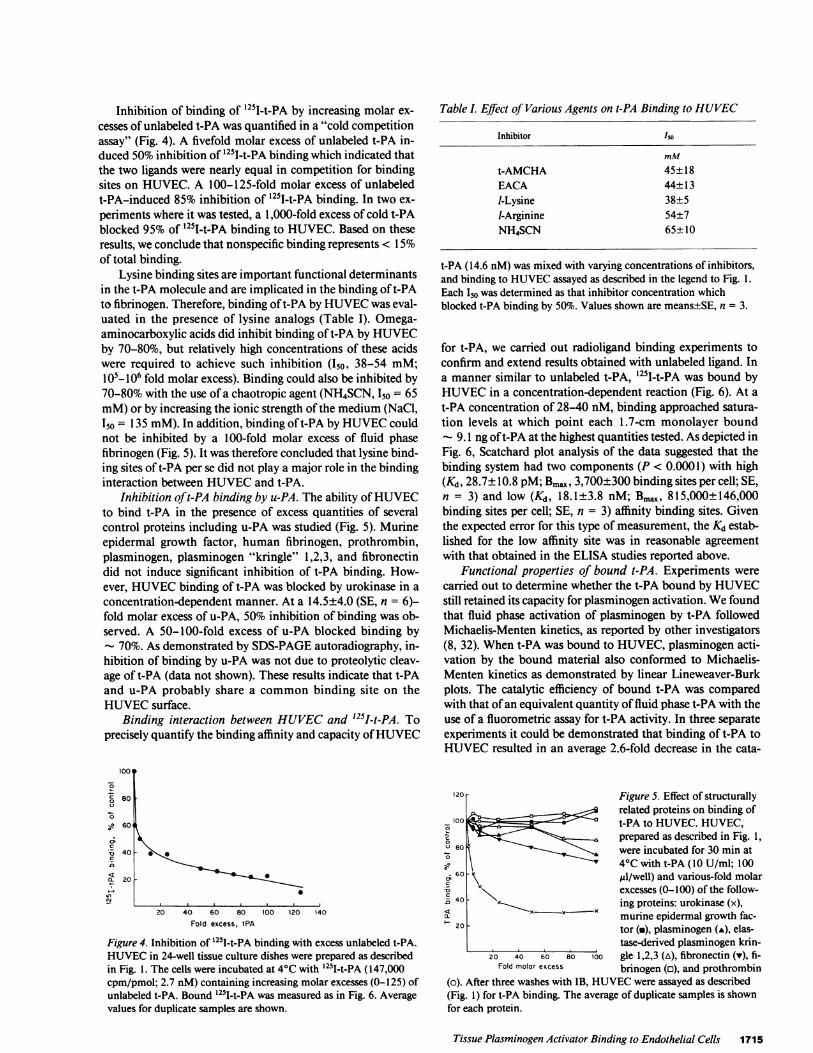
Inhibition of binding of '251I-t-PA by increasing molar ex-cesses of unlabeled t-PA was quantified in a "cold competitionassay" (Fig. 4). A fivefold molar excess of unlabeled t-PA in-duced 50% inhibition of 1251I-t-PA binding which indicated thatthe two ligands were nearly equal in competition for bindingsites on HUVEC. A 100-125-fold molar excess of unlabeledt-PA-induced 85% inhibition of '251-t-PA binding. In two ex-periments where it was tested, a 1,000-fold excess of cold t-PAblocked 95% of '251I-t-PA binding to HUVEC. Based on theseresults, we conclude that nonspecific binding represents < 15%of total binding.
Lysine binding sites are important functional determinantsin the t-PA molecule and are implicated in the binding of t-PAto fibrinogen. Therefore, binding oft-PA by HUVECwas eval-uated in the presence of lysine analogs (Table I). Omega-aminocarboxylic acids did inhibit binding of t-PA by HUVECby 70-80%, but relatively high concentrations of these acidswere required to achieve such inhibition (150, 38-54 mM;l05- 106 fold molar excess). Binding could also be inhibited by70-80% with the use of a chaotropic agent (NH4SCN, I50 = 65mM)or by increasing the ionic strength of the medium (NaCl,150 = 135 mM). In addition, binding of t-PA by HUVECcouldnot be inhibited by a 100-fold molar excess of fluid phasefibrinogen (Fig. 5). It was therefore concluded that lysine bind-ing sites of t-PA per se did not play a major role in the bindinginteraction between HUVECand t-PA.
Inhibition of t-PA binding by u-PA. The ability of HUVECto bind t-PA in the presence of excess quantities of severalcontrol proteins including u-PA was studied (Fig. 5). Murineepidermal growth factor, human fibrinogen, prothrombin,plasminogen, plasminogen "kringle" 1,2,3, and fibronectindid not induce significant inhibition of t-PA binding. How-ever, HUVECbinding of t-PA was blocked by urokinase in aconcentration-dependent manner. At a 14.5±4.0 (SE, n = 6)-fold molar excess of u-PA, 50% inhibition of binding was ob-served. A 50-100-fold excess of u-PA blocked binding by
- 70%. As demonstrated by SDS-PAGEautoradiography, in-hibition of binding by u-PA was not due to proteolytic cleav-age of t-PA (data not shown). These results indicate that t-PAand u-PA probably share a common binding site on theHUVECsurface.
Binding interaction between HUVECand '25I-t-PA. Toprecisely quantify the binding affinity and capacity of HUVEC
1001
80
60
40
7E
0
.,S
.c
to_~
200
0
20 40 60 80 100 120 140Fold excess, tPA
Figure 4. Inhibition of 251I-t-PA binding with excess unlabeled t-PA.HUVECin 24-well tissue culture dishes were prepared as describedin Fig. 1. The cells were incubated at 4°C with 251I-t-PA (147,000cpm/pmol; 2.7 nM) containing increasing molar excesses (0-125) ofunlabeled t-PA. Bound 125I-t-PA was measured as in Fig. 6. Averagevalues for duplicate samples are shown.
Table I. Effect of Various Agents on t-PA Binding to HUVEC
Inhibitor ISO
mM
t-AMCHA 45±18EACA 44± 13I-Lysine 38±5I-Arginine 54±7NH4SCN 65± 10
t-PA (14.6 nM) was mixed with varying concentrations of inhibitors,and binding to HUVECassayed as described in the legend to Fig. 1.Each Io was determined as that inhibitor concentration whichblocked t-PA binding by 50%. Values shown are means±SE, n = 3.
for t-PA, we carried out radioligand binding experiments toconfirm and extend results obtained with unlabeled ligand. Ina manner similar to unlabeled t-PA, '251-t-PA was bound byHUVECin a concentration-dependent reaction (Fig. 6). At at-PA concentration of 28-40 nM, binding approached satura-tion levels at which point each 1.7-cm monolayer bound
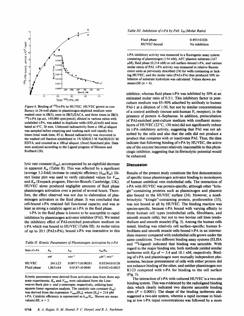
- 9.1 ng oft-PA at the highest quantities tested. As depicted inFig. 6, Scatchard plot analysis of the data suggested that thebinding system had two components (P < 0.0001) with high(&, 28.7±10.8 pM; Bmax, 3,700±300 binding sites per cell; SE,n = 3) and low (Kd, 18.1±3.8 nM; Bmax, 815,000±146,000binding sites per cell; SE, n = 3) affinity binding sites. Giventhe expected error for this type of measurement, the Kd estab-lished for the low affinity site was in reasonable agreementwith that obtained in the ELISA studies reported above.
Functional properties of bound t-PA. Experiments werecarried out to determine whether the t-PA bound by HUVECstill retained its capacity for plasminogen activation. Wefoundthat fluid phase activation of plasminogen by t-PA followedMichaelis-Menten kinetics, as reported by other investigators(8, 32). Whent-PA was bound to HUVEC, plasminogen acti-vation by the bound material also conformed to Michaelis-Menten kinetics as demonstrated by linear Lineweaver-Burkplots. The catalytic efficiency of bound t-PA was comparedwith that of an equivalent quantity of fluid phase t-PA with theuse of a fluorometric assay for t-PA activity. In three separateexperiments it could be demonstrated that binding of t-PA toHUVECresulted in an average 2.6-fold decrease in the cata-
120 - Figure 5. Effect of structurallyrelated proteins on binding of
_00 lot-PA to HUVEC. HUVEC,°0 1:= - prepared as described in Fig. 1,t'80 0 were incubated for 30 min at
4°C with t-PA (10 U/ml; 100; 60 A uZl/well) and various-fold molar
Xx . excesses (0-100) of the follow--Z40 - xing proteins: urokinase (x),
20 x xmurine epidermal growth fac-tor (-), plasminogen (A), elas-tase-derived plasminogen krin-
20 40 60 80 100 gle 1,2,3 (A), fibronectin (v), fi-Fold molar excess brinogen (o), and prothrombin
(o). After three washes with IB, HUVECwere assayed as described(Fig. 1) for t-PA binding. The average of duplicate samples is shownfor each protein.
Tissue Plasminogen Activator Binding to Endothelial Cells 1715
57-
0 S
00.0498-5 0.0427
- / 0.0356
=3 LL 0.0285
3 V 0.0213
-s) 2 _ 0C 0.0142
Nr 0.0071
00000. .019 .058 .097 .136 .165 .195Bound, nM
10 20 30 40 50
Total 251-tPA,nM
Figure 6. Binding of '25T-t-PA to HUVEC. HUVECgrown to con-fluency in 24-well plates in plasminogen-depleted medium werewashed once in IB(5), once in IB(5)/EACA, and three times in IB(5).'251-t-PA (sp act, 149,000 cpm/pmol), diluted in various ratios withunlabeled t-PA, was added to duplicate wells (450 il/well) and incu-bated at 4°C, 30 min. Unbound radioactivity from a 100-yl aliquotwas sampled before emptying and washing each well rapidly fivetimes (total wash time, 45 s). Bound radioactivity was recovered inthe washed cell fraction solubilized in 1% SDS/0.5 MNaOH/0.01 MEDTA, and counted as a 100-Al aliquot. (Inset) Scatchard plot. Datawere analyzed according to the Ligand program of Munson andRodbard (28).
lytic rate constant (kcA), accompanied by an eightfold decreasein apparent Km (Table TI). This was reflected by a significant(average 3.2-fold) increase in catalytic efficiency (kcadKm). Di-rect linear plot was used to verify calculated values for Vma.and Km(Enzpack program, Elsevier-Biosoft; Cambridge, UK).HUVECalone produced negligible amounts of fluid phaseplasminogen activation over a period of several hours. There-fore, the effect observed was not due to elaboration of plas-minogen activators in the fluid phase. It was concluded thatcell-bound t-PA retained full functional capacity and was atleast as strong a catalytic agent as t-PA in the fluid phase.
t-PA in the fluid phase is known to be susceptible to rapidinhibition by plasminogen activator inhibitor (PAI). Wetestedthe inhibitory effect of PAI-enriched postculture medium ont-PA which was bound to HUVEC(Table III). At molar ratiosof up to 20:1 (PAI:t-PA), bound t-PA was insensitive to this
Table II. Kinetic Parameters ofPlasminogen Activation by t-PA
State of t-PA Km ktca ktdKm
nM min-' pMM', min-'
HUVEC 241±23 0.00717±0.00261 0.0324±0.0126Fluid phase 1,863±64 0.0187±0.0040 0.0102±0.0023
Kinetic parameters were derived from activation data from three sep-arate experiments. Kmand Vma. were calculated from the Line-weaver-Burk plot x- and y-intercepts, respectively, utilizing leastsquares linear regression analysis. The catalytic rate constant (kij)was derived from the expression Vma.,[Eo], where [EO] = 214 pMt-PA. Catalytic efficiency is represented as k,_/Km. Shown are meanvalues±SE, n = 3.
Table III. Inhibition of t-PA by PAL: I50 (Molar Ratio)
Fluid phase 0.493±0.026HUVEC-bound No inhibition
t-PA inhibitory activity was measured in a fluorogenic assay systemconsisting of plasminogen (154 nM), AFCplasmin substrate (167MM), fluid phase (0.214 nM) or cell surface-bound t-PA, and variousmolar ratios of PAL. t-PA activity was measured in relative fluores-cence units as previously described (24) for wells containing or lack-ing HUVEC, and the molar ratio (PAI:t-PA) that produced 50% in-hibition of substrate hydrolysis was calculated. Values shown aremean±SE (n = 4).
inhibitor, whereas fluid phase t-PA was inhibited by 50% at anestimated molar ratio of 0.5:1. This inhibitory factor in post-culture medium was 85-90% adsorbed by antibody to humanPAI-I at a dilution of 1:50, but not by similar concentrationsof a control antibody (mouse anti-human F, receptor), in thepresence of protein A-Sepharose. In addition, preincubationof PAI-enriched post-culture medium with confluent mono-layers of HUVEC(220C, 150 min) did not significantly reduceits t-PA-inhibitory activity, suggesting that PAI was not ad-sorbed by the cells and also that the cells did not produce aproduct that competes with or inactivates PAL. Thus, the dataindicate that following binding of t-PA by HUVEC, the activesite of the enzyme becomes relatively inaccessible to this physi-ologic inhibitor, suggesting that its fibrinolytic potential wouldbe enhanced.
Discussion
Results of the present study constitute the first demonstrationof specific tissue plasminogen activator binding to monolayersof human umbilical vein endothelial cells. The interaction oft-PA with HUVECwas protein-specific, although other "krin-gle"-containing proteins such as plasminogen and plasminalso bound to the HUVECsurface (24). However, a nonfi-brinolytic "kringle"-containing protein, prothrombin (33),was not bound at all by HUVEC. The binding reaction wasspecies-specific, because t-PA bound with varying affinity tothree human cell types (endothelial cells, fibroblasts, andsmooth muscle cells), but not to two bovine cell lines (endo-thelium and smooth muscle) (Fig. 3). Among the human cellstested, binding was relatively cell surface-specific; human fi-broblasts and smooth muscle cells bound t-PA in an interme-diate manner compared with endothelial cells grown under thesame conditions. Two different binding assay systems (ELISAand '251-ligand) indicated that binding was saturable. Withregard to the major binding site, both methods yielded similarisotherms with Kds of - 5.4 and 18.1 nM, respectively. Bind-ing of t-PA and plasminogen were mutually independent phe-nomena, because pretreatment of cells with either protein didnot enhance binding of the other, and neither plasminogen norK123 competed with t-PA for binding to the cell surface(Fig. 5).
The interaction of t-PA with cultured HUVECis a two-sitebinding system. This was evidenced by the radioligand bindingdata which clearly indicated two discrete saturable bindingsites (P < 0.0001). The shape of the binding isotherms alsosuggested a two-site system, wherein a rapid increase in bind-ing at low t-PA input concentrations was followed by a more
1716 K A. Hajjar, N. M. Hamel, P. C. Harpel, and R. L. Nachman
gradual increase in binding as higher concentrations were ap-proached (Fig. 1, 3, and 6). Furthermore, the shape of thebinding dissociation curve also suggested a two-phase processwith different rates of dissociation (Fig. 2). Even though theradioligand binding studies were not carried out under strictequilibrium conditions, reasonable estimates of binding affin-ity and capacity were provided via excellent agreement withthose obtained by ELISA. Finally, the cold competition assaysystem validated the '25I-t-PA data by providing verificationthat labeled and unlabeled ligand competed for the cell surfacewith approximately equal affinity (Fig. 4).
Under baseline conditions, the level of t-PA in humanplasma is 50-100 pM (14, 34, 35); following physical exercise,vaso-occlusion, or infusion of l-deamino-8-D-arginine vaso-pressin (DDAVP), plasma levels of t-PA increase threefold (34,35). Because our binding studies have demonstrated a highaffinity Kd for the t-PA-HUVEC interaction (- 29 pM), cir-cumstances in which t-PA is released from the vascular wallwould result in plasma t-PA levels which would equal or ex-ceed this value. In addition, levels of t-PA in the endothelialcell microenvironment might transiently reach even higherlevels and therefore exert a favorable influence upon bindingof t-PA to the endothelial cell surface. Thus the interactionsobserved in our experiments are of possible physiological rele-vance especially under conditions promoting t-PA release,such as venous occlusion or physical stress.
The precise mechanism whereby HUVECbound t-PA totheir surface remains to be elucidated. Although the fibrin-binding domain oft-PA involves the "kringle"-2 segment (andto a lesser extent the fibronectin "finger" and epidermalgrowth factor regions [36, 37]), it is unlikely that the fibrinbinding domain of t-PA is germane to the interactions westudied. This is evidenced by our demonstration that theHUVECemployed in these experiments contained insuffi-cient surface fibrinogen to account for the degree of bindingattained. Furthermore, binding of t-PA to the HUVECsurfacewas not blocked by fluid phase fibrinogen (Fig. 5). In addition,relatively high (mM) concentrations of lysine analogs are re-quired to induce significant inhibition of t-PA binding, and theinhibition that did occur may have been a reflection of anincrease in ionic strength of the medium (Table I). Whereasplasminogen does exhibit a lysine binding site-dependent in-teraction with the HUVECsurface (24), neither plasminogennor its elastase-derived fragment, "kringle 1,2,3", blockedbinding of t-PA by HUVEC, even when present in 100-foldmolar excess (Fig. 5).
Although urokinase strongly inhibits t-PA binding toHUVEC, other proteins which are structurally homologous(epidermal growth factor, plasminogen, plasminogen-derived"kringle" 1,2,3, fibronectin, fibrinogen, and prothrombin) didnot do so (Fig. 5). Both urokinase and t-PA both possessgrowth factor, "kringle," and catalytic domains which demon-strate varying degrees of homology (6, 7). The amino terminalfragment of u-PA which contains the growth factor and "krin-gle" domains appears to mediate u-PA binding to humanmonocytic and epidermoid carcinoma cell lines (38). As de-picted in Fig. 5, one of these two or possibly an as yet unde-fined domain may be responsible for the binding function oft-PA by HUVEC.
It is important to note that t-PA retains its activity as acatalytic agent after it has been bound by HUVEC(Table II).
This did not represent release of a soluble endothelial cell com-ponent(s) because endothelial cell postculture medium wasincapable of activating plasminogen in the time frame of thisassay. Upon binding of t-PA by HUVEC, we measured a 3.2-fold increase in its catalytic efficiency (Table II). Upon bindingof t-PA to fibrin, the same reaction proceeds - 500 timesmore efficiently (8), suggesting that the endothelial cell mayhave a fibrinlike effect, albeit of much lower magnitude. Thebasis of the HUVEC-induced increase in catalytic efficiency,like that conferred by fibrin, was a decrease (eightfold) in theapparent Km. Wehave previously demonstrated (24) thatplasminogen also binds to HUVEC, and that binding aug-ments its activatability by 12-fold, based on a proportionatedecrease in apparent Km. The concentrations of plasminogenused in the t-PA activation experiments were at or below theKd for the interaction of plasminogen with HUVEC. Thus,under these assay conditions, plasminogen binding sites couldbe partially occupied, and the eightfold decrease in Km ob-served for plasminogen activation by cell surface t-PA mayreflect, at least in part, a contribution from plasminogenbound to the cell surface.
The endothelial cell surface appears to confer protection oft-PA with respect to its physiologic inhibitor, PAL. Althoughthe mechanism for this protection is unclear, this phenome-non has been reported in other enzyme systems. Thus, plas-min bound to a fibrin surface is sequestered from the action ofcirculating antiplasmin (39), and acrosin from ram seminalvesicle is protected from its inhibitor in seminal plasma (40).In cellular systems, urokinase which becomes macrophage-as-sociated is not susceptible to the action of PAI, whereas plas-minogen activator in the fluid phase is exquisitely sensitive toPAI (41, 42). Therefore, active site protection, whereby pro-teases acquire increased catalytic activity upon associationwith physiologically relevant surfaces, may represent a broadphysiologic mechanism. This protection is of pertinence in oursystem, because the phenomenon augments fibrinolytic po-tential in a plasma milieu where a thrombus might be locatedin the in vivo situation.
In previous studies (24), we demonstrated that plasmino-gen, the major substrate for t-PA, bound in a specific mannerand with high affinity to cultured HUVECin physiologic con-centrations. Data presented in the experiments described haveindicated that t-PA was also bound in vitro under comparablecircumstances. Similar data concerning binding of plasmino-gen and urokinase to the promyeloid leukemic cell line (U937)and a fetal lung fibroblast cell line (GM1380) have been re-cently reported by Plow et al. (43). The binding described inthe present study may be physiologically relevant in the courseof events such as venous occlusion or physical stress. Althoughthe t-PA binding site was not identical to the plasminogenbinding site or plasminogen itself, the reaction allowed t-PA toretain its ability to activate plasminogen and the bound t-PAwas inaccessible to its rapidly-acting inhibitor, PAL. Therefore,the endothelial cell, by virtue of its ability to provide specificbinding sites for the assembly of proteins of the fibrinolyticsystem, may serve as a focal point for plasmin generation inthe blood vessel microenvironment.
AcknowledgmentsDr. Hajjar is the recipient of Clinical Investigator Award K08HL-0 1352 from the National Institutes of Health, and an Andrew W.
Tissue Plasminogen Activator Binding to Endothelial Cells 1717
Mellon Foundation Teacher-Scientist Award. This work was also sup-ported by grant HL- 1 8828 (Specialized Center of Research in Throm-bosis) from the National Institutes of Health.
References
1. Bachmann, F., and E. K. 0. Kruithof. 1984. Tissue plasminogenactivator: chemical and physiological aspects. Semin. Thromb. Hemo-stasis. 10:6-17.
2. Wallen, P., M. Ranby, N. Bergsdorf, and P. Kok. 1981. Purifica-tion and characterzation of tissue plasminogen activator: on the oc-currence of two different'forms and their enzymatic properties. InProgress in Fibrinolysis. J. F. Davidson, I. M. Nilsson, and B. Astedt,editors. Churchill/Livingstone, Edinburgh. 16-23.
3. Rijken, D. C., and E. Groeneveld. 1986. Isolation and functionalcharacterization of the heavy and light chains of human tissue-typeplasminogen activator. J. Biot. Chem. 261:3098-3102.
4. Banyai, L., A. Varadi, and L. Patthy. 1983. Commonevolution-ary origin of the fibrin-binding structures of fibronectin and tissue-typeplasminogen activator. FEBS(Fed. Eur. Biochem. Soc.) Lett. 163:37-41.
5. Pennica, D., W. E. Holmes, W. J. Kohr, R. N. Harkins, G. A.Vehar, C. A. Ward, W. E. Bennett, E. Yelverton, P. H. Seeburg, H. L.Heyneker, D. V. Goeddel, and D. Collen. 1983. Cloning and expres-sion of human tissue-type plasminogen activator cDNA in E. Coli.Nature (Lond.). 301:214-221.
6. Strassburger, W., A. Wollmer, J. E. Pitts, I. D. Glover, I. J.Tickle, T.' L. Blundell, G. J. Steffens, W. A. Gunzler, F. Otting, and L.Flohe. 1983. Adaptation of plasminogen activator sequences to knownprotease structures. FEBS (Fed. Eur. Biochem. Soc.) Lett. 157:219-223.
7. Ny, T., F. Elgh, and B. Lund. 1984. The structure of the humantissue-type plasminogen activator gene: correlation of intron and exonstructures to functional and structural domains. Proc. Natl. Acad. Sci.USA. 81:5355-5359.
8. Hoylaerts, M., D. C. Rijken, H. R. Lijnen, and D. Collen. 1982.Kinetics of the activation of plasminogen by human tissue plasmino-gen activator. J. Biol. Chem. 257:2912-2919.
9. Silverstein, R. L., R. L. Nachman, L. L. K. Leung, and P. C.Harpel, 1985. Activation of immobilized plasminogen by tissue acti-vator: multimolecular complex formation. J. Biol. Chem. 260:10346-10352.
10. Loskutoff, D. J., and T S. Edgington. 1977. Synthesis of afibrinolytic activator and inhibitor by endothelial cells. Proc. Natl.Acad. Sci. USA. 74:3903-3907.
11. Levin, E. G., and D. J. Loskutoffi 1982. Cultured bovine endo-thelial cells produce both urokinase and tissue-type plasminogen acti-vators. J. Cell Biol. 94:631-636.
12. Booyse, F. M., G. Osikowicz, S. Feder, and J. Scheinbuks.1984. Isolation and characterization of a urokinase-type plasminogenactivator (Mr = 54,000) from cultured human endlothelial cells indis-tinguishable from urinary urokinase. J. Biol. Chem. 259:7198-7205.
13. Van Mourik, J. A., D. A. Lawrence, and D. J. Loskutoff. 1984.Purification of an inhibitor of plasminogen activator (antiactivator)synthesized by endothelial cells. J. Biol. Chem. 259:14914-14921.
14. Erickson, L. A., R. R'. Schleef, T. Ny, and D. J. Loskutoff. 1985.The fibrinolytic system of the vascular wall. Clin. Haematol. 14:513-530.
15. Moscatelli, D. 1986. Urokinase-type and tissue-type plasmino-gen activators have different distributions in cultured bovine capillaryendothelial cells. J. Cell. Biochem. 30:19-29.
16. Vassalli, J.-D., D. Baccino, and D. Belin. 1985. A cellularbinding site for the Mr 55,000 form of the human plasminogen activa-tor, urokinase. J. Cell Biol. 100:86-92.
17. Stoppelli, M. P., A. Corti, A. Soffienrini, G. Cassani, F. Blasi,
and R. K. Assoian. 1985. Differentiation-enhanced binding of theamino-terminal fragment of human urokinase plasminogen activatorto a specific receptor on U937 monocytes. Proc. NatL. Acad. Sci. USA.82:4939-4943.
18. Del Rosso, M., G. Dini, and G. Fibbi. 1985. Receptors forplasminogen activator, urokinase, in normal and Rous sarcoma virus-transformed mouse fibroblasts. Cancer Res. 45:630-636.
19. Keski-Oja, J., and A. Veheri. 1982. The cellular target for theplasminogen activator, urokinase, in human fibroblasts -66,000-dal-ton protein. Biochim. Biophys. Acta. 720:141-146.
20. Fibbi, G., G. Dini, F. Pasquali, M. Pucci, and M. Del Rosso.1986. The Mr 17,500 region of the A chain of u'rokinase is required forinteraction with a specific receptor in A431 cells. Biochim. Biophys.Acta. 885:301-308.
21. Stoppelli, M., C. Tacchetti, M. V. Cubellis, A. Corti, V. J.Hearing, G. Cassani, E. Appella, and F. Blasi. 1986. Autocrine satura-tion of pro-urokinase receptors on human A431 cells. Cell. 45:675-684.
22. Gross, J. L., M. N. Krupp, D. B. Rifkin, and M. D. Lane. 1983.Down-regulation of epidermal growth factor receptor correlates withplasminogen activator activity in human A431 epidermoid carcinomacells. Proc. Natl. Acad. Sci. USA. 80:2276-2280.
23. Shuman, M. A., and C. H. Merkel. 1985. Urokinase binding tobovine corneal endothelial cells. Exp. Eye Res. 41:371-382.
24. Hajar, K. A., P. C. Harpel, E. A. Jaffe, and R. L. Nachman.1986. Binding of plasminogen to cultured human endothelial cells. J.Bio. Chem. 261:11656-11662.
25. Sottrup-Jensen, L., H. Claeys, M. Zajdel, T. E. Petersen, and S.Magnusson. 1978. The primary structure of human plasminogen: iso-lation of two lysine-binding fragments and one "mini-" plasminogen(MW, 38,000) by elastase-catalyzed-specific limited proteolysis. InProgress in Chemical Fibrinolysis and Thrombolysis. Vol. 3. J. F.Davidson, R. M. Rowan, M. M. Samama, and P.'C. Edsnoyers, edi-tors. Raven Press, NewYork. 191-209.
26. Martin, B. M., W. W. Wasiewski, J. W. Fenton, and T. C.Detwiler. 1976. Equilibrium binding of thrombin to platelets. Bio-chemistry. 15:4886-4893.
27. Laemmli, U. K. 1970. Cleavage of structural proteins duringthe assembly of the head of bacteriophage T4. Nature (Lond.).227:680-684.
28. Munson, P. J., and D. Rodbard. 1980. Ligand: a versatile com-puterized approach for characterization of ligand-binding systems.Anal. Biochem. 107:220-239.
29. Nachman, R. L., K. A. Hajar, R. L. Silverstein, and C. A.Dinarello. 1986. Interleukin 1 induces endothelial cell synthesis ofplasminogen activator inhibitor. J. Exp. Med. 163:1595-1600.
30. Andreasen, P. A., L. S. Nielsen, P. Kristensen, J. Grondahl-Hansen, L. Skriver, and K. Dano. 1986. Plasminogen activator inhibi-tor from human fibrosarcoma cells binds urokinase-type plasminogenactivator, but not its proenzyme. J. Biot. Chem. 261:7644-7651.
31. Green, G. D. J., and E. Shaw. 1979. Thiobenzyl benzyloxycar-bonyl-L-lysinate, substrate for a sensitive colorimetric assay for tryp-sin-like enzymes. Anal. Biochem. 93:223-226.
32. Ranby, M. 1982. Studies on the kinetics of plasminogen acti-vation by tissue plasminogen activator. Biochim. Biophys. Acta.704:461-469.
33. Magnusson, S., T. E. Petersen, L. Sottrup-Jensen, and H.Claeys. 1975. Complete primary structure of prothrombin. In Pro-teases and Biologic Control. E. Reich, D. B. Rifkin, and E. Shaw,editors. Cold Spring Harbor Laboratories, Cold Spring Harbor, NY.123-149.
34. Bergsdorf, N., T. Nilsson, and P. Wallen. 1983. An enzymelinked immunosorbent assay for determination of tissue plasminogenactivator applied to patients with thromboembolic diseases. Thromb.Haemostasis. 50:740-744.
35. Rijken, D. C., I. Juhan-Vague, F. DeCock, and D. Collen. 1983.
1718 K. A. Hajjar, N. M. Hamel, P. C. Harpel, and R. L. Nachman
Measurement of human tissue-type plasminogen activator by a two-site immunoradiometric assay. J. Lab. Clin. Med. 101:274-284.
36. Ichinose, A., K. Takio, and K. Fujikawa. 1986. Localization ofthe binding site of tissue-type plasminogen activator to fibrin. J. Clin.Invest. 78:163-169.
37. Van Zonneveld, A., H. Veerman, and H. Pannekoek. 1986.Autonomous function of structural domains on human tissue-typeplasminogen activator. Proc. Nat!. Acad. Sci. USA. 83:4670-4674.
38. Cubellis, M. V., M. L. Nolli, G. Cassani, and F. Blasi. 1986.Binding of single-chain prourokinase to the urokinase receptor ofhuman U937 cells. J. Biol. Chem. 261:15819-15822.
39. Wiman, B., and D. Collen. 1978. Molecular mechanism ofphysiologic fibrinolysis. Nature (Lond.). 272:549-550.
40. Brown, C. R., and E. F. Hartree. 1976. Effects of acrosin inhibi-
tors on the soluble and membrane-bound forms of ram acrosin, and areappraisal of the role of the enzyme in fertilization. Hoppe-Seyler's Z.Physiol. Chem. 357:57-65.
41. Chapman, H. A., Z. Vavrin, and J. B. Hibbs. 1982. Macrophagefibrinolytic activity: identification of two pathways of plasmin forma-tion by intact cells and of a plasminogen activator inhibitor. Cell.28:653-662.
42. Chapman, H. A., 0. L. Stone, and Z. Vavrin. 1984. Degrada-tion of fibrin and elastin by intact human alveolar macrophages invitro. J. Clin. Invest. 73:806-815.
43. Plow, E. F., D. E. Freaney, J. Plescia, and L. A. Miles. 1986.Evidence for plasminogen and urokinase receptors on the same celltype. J. Cell Biol. 103:2411-2420.
Tissue Plasminogen Activator Binding to Endothelial Cells 1719