Bile Resistance in Lactobacillus. Rhamnosus GG: Stability ... · Tiivistelmä -Referat -Abstract...
72
Bile Resistance in Lactobacillus rhamnosus GG: Stability and Mechanisms Kun Xiao Master’s Thesis University of Helsinki MBIOT Biotechnology September 2014
Transcript of Bile Resistance in Lactobacillus. Rhamnosus GG: Stability ... · Tiivistelmä -Referat -Abstract...

Bile Resistance in Lactobacillus rhamnosus GG: Stability and Mechanisms
Kun Xiao Master’s Thesis University of Helsinki MBIOT Biotechnology September 2014

HELSINGIN YLIOPISTO - HELSINGFORS UNIVERSITET - UNIVERSITY OF HELSINKI
Tiedekunta/Osasto - Fakultet/Sektion -Faculty
Agriculture and Forestry
Laitos - Institution -Department
MBIOT Master’s Degree Programme in
Biotechnology Tekijä -Författare – Author
Kun Xiao
Työn nimi -Arbetets titel - Title
Bile Resistance in Lactobacillus rhamnosus GG: Stability and Mechanisms
Oppiaine -Läroämne -Subject
Biotechnology
Työn laji -Arbetets art -Level
Master’s Thesis Aika -Datum - Month and year
September 2014 Sivumäärä - Sidoantal - Number of pages
72
Tiivistelmä -Referat -Abstract
Lactobacillus rhamnosus GG is a lactic acid bacterium that is widely used as probiotic products in
the dairy industry. To gain insights into the genome stability of the L. rhamnosus GG in the human
gastrointestinal tract and the possible adaption mechanism under different stresses, we first
examined the genotype and phenotype of the L. rhamnosus GG grown over 1000 generations under
various stresses, including bile salts, osmotic stress or shearing forces. Immunoblotting analysis of
L. rhamnosus grown over 1000 generations showed that the production of mucus -binding pili by L.
rhamnosus GG was the most impaired when exposed to bile salts. Complementary PCR screening
of 13 highly variable chromosomal regions in GG confirmed that the pilus gene cluster had been
lost when exposed to bile stress over time. In vitro bile-induced genomic changes observed in GG
possibly reflects the genome plasticity and stability of GG in the human intestinal tract. Still, we
showed that these changes only occurred after more than 100 generations, a period of time
relatively long compared to the observed time of persistence and transit of GG in the intestinal
tract. Although damages and stresses may be caused by bile salts, L. rhamnosus GG still has the
ability to tolerate bile salts. The resistance mechanism is still unclear but, based on previous
studies, we identified one ABC transporter encoded by the gene operon called tauABC that may be
involved in bile resistance. In an effort to demonstrate its function, a tauB-null mutant derivative
was generated and phenotypically characterized in terms of metabolic, signaling and functional
properties. The data revealed that the tauB-null mutant significantly grow slower than L.
rhamnosus GG wild-type strain in the presence of ox bile extracts. Additional screenings using
various bile conjugates specifically revealed that two compounds of bile salts, i.e. taurodeoxycholic
and taurochenodeoxycholic acid, may be processed by the TauABC transporter, contributing at
least partially to the tolerance of GG to bile salts. Overall, we showed that bile salts constitute an
important stress factor for GG that causes genomic alterations, although it has bile tolerance
mechanisms to bile, such as the newly-characterized tauABC operon.
Avainsanat – Nyckelord- Keywords Lactobacillus rhamnosus GG pili bile stress genomic stability ABC transporter
Säilytyspaikka - Förvaringsställe -Where deposited
Viiki library
Muita tietoja -Övriga uppgifter -Further information
Supervisor and responsible professor: Academy Professor De Vos. Willem, Dr François P.
Douillard

ABSTRACT
Lactobacillus rhamnosus GG is a lactic acid bacterium that is widely used as probiotic products in
the dairy industry. To gain insights into the genome stability of the L. rhamnosus GG in the human
gastrointestinal tract and the possible adaption mechanism under different stresses, we first
examined the genotype and phenotype of the L. rhamnosus GG grown over 1000 generations under
various stresses, including bile salts, osmotic stress or shearing forces. Immunoblotting analysis of
L. rhamnosus grown over 1000 generations showed that the production of mucus-binding pili by L.
rhamnosus GG was the most impaired when exposed to bile salts. Complementary PCR screening
of 13 highly variable chromosomal regions in GG confirmed that the pilus gene cluster had been
lost when exposed to bile stress over time. In vitro bile-induced genomic changes observed in GG
possibly reflects the genome plasticity and stability of GG in the human intestinal tract. Still, we
showed that these changes only occurred after more than 100 generations, a period of time
relatively long compared to the observed time of persistence and transit of GG in the intestinal tract.
Although damages and stresses may be caused by bile salts, L. rhamnosus GG still has the ability to
tolerate bile salts. The resistance mechanism is still unclear but, based on previous studies; we
identified one ABC transporter encoded by the gene operon called tauABC that may be involved in
bile resistance. In an effort to demonstrate its function, a tauB-null mutant derivative was generated
and phenotypically characterized in terms of metabolic, signaling and functional properties. The
data revealed that the tauB-null mutant significantly grow slower than L. rhamnosus GG wild-type
strain in the presence of ox bile extracts. Additional screenings using various bile conjugates
specifically revealed that two compounds of bile salts, i.e. taurodeoxycholic and
taurochenodeoxycholic acid, may be processed by the tauABC transporter, contributing at least
partially to the tolerance of GG to bile salts. Overall, we showed that bile salts constitute an
important stress factor for GG that causes genomic alterations, although it has bile tolerance
mechanisms to bile, such as the newly-characterized tauABC operon.
Key words: Lactobacillus rhamnosus GG pili bile stress genomic stability ABC transporter
TABLE OF CONTENTS
1 INTRODUCTION ........................................................................................................................ 9
1.1 Lactic acid Bacteria: a Highly Versatile Group ......................................................... 9
1.1.1 Lactic acid bacteria in human body .................................................................... 9
1.1.2 Possible origins of LAB present in the human body ...................................... 12
1.1.3 Adaption of LAB to the human gut .................................................................... 12
1.2 Lactobacillus rhamnosus Strains, Industrial use and Probiotic properties ........ 14
1.2.1 Lactobacillus rhamnosus .................................................................................... 14
1.2.2 Genotype and phenotype of the Lactobacillus rhamnosus species ............ 14
1.2.3 Industrial use of Lactobacillus rhamnosus strains.......................................... 15
1.2.4 Probiotic properties of Lactobacillus rhamnosus strains ............................... 15
1.3 Lactobacillus rhamnosus GG: Multifaceted Bacteria. ........................................... 16
1.3.1 A multifaceted bacterium .................................................................................... 16
1.3.2 The application of LGG....................................................................................... 17
1.4 Pili and genome stability in L. rhamnosus GG ....................................................... 18
1.4.1 Pili .......................................................................................................................... 18
1.4.2 Pilus gene cluster in L. rhamnosus GG ........................................................... 19
1.5 Bile and Bile Acids ...................................................................................................... 22
1.6 Bile Response in the Lactobacillus rhamnosus Strains ....................................... 24
1.7 Bile Resistance in Lactobacillus rhamnosus Strain GG ....................................... 26
1.8 ABC Transporters ....................................................................................................... 27
1.8.1 Structure of ABC transporter.............................................................................. 27
1.8.2 Function of ABC transporter .............................................................................. 28
1.9 The tauABC Operon in Lactobacillus rhamnosus Strain GG .............................. 28
2 OBJECTIVES ........................................................................................................................... 32
3 METHODS ................................................................................................................................. 33
3.1 Bacterial Strains and Growth Media ................................................................ 33
3.2 Immunoblotting Analysis..................................................................................... 33
3.3 Colony PCR Analysis .......................................................................................... 34
3.4 Analysis of Hyper Variable Chromosomal Regions by PCR......................... 34
3.5 Immuno-gold Staining Transmission Electron Microscopy Analysis ........... 35 3.6 Co-culturing of the Wild-type L. rhamnosus GG and a Pili Negative Mutant
Derivative .......................................................................................................................... 36
3.7 Fluorescence Microscopy .................................................................................. 36
3.8 Construction of L. rhamnosus GG tauB-null Mutant ...................................... 37
3.9 Complementation of the tauB-null Mutant Strains.......................................... 38 3.10 Sensitivity of Wild-type L. rhamnosus GG and tauB-null Mutant Strain in
the Presence of Ox Bile Extracts .................................................................................. 39
3.11 Sensitivity of Wild-type L. rhamnosus GG and tauB-null Mutant Strain in
the Presence of Pig Bile ................................................................................................. 39 3.12 Sensitivity of Wild-type L. rhamnosus GG and tauB-null Mutant Strain in
the Presence of Human Bile .......................................................................................... 39 3.13 Sensitivity of Wild-type L. rhamnosus GG and tauB-null Mutant Strain in
the Presence of Bile Components ................................................................................ 40
4 Results ...................................................................................................................................... 41
4.1 Immunoblotting Analysis of the SpaCBA Pili in L. rhamnosus GG .............. 41
4.2 PCR Analysis of 13 Hyper Variable Regions Over Time ............................... 44
4.3 Co-culturing of the L. rhamnosus GG and a Pili Negative Mutant ............... 45
4.4 Generation of tauB-null Mutant Strain .............................................................. 47

4.5 Complementation of tauB-null Mutant .............................................................. 47
4.6 Effects of Ox Gall on tauB-null Strain vs. L. rhamnosus GG ........................ 52
4.7 Effects of Pig Bile Salts on tauB-null Strain vs L. rhamnosus GG ............... 52
4.8 Effects of Human Bile Salts on tauB-null Strain vs L. rhamnosus GG........ 55 4.9 Comparative Curve of GG WT vs. tauB-null Strain in the Presence of Bile
Components ..................................................................................................................... 57
5 DISCUSSION ............................................................................................................................ 61
6 CONCLUSION .......................................................................................................................... 64
7 ACKNOWLEDGMENTS ......................................................................................................... 64
8 REFERENCES ......................................................................................................................... 65

List of Abbreviations
MRS Man-Rogosa- Sharpe Medium
LB Lysogeny broth
BSA Bovine Serum Albumin
LGG Lactobacillus rhamnosus GG
TCA Taurocholic Acid
TDCA Taurodeoxycholic Acid
GCDA Glycochenodeoxycholic Acid
TCDA Taurochenodeoxycholic Acid
GCLA Glycolithocholic Acid
TLCA Taurolithochoic Acid
PTS Phosphotransferase System
CRISPR Cluster Regularly Interspaced Short Palindromic Repeat
BSH Bile Salt Hydrolase
EPS Extracellular Polymeric Substance
LAB Lactic Acid Bacteria
CFU Colony Forming Unit
API Active Pharmaceutical Ingredient
TEM Transmission Electron Microscopy.
LPXTG Leucine-Proline-any-Threonine-Glycine
BSH Bile Salt Hydrolase
GI Gastrointestinal
IS Insertion Sequence

List of Figures
Figure 1: One example of pili by transmission electron microscopy ………………………...……18
Figure 2: Mutations detected in LGG isolated. ……………………………………………………20
Figure 3: Schematic model of Lactobacillus rhamnosus GG SpaCBA pili. ………………….……21
Figure 4: Synthesis of the 12 bile acids present in human bile from the two primary bile acids….22
Figure 5: Structure of conjugated bile acid……………………...………………………………….23
Figure 6: Bile resistance distribution among L. rhamnosus isolates from different niches. ….……25
Figure 7: Model of the physiological response of L. rhamnosus GG to bile stress. …………..……29
Figure 8: One example of the ABC transporters……………………………………………………29
Figure 9: Organization of ABC transporters. …………………………………………………....…30
Figure 10: Taurine ABC transporter cluster in L. rhamnosus GG ………………………….....……30
Figure 11: String analysis of the tauABC gene. ………………………………………….……..…31
Figure 12: Results of genome stability of LGG under top fraction treatment ….………………..…42
Figure 13: One membrane example of immunoblotting with monoclonal SpaC antibody…..….…42
Figure 14: Results of genome stability of LGG under bile treatment ……………………...………43
Figure 15: Results of genome stability of LGG under salt treatment……………...……………….43
Figure 16: Mutation Percentage of 13 hyper variable regions under bile and vortex treatment……44
Figure 17: Summary of L. rhamnosus GG genome stability under different conditions………...…45
Figure 18: Competitive assay between the wild type and pili negative mutant. ….......................…46
Figure 19: One example of fluorescent Microscopy. ………………………………………………46
Figure 20: Model of the knockout tauB mutant ……………………………………………………48
Figure 21: Model of the complementary tauB-CM ……………………………………………..…48
Figure 22: One example of PCR production …………………………………………….……...….49
Figure 23: The electron microscopy results of the knockout strain. …………………….…………49
Figure 24: Generation time of three strains in the presence of 3% ox gall…………………………53
Figure 25: Generation time of the WT VS tauB-null in the present of 0.05% porcine…………..…53
Figure 26: Example of Cell sedimentation …...……………………………………………………54
Figure 27: OD600 value of WT, mutant strain and Complementary strain …………………….……54
Figure 28: OD600 value of the wild type LGG and the mutant strain after 24 h incubation in different
concentrations of porcine bile……………………………………………………………..…..……54
Figure 29: The concentration of different compounds in the 4 samples……………………………55
Figure 30: OD600 after 24 h at the present of 3% of human bile ……………………………..….…55
Figure 31: Generation time of three strains in the present of 3% of each subject…………………56

Figure 32: Generation time of three strains in the present of 3% of human bile of subject 5……...56
Figure 33: Generation time in different component. ……………………………………...……58-59
Figure 34: Generation time of WT, tauB-null and complementary strains at the 8mM of the
Taurodeoxycholic acid………………………………………………………………………………60
Figure 35: Generation time of WT, tauB-null and complementary strains at 5mM of the
TCDA. …………………………………………………………………………………………...…60
Figure 36: The structure of TDCA and TDCA……………………………………………...………63
Figure 37: Hypothesis model of the TauABC transporter mechanism…………………….…..……67
List of Tables
Table 1: Five families of the lactic acid bacteria……………………………………………..……..11
Table 2: Strains and plasmid used in this study……………………………………………………..34
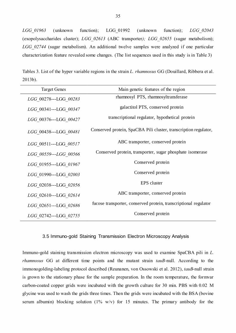
Table 3: List of the hyper variable region in the strain L. rhamnosus GG …………………………35
Table 4: Different concentration tested in the bile components…………………………………….40
Table 5: All kinds of sugar metabolized between the knockout strains and wild type LGG…..……50

9
1 INTRODUCTION
1.1 Lactic acid Bacteria: a Highly Versatile Group
1.1.1 Lactic acid bacteria in human body
The human gastrointestinal tract (GI) microbiota consists of over 100 trillion (1014) of microbes and
even outnumbers the number of our body cells (Qin, Li et al. 2010). Up to date, more than 1000
bacterial species from the human intestinal have been isolated and cultured (Rajilic-Stojanovic & de
Vos 2014), but these species are still a small fraction of all the bacteria present in the GI tract.
Extensive characterization of the genotype and phenotype of these intestinal species, along with the
development and use of molecular metagenomic approaches constitutes therefore an important step
towards the understanding of the complexity of the lactic acid bacteria (LAB) in the GI tract.
In general, Lactic acid bacteria (LAB) are found or isolated in ecological habitats with a rich
supply of nutrients and they occur on decomposing plant materials and fruits, in dairy products,
fermented meat and fish (Aguirre & Collins 1993). LAB are found in a large range of human
cavities such as the skin, the mouth, the stomach and the GI tract. Typically, LAB can be detected at
the amounts of 104 CFU and 106 CFU per gram in the small intestine and fecal samples,
respectively (Dal Bello, Walter et al. 2003, Walter 2008). In comparison to other gut bacterial
communities, LAB represent a relatively small fraction of the overall gut microbiota (0.01-0.1%)
(Louis, Scott et al. 2007). Although their quantity is low in the human GI tract, this does not
exclude them to have a significant impact on the human host and its health - see Section 1.2.
Lactic acid bacteria (LAB) are one of the bacterial groups found in the GI tract that has been
well-researched and documented. The first isolation of the lactic acid bacteria was performed by J.
Lister in 1890 (Leroy & De Vuyst 2004). Since then, LAB have been gradually introduced in
production processes as the starter culture for cheese or sour milk products (Leroy & De Vuyst
2004). Typically, LAB under standard growth conditions (non-limiting amount of nutrients,
presence of growth factors) are anaerobic Gram-positive, non-spore, acid tolerant, aero-tolerant, rod
or coccus bacteria (Leroy & De Vuyst 2004, Saxelin, Tynkkynen et al. 2005). LAB are
characterized by the production of lactic acid as a major catabolic end product from the glucose.
But the metabolic functions of LAB may vary depending on environmental conditions. Thus, in the
presence of hemes, the production of catalase and cytochromes may result in the use of lactic acid
that can be then further metabolized, resulting in low lactic acid concentration (Leroy & De Vuyst
2004, Saxelin, Tynkkynen et al. 2005).

10
Lactic acid bacteria are a group in the Clostridium branch of the Gram-positive bacteria and are
grouped in one order and six families (Table 1) (Agostoni, Axelsson et al. 2004). In Table 1, a large
number of bacterial species constitutes the LAB group. Many of these bacterial species can be
found in different parts of the human body. In the oral cavity, the genera of the Lactobacillus have
been reported to teeth, saliva and the tongue (Badet & Thebaud 2008). In the human intestine, some
species such as Lactobacillus gasseri, Lactobacillus salivarius, Lactobacillus ruminis,
Lactobacillus reuteri or Lactobacillus crispatus strains were found to be persistent. In the human
vaginal cavity, Lactobacillus crispatus, Lactobacillus gasseri, Lactobacillus iners and
Lactobacillus jensenii are commonly detected (Douillard & de Vos 2014).
Along with the variety of LAB species, one would expect that they display distinctive
functions, including probiotic functions. Some of them have been used as probiotics, such as
Lactobacillus plantarum, Lactobacillus casei and Lactobacillus rhamnosus (Saxelin, Tynkkynen et
al. 2005). Others have been utilized in industry as the dairy and meat starter cultures, such as
Lactococcus lactis, Streptococcus thermophilus and Pediococcus acidilacti (Leroy & De Vuyst
2004) – see details Section 1.2.3.
In some cases, the differences observed between the LAB members at genotypic and
phenotypic levels are reflected by their ecological fitness (Kalliomaki, Salminen et al. 2001, Walter
2008). Thus, Lactobacillus iners is a member of the vaginal microbiota, but it cannot be detected in
other ecological niches (Macklaim, Gloor et al. 2011). This supports that some members of the LAB
group have evolved towards a better adaptation to their environment, but in the meantime this
dependency to the host, i.e. L. iners, illustrates the inability to colonize other niches. There are
ecological adaptation patterns between species but also within LAB species. An example is the
Lactobacillus rhamnosus species that is found in many different human body sites. Lactobacillus
rhamnosus strains isolated in the small intestine, such as Lactobacillus rhamnosus GG, possess a
mucus-binding pilus gene cluster that may contribute to persistence in the gut environment
(Kankainen, Paulin et al. 2009). In contrast, this pilus gene cluster is not present in the genomes of
isolates from the oral or vaginal cavity (Kankainen, Paulin et al. 2009, Douillard & de Vos 2014).
Further comparative analysis revealed that some chromosomal regions called lifestyle islands
concentrate most of the genes that ensure the ecological fitness of the bacteria (Douillard, Ribbera
et al. 2013b). The high diversity of LAB illustrates is remarkable and LAB members greatly vary at
both species and strain levels, resulting in distinctive ecological fitness and adaption to specific
habitats (Douillard, Ribbera et al. 2013b).

11
Table 1. Six families of the lactic acid bacteria (Agostoni, Axelsson et al. 2004).
Family Genus
I. Lactobacillaceae
I. Lactobacillus
II. Paralactobacillus III. Pediococcus
II. “Aerococcaceae”
I. Carnobacterium
II. Agitococcus
III. Alkalibacterium
IV. Allofustis
V. Alloiococcus
VI. Desemzia
VII. Dolosigranulum
VIII. Granulicatella
IX. Isobaculum
X. Lactosphaera
XI. Marinilactibacillus
XII. Trichococcus
III. Carnobacteriaceae
I. Carnobacterium
II. Agitococcus
III. Alkalibacterium
IV. Allofustis
V. Alloiococcus
VI. Desemzia
VII. Dolosigranulum
VIII. Granulicatella
IX. Isobaculum
X. Lactosphaera
XI. Marinilactibacillus XII. Trichococcus
IV. Leuconostocaceae
I. Enterococcus
II. Atopobacter
III. Melissococcus
IV. Tetragenococcus V. Vagococcus
V. Enterococcaceae I. Leuconostoc
II. Oenococcus
III. Weissella
VI. Streptococcaceae I. Streptococcus
II. Lactococcus
12
1.1.2 Possible origins of LAB present in the human body
Just like described above, many of the LAB can be found in different parts of human body. Mouth
is one of the main ecological niches for the lactic acid bacteria to colonize. But it is hard to define
the LAB species whether it is derived from oral cavity or from the food products. For instance,
some species of L. plantarum used in the food and dairy industry also can be found in the oral
cavity (Siezen & van Hylckama Vlieg 2011). In terms of the genotypic and phenotypic
characterization, some cheese isolates are found closely related with the Lactobacillus rhamnosus
that are present in the oral (Douillard, Ribbera et al. 2013b). So there should be more work to define
the origin of the LAB in the human body.
Another main part for LAB to colonize in the human body is the small intestine. Some of the
bacteria present in this part are the oral remains after going through the GI tract (Lahti, Salonen et
al. 2013). Some of them are the endogenous inhabitants. But it is also hard to clarify whether the
LAB present in the small intestine are the endogenous or passenger strains. Due to the severe
condition in the small intestine, these isolates present in this part possess some special
characterization that cannot be found in the isolates from other part of the body. For example, some
strains of Lactobacillus rhamnosus isolated from the small intestine possess a pilus gene cluster,
which is essential for it to bind to intestinal mucosa (Douillard, Ribbera et al. 2013a). However, this
gene cluster cannot be found in the isolates from the oral cavity (Douillard, Ribbera et al.
2013b)(See details in the section 1.1.4).
At last but not least, some species can also be found in the vaginal cavity (Pendharkar,
Magopane et al. 2013). They are probably the endogenous inhabitants, or could be derived from the
food or the oral cavity after going through the gastro-intestinal stresses (bile resistant, antimicrobial
activity) in the GI tract (Mastromarino, Vitali et al. 2013, Douillard & de Vos 2014). To sum up, the
lactic acid bacteria in the gastrointestinal tract can be found in different niches, such as the oral,
small intestine, as well as the vaginal.
1.1.3 Adaption of LAB to the human gut
To survive in the GI tract, bacteria should endure various chemical and physical stresses, i.e.
exposure to low pH, bile salts, low oxygen level or scarce nutrition availability (Lee & Pi 2010,
Douillard, Ribbera et al. 2013b). The rod or spherical shaped with thick cell wall could increase its
tolerance to the extreme environment. Moreover, there are also some other factors that affect their
adaption in the host as discussed below.

13
Firstly, in order to survive in the severe conditions of the GI tract, lactic acid bacteria should
utilize a wider range of nutrients. Compared to most dairy strains, which are able to ferment D-
lactose and D-maltose, isolates from the intestinal tract can metabolize complex sugars (Douillard,
Ribbera et al. 2013b). For example, L. rhamnosus GG bacteria are able to utilize L-fucose and D-
arabinose (Douillard, Ribbera et al. 2013a). The different abilities to metabolize the sugar could be
associated with the decay, loss or mutant of the carbohydrate -related genes (Makarova, Slesarev et
al. 2006). These metabolic disparities reflect the strain’s ecological adaptation abilities to different
environments. Secondly, to colonize in the human intestine, LAB also should compete or co-exist
with other bacteria in the GI tract. Their production of lactic acid can lower the pH value near their
colonization site, which can inhibit some bacteria growing. They can also live together with other
bacteria. For instance, sugars in the small intestine are converted to lactic acid by Streptococcus
spp. phosphotransferase systems. Then, the lactic acid can be used by Veillonella spp. to convert it
into the propionic and acetic acid, which provide the energy to Lactobacillus spp. to survive in the
lower intestine (Zoetendal, Raes et al. 2012). Thirdly, LAB are the anaerobic and acid tolerant
bacteria, so the low pH and low oxygen are not strong stress factors for LAB. The microbes can
colonize the upper intestine with the density reaching 103 to 106, although the pH is low (Heilig,
Zoetendal et al. 2002). In the human GI tract, bile is another harsh stress for LAB. Nowadays,
research work has been focused on the exposure of these strains to the bile acids during the
colonization in the GI tract (Koponen, Laakso et al. 2012). Proteomic and transcriptomic analysis of
the intestinal Lactobacillus rhamnosus GG under bile stress reveals the activation of some genes
related to cell wall functions and possibly are involved in the bile resistance (Koskenniemi, Laakso
et al. 2011)(See details in section 1.6).
To sum up, as the increasing consumption of LAB as the probiotic products in the market, the
host interaction has been studied actively. Different kinds of models have been used in order to
clarify the mechanism of the LAB binding to the human intestine and some mechanism have been
already demonstrated: in order to survive in the severe environment in the GI tract, some metabolic
routes could be changed; the construction of the cell wall can be altered or the activation of the
stress-related mechanism are induced (Koskenniemi, Laakso et al. 2011, Koponen, Laakso et al.
2012, Douillard, Ribbera et al. 2013b). However, due to the technique limitations and the
difficulties to represent the entire in vivo physiological conditions, the resistance mechanisms and
stress responses are not clearly comprehended.

14
1.2 Lactobacillus rhamnosus Strains, Industrial use and Probiotic properties
1.2.1 Lactobacillus rhamnosus
Nowadays, the most studied lactic acid bacteria in the probiotic market are the Lactobacillus
rhamnosus strains. In 1983, it was first isolated from a man subject by Conway group (Conway,
Gorbach et al. 1987). Since then, their remarkable tolerance in the acid environment was well
known. Lactobacillus belongs to a genus of Gram-positive, anaerobic bacteria. They can be found
in nature for example in the leaves or in the human body: the oral, the small intestinal, or the vagina
(Human Microbiome Project 2012).
1.2.2 Genotype and phenotype of the Lactobacillus rhamnosus species
Within the Lactobacillus genus, the characterizations of the genotypes and phenotypes in different
species are variable. For example, the subset of GI tract isolates harbor more prevalently genes
associated with the carbohydrate pathways and the immunity system and biofilm formation than the
ones from the oral (Kankainen, Paulin et al. 2009, Douillard, Ribbera et al. 2013b). These traits
likely provide stronger capacity to colonize and survive in the GI tract (Douillard, Ribbera et al.
2013b). Another remarkable example in different species is the variation of SpaCBA genes cluster,
which encodes the pili in the L. rhamnosus GG (Kankainen, Paulin et al. 2009). The percentage of
this gene cluster present in the L. rhamnosus GG isolates in the human intestinal is much higher
(56%) than the ones isolated from the dairy isolates (13%) (Douillard, Ribbera et al. 2013a). This
indicates that the pilus gene cluster associated to the mucus binding is a trait that varies between
strains in the species L. rhamnosus GG.
In addition, comparative genomic analysis of the 100 L. rhamnosus strains also indicates the
association of different genomic islands to niche adaption (Douillard, Ribbera et al. 2013b). It
revealed the same variations in terms of genomic adaptation and content in L. rhamnosus, L. casei
and L. paracasei.
To sum up, in order to colonize or be persistent in the certain environment, L. rhamnosus strain
shows significant differences between of isolates in terms of the genotypes and therefore
phenotypes, illustrating their ecological fitness.

15
1.2.3 Industrial use of Lactobacillus rhamnosus strains
Traditionally, in the third century, China has already used methods for fermentation of vegetables.
At that time, different European countries have used the LAB to improve the storage qualities and
the nutrition value such as milk, meat, fish and vegetables (Saxelin 2008). Today LAB are widely
used in the industrial fermentations in the dairy industry (Conway, Gorbach et al. 1987, Leroy & De
Vuyst 2004). For example, in the sour cream ripening step, LAB are used for providing the
production of lactic acid in the pasteurized cream (Leroy & De Vuyst 2004). Because of LAB
remarkable acid tolerance ability, they are also used as a natural preservative in the market,
extending the shelf-life of dairy products (Leroy & De Vuyst 2004, Bernardeau, Guguen et al.
2006).
1.2.4 Probiotic properties of Lactobacillus rhamnosus strains
Probiotics can be simply defined as the microorganisms that offer health benefits for the human
body (Parvez, Malik et al. 2006). In the gastrointestinal tract, they can prevent some harmful
bacteria growing in the tract (competitive exclusion) and possibly activate the host immune system
(Parvez, Malik et al. 2006, Lebeer, Claes et al. 2011). They can also compete with other bacteria by
producing antimicrobial compounds (Lebeer, Claes et al. 2011). Lactic acid bacteria are one of the
most used probiotics. Nowadays, three specific strains are widely used in the market: L.
acidophilus, L. paracasei and L. rhamnosus (Saxelin, Tynkkynen et al. 2005). They have several
remarkable benefits as the probiotic.
The first benefit is that they possibly involve in maintaining healthy microbiota (Conway,
Gorbach et al. 1987). According to the studies published by Conway group, these species can
survive in a low pH environment, which confers to them the competitive ability to inhibit and
conquer some pathogens in the small intestinal tract (Conway, Gorbach et al. 1987). The second
benefit is that they can reduce the extent of diarrhea, which was supported by the study that the
duration of the pervasive diarrhea can be shortened by at least one day after consuming the L.
rhamnosus GG in the diarrhea-suffering children (Szajewska, Kotowska et al. 2001).
Furthermore, L. rhamnosus species in some level can inhibit the bacteria infection in the
patients with kidney problems, which was supported by the study that the pathogen transportation
was disturbed by consuming L. rhamnosus in the kidney related patients (Vanderhoof, Whitney et
al. 1999). Moreover, there are some studies indicating that L. rhamnosus would also play a defender
role in the urogenital tract (Schieszer 2009). For example, the suffering would be relieved after

16
daily consuming the L. rhamnosus for the women whose urinary tract are infected. This mechanism
probably is due to that L. rhamnosus can inhibit the pathogen adhesion to the tract (Schieszer 2009).
Just like description above, L. rhamnosus can survive in extremely acidic environment. This acidic
property confers to them the ability to induce the immune system to produce antibodies and play a
helpful role in the process of phagocytosis (Galdeano & Perdigon 2006). Therefore, it looks like
they can also help the human body to build a superior immune system.
Finally, they probably also involve in the digestion of dairy products. It can be supported by the
evidence that the persons consuming the L. rhamnosus GG did not present the inflammatory
response, however, it happened in the persons who belonged to placebo group (Kekkonen,
Lummela et al. 2008). In addition, the immune system seems to be enhanced in the consuming
group, which was reflected by the fact that consuming the probiotic milk can relieve inflammatory
reaction (Kekkonen, Lummela et al. 2008).
To sum up, the probiotic-host interactions of the L. rhamnosus GG are assumed to play an
important role in improving health effects, possibly by stimulating the host immune system,
protecting the host from pathogenic bacteria, facilitating the intestinal transit, or combatting with
intestinal and urinary tract pathogens.
1.3 Lactobacillus rhamnosus GG: Multifaceted Bacteria.
1.3.1 A multifaceted bacterium
Lactobacillus rhamnosus GG was first discovered in the human digestive system, then it was also
found existing in the urinary and genital tract (Schieszer 2009). Today, it has been well known as a
multifaceted bacterium: Generally, as it belongs to one group of the lactic acid bacteria and
possesses the non-pathogenicity property, it is widely used in the fermentation products in the
market, such as the milk and the fermented cheese (Leroy & De Vuyst 2004). Moreover, due to the
ability to colonize in the small intestine and produce some compounds to inhibit the pathogens
growing and reduce them to adhere (Marco, Pavan et al. 2006, von Ossowski, Satokari et al. 2011),
as well as the adhering of it could also induce some specific interaction between the host and them,
it is particularly used in the probiotic markets (See details in section 1.3.2). In the medical market,
L. rhamnosus GG was also discovered with the corresponding function with some bioactive
compounds and pharmaceuticals in medicine treatment (van Baarlen, Troost et al. 2011). Therefore,
all these multifaceted characters make L. rhamnosus GG becoming one of the most widely studied
and widely used bacteria.
17
1.3.2 The application of LGG
The most well-known application of the LGG is the industrial use of its production (Described in
section 1.2.3). Besides these industrial applications, L. rhamnosus GG was also discovered that they
have the probiotic ability to treat some diseases. For example, they are widely used in reducing the
risk of the atopic disease in infants (Kalliomaki, Salminen et al. 2003). Recently, the diarrhea
treatment of the LGG was also intensively studied (Szajewska, Kotowska et al. 2001). The study
revealed that the duration time of the diarrhea would be shorter in the group who consuming the
probiotic L. rhamnosus GG, comparing to the placebo group (Szajewska, Kotowska et al. 2001).
Some studies also demonstrated that the severity of the symptoms could also be substantially
decreased, comparing to the ones who did not take LGG (Goldin & Gorbach 2008). Another
remarkable evidence was that the occurrence of the diarrhea in the children (6-24 month) who
consuming LGG supplements was significantly less than the children without taking the probiotic
(Goldin & Gorbach 2008). All these facts have indicated that LGG have some treatment functions
for the normal diarrhea.
Additionally, it is common for the travelers in the developing countries or the warmer climate
cities to experience the debilitating symptoms because of the diarrhea. One study shows that the
number of people who consumed LGG supplements suffering the diarrhea was half less than the
ones who did not consume LGG probiotic powder (Sazawal, Hiremath et al. 2006). Similarly, the
same treatment functions of LGG are also found in the traveler-related diarrhea. Moreover, we
know that one kind of diarrhea is related with the antibiotic consumption, which is called the
antibiotic-associated diarrhea (Parvez, Malik et al. 2006). This diarrhea is caused by the decreased
ability of colonizing and imbalance of microbiota in the gastrointestinal ecosystem (Parvez, Malik
et al. 2006). Today, LGG are also found with the treatment function in the antibiotic-associated
diarrhea. One study shows that almost two-thirds of the antibiotic-associated diarrhea patients
presented a diarrhea symptom reduction after consuming the LGG (Sazawal, Hiremath et al. 2006).
Supportively, the number of diarrhea-related people who did not consume LGG supplements was
triple than the ones who took the LGG supplements (Parvez, Malik et al. 2006). Not only the
treatment function was discovered in the diarrhea, but some studies also demonstrated that patients
who received LGG treatment could kill Helicobacter pylori and experience significantly less nausea
and diarrhea (Marteau, de Vrese et al. 2001).
LGG are shown with the corresponding functions for some diarrhea-related diseases and some
studies also demonstrated that some therapies with this specific microbe are more effective than the
18
pharmaceutical treatment (van Baarlen, Troost et al. 2011). However, the medical applications of
LGG in the markets need more intensive study, because of the mixed outcome from different
studies. Some study shows that LGG have some treatment functions in the gastrointestinal disease
and infection (Reid, Jass et al. 2003, van Baarlen, Troost et al. 2011). Conversely, the treatment for
the disease from other part of human body, such as the kidney, cannot give the similar results (Reid,
Jass et al. 2003). Therefore, the medical research for these bacteria is relatively new.
To sum up, L. rhamnosus GG are one of the mostly intensively studied probiotic bacteria. The
possible role of LGG on the human health and the correlated human disease started to be revealed
by comparative and metagenomic study, especially its treatments in gastrointestinal disorder and
immune system are well documented and under intensive research (Koskenniemi, Koponen et al.
2009). However, the interaction mechanisms between this probiotic strain and the human gut,
especially the mechanisms in the pharmaceutical treatment, have only recently been uncovered.
1.4 Pili and genome stability in L. rhamnosus GG
1.4.1 Pili
A pilus appendage structure can be found on the surface of many bacteria. It is commonly found in
pathogenic Gram-positive bacteria, such as Streptococcus agalactiae (Doran & Nizet 2004) or
Streptococcus pneumoniae (Kadioglu, Weiser et al. 2008), as they are an essential organelle in
pathogenesis (Tonjum & Koomey 1997, Proft & Baker 2009) (Figure 1 A). Image of the bacteria
shows dozens of this structure existing on the surface of cell. The cell-surface pilus organelle is not
only present in these pathogens, but it is also found in the commensal Gram-positive bacteria LGG
(Tripathi, Beaussart et al. 2013). It is the first description of such organelle in the probiotic LGG.
This fact probably indicates that some non-pathogenic Gram-positive bacteria might also display
pili on the cell surface to adhere and colonize the host intestine tissues (Koskenniemi, Koponen et
al. 2009). The electron microscopy (Figure 1 B) shows the Lactobacillus rhamnosus GG multiple
pili structures near the cell poles (Koskenniemi, Koponen et al. 2009). Pili can be treated as one of
the important components located in the surface of LAB. They confer to LGG the remarkable
ability to adhere to the human intestinal mucosa. It plays an important role in the interaction with
the host tissues (Section 1.4.2).

19
Figure 1. (A) One example of pili extending from the surface of Streptococcus pneumoniae as seen
by transmission electron microscopy (Kadioglu, Weiser et al. 2008). (B) Electron micrograph of L.
rhamnosus GG showing multiple pili (Koskenniemi, Koponen et al. 2009).
1.4.2 Pilus gene cluster in L. rhamnosus GG
In order to characterize the pili adhesion mechanisms between Lactobacillus rhamnosus GG and the
human host, the complete genome of LGG was sequenced (Morita, Toh et al. 2009). It sized at 3.01
Mbp (average Lactobacillus species genomes are ~2Mbp), 2944 genes, and average gene size of
873 bps, and no plasmids. Five genomic islands have been identified with different functions: the
GGISL1 and ICISL2 were known as the function to transport or metabolic some sugar; the GGISL3
were considered as phage-related genes (Koskenniemi, Koponen et al. 2009). Generally, almost 80
unique proteins encoded by these five islands, in which there are some remarkable proteins called
SpaCBA have been intensively studied. These proteins were encoded by the genes in the GGISl2
Island (Koskenniemi, Koponen et al. 2009). Another sortase protein which combing with the
SpaCBA proteins were identified with the function to constitute the pili structure (Koskenniemi,
Koponen et al. 2009).
Generally, there are three pilin proteins and pilin related sortase proteins encoded by the genes
in the GGISL2 Island. They are called SpaC, SpaB, SpaA and SrtC (Figure 2) (Kankainen, Paulin et
al. 2009, Reunanen, von Ossowski et al. 2012). The Justus Reunanen group revealed the pili
heterotrimic structure (Figure 3) (Reunanen, von Ossowski et al. 2012). The SpaC subunit can be
found on the surface of the pili, and SpaA subunit form the backbone of the pili, as well as the SpaB
minor pilin located at the pilus base (Reunanen, von Ossowski et al. 2012). The tip pilin subunit
SpaC has mucosa binding capability and is required for adhesion to the intestinal mucosa through
B A

20
electrostatic interaction (Reunanen, von Ossowski et al. 2012). Sortase C protein is also important
in for the LGG to construct the pili structure (Reunanen, von Ossowski et al. 2012, Douillard,
Ribbera et al. 2013a).
Today we already know that the adhesion and colonization of lactobacilli is mediated, not
exclusively, by exopolysaccharides, LPXTG-like proteins and S-layer proteins (von Ossowski,
Satokari et al. 2011). Additionally, two other proteins MabA and MBFF were also found to display
adhesion and biofilm formation properties (Lebeer, Claes et al. 2012). Some studies also show that
there are 108 predicted secreted proteins possibly involved in the adhesion through the genome
wide analysis of Lactobacillus salivarius UCC118 (Claesson, Li et al. 2006). Gene deletion of one
protein related sortase significantly reduced the binding ability of this strain (Claesson, Li et al.
2006). This fact indicates that some other proteins possibly are also involved in the pili adhesion.
But, at least, the SpaCBA pili still remain the main player to the adhesion to mucus.
Besides the pili gene regions, there are also some other regions related with the general life in
the Lactobacillus species. According to the genomic sequence study for the L. rhamnosus, these
genes play an essential role for the LGG to colonize in the human gastrointestinal tract. For
example, the CRISPR-based immunity system can provide protection and immunity against
bacteriophages and other foreign DNA elements (Douillard, Ribbera et al. 2013b). There are also
some other genes were considered to encode the glycosidase, which could be used as to metabolize
the sugar (Douillard, Ribbera et al. 2013a). However, there still some variable regions related to
carbohydrate metabolism genes (PTS and ABC transporter), host interaction and signaling and
immunity system responding (Saxelin, Tynkkynen et al. 2005, Lebeer, Claes et al. 2011).
Due to the adhesion properties of the pili in the human GI tract, it plays a major role in the
interaction of the host-probiotic bacteria. They significantly impact on the gastro-intestinal (GI)
macrobiotic, i.e. displacement of pathogenic bacteria (Vesterlund, Karp et al. 2006) and beneficially
contribute to the host defenses (Johnson-Henry, Donato et al. 2008). It is therefore no surprise that
mechanism of the adhesion of the pili in probiotic lactobacilli are increasingly studied, especially,
examining the pilus biogenesis in LGG (Douillard, Rasinkangas et al. 2014). But the molecular
mechanism of pilus adhesion with human intestinal mucosa is not completely understood. In
addition, because the host-probiotic bacteria interaction has a pivotal role in resulting health-
promoting effects for the host (Description in section 1.3.2), there are also much research efforts
focused on identifying the other different components, interaction partners or macromolecules
involved. However, the mechanistic aspects of these host-probiotic lactobacilli interactions are not
fully comprehended at the molecular level.

21
Figure 2. Panel A shows the mutations detected in LGG. The different color dots show the
possible mutations site in the genome. The purple bar shows the pili gene cluster site.
Panel B shows the structure of the pili gene cluster. It includes the region spaC, spaB spaA, and
srtC, which encode the subunits of the pili.
Figure 3. Schematic model of Lactobacillus rhamnosus GG SpaCBA pili. The SpaC subunits are
located on the surface of the pili, and SpaA subunits form the backbone of the pile, as well as the
SpaB minor pilin located at the pilus base (Reunanen, von Ossowski et al. 2012).

22
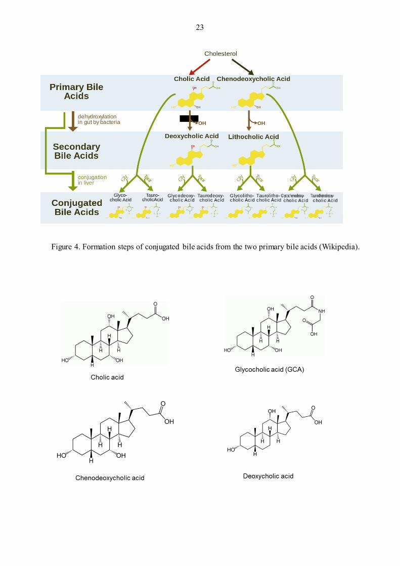
1.5 Bile and Bile Acids
Generally, bile is produced by liver and stored in the gall bladder, which can be used as the
biological detergent to solubilize fat in the gastrointestinal tract (Hofmann, Hagey et al. 2010).
There are many different compounds in the bile, for example, proteins (7%), fatty acids (12%) and
the bile acids (60%). We can see that the bile acids consist a large part of the bile. Briefly, these bile
acids are formed in the liver by multiple enzymes, secreted in the bile canaliculi and finally stored
in the gall bladder (Hofmann 1999, Hofmann, Hagey et al. 2010). Generally, there are two steps to
form the bile acids (Figure 4). Firstly, in the liver, the bile acids are conjugated with glycine or
taurine to form the conjugated bile acid. For example, glycocholic acid is the product after the
cholic acid conjugate with glycine. After the secretion in the gut tract, the conjugated bile acids will
be modified by altering or removing the hydroxylation groups or the conjugated groups with some
bacteria in the small intestine tract (Hofmann & Mysels 1992, Begley, Gahan et al. 2005). These
modification results in the cleavage of the amino acid group chains unconjugated with the bile acid.
During emulsifying the fat in the gut tract, these modified bile acids will be re-absorbed by passive
diffusion in the whole gut tract or be actively transported in the distal ileum by the enterohepatic
circulation (Huijghebaert & Hofmann 1986, Grill, Schneider et al. 1995, Mills, Mushtaq et al.
1998). According to the study, 95% of bile acids can be recycled used in the gut tract, while only
0.4-0.6 g per day were lost (Begley, Gahan et al. 2005).
To sum up, there are two mechanisms for the bile acid formation in the gut tract. The first one is
that the primary bile acids will be altered by the dehydroxylation function in the gut by bacteria to
form the secondary bile acid, i.e. deoxycholic acid is the product after the cleavage of the hydroxyl
group from the cholic acid. Then these secondary bile acids present in the live will be conjugated
either with the glycine or the taurine to form the conjugated bile acids (Grill, Schneider et al. 1995).
Generally, there are eight conjugated bile acids playing the main emulsification function in the
human gut. They are glycocholic acid (GCA), taurocholic acid (TCA), glycodeoxycholic acid
(GDCA), taurodeoxycholic acid (TDCA), glycolithocholic acid (GCLA), taurolithochoic acid
(TLCA), glycochenodeoxycholic acid (GCDA), and the taurochenodeoxycholic acid (TCDA)
(Figure 5). The components in bile acids vary in different species. The exact function of them is not
clear yet.
23
Figure 4. Formation steps of conjugated bile acids from the two primary bile acids (Wikipedia).

24
Figure 5. Structure of conjugated bile acid. (* Due to the limitation of the GCDA, GCLA, TLCA
used in this study, the structures are not shown here) (Wikipedia).
1.6 Bile Response in the Lactobacillus rhamnosus Strains
Bile present in the gut tract acts as a detergent to solubilize the fat. It is a harsh condition for the
bacteria colonizing in the gastrointestinal tract. Therefore, to survive in the human body, bile acid is
a vital condition for the bacteria to conquer. After secretion in the human gut, the concentration of
the bile present varies. According to the recent study, when the bacteria present in high
concentrations of the bile, the membrane, in which the fatty acid consist a large part of it, are
quickly dissolved, which results in the isolation of the integral proteins in the membrane and the
death of the cells (Coleman, Lowe et al. 1980, Heuman, Bajaj et al. 1996). When exposed to a low
concentration of the bile, the activity of the enzyme associated on the membrane will be altered and
in some level, the flux of different compounds to be transported through the membrane is also be
modified (Fujisawa & Mori 1996, Heuman, Bajaj et al. 1996). If some compounds that are essential
for the bacteria to survive are not provided efficiently, the metabolism and the growth of the cells
will be modified by the bile acid. In other words, the bile acids are hard conditions for the bacteria
to survive. Additionally, a study shows that when bacteria are pre-exposed to the bile acid (un-lethal

25
level), it can increase the rate of the bacteria to survive. The electron microscope investigation
shows that the cells that are not pre-exposed in the bile become empty or shrunk (Leverrier, Dimova
et al. 2003, Leverrier, Vissers et al. 2004). This fact can indicate that pre-exposure to the bile stress
can help the bacteria to survive in the stress. The mechanism behind it would be like this: under the
bile stress (un-lethal level), the structures of the proteins binding on the surface of the membrane
would be altered and then the property of the membrane will be changed by the pre-exposure to the
bile, and this alteration would prevent the lethal level bile to affect the cell.
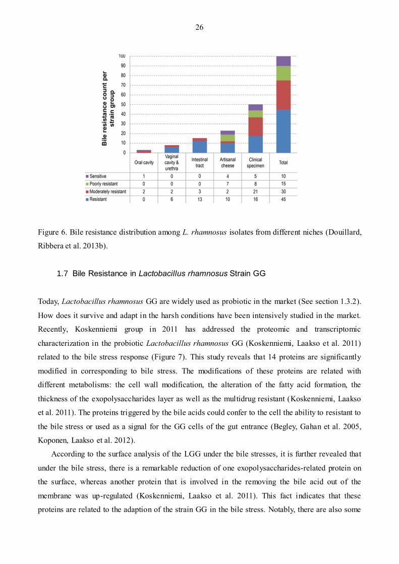
Nowadays, bile resistance of all 100 strains of species in the L. rhamnosus was examined
(Figure 6) (Douillard, Ribbera et al. 2013b). It shows that half of the strains in the L. rhamnosus
species are bile resistant (45%). It is no surprise to observe this fact, because most of them can
survive in the human gastrointestinal tract and it also indicates that the resistance to bile salts is an
essential character for the species in the human gut tract. It is consistent to the ecological fitness and
adaptions. However, the mechanism behind these bacteria is not clear yet. Recently, the whole
proteome study of the L. rhamnosus reveals that there are complex global regulation systems for L.
rhamnosus strains in the bile resistance mechanism (Koskenniemi, Koponen et al. 2009). It possibly
includes modification of the pathway of the carbohydrate metabolism, alteration of protein
biosynthesis or triggering of the expression of some genes, i.e. in the species of the Lactobacillus
reuteri, there are almost 28 genes triggered when exposed to the bile stress. The genes are related
with the metabolism pathway or the pH homeostasis (Lee & Pi 2010). There is another remarkable
example in the species Lactobacillus plantarum WCFS1. It is observed that over hundreds genes
are triggered when exposed to the bile stress (Troost, van Baarlen et al. 2008). This fact indicates
that in order to survive in the bile condition, some genes related to the bile resistance are triggered
in the L. rhamnosus strains.
To sum up, in order to survive in the bile stress condition, a lot of genes in the Lactobacillus
species would be triggered to express the proteins which are involved in the ATP-binding
transporter or the carbohydrate metabolism modification, or on the cell wall formation. Moreover,
the bile acids could also alter RNA structures or trigger DNA-repairing in the bacterial cells
(Begley, Gahan et al. 2005). The bile resistance proteins in the gut tract can also play a role in
improving the signal or the interaction between the bacteria and host. However, the exact
mechanism is not clear yet.
26
Figure 6. Bile resistance distribution among L. rhamnosus isolates from different niches (Douillard,
Ribbera et al. 2013b).
1.7 Bile Resistance in Lactobacillus rhamnosus Strain GG
Today, Lactobacillus rhamnosus GG are widely used as probiotic in the market (See section 1.3.2).
How does it survive and adapt in the harsh conditions have been intensively studied in the market.
Recently, Koskenniemi group in 2011 has addressed the proteomic and transcriptomic
characterization in the probiotic Lactobacillus rhamnosus GG (Koskenniemi, Laakso et al. 2011)
related to the bile stress response (Figure 7). This study reveals that 14 proteins are significantly
modified in corresponding to bile stress. The modifications of these proteins are related with
different metabolisms: the cell wall modification, the alteration of the fatty acid formation, the
thickness of the exopolysaccharides layer as well as the multidrug resistant (Koskenniemi, Laakso
et al. 2011). The proteins triggered by the bile acids could confer to the cell the ability to resistant to
the bile stress or used as a signal for the GG cells of the gut entrance (Begley, Gahan et al. 2005,
Koponen, Laakso et al. 2012).
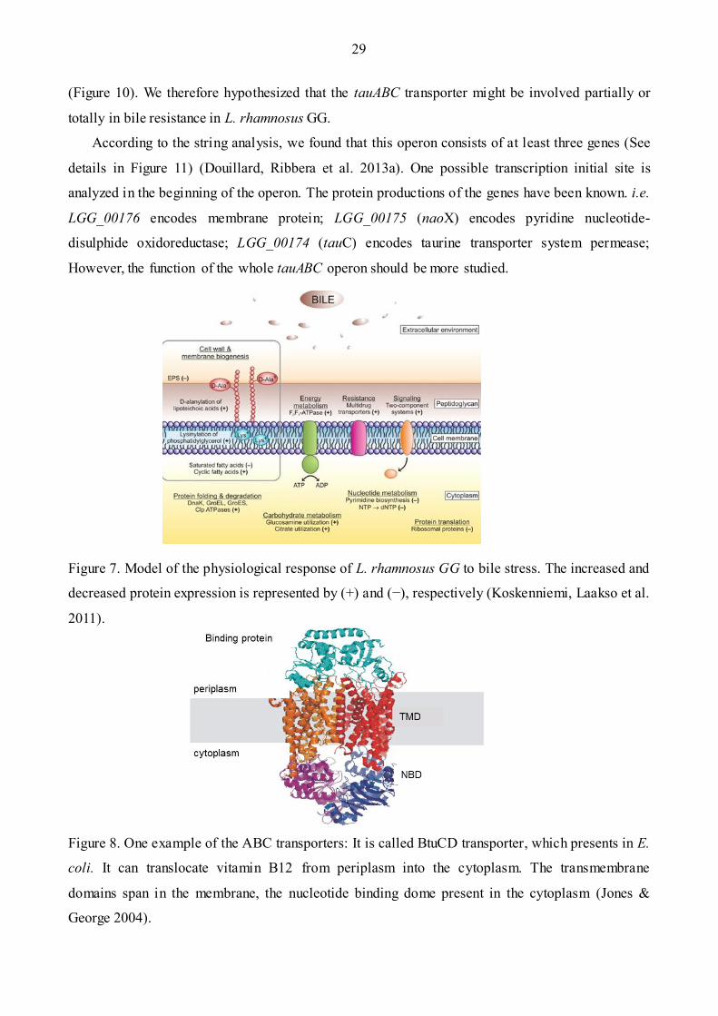
According to the surface analysis of the LGG under the bile stresses, it is further revealed that
under the bile stress, there is a remarkable reduction of one exopolysaccharides-related protein on
the surface, whereas another protein that is involved in the removing the bile acid out of the
membrane was up-regulated (Koskenniemi, Laakso et al. 2011). This fact indicates that these
proteins are related to the adaption of the strain GG in the bile stress. Notably, there are also some

27
transporters involved in the bile stress, by which the bile acid compounds can be transported outside
of the cell membrane. To gain insight into the mechanism behind the resistant of the LGG to the
bile, one region called tauABC operon was examined. The details of this transporter will be
described in section 1.9.
1.8 ABC Transporters
ABC transporter is a group of protein family, which plays important physiological functions in
different prokaryotic and eukaryotic species. They can be found almost in all the species from the
prokaryotes to human being. It is well known for their function that it can uptake the nutrition in
some bacteria (Jones & George 2004). After many years study, it has become clear that they are a
multiple system to uptake the nutrition (Davidson, Dassa et al. 2008). Their subunits belong to the
superfamily of proteins. Based on their sequences and the structures of these subunits, the ABC
transporters are defined (van Veen & Konings 1998). Nowadays, the structure and function of the
ABC transporters in different species have been intensively studied.
1.8.1 Structure of ABC transporter
The subunit of the ABC transporters is a large superfamily of proteins (van Veen & Konings 1998).
Generally, each ABC transporter consists of four sub -units that are presented in Figure 8. There are
two subunits which are transmembrane, and called as the transmembrane domains (TMD), whereas,
the other two domains are present in the intercellular for the nucleotide binding, designated as the
nucleotide binding domains (NBD) (Davidson, Dassa et al. 2008).
Today, there is a hypothesis about the construction of these subunits. Briefly, there are 6 α-
helices present in each transmembrane domain; therefore, there are 12 α-helices present in one ABC
transporters. Some of the α-helices would play a core role in the ABC transporter, for example, they
would be the specific site for the substrate binding; some of them could act as an assistant for the
binding function. However, the details of these α-helices are not clear yet. The other two nucleotide
binding domains are always present in the cytoplasmic side, and they are hydrophilic and quite
conserved. The conserved area is formed by almost 215 amino acids, which determines the family
of the transporters. These two domains may span the membrane, but some study shows that they do
not exist on the outside of the membrane (Higgins, Hiles et al. 1986, Davidson, Dassa et al. 2008,
Michaki, Guix et al. 2012). Generally, these four domains are formed separately. However, there are
still some kinds of structures that are formed by different combination between these domains. For

28
example, the multi domains polypeptides could be formed by combining with the other two separate
domains. The different models are presented in Figure 9. In the case one domain is absent, the
others can act as single dimer to maintain the function of transporters (Higgins 2001). The
structures of these different domains are clearly demonstrated, but the detailed structures of the
ABC transporters are not quite clear.
1.8.2 Function of ABC transporter
Generally, ABC transporter uses ATP as energy to translocate some substrate out of membrane
(Davidson, Dassa et al. 2008). Briefly, substrate uptake is the main function for transporters, but
nowadays, it appears that many of the transporters can also act as exporter (Davidson, Dassa et al.
2008). It means that when the cell exposed to the toxic environment, some transporters can
translocate these substrates out of the membrane. Therefore, elimination of toxic and waste
compounds outside of the cell is another important function for most ABC transporters (van Veen,
Margolles et al. 2000). Most of the ABC transporters are specific to transport one certain
compound, e.g. small, large, and highly charged molecules, as well as some inorganic ions,
polysaccharides (Chen, Sharma et al. 2001, Davidson, Dassa et al. 2008). But some other
transporters are multiply specific to different compounds. For example, the oligo-peptide
transporter, which is found on the surface of the membrane not only can transport the oligo -peptide,
but also can translocate the di- or the tripeptide (Higgins 1992). Other example is that one
transporter called LmrA discovered from Lactococcus lactis is extremely specific for the
hydrophobic compounds in the transportation process (van Veen, Margolles et al. 2000). The
characterization of ABC transporters is based on their specific substrates. The different specific
substrates can reflect the different characterization of the transporters.
1.9 The tauABC Operon in Lactobacillus rhamnosus Strain GG
The ABC transporters not only play a role in up-taking substrates but also act as an exporter in some
bacteria. In order to clarify the possibility of one transporter involved in the bile resistance of the
strain Lactobacillus rhamnosus GG, 100 L. rhamnosus strains were tested for bile resistance
(Douillard, Ribbera et al. 2013b). Combined with genomic data, the results show that one hyper-
variable region was absent in nine strains out of the twenty-five bile sensitive strains. This result
indicates that this gene cassette may affect the bile sensitivity in L. rhamnosus GG. The tauABC
operon is this gene cassette, which belongs to a kind of the ABC transporter (ATP binding cassette)
29
(Figure 10). We therefore hypothesized that the tauABC transporter might be involved partially or
totally in bile resistance in L. rhamnosus GG.
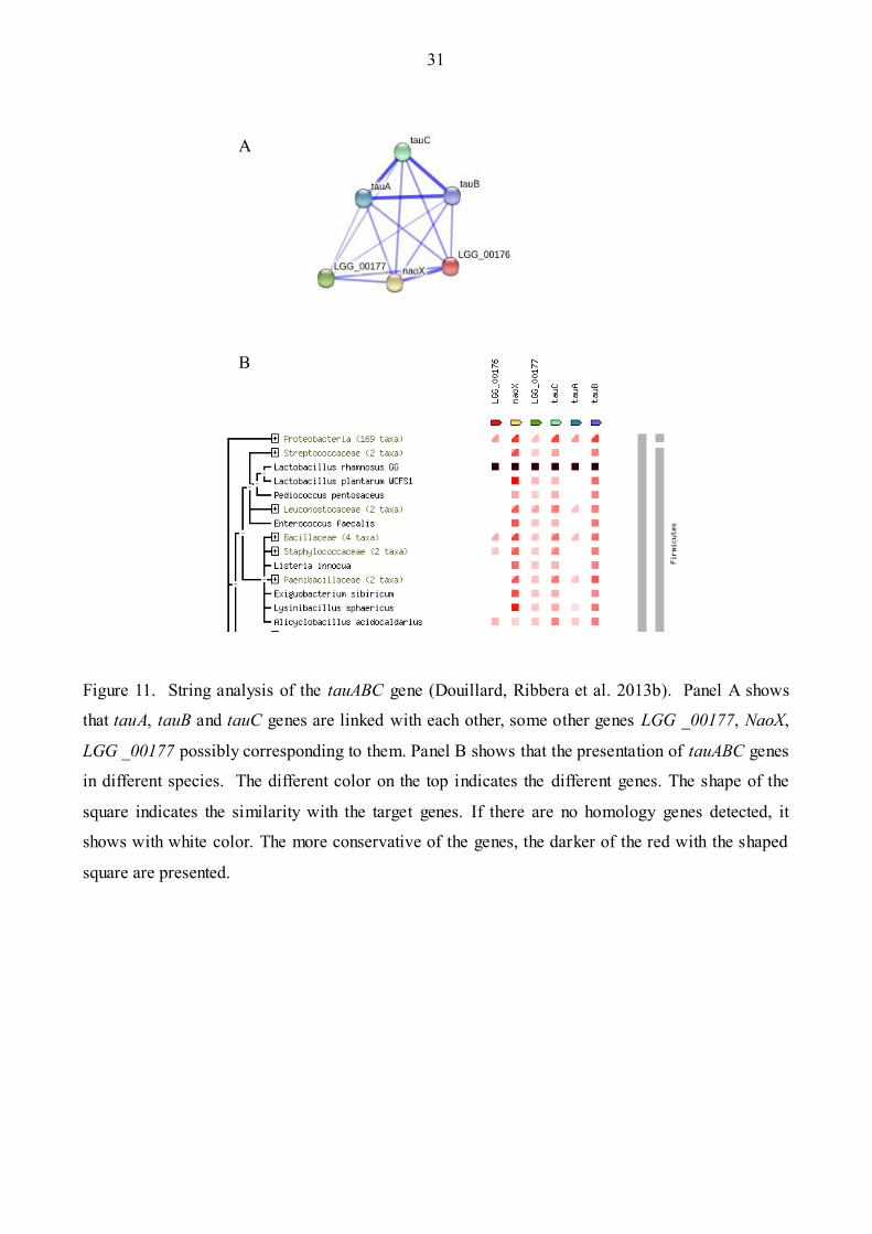
According to the string analysis, we found that this operon consists of at least three genes (See
details in Figure 11) (Douillard, Ribbera et al. 2013a). One possible transcription initial site is
analyzed in the beginning of the operon. The protein productions of the genes have been known. i.e.
LGG_00176 encodes membrane protein; LGG_00175 (naoX) encodes pyridine nucleotide-
disulphide oxidoreductase; LGG_00174 (tauC) encodes taurine transporter system permease;
However, the function of the whole tauABC operon should be more studied.
Figure 7. Model of the physiological response of L. rhamnosus GG to bile stress. The increased and
decreased protein expression is represented by (+) and (−), respectively (Koskenniemi, Laakso et al.
2011).
Figure 8. One example of the ABC transporters: It is called BtuCD transporter, which presents in E.
coli. It can translocate vitamin B12 from periplasm into the cytoplasm. The transmembrane
domains span in the membrane, the nucleotide binding dome present in the cytoplasm (Jones &
George 2004).

30
Figure 9. Organization of ABC transporters (Higgins 2001). Two transmembrane domains are
presented by the shaded squares. The nucleotide binding domains are depicted with the white oval.
(a) The transporters can be formed by four polypeptides separately (b) The nuclear binding domains
(NBD) can combine together (c) The transmembrane domains (TMD) can combine together (d)
Two fused TMD and NBD, they act as a homodimer (e) One homodimer (One fused TMD and
NBD), the other two TMD and NBD are separated polypeptides (f) One polypeptides formed by the
combination of the one TMD and NBD.
Figure 10. Taurine ABC transporter cluster in L. rhamnosus GG LGG_00177 encodes LysR family
transcriptional regulator; LGG_00176 encodes Membrane protein; LGG_00175 (naoX) encodes
pyridine nucleotide-disulphide oxidoreductase; LGG_00174 (tauC) encodes taurine transporter
system permease; LGG_00173 (tauA) encodes aliphatic sulfonate ABC transporter. LGG_00172
(tauB) encodes taurine ABC transporter ATP-binding protein (Douillard, Ribbera et al. 2013a).
31
A
B
Figure 11. String analysis of the tauABC gene (Douillard, Ribbera et al. 2013b). Panel A shows
that tauA, tauB and tauC genes are linked with each other, some other genes LGG _00177, NaoX,
LGG _00177 possibly corresponding to them. Panel B shows that the presentation of tauABC genes
in different species. The different color on the top indicates the different genes. The shape of the
square indicates the similarity with the target genes. If there are no homology genes detected, it
shows with white color. The more conservative of the genes, the darker of the red with the shaped
square are presented.
32
2 OBJECTIVES
In the present study, the expression and the integrity of the spaCBA-srtC pili cluster in LGG was
examined over a long period of time (growth of L. rhamnosus GG for 1000 generations) by PCR,
immunoblotting analysis and immune-gold staining electron microscopy. L. rhamnosus GG was
propagated in four different conditions for 1000 generations: exposure to salt (NaCl), bile salts,
vortexing treatment, sampling (top fraction). From generation 100 to 1000, aliquots were stored at -
80°C for further analyses, allowing us to study the possible genome changes in LGG at different
time points. The data collected aim at bringing insights into the long-term genome stability of L.
rhamnosus strain GG, with a particular focus on the region harboring the pilus gene cluster but also
other hyper variable chromosomal regions associated with sugar metabolism, exopolysaccharides
production, stress resistance mechanism and phages.
The bile salts significantly impact on the genome stability of L. rhamnosus GG, which is
particularly relevant in regard to its probiotic use and functions. Although bile constitutes an
important stress for L. rhamnosus GG, it is noteworthy that L. rhamnosus GG is bile resistant. We,
therefore, analyzed genes that may be involved in bile resistance, more specifically the tauABC
operon. To screen the different bile compounds and their associated effects on L. rhamnosus strain
GG and some derivatives knockout mutant. Possibly start with the construction of the knockout
mutants using homologous recombination for the tauA, tauB, and tauC, NaoX, and then to test the
bile resistant in in bovine, porcine bovine, and human bile. Finally, to characterize novel the
tauABC operon possibly involved in the bile resistant.

33
3 METHODS
3.1 Bacterial Strains and Growth Media
Several bacterial strains and plasmid studied in this research (listed in table 2.). L. rhamnosus GG
(ATCC 53103) was provided by Valio Company (Valio Ltd, Helsinki, Finland). The tauB-null strain
and complementary strain tauB-CM were developed in this study.
To examine the genome stability of GG under various stresses, Lactobacillus rhamnosus strain
GG was serially grown and transferred for an estimated time of 1000 generations under different
conditions: 3% (w/v) NaCl (Sigma, USA), 0.25% (w/v) ox gall (Sigma) bile salts, daily vortex
treatment (30s) and subculturing using top fraction. In the all four conditions, GG was grown at
37°C anaerobically in Man-Rogosa-Sharpe (MRS) (Oxoid, USA) liquid medium in static
conditions. Isolation of the colonies was performed by the serial diluting and plating on the MRS
solidified with 1% (w/v) agar.
In the bile resistance test, wild type Lactobacillus rhamnosus strains GG and mutant tauB-null
(tauB gene are deleted) was used. E. coli TOP100 with the plasmid pNZ19 harboring erythromycin
antibiotic resistance gene was used to generate the tauB-null strain. L. lactis subsp. cremoris
NZ9000 was used to produce the complementary strain (Details presented in section 3.8). When the
tauB-null strain was incubated, 5 μg/ml of the erythromycin was supplemented with the MRS
media (De Keersmaecker, Braeken et al. 2006) (Sigma, MO, USA). 10 μg/ml of the ampicillin was
used when we constructed the tauB-null strain (Details in section 3.7). 10 μg/ml of the
chloramphenicol was added to the media when the complementary strain tauB-CM was incubated
(Details presented in section 3.8).
3.2 Immunoblotting Analysis
From an overnight culture of L. rhamnosus GG (from a glycerol stock at generation n), 100 µL
aliquot was serially diluted (10-4-10-5), plated on MRS plates and further incubated at 37°C
overnight. Ninety-six single colonies were separately picked from the overnight MRS plates and
inoculated in 200 μl MRS into the 96-well plate. Next, immunoblotting analysis was carried out on
the 96-plate samples, i.e. using SpaC monoclonal antibody as a primary antibody (dilution 1:25
000), and a HRP-conjugated goat anti-mouse antibody as the secondary antibody (Biorad, CA,
USA, dilution 1:100 000). Membrane detection was done using the ECL Prime Western Blotting kit

34
(GE Healthcare, UK). If needed, SpaA polyclonal antibody was also used as the primary antibody
(dilution 1:25 000), following the same protocol.
3.3 Colony PCR Analysis
Twenty single colonies of L. rhamnosus GG grown in 96-well plates (see Section 3.1.2 above) were
sampled, transferred into strip PCR tubes, and heated in a microwave oven at full power for 3 min
(Douillard, Ribbera et al. 2013b). Then, DreamTaq Master Mix (Thermo scientific, MA)
supplemented with the appropriate primer pair was added to the PCR tubes according to the
manual’s instructions. The PCR mixture was run in a PCR thermal cycler (Thermo Scientific, MA).
Subsequently, the PCR mixture was analyzed by DNA gel electrophoresis.
Tables 2. Strains and plasmids used in this study
Strains or plasmids relevant characteristics Reference
L. rhamnosus GG Wild type
tauB-null Deletion of tauB gene in L. rhamnosus GG This study
tauB-CM Complementary strain for the tauB-null strain This study
E .coli TOP100 harbouring the pUC19-EryR plasmid Life technologies, USA
E. coli TOP10 Competent cells Life technologies, USA
pUC19 harbouring erythromythin resistance cassette
pNZ44 harbouring chloramphenicol resistance
cassette
3.4 Analysis of Hyper Variable Chromosomal Regions by PCR
Thirteen hyper variable chromosomal regions have been identified in L. rhamnosus GG (Douillard,
Ribbera et al. 2013b) and were therefore analyzed by PCR amplification, in order to examine if
these regions may have also been altered by stresses over time. For each stress condition at
generation 1000, 4 single colonies were analyzed by PCR on the following genes: LGG_00281
(sugar metabolism); LGG_00341 (sugar metabolism); LGG_00397 (sugar metabolism);
LGG_00442 (pilin protein); LGG_00512 (ABC transporter); LGG_00561 (sugar metabolism);
35
LGG_01963 (unknown function); LGG_01992 (unknown function); LGG_02043
(exopolysaccharides cluster); LGG_02613 (ABC transporter); LGG_02655 (sugar metabolism);
LGG_02744 (sugar metabolism). An additional twelve samples were analyzed if one particular
characterization feature revealed some changes. (The list sequences used in this study is in Table 3)
Tables 3. List of the hyper variable regions in the strain L. rhamnosus GG (Douillard, Ribbera et al.
2013b).
3.5 Immuno-gold Staining Transmission Electron Microscopy Analysis
Immuno-gold staining transmission electron microscopy was used to examine SpaCBA pili in L.
rhamnosus GG at different time points and the mutant strain tauB-null. According to the
immonogolding-labeling protocol described (Reunanen, von Ossowski et al. 2012), tauB-null strain
is grown to the stationary phase for the sample preparation. In the room temperature, the formvar
carbon-coated copper grids were incubated with the growth culture for 30 min. PBS with 0.02 M
glycine was used to wash the grids three times. Then the grids were incubated with the BSA (bovine
serum albumin) blocking solution (1% w/v) for 15 minutes. The primary antibody for the
Target Genes Main genetic features of the region
LGG_00278---LGG_00283 rhamnosyl PTS, rhamnosyltransferase
LGG_00341---LGG_00347 galactitol PTS, conserved protein
LGG_00376---LGG_00427 transcriptional regulator, hypothetical protein
LGG_00438---LGG_00481 Conserved protein, SpaCBA Pili cluster, transcription regulator,
LGG_00511---LGG_00517 ABC transporter, conserved protein
LGG_00559---LGG_00566 Conserved protein, transporter, sugar phosphate isomerase
LGG_01955---LGG_01967 Conserved protein
LGG_01990---LGG_02003 Conserved protein
LGG_02038---LGG_02056 EPS cluster
LGG_02610---LGG_02614 ABC transporter, conserved protein
LGG_02651---LGG_02686 fucose transporter, conserved protein, transcriptional regulator
LGG_02742---LGG_02755 Conserved protein

36
incubation is the polyclonal SpaA with the concentration of 1:100 in the BSA blocking solution.
After one hour incubation, 0.1% (w/v) BSA was used to wash the grids for many times. Then the
grids were incubated with the protein A gold conjugate for 20 minutes. After that PBS was used to
wash the grids many times and the grids were fixed with 1% (w/v) glutaraldehyde for 5 minutes.
The last step was using the 1.8% methylcellulose and the 0.4% (w/v) uranyl acetate solution to stain
the grids. Finally, the grids were observed in the Jeol 1200 EX II transmission electron microscope
(Jeol Ltd. Japan).
3.6 Co-culturing of the Wild-type L. rhamnosus GG and a Pili Negative Mutant
Derivative
L. rhamnosus GG wild type strain and a spontaneous mutant LGG-B-1000 (pili negative, bile,
generation 1000) were grown anaerobically in 2 ml of MRS liquid medium. From an overnight
culture, the OD600 were measured, adjusted and equivalent numbers of bacterial cells of GG and
LGG-B-1001 were inoculated overnight into 2 ml of fresh MRS broth. Hundred microliters of the
cell suspension was serially diluted and plated on MRS plates and incubated overnight for
subsequent colony PCR screening, as described above. Twenty microliters of culture were sub-
cultured into 1980 μl of fresh MRS medium. The same procedure (culturing, colony picking & PCR
screening) was performed for 7 days.
3.7 Fluorescence Microscopy
L. rhamnosus strains were grown overnight in MRS. Bacterial cells were pelleted by centrifugation
for 5 min at 4,500 rpm. Next, cells were washed with PBS and the OD600 was adjusted to 1.0 and
200 μl cell suspension was incubated with anti-SpaA rabbit polyclonal antibodies for at least 30 min
at room temperature. Alexa-488-conjugated anti-rabbit goat antibodies were then diluted to 1-2
μg/ml and cells were incubated for 30 min. The whole procedure was carried out in dark. Cells were
finally washed with PBS and 10 μl of the cell preparation was analyzed by fluorescence microscopy
(Jeol Ltd. Japan).

37
3.8 Construction of L. rhamnosus GG tauB-null Mutant
A 449-bp fragment of tauB (position 138-583) was amplified by PCR using L. rhamnosus GG
genomic DNA. The primers used were: F-tauB: 5’- CG GGATCC TCC GCT GGC CGA GAA ACA
AC-3’ and R-tauB: 5’-GC CTCGAG TCT GAT TGC CGG CTA CCT GC-3’. They both contained
restriction sites (underlined sequences) that were used for subsequent cloning: BamHI and XhoI.
Plasmid Extraction: E. coli TOP100 harboring pUC19-Ery was grown in 15 ml LB broth
containing 100 μg/ml ampicillin at 37ºC overnight under agitation. QIAPrep Plasmid Maxi Kit
(QIAGEN, USA) was then used to extract the plasmid pUC19-Ery from E. coli TOP100 liquid
cultures.
Primer Design: The primers were designed using Primer Designer software to amplify the 449-
bp long fragment and introduce BamHI and XhoI restriction sites to both ends of the amplicons.
Restriction Digestion: The purified PCR amplicon tauB (138-583) was digested by BamHI and
XhoI, as follows: 20.5 μl DNA, 1 μl BamHI (Thermo Scientific, USA), 1 μl XhoI (Thermo
Scientific) and 2.5 μl 10X Fast Digest Buffer (Thermo Scientific). The mixture was incubated at
37ºC for 2 hours. Similarly, the plasmid pUC19-Ery was digested with BamHI and XhoI.
Ligation Reaction: The digested and purified tauB fragment and pUC19 plasmid were ligated,
as follows: 5 μl plasmid, 7 μl insert DNA and 10X Ligation Buffer (Promega, WI, USA) and 2 μl
T4 ligase (Promega). The ligation mixture was incubated at 4 ºC overnight.
Transformation by Heat Shock: The ligation product was transformed into One-shot E. coli
competent cells TOP10 (Invitrogen, USA) as per manual’s instructions. Briefly, the competent cells
were thawed on ice and incubated for 30 minute with 5 μl DNA. The vial was incubated for 30s at
42 ºC and 2 minute on ice. Next, 250 μl pre-warmed S.O.C medium was added into the vial. The
cell suspension was incubated at 37 ºC for 1 h at 250 rpm. 80 μl of cells were plated on pre-warmed
selective LB agar plates, containing 100 μg/mL of ampicillin and incubated overnight at 37ºC.
Colonies were screened by PCR and positive transformants were analyzed by sequencing. This
yielded to plasmid pUC19-Ery-tauB that was subsequently used for inactivating the tauB in GG.
Electroporation: Wild-type L. rhamnosus strain GG and all the mutant strains were cultured
overnight in 5 ml pre-warmed MRS medium supplemented with 2% (w/v) glycine at 37 ºC. Then,
GG cells were inoculated (1/25) into 100 ml pre-warmed MRS medium supplemented with 2%
(w/v) glycine at 37 ºC. Ampicillin was added to the final concentration of 10 μg/ml when the OD600
reached 0.2 to 0.3. When OD600 was 0.4-0.5, cells are harvested by centrifuging 10 min at 4,000
rpm. The cells were washed twice at room temperature with electroporation buffer (0.5 M Sucrose,

38
7 mM potassium phosphate, 1 mM MgCl2), then resuspended in 1 ml of the electroporation buffer
and put on ice prior to electroporation. Hundred microliters of cell suspension and 4 μl DNA of the
vector pUC19-Ery-tauB pipetted into a pre-chilled electroporation cuvette with 0.2-cm electrode
gap and electroporated using the following setting: 1.7 kV (voltage), 25 μF (capacitance) and 200 Ω
(resistance), then 1 ml of ice-cold MRS medium containing 2 mM Cacl2 and 20mM Mgcl2 was
immediately added to the cuvette, which was incubated at 37 ºC for 3 h. The cell suspension was
plated onto MRS agar plates containing 5 μg/ml of the erythromycin (Sigma, MO, USA) and
incubated in anaerobic conditions at 37ºC for 48 to 72 h.
Screening: Positive transformants were identified by PCR using the following primer: F-tauB:
5’-CG GGATCC TCC GCT GGC CGA GAA ACA AC-3’ and R-tauB: 5’-GC CTCGAG TCT GAT
TGC CGG CTA CCT GC-3’. If the colony on the MRS plate with erythromycin was L. rhamnosus
GG tauB-null mutant, the PCR product was totally 4727-bp (449-bp homology sequence and 3829-
bp pUC-Em and 449-bp homology sequences). This PCR product was sequenced and the results
showed that it included the whole plasmid pUC-Ery-tauB sequence, indicating the pUC19-Ery-tauB
has been inserted into the chromosome.
3.9 Complementation of the tauB-null Mutant Strains
The gene tauB was amplified by PCR from L. rhamnosus strain GG using the primer pairs: F-tauB:
5’-AGC CAT GGA AGG AGG CAC TCA CCA TGT CAT TAC TGA CAC TTG ATC AT-3’, R-
tauB: 5’- AGC ACT AGT TTA GCT AAT CCC ATT CAA CAA TTC C-3’. Restriction sites used
for subsequent cloning are underlined: NcoI and SpeI. The PCR production was digested by NcoI
and SpeI and then insert into the similarly digested plasmid pNZ44. The ligation mixture was
electroporated into L. lactis subsp. cremoris NZ9000 as previously described (Douillard, Ribbera et
al. 2013a). Following colony PCR screening and sequencing analysis, we obtained the plasmid
pNZ44-tauB, which was introduced into the tauB-null strain, as done in Section 3.8.
Transformants were verified by colony PCR using the primer F-tauB: 5’-AGC CAT GGA AGG
AGG CAC TCA CCA TGT CAT TAC TGA CAC TTG ATC AT-3’ (NcoI), R-tauB: 5’-AGC ACT
AGT TTA GCT AAT CCC ATT CAA CAA TTC C-3’ (SpeI). If the colony on the MRS with
erythromycin and chloramphenicol antibiotics is the complementary strain, the PCR product should
be the 732 bps, which are the whole sequences of tauB. The PCR product was sequenced with
results shows that the combined plasmid was present into the tauB-null strain. The complemented
strain was then analyzed for bile resistance.

39
3.10 Sensitivity of Wild-type L. rhamnosus GG and tauB-null Mutant Strain in the
Presence of Ox Bile Extracts
Two milliliters of wild-type L. rhamnosus GG cells and tauB-null cells were propagated in MRS
liquid medium with 3% (w/v) ox gall bile salt (Sigma) in static conditions anaerobically at 37 C.
The tauB-null culture was supplemented with 5 μg/ml erythromycin. After overnight, the OD600
were measured and the same numbers of GG and mutant cells were inoculated into separate 15 mL
Falcon tubes were kept at 37ºC in a water bath. Every 30 min, the OD600 values were measured until
the cultures reached the stationary phase and then after 24 h and 48 h. The growth rates of each
sample were calculated, resulting in generation times of the tested strains. The complementary
strains tauB-CM was also performed in the resistant test. The protocol is the same with the former
two strains, except supplied with the 5 μg/ml erythromycin (Sigma, MO, USA) and 5 μg/ml
chloramphenicol (Sigma, MO, USA) antibiotic (De Keersmaecker, Braeken et al. 2006).
3.11 Sensitivity of Wild-type L. rhamnosus GG and tauB-null Mutant Strain in the
Presence of Pig Bile
Two milliliters of wild type L. rhamnosus GG cells and tauB-null cells was incubated anaerobically
in MRS liquid medium with either 0.3%, 0.2%, 0.1% or 0.05% (w/v) porcine extracts (Sigma) in
static conditions. The tauB-null culture was supplemented with 5 μg/ml erythromycin. The tauB-
CM was also tested when needed. The rest of the protocol was identical as described in Section 3.9.
3.12 Sensitivity of Wild-type L. rhamnosus GG and tauB-null Mutant Strain in the
Presence of Human Bile
There are four kinds of human bile extracted from four subjects (Groningen NL). Similarly, two
milliliters of wild type L. rhamnosus GG cells and tauB-null cells was incubated anaerobically in
MRS liquid medium with 2% (w/v) human bile extracts in static conditions. The tauB-null culture
was supplemented with 5 μg/ml erythromycin. After overnight, the OD600 were measured and the
same amount of culture for LGG and mutant will be inoculated into 2 ml cuvettes in the 37ºC
separately. Then every 30 minutes the OD600 value was measured until the growth phase started in
stationary phase. After 24 h, the OD600 of different cultures were also measured. According to the
results, subject 2 and subject 5 have been performed in triplicate. And the complementary strain
tauB-CM was also tested.

40
3.13 Sensitivity of Wild-type L. rhamnosus GG and tauB-null Mutant Strain in the
Presence of Bile Components
Two milliliters of wild type L. rhamnosus GG cells and tauB-null cells was incubated anaerobically
in MRS liquid medium with on the compounds present in bile salts in static conditions. The tauB-
null culture was supplemented with 5 μg/ml erythromycin. The components tested were glycocholic
acid (GCA), taurocholic acid (TCA), glycodeoxycholic acid (GDCA), taurodeoxycholic acid
(TDCA), glycochenodeoxycholic acid (GCDA), and taurochenodeoxycholic acid (TCDA). Firstly,
5 mM of each component were used to grow the wild type LGG and LGG tauB-null. However,
LGG and tauB-null strains cannot grow in such concentrations of some components. Therefore,
different concentrations have been tes ted in different concentration. (The details are in the Table 4).
The complementary tauB-CM was also tested at 8 mM and 5 mM of the components TDCA and
TCDA separately. The rest of the protocol was identical, as described in Section 3.9.
Table 4. Different concentration tested in the bile components
Components Concentrations used for testing
GCA 5 mM, 8 mM
GDCA 0.5 mM, 1 mM, 1.5 mM, 2 mM
TCA 5 mM, 10 mM
TDCA* 5 mM, 8 mM
GCDA 5 mM
TCDA* 5 mM, 8 mM
Taurine 5 mM, 200 mM, 300 mM
All the tests in the different concentrations were performed twice, TDCA* and TCD * were
performed in triplicate.

41
4 Results
4.1 Immunoblotting Analysis of the SpaCBA Pili in L. rhamnosus GG
L. rhamnosus GG cells at generation 1000 were first analyzed to examine whether the SpaCBA pili
were lost in the four growth conditions. Then immunoblotting of the cells at generation 900, 800,
700 till 100 were performed or until we observed 100% pili-positive. The immunoblotting results at
each time point were also verified by PCR using spaB and spaA primer pairs.
The immunoblotting and PCR analysis for top fraction condition (Figure 12, Panel A) showed
that L. rhamnosus GG from the top fraction did not lose pili over 1000 generations. The results were
confirmed by PCR using spaB primer pairs. At generation 1000, immunoblotting data were also
confirmed using polyclonal anti-SpaA antibody. The slightly higher data of the PCR result than the
immunoblotting is possibly due to either the technical variation or the small scale of samples being
taken for the PCR analysis that can demonstrate higher scale percentage of the result. In contrast,
Figure 12 (Panel B) showed that vortex treatment substantially affected the presence of the SpaCBA
proteins in L. rhamnosus GG over time. This was confirmed by immunoblotting with both
polyclonal and monoclonal anti-SpaC antibodies. One example of the immunoblotting membrane
was shown in Figure 13. The same results based on immunoblotting with polyclonal anti-SpaA
antibody were obtained (data not shown).
The immunoblotting results under bile exposure (Figure14, Panel A) demonstrated that some L.
rhamnosus GG cells started to lose the SpaCBA pili after 200 generations. No pili-carrying colonies
were detected after 500 generations. The PCR results, obtained with spaB primer pair confirmed
immunoblotting data. After 1000 generation, L. rhamnosus GG cells observed by immuno-electron
microscopy had no pili (Figure 14, Panel B).
Under the salt condition, all the PCR results demonstrated that all cells possessed pili at
generation 1000, whereas the immunoblotting results were different. The pili started to be lost from
generation 750 (Figure 15, Panel A). Although monoclonal SpaC immunoblotting data showed a
loss of SpaC pili after 750 generation, PCR, polyclonal SpaA and SpaC immunoblotting were
congruent, and indicated that a majority of L. rhamnosus GG cells display pili over 1000
generations (Figure 15, Panel B). The discrepancy with monoclonal SpaC immunoblotting data may
be due to some technical artifacts. We cannot exclude the possibility that a mutation occurred in the
epitope region of the SpaC protein.
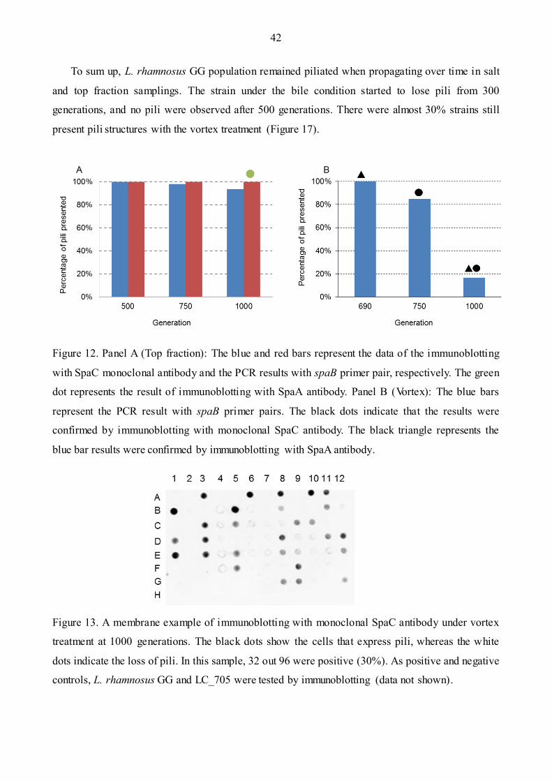
42
To sum up, L. rhamnosus GG population remained piliated when propagating over time in salt
and top fraction samplings. The strain under the bile condition started to lose pili from 300
generations, and no pili were observed after 500 generations. There were almost 30% strains still
present pili structures with the vortex treatment (Figure 17).
Figure 12. Panel A (Top fraction): The blue and red bars represent the data of the immunoblotting
with SpaC monoclonal antibody and the PCR results with spaB primer pair, respectively. The green
dot represents the result of immunoblotting with SpaA antibody. Panel B (Vortex): The blue bars
represent the PCR result with spaB primer pairs. The black dots indicate that the results were
confirmed by immunoblotting with monoclonal SpaC antibody. The black triangle represents the
blue bar results were confirmed by immunoblotting with SpaA antibody.
Figure 13. A membrane example of immunoblotting with monoclonal SpaC antibody under vortex
treatment at 1000 generations. The black dots show the cells that express pili, whereas the white
dots indicate the loss of pili. In this sample, 32 out 96 were positive (30%). As positive and negative
controls, L. rhamnosus GG and LC_705 were tested by immunoblotting (data not shown).
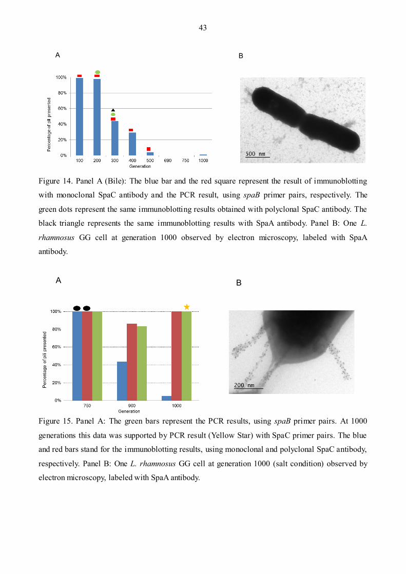
43
Figure 14. Panel A (Bile): The blue bar and the red square represent the result of immunoblotting
with monoclonal SpaC antibody and the PCR result, using spaB primer pairs, respectively. The
green dots represent the same immunoblotting results obtained with polyclonal SpaC antibody. The
black triangle represents the same immunoblotting results with SpaA antibody. Panel B: One L.
rhamnosus GG cell at generation 1000 observed by electron microscopy, labeled with SpaA
antibody.
Figure 15. Panel A: The green bars represent the PCR results, using spaB primer pairs. At 1000
generations this data was supported by PCR result (Yellow Star) with SpaC primer pairs. The blue
and red bars stand for the immunoblotting results, using monoclonal and polyclonal SpaC antibody,
respectively. Panel B: One L. rhamnosus GG cell at generation 1000 (salt condition) observed by
electron microscopy, labeled with SpaA antibody.
A B
A B

44
4.2 PCR Analysis of 13 Hyper Variable Regions Over Time
Exposure to bile salts and vortex trigger the loss pili in LGG at different extent. The results (Figure
16) show that the gene (LGG_00442) was substantially altered in both conditions. This gene is
located within the genomic island, which also encodes the pili protein (including LGG_00442).
These genes were not altered under the salt and top fraction conditions (data not shown). Based on
the PCR results, exposure to vortex and bile salts trigger the loss of the pili in L. rhamnosus GG
from generations 500 and 100, respectively. In contrast, the pili were maintained under salt
exposure and top fraction sampling (Figure 17).
Figure 16. Mutation percentage of 13 hyper variable regions under bile and vortex treatment over
1000 generations. Panel A shows that the genes LGG_00397 (sugar metabolism) –and LGG_00442
(pilin gene) which were in one of the genome islands almost mutated under vortex treatment. The
same regions are mutated under bile treatment (Panel B).
A B

45
Figure 17. Summary of L. rhamnosus GG genome stability under different conditions. The strains
exposed in the bile condition start to lose pili from 300 generation, and no pili observed start from
500 generations. Almost all the pili are present on the surface of the bacteria in the salt condition
and with the top fraction treatment. There are about 30% strains presenting the pili at 1000
generations with vortex treatment. (This data is based on the immunoblotting with the monoclonal
antibody, which is consistent with the PCR results.)
4.3 Co-culturing of the Wild-type L. rhamnosus GG and a Pili Negative
Mutant
In order to test the growth ability of the wild type and LGG-B-1001, co-culturing of the two strains
was performed. The competitive assay was conducted for 8 days (the equivalent of 5 generations
per overnight culture) (Figure 18). The results indicated that the mutated strain grew faster than the
wild type, as shown by immunoblotting analysis with monoclonal anti -SpaC antibody, PCR analysis
(25 and 30 generations), and fluorescent microscopy (30 generations).
On day 6 (30 generations), an aliquot of L. rhamnosus GG cells was observed by fluorescence
microscopy (Figure 19). Cells showing green fluorescent possess pili on their surface, whereas
other cells had no pilin protein display on the cell surface. When comparing cells with and without
fluorescence (same area), the ratio of pili (+) vs. pili (-) cells were about 20%:80%, which is in
agreement with the colony PCR results.

46
A B
Figure 18. Competitive assay between the wild type L. rhamnosus GG and pili negative mutant. The
black dots stand for the same results detained in the second time of co-culture. The green triangle
standing for the L. rhamnosus GG cells at 30 generations was observed by fluorescent microscopy.
Figure 19. Fluorescent microscopy at day 6. Panel A: The cells appearing as green fluorescent
represent the ones that have the pilin protein. The number of the cells was about 10. Panel B: All
cells were counted as 39 under the microcopy. The rate between wild type and the mutant are also
20%: 80%.
47
4.4 Generation of tauB-null Mutant Strain
The knockout tauB-null strain was constructed with the homologous recombination technique.
Briefly, the proposed model of the knockout gene should be the plasmid with antibiotic resistance
gene inserted into the tauB gene (Figure 20). The PCR production of the tauB-null with the primer
F-tauB: AGC CAT GGA AGG AGG CAC TCA CCA TGT CAT TAC TGA CAC TTG ATC AT
(NcoI), R-tauB: AGC ACT AGT TTA GCT AAT CCC ATT CAA CAA TTC C (SpeI) should be
around 4900bp. This size should include 449bp similar genes with tauB gene; insert plasmid and
732bp tauB genes. This knockout strain was confirmed by PCR production (Figure 21). The PCR
product was also sequenced with results showing that the plasmid was present into tauB gene.
In order to clarify the phenotype of the tauB-null strain, API 50CH test and TEM was analyzed.
The API test indicated that there was no different for the sugar consuming between the knockout
tauB strain tauB-null and the wild type (Table 5). The electron microscope analysis showed that the
pili was still present in the knockout tauB strains, which indicates that probably the tauB genes were
not responding to the loss of pili in the present of bile (Figure 23). The characterization of its bile
resistant phenotype was described in section 4.6, 4.7, 4.8, 4.9.
4.5 Complementation of tauB-null Mutant
To get the complementation of tauB-null mutant tauB-CM, we cloned the full tauB genes (732 bp
region) into plasmid pNZ44, and then introduced it into the knockout tauB strain (Figure 20). The
model of the complementation strain contains the knockout tauB chromosome, as well as the
combined plasmid pNZ44 with chloramphenicol antibiotic resistance genes (Figure 21). Therefore,
the tauB-CM strain is resistant to two antibiotics: chloramphenicol and erythromycin. The PCR
product of the plasmid was sequenced with results showing that pNZ44 was harboring the antibiotic
gene, which were inserted into the tauB-null strain (Data not presented).

48
Figure 20. Model of the knockout tauB mutant with the insertion of plasmid pUC19-Ery with the
erythromycin antibiotic gene. The purple bar presents the erythromycin resistance gene.
Figure 21. Complementation knockout tauB strain (tauB-CM). It includes the pUC-EM2.0 (purple
bar present), which contains the erythromycin resistance antibiotic. It is constructed with the tauB
sequences with the homologous sequence (Blue bar), and the constructed plasmid pNZ44 with the
chloramphenicol antibiotic (Yellow bar).
pNZ44-tauB
tauB tauB
pUC19-Ery tauB

49
Figure 22. PCR production with the primer of F-tauB, R-tauB. The tauB product size is 4900-bp.
(The PCR product should be 732-bp tauB sequence and 3829-bp pUC-Em and 449-bp homology
sequences). The control LGG product size is 732bp.
Figure 23. Electron microscopy of the knockout cells. The phenotype of the pili is similar with the
wild type, which indicates that the tauB-null strain is not responding to pili lost in the present of bile
(Using the antibody SpaA).
3000bp
5000bp
750bp
tauB-null LGG
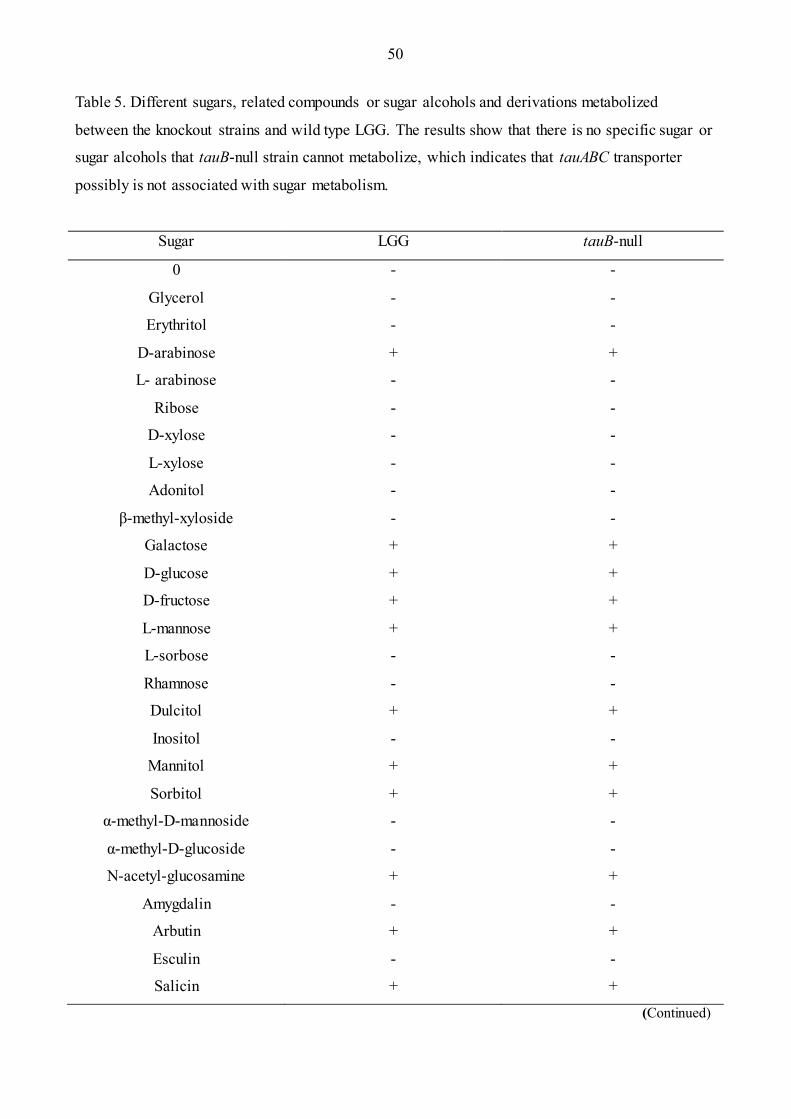
50
Table 5. Different sugars, related compounds or sugar alcohols and derivations metabolized
between the knockout strains and wild type LGG. The results show that there is no specific sugar or
sugar alcohols that tauB-null strain cannot metabolize, which indicates that tauABC transporter
possibly is not associated with sugar metabolism.
Sugar LGG tauB-null
0 - -
Glycerol - -
Erythritol - -
D-arabinose + +
L- arabinose - -
Ribose - -
D-xylose - -
L-xylose - -
Adonitol - -
β-methyl-xyloside - -
Galactose + +
D-glucose + +
D-fructose + +
L-mannose + +
L-sorbose - -
Rhamnose - -
Dulcitol + +
Inositol - -
Mannitol + +
Sorbitol + +
α-methyl-D-mannoside - -
α-methyl-D-glucoside - -
N-acetyl-glucosamine + +
Amygdalin - -
Arbutin + +
Esculin - -
Salicin + +
(Continued)

51
(Continued)
Sugar LGG tauB-null
Cellobiose + +
Maltose - -
Saccharose - -
Trehalose + +
Inulin - -
Melezitose - -
D-raffinose - -
Amidon - -
Glycogene - -
Xylitol - -
β-gentiobiose - -
D-turanose + +
D-lyxose - -
D-tagatose + +
D-fucose - -
L-fucose + +
D-arabitol - -
L-arabitol - -
Gluconate + +
2-keto-gluconate - -
2-keto-gluconate + +
52
4.6 Effects of Ox Gall on tauB-null Strain vs. L. rhamnosus GG
Wild type LGG and tauB-null mutant were incubated in the presence of 3% (w/v) ox gall
separately. In addition, two other groups were performed as control without ox gall. The results
showed that in the presence of ox gall, the WT and mutant tauB strains grew slower than the ones in
the MRS medium without ox gall. The tauB knockout strains grew significantly slower (about 40
minutes longer generation time) than the wild type in the presence of ox gall (Figure 24). The
complementary strain grew at a comparable rate as the wild type, indicating that the introduction of
a functional copy of the tauB gene in L. rhamnosus tauB-null mutant restored the wild-type
phenotype. The observed mutant phenotype is therefore clearly due the alteration of tauB gene.
The phenotypes of the mutant strains and the WT were also tested: The control group and the
two test groups were analyzed by cell sedimentation for 24 and 48 hours in the presence of 3%
(w/v) ox gall. The amount of cells of tauB-null strain was significantly less than the wild type after
the 24 hours. The complementary strain grew at similar amount as wild type (Figure 26 A). The
difference between the wild type GG and tauB-null strain still existed after 48 hours, but the
difference was smaller (Figure 26 B). This result is consistent with the OD600 value after overnight
incubation, which is 3.2 for the wild type LGG and 2.3 for the knockout tauB-null strain.
The OD600 and pH value were also analyzed after 24h and 48 hours. The result shows that
OD600 value of the mutant strain was always lower than the wild type (Figure 27). The similar pH
value for three strains (not presented) indicates that the phenotype was not induced by the pH,
probably other reasons.
4.7 Effects of Pig Bile Salts on tauB-null Strain vs L. rhamnosus GG
Different percentages of the porcine bile were tested (5% - 0.05%). Only the result by 0.05% (w/v)
presented here (Figure 25). (The other results do not present here, because these two strains cannot
grow above this concentration). In the 0.05% (w/v) concentration, we found that there were no
differences of the generation time between these two strains. If there is one specific component
sensitive for the knockout tauB, we assume that the amount of it would be much lower than the
amount that could induce the bile sensitivity.

53
Figure 24. Generation time of the WT and mutant tauB-null and complementation strain in the
presence of 3% ox gall. p value < 0.002 (* This experiment was performed three times)
Figure 25. Generation time of the WT VS tauB-null in the present of 0.05% porcine bile.
p value < 0.002 (* This experiment was performed three times)
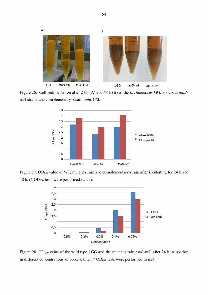
54
Figure 26. Cell sedimentation after 24 h (A) and 48 h (B) of the L. rhamnosus GG, knockout tauB-
null strain, and complementary strain tauB-CM.
Figure 27. OD600 value of WT, mutant strain and complementary strain after incubating for 24 h and
48 h. (* OD600 tests were performed twice).
Figure 28. OD600 value of the wild type LGG and the mutant strain tauB-null after 24 h incubation
in different concentrations of porcine bile. (* OD600 tests were performed twice).
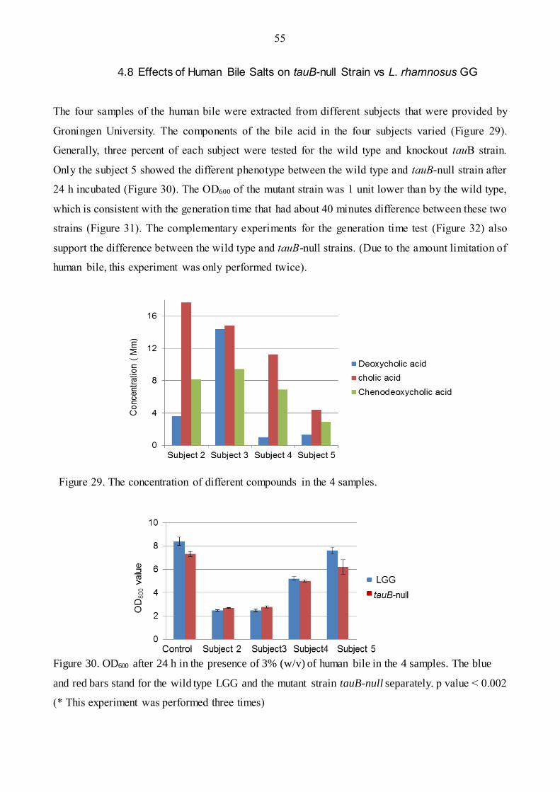
55
4.8 Effects of Human Bile Salts on tauB-null Strain vs L. rhamnosus GG
The four samples of the human bile were extracted from different subjects that were provided by
Groningen University. The components of the bile acid in the four subjects varied (Figure 29).
Generally, three percent of each subject were tested for the wild type and knockout tauB strain.
Only the subject 5 showed the different phenotype between the wild type and tauB-null strain after
24 h incubated (Figure 30). The OD600 of the mutant strain was 1 unit lower than by the wild type,
which is consistent with the generation time that had about 40 minutes difference between these two
strains (Figure 31). The complementary experiments for the generation time test (Figure 32) also
support the difference between the wild type and tauB-null strains. (Due to the amount limitation of
human bile, this experiment was only performed twice).
Figure 29. The concentration of different compounds in the 4 samples.
Figure 30. OD600 after 24 h in the presence of 3% (w/v) of human bile in the 4 samples. The blue
and red bars stand for the wild type LGG and the mutant strain tauB-null separately. p value < 0.002
(* This experiment was performed three times)

56
Figure 31. Generation time of WT, mutant strain tauB-null, and the complementary strain tauB-CM
growing in the presence of 3% (w/v) bile of each subject. The blue, red, green bar stands for the
wild type LGG, the mutant strain, and the complementary strain separately. p value < 0.002 (* This
experiment was performed three times)
Figure 32. Generation time of WT, mutant strain tauB-null, complementary strain tauB-CM growing
in the presence of 3% (w/v) of human bile (subject 5). (*Sensitive tests were performed twice)
57
4.9 Comparative Curve of GG WT vs. tauB-null Strain in the Presence of Bile
Components
All the bile salts were tested for the two strains. The salts included the primary bile acids: cholic
acid and chenodeoxycholic acid (CDC); the secondary bile acid: deoxycholic acid, lithocholic acid,
and the conjugated bile acids: glycocholic acid (GCA), taurocholic acid (TCA), glycodeoxycholic
acid (GDCA), taurodeoxycholic acid (TDCA), taurilithochoic acid (TLCA), and
taurochenodeoxycholic acid (TCDA).
Bile sensitivity of the tauB-null strain and wild type were tested in all components. (Results
were shown in Figure 33). Generally, 5 mM was used to test all components, but LGG and tauB-
null could not grow well in GDCA component. Therefore, the concentrations chosen to test varied
among the different components (shown in Figure 33). According to the final results, we found that
GCA, GDCA, taurine, and TCA did not present a different phenotype between the LGG and tauB-
null strain. Additionally, the raising of the concentration would decrease the generation time for
both strains in taurine test.
There were significant differences of the generation times between the wild type and tauB-null
strain in the presence of TDCA. Figure 33 (Panel E) shows that as the increase of concentration of
TDCA (5 mM to 8 mM), the time differences from around 30 minutes increased to 40 minutes. In
order to confirm this difference, complementary experiments were performed in 8 mM TDCA
(shown in Figure 34). The complementary strain grew at comparable rate as the wild type, but much
faster than the mutant strain. Another component TCDA at 5 mM also resulted in time differences
between these two strains. Complementary data also confirmed the results (Figure 35).
To sum up, there are two components, namely TDCA and TCDA that cause the generation time
differences between the wild type GG and the tauB-null. This significant difference can be around
40 minutes in both of the components, which is supported by the complementary experiment. As all
the tests were preliminary experiments, all of them were performed twice. Due to the limitation of
the components, TLCA and CDC were only tested one time (The results are not presented here

58
B
C
A A
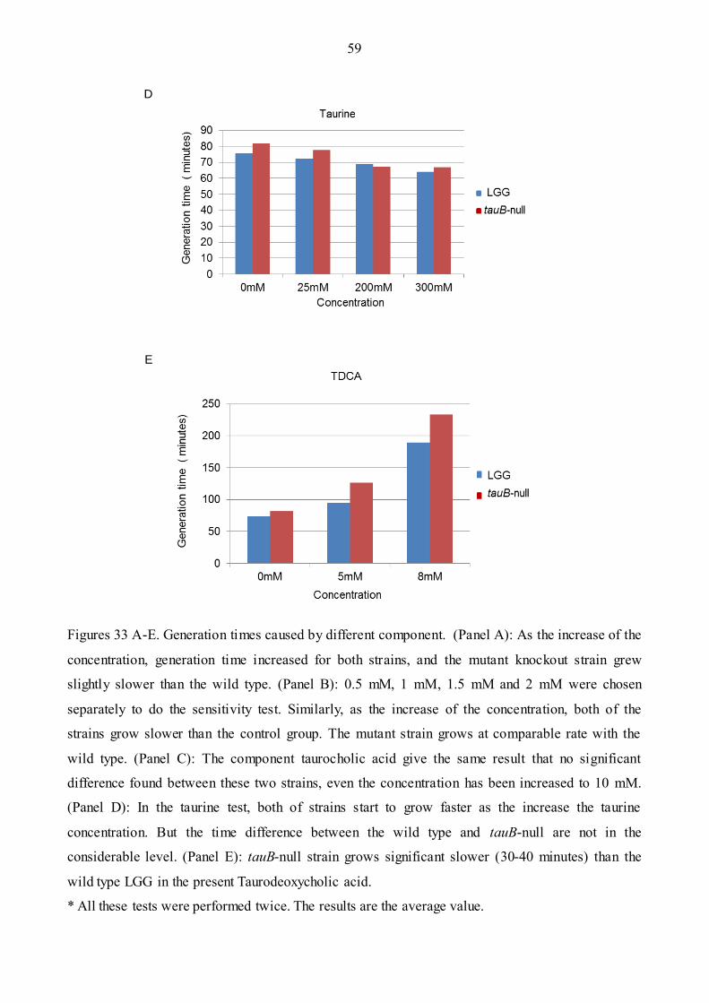
59
Figures 33 A-E. Generation times caused by different component. (Panel A): As the increase of the
concentration, generation time increased for both strains, and the mutant knockout strain grew
slightly slower than the wild type. (Panel B): 0.5 mM, 1 mM, 1.5 mM and 2 mM were chosen
separately to do the sensitivity test. Similarly, as the increase of the concentration, both of the
strains grow slower than the control group. The mutant strain grows at comparable rate with the
wild type. (Panel C): The component taurocholic acid give the same result that no significant
difference found between these two strains, even the concentration has been increased to 10 mM.
(Panel D): In the taurine test, both of strains start to grow faster as the increase the taurine
concentration. But the time difference between the wild type and tauB-null are not in the
considerable level. (Panel E): tauB-null strain grows significant slower (30-40 minutes) than the
wild type LGG in the present Taurodeoxycholic acid.
* All these tests were performed twice. The results are the average value.
E
D

60
Figure 34. Generation time of WT, tauB-null and complementary strains at the 8 mM of the
Taurodeoxycholic acid. The first three blue bars present the control group in the absence of TDCA.
The last three blue bars present the wild type, mutant, complementary strains separately at the
present of the 8 mM TDCA. (*Tests were performed twice. The results are the average values.)
Figure 35. Generation time of WT, tauB-null and complementary strains at the 5 mM of the TCDA.
The first three blue bars present the control group in the absence of TCDA. The last three blue bars
present the wild type, mutant, complementary strains separately at the present of the 5 mM TCDA.
(*Tests were performed twice. The results are the average value.)

61
5 DISCUSSION
To conclude, we explored the long-term effect of four stresses on L. rhamnosus strain GG using
complementary methods such as immunoblotting analysis, PCR, and fluorescence microscopy.
Long-term exposure to bile salts or shearing force treatment impacted the most of GG genome,
more specifically on the genomic island encoding pilin proteins. The latter was found to be lost in
significant proportions. In contrast, there were no genomic changes induced by salt and top fraction
sampling. The most susceptible genomic region to stress was the genomic island encoding the pilus
gene cluster SpaCBA-srtC, but also genes associated with carbohydrate transport and IS elements.
PCR analysis of 13 hyper variable chromosomal regions did confirm that the genes near the
pilin gene cluster were also lost. Such important genomic rearrangements could be linked to the
high number of IS elements presents throughout this particular genomic island. Both phenotypic
and genotypic data could be correlated and pointing once again to this genomic island. It is
noteworthy that the analysis of salt exposure to GG (Figure 15, panel A) did reveal discrepancies,
especially in the data obtained with the monoclonal antibody anti -SpaC. Although the differences
may be due to the technical artifacts, we cannot exclude that the mutations occurred in the epitope
region of SpaC protein. Taken together, we showed that, salt exposure and top fraction preferential
sampling did not alter the pili operon integrity and more generally the genomic coding capacity of
GG, whereas effects related to bile salts or vortexing were more substantial. This finding is relevant
regarding to the use of L. rhamnosus GG as probiotic bacterium, since probiotic
functions/properties may be altered, i.e. the stability of mucus-binding pili in L. rhamnosus GG. L.
rhamnosus GG did show a relatively high genomic stability for all four tested conditions up to 200
generations, which is a long time from an industrial perspective and also in ecological niches. The
daily subculturing of GG underlines that the cells were alternating growth phases and stationary
phases for few hours every day. It would be interesting now to test how stable is the genome of L.
rhamnosus GG in non-growth state for an extended period of time, since this would parallel with
the conditions found in gut or packaged dairy products.
Bile salts induced a significant stress response to L. rhamnosus GG, although it has been
shown to display bile resistance. Within the L. rhamnosus species, not all strains are bile resistant.
Bile sensitivity screening revealed that some strains could not survive in the presence of bile salts
and that the phenotype could be associated in some cases with one of the lifestyle islands that
contain genes encoding a taurine transporter operon tauABC. We therefore hypothesized that the
tauABC transporter might be involved partially or totally in bile resistance in L. rhamnosus GG. A

62
mutant derivative, where the gene tauB was knock-out, was phenotypically analyzed and presented
higher bile sensitivity than the wild type LGG. Specifically, the mutant tauB-null strain grew
significantly slower than the wild type in the presence of 3% (w/v) ox gall. The mutant phenotype
was indeed due to the impairment of the TauABC transporter, since the complemented strain grew
at a comparable rate than the wild type strain. The results indicate that the tauABC operon was
involved into the bile resistance mechanisms in L. rhamnosus GG.
Data observed with ox bile salts showing that tauB mutant strain grew significantly more slowly
than the wild type LGG were similarly found, using human bile samples, although it is known that
the composition of bile varies between individuals. Variations were noted between the five human
bile samples extracted from different patients and may relate to bile composition variations
associated to particular medical conditions. This applies specifically for the bile sample 5, which
gave different outcome.
According to Figure 29, we know that in the subject 5, three components belonging to the
primary bile acids were at the minimum amount. According to the formation of the conjugated bile
acids (Figure 4), it means most of them would have already been conjugated with taurine or glycine
to form the secondary conjugated bile acids, in which, we believe there should be one or more
substrates specific to the mutant strain. Which components are specific to the mutant strain? From
the composition of the bile, we found that the amount of deoxycholic was low in subject 2, subject 4
and subject 5, but the strain in bile of subject 2 and 4 did not present the phenotype difference in
tauB-null strain. This result would probably indicate that the target components for the tauB gene
are not deoxycholic acids, but other cholic acid and chenodeoxycholic acids. Of course, we cannot
exclude that there are two or three components working together to induce the differences observed
in the phenotype or there are some pathological changing of the bile in subject 5.
Data obtained from human bile samples indicated that one or more components specifically
affected the tauB-null mutant. To further examine the possible candidates, we tested a variety of
purified bile acids obtained from different manufactory. Among them, we found that GCA, GDCA,
TCA, taurine, cholic acid, chenodeoxycholate acid, deoxycholic acid, and lithocholic acid did not
inhibit the growth of the mutant strain compared to the control, indicating that these components
were not the components transported by tauABC. More interestingly, in the presence of
taurodeoxycholic acid (TDCA) and taurochenodeoxycholic acid (TCDA), the mutant strain of
tauABC operon grew significantly slower than the wild type strain. These two components are
secondary bile acids derived from cholic acid and chenodeoxycholic acids.
The chemical structures of these two components are both conjugated with taurine and flanked
with two-hydroxyl groups to the cholesterol backbone (Figure 36), suggesting that the components

63
TCDA and TDCA could be transported through the membrane by unknown transporter or diffusion
and then cleaved by the BSH activity (Begley, Hill et al. 2006). The by-products are pumped
outside of the cell by tauABC transporter. In the mutant strain, the lack of tauB may result in the
impairment of this transporter and therefore the accumulation of CDA and DCA, possibly resulting
in toxicity and therefore a slower growth of mutant compared to the wild type (See details in Figure
37).
This hypothetical mechanism may explain the differences observed between ox gall and human
bile salts. Notably, porcine did not impact on the phenotypes between mutant and wild-type strains.
This may suggest that porcine bile used in the present study may have a low concentration in TCDA
and TDCA. Bile resistance mechanisms are therefore host specific and should be further
investigated in relation with the ecological fitness and adaptation of bacterial species.
Figure 36: Chemical structure of A (TCDA) and B (TDCA)
Figure 37: Proposed model of the TauABC transporter mechanism
64
6 CONCLUSION
In the present study, we analyzed the genome stability of Lactobacillus rhamnosus strain GG when
exposed to different stresses. Our data indicated that under the shearing forces and the top fraction
subculture treatment, the bacteria remained piliated. In contrast, when exposed to the bile salts, the
gene cluster related with the pili started to get lost. As well as the 13 hyper regions screening result
also showed that some regions related with the pili also mutated or were lost. This fact indicates that
bile stress in the gastrointestinal tract is a strong stress factor for the probiotic bacteria.
To characterize the novel tauABC operon in in vitro conditions, the result shows that at least the
tauB genes were possibly involved in the bile resistance in the L. rhamnosus GG. More work is
needed to characterize the other genes, namely tauA, tauC genes.
Finally, the screening of the bile components for the L. rhamnosus GG resistance shows that the
taurodeoxycholic acid and taurochenodeoxycholic acid were specific to the tauB genes mutant. It
possibly indicates that tauB gene function is related with the property of these two components. The
detailed mechanism needs to be studied in the future.
7 ACKNOWLEDGMENTS
The present study was financially supported by the ERC grant Microbes Inside (Grant 250172), the
Center of Excellence in Microbial Food Safety Research (CoE-MiFoSa), the Academy of Finland
(grant 141140), and the University of Helsinki. I am grateful to Academy Prof. Willem de Vos and
Dr. François Douillard to support my thesis work. I also want to thank Pia Rasinkangas for her help
in the lab. I also give my thanks to the people who initiated this work in Wageningen University
(Dr. Yanling Hao and Dr. Angela Ribbera).
65
8 REFERENCES
Agostoni, C., I. Axelsson, C. Braegger, O. Goulet, B. Koletzko, K. F. Michaelsen, J. Rigo, R.
Shamir, H. Szajewska, D. Turck, L. T. Weaver and E. C. o. Nutrition. 2004. Probiotic
bacteria in dietetic products for infants: a commentary by the ESPGHAN Committee on
Nutrition. Journal of Pediatric Gastroenterology and Nutrition. 38: 365-374.
Aguirre, M. and M. D. Collins. 1993. Lactic acid bacteria and human clinical infection. Journal of
Applied Bacteriology. 75: 95-107.
Badet, C. and N. B. Thebaud. 2008. Ecology of lactobacilli in the oral cavity: a review of literature.
The Open Microbiology Journal. 2: 38-48.
Begley, M., C. G. M. Gahan and C. Hill. 2005. The interaction between bacteria and bile. FEMS
Microbiology Reviews. 29: 625-651.
Begley, M., C. Hill and C. G. Gahan. 2006. Bile salt hydrolase activity in probiotics. Applied and
Environmental Microbiology. 72: 1729-1738.
Bernardeau, M., M. Guguen and J. P. Vernoux. 2006. Beneficial lactobacilli in food and feed: long-
term use, biodiversity and proposals for specific and realistic safety assessments. FEMS
Microbiology Reviews. 30: 487-513.
Chen, J., S. Sharma, F. A. Quiocho and A. L. Davidson. 2001. Trapping the transition state of an
ATP-binding cassette transporter: evidence for a concerted mechanism of maltose transport.
Proceedings of the National Academy of Sciences of the United States of America. 98:
1525-1530.
Claesson, M. J., Y. Li, S. Leahy, C. Canchaya, J. P. van Pijkeren, A. M. Cerdeno -Tarraga, J.
Parkhill, S. Flynn, G. C. O'Sullivan, J. K. Collins, D. Higgins, F. Shanahan, G. F. Fitzgerald,
D. van Sinderen and P. W. O'Toole. 2006. Multireplicon genome architecture of
Lactobacillus salivarius. Proceedings of the National Academy of Sciences U S A. 103:
6718-6723.
Coleman, R., P. J. Lowe and D. Billington. 1980. Membrane lipid composition and susceptibility to
bile salt damage. Biochimica et Biophysica Acta 599: 294-300.
Conway, P. L., S. L. Gorbach and B. R. Goldin. 1987. Survival of lactic acid bacteria in the human
stomach and adhesion to intestinal cells. Journal of Dairy Science. 70: 1-12.
Dal Bello, F., J. Walter, W. P. Hammes and C. Hertel. 2003. Increased complexity of the species
composition of lactic acid bacteria in human feces revealed by alternative incubation
condition. Microbial Ecology. 45: 455-463.
66
Davidson, A. L., E. Dassa, C. Orelle and J. Chen. 2008. Structure, function, and evolution of
bacterial ATP-binding cassette systems. Microbiology and Molecular Biology Reviews. 72:
317-364.
De Keersmaecker, S. C., K. Braeken, T. L. Verhoeven, M. Perea Velez, S. Lebeer, J. Vanderleyden
and P. Hols. 2006. Flow cytometric testing of green fluorescent protein-tagged Lactobacillus
rhamnosus GG for response to defensins. Applied and Environmental Microbiology. 72:
4923-4930.
Doran, K. S. and V. Nizet. 2004. Molecular pathogenesis of neonatal group B streptococcal
infection: no longer in its infancy. Molecular Microbiology. 54: 23-31.
Douillard, F. P. and W. M. de Vos. 2014. Functional genomics of lactic acid bacteria: from food to
health. Microbial Cell Factories. 13 Supplement 1: S8.
Douillard, F. P., P. Rasinkangas, I. von Ossowski, J. Reunanen, A. Palva and W. M. de Vos. 2014.
Functional identification of conserved residues involved in Lactobacillus rhamnosus strain
GG sortase specificity and pilus biogenesis. The Journal of Biological Chemistry. 289:
15764-15775.
Douillard, F. P., A. Ribbera, H. M. Jarvinen, R. Kant, T. E. Pietila, C. Randazzo, L. Paulin, P. K.
Laine, C. Caggia, I. von Ossowski, J. Reunanen, R. Satokari, S. Salminen, A. Palva and W.
M. de Vos. 2013a. Comparative genomic and functional analysis of Lactobacillus casei and
Lactobacillus rhamnosus strains marketed as probiotics. Applied and Environmental
Microbiology. 79: 1923-1933.
Douillard, F. P., A. Ribbera, R. Kant, T. E. Pietila, H. M. Jarvinen, M. Messing, C. L. Randazzo, L.
Paulin, P. Laine, J. Ritari, C. Caggia, T. Lahteinen, S. J. Brouns, R. Satokari, I. von
Ossowski, J. Reunanen, A. Palva and W. M. de Vos. 2013b. Comparative genomic and
functional analysis of 100 Lactobacillus rhamnosus strains and their comparison with strain
GG. PLOS Genetics. 9: e1003683.
Fujisawa, T. and M. Mori. 1996. Influence of bile salts on beta -glucuronidase activity of intestinal
bacteria. Letters in Applied Microbiology 22: 271-274.
Galdeano, C. M. and G. Perdigon. 2006. The probiotic bacterium Lactobacillus casei induces
activation of the gut mucosal immune system through innate immunity. Clinical and Vaccine
Immunology. 13: 219-226.
Goldin, B. R. and S. L. Gorbach. 2008. Clinical indications for probiotics: an overview. Clinical
Infectious Diseases. 46 Supplement 2: S96-100; discussion S144-151.

67
Grill, J., F. Schneider, J. Crociani and J. Ballongue. 1995. Purification and Characterization of
Conjugated Bile Salt Hydrolase from Bifidobacterium longum BB536. Applied and
Environmental Microbiology. 61: 2577-2582.
Heilig, H. G., E. G. Zoetendal, E. E. Vaughan, P. Marteau, A. D. Akkermans and W. M. de Vos.
2002. Molecular diversity of Lactobacillus spp. and other lactic acid bacteria in the human
intestine as determined by specific amplification of 16S ribosomal DNA. Applied and
Environmental Microbiology. 68: 114-123.
Heuman, D. M., R. S. Bajaj and Q. Lin. 1996. Adsorption of mixtures of bile salt taurine conjugates
to lecithin-cholesterol membranes: implications for bile salt toxicity and cytoprotection. The
Journal of Lipid Research. 37: 562-573.
Higgins, C. F. 1992. ABC transporters: from microorganisms to man. Annual Review of Cell and
Developmental Biology. 8: 67-113.
Higgins, C. F. 2001. ABC transporters: physiology, structure and mechanism--an overview.
Research in Microbiology. 152: 205-210.
Higgins, C. F., I. D. Hiles, G. P. Salmond, D. R. Gill, J. A. Downie, I. J. Evans, I. B. Holland, L.
Gray, S. D. Buckel, A. W. Bell and et al. 1986. A family of related ATP -binding subunits
coupled to many distinct biological processes in bacteria. Nature. 323: 448-450.
Hofmann, A. F. 1999. Bile Acids: The Good, the Bad, and the Ugly. News in Physiological
Sciences. 14: 24-29.
Hofmann, A. F., L. R. Hagey and M. D. Krasowski. 2010. Bile salts of vertebrates: structural
variation and possible evolutionary significance. The Journal of Lipid Research. 51: 226-
246.
Hofmann, A. F. and K. J. Mysels. 1992. Bile acid solubility and precipitation in vitro and in vivo:
the role of conjugation, pH, and Ca2+ ions. The Journal of Lipid Research. 33: 617-626.
Huijghebaert, S. M. and A. F. Hofmann. 1986. Influence of the amino acid moiety on deconjugation
of bile acid amidates by cholylglycine hydrolase or human fecal cultures. The Journal of
Lipid Research. 27: 742-752.
Human Microbiome Project, C. 2012. Structure, function and diversity of the healthy human
microbiome. Nature. 486: 207-214.
Johnson-Henry, K. C., K. A. Donato, G. Shen-Tu, M. Gordanpour and P. M. Sherman. 2008.
Lactobacillus rhamnosus strain GG prevents enterohemorrhagic Escherichia coli O157:H7-
induced changes in epithelial barrier function. Infection and Immunity. 76: 1340-1348.
Jones, P. M. and A. M. George. 2004. The ABC transporter structure and mechanism: perspectives
on recent research. Cellular and Molecular Life Sciences. 61: 682-699.
68
Kadioglu, A., J. N. Weiser, J. C. Paton and P. W. Andrew. 2008. The role of Streptococcus
pneumoniae virulence factors in host respiratory colonization and disease. Nature Reviews
Microbiology. 6: 288-301.
Kalliomaki, M., S. Salminen, H. Arvilommi, P. Kero, P. Koskinen and E. Isolauri. 2001. Probiotics
in primary prevention of atopic disease: a randomised placebo-controlled trial. Lancet. 357:
1076-1079.
Kalliomaki, M., S. Salminen, T. Poussa, H. Arvilommi and E. Isolauri. 2003. Probiotics and
prevention of atopic disease: 4-year follow-up of a randomised placebo-controlled trial. The
Lancet. 361: 1869-1871.
Kankainen, M., L. Paulin, S. Tynkkynen, I. von Ossowski, J. Reunanen, P. Partanen, R. Satokari, S.
Vesterlund, A. P. Hendrickx, S. Lebeer, S. C. De Keersmaecker, J. Vanderleyden, T.
Hamalainen, S. Laukkanen, N. Salovuori, J. Ritari, E. Alatalo, R. Korpela, T. Mattila-
Sandholm, A. Lassig, K. Hatakka, K. T. Kinnunen, H. Karjalainen, M. Saxelin, K. Laakso,
A. Surakka, A. Palva, T. Salusjarvi, P. Auvinen and W. M. de Vos. 2009. Comparative
genomic analysis of Lactobacillus rhamnosus GG reveals pili containing a human- mucus
binding protein. Proceedings of the National Academy of Sciences USA. 106: 17193-17198.
Kekkonen, R. A., N. Lummela, H. Karjalainen, S. Latvala, S. Tynkkynen, S. Järvenpää, H.
Kautiainen, I. Julkunen, H. Vapaatalo and R. Korpela. 2008. Probiotic intervention has
strain-specific anti-inflammatory effects in healthy adults. World Journal of
Gastroenterology. 14: 2029-2036.
Koponen, J., K. Laakso, K. Koskenniemi, M. Kankainen, K. Savijoki, T. A. Nyman, W. M. de Vos,
S. Tynkkynen, N. Kalkkinen and P. Varmanen. 2012. Effect of acid stress on protein
expression and phosphorylation in Lactobacillus rhamnosus GG. Journal of Proteome
Research. 75: 1357-1374.
Koskenniemi, K., J. Koponen, M. Kankainen, K. Savijoki, S. Tynkkynen, W. M. de Vos, N.
Kalkkinen and P. Varmanen. 2009. Proteome analysis of Lactobacillus rhamnosus GG using
2-D DIGE and mass spectrometry shows differential protein production in laboratory and
industrial-type growth media. Journal of Proteome Research. 8: 4993-5007.
Koskenniemi, K., K. Laakso, J. Koponen, M. Kankainen, D. Greco, P. Auvinen, K. Savijoki, T. A.
Nyman, A. Surakka, T. Salusjarvi, W. M. de Vos, S. Tynkkynen, N. Kalkkinen and P.
Varmanen. 2011. Proteomics and transcriptomics characterization of bile stress response in
probiotic Lactobacillus rhamnosus GG. Molecular & Cellular Proteomics. 10: M110
002741.

69
Lahti, L., A. Salonen, R. A. Kekkonen, J. Salojarvi, J. Jalanka-Tuovinen, A. Palva, M. Oresic and
W. M. de Vos. 2013. Associations between the human intestinal microbiota, Lactobacillus
rhamnosus GG and serum lipids indicated by integrated analysis of high-throughput
profiling data. PeerJ. 1: e32.
Lebeer, S., I. Claes, H. L. Tytgat, T. L. Verhoeven, E. Marien, I. von Ossowski, J. Reunanen, A.
Palva, W. M. Vos, S. C. Keersmaecker and J. Vanderleyden. 2012. Functional analysis of
Lactobacillus rhamnosus GG pili in relation to adhesion and immunomodulatory
interactions with intestinal epithelial cells. Applied and Environmental Microbiology. 78:
185-193.
Lebeer, S., I. J. Claes, T. L. Verhoeven, J. Vanderleyden and S. C. De Keersmaecker. 2011.
Exopolysaccharides of Lactobacillus rhamnosus GG form a protective shield against innate
immune factors in the intestine. Microbial Biotechnology. 4: 368-374.
Lee, K. and K. Pi. 2010. Effect of transient acid stress on the proteome of intestinal probiotic
Lactobacillus reuteri. Biochemistry (Mosc). 75: 460-465.
Leroy, F. and L. De Vuyst. 2004. Lactic acid bacteria as functional starter cultures for the food
fermentation industry. Trends in Food Science & Technology. 15: 67-78.
Leverrier, P., D. Dimova, V. Pichereau, Y. Auffray, P. Boyaval and G. Jan. 2003. Susceptibility and
adaptive response to bile salts in Propionibacterium freudenreichii: physiological and
proteomic analysis. Applied and Environmental Microbiology. 69: 3809-3818.
Leverrier, P., J. P. Vissers, A. Rouault, P. Boyaval and G. Jan. 2004. Mass spectrometry proteomic
analysis of stress adaptation reveals both common and distinct response pathways in
Propionibacterium freudenreichii. Archives of Microbiology. 181: 215-230.
Louis, P., K. P. Scott, S. H. Duncan and H. J. Flint. 2007. Understanding the effects of diet on
bacterial metabolism in the large intestine. Journal of Applied Microbiology. 102: 1197 -
1208.
Macklaim, J. M., G. B. Gloor, K. C. Anukam, S. Cribby and G. Reid. 2011. At the crossroads of
vaginal health and disease, the genome sequence of Lactobacillus iners AB-1. Proc Natl
Acad Sci U S A. 108 Supplement 1: 4688-4695.
Makarova, K., A. Slesarev, Y. Wolf, A. Sorokin, B. Mirkin, E. Koonin, A. Pavlov, N. Pavlova, V.
Karamychev, N. Polouchine, V. Shakhova, I. Grigoriev, Y. Lou, D. Rohksar, S. Lucas, K.
Huang, D. M. Goodstein, T. Hawkins, V. Plengvidhya, D. Welker, J. Hughes, Y. Goh, A.
Benson, K. Baldwin, J. H. Lee, I. Diaz-Muniz, B. Dosti, V. Smeianov, W. Wechter, R.
Barabote, G. Lorca, E. Altermann, R. Barrangou, B. Ganesan, Y. Xie, H. Rawsthorne, D.
Tamir, C. Parker, F. Breidt, J. Broadbent, R. Hutkins, D. O'Sullivan, J. Steele, G. Unlu, M.

70
Saier, T. Klaenhammer, P. Richardson, S. Kozyavkin, B. Weimer and D. Mills. 2006.
Comparative genomics of the lactic acid bacteria. Proceedings of the National Academy of
Sciences of the United States of America. 103: 15611-15616.
Marco, M. L., S. Pavan and M. Kleerebezem. 2006. Towards understanding molecular modes of
probiotic action. Current Opinion in Biotechnology. 17: 204-210.
Marteau, P. R., M. de Vrese, C. J. Cellier and J. Schrezenmeir. 2001. Protection from
gastrointestinal diseases with the use of probiotics. The American Journal of Clinical
Nutrition. 73: 430S-436S.
Mastromarino, P., B. Vitali and L. Mosca. 2013. Bacterial vaginosis: a review on clinical trials with
probiotics. New Microbiologica. 36: 229-238.
Michaki, V., F. X. Guix, K. Vennekens, S. Munck, C. Dingwall, J. B. Davis, D. M. Townsend, K. D.
Tew, F. Feiguin, B. De Strooper, C. G. Dotti and T. Wahle. 2012. Down-regulation of the
ATP-binding cassette transporter 2 (Abca2) reduces amyloid-beta production by altering
Nicastrin maturation and intracellular localization. The Journal of Biological Chemistry.
287: 1100-1111.
Mills, K. A., I. Mushtaq, A. W. Johnson, P. D. Whitfield and P. T. Clayton. 1998. A method for the
quantitation of conjugated bile acids in dried blood spots using electrospray ionization-mass
spectrometry. Pediatric Research. 43: 361-368.
Morita, H., H. Toh, K. Oshima, M. Murakami, T. D. Taylor, S. Igimi and M. Hattori. 2009.
Complete genome sequence of the probiotic Lactobacillus rhamnosus ATCC 53103. Journal
of Bacteriology. 191: 7630-7631.
Parvez, S., K. A. Malik, S. Ah Kang and H. Y. Kim. 2006. Probiotics and their fermented food
products are beneficial for health. Journal of Applied Microbiology. 100: 1171-1185.
Pendharkar, S., T. Magopane, P. G. Larsson, G. de Bruyn, G. E. Gray, L. Hammarstrom and H.
Marcotte. 2013. Identification and characterisation of vaginal lactobacilli from South
African women. BMC Infectious Diseases. 13: 43.
Proft, T. and E. N. Baker. 2009. Pili in Gram-negative and Gram-positive bacteria - structure,
assembly and their role in disease. Cellular and Molecular Life Sciences. 66: 613-635.
Qin, J., R. Li, J. Raes, M. Arumugam, K. S. Burgdorf, C. Manichanh, T. Nielsen, N. Pons, F.
Levenez, T. Yamada, D. R. Mende, J. Li, J. Xu, S. Li, D. Li, J. Cao, B. Wang, H. Liang, H.
Zheng, Y. Xie, J. Tap, P. Lepage, M. Bertalan, J. M. Batto, T. Hansen, D. Le Paslier, A.
Linneberg, H. B. Nielsen, E. Pelletier, P. Renault, T. Sicheritz-Ponten, K. Turner, H. Zhu, C.
Yu, S. Li, M. Jian, Y. Zhou, Y. Li, X. Zhang, S. Li, N. Qin, H. Yang, J. Wang, S. Brunak, J.
Dore, F. Guarner, K. Kristiansen, O. Pedersen, J. Parkhill, J. Weissenbach, H. I. T. C. Meta,
71
P. Bork, S. D. Ehrlich and J. Wang. 2010. A human gut microbial gene catalogue established
by metagenomic sequencing. Nature. 464: 59-65.
Rajilic-Stojanovic, M. and W. M. de Vos. 2014. The first 1000 cultured species of the human
gastrointestinal microbiota. FEMS Microbiology Reviews 38: 996-1047.
Reid, G., J. Jass, M. T. Sebulsky and J. K. McCormick. 2003. Potential uses of probiotics in clinical
practice. Clinical Microbiology Reviews. 16: 658-672.
Reunanen, J., I. von Ossowski, A. P. Hendrickx, A. Palva and W. M. de Vos. 2012. Characterization
of the SpaCBA pilus fibers in the probiotic Lactobacillus rhamnosus GG. Applied and
Environmental Microbiology. 78: 2337-2344.
Saxelin, M. 2008. Probiotic formulations and applications, the current probiotics market, and
changes in the marketplace: a European perspective. Clinical Infectious Diseases. 46
Supplement 2: S76-79; discussion S144-151.
Saxelin, M., S. Tynkkynen, T. Mattila-Sandholm and W. M. de Vos. 2005. Probiotic and other
functional microbes: from markets to mechanisms. Current Opinion in Biotechnology. 16:
204-211.
Sazawal, S., G. Hiremath, U. Dhingra, P. Malik, S. Deb and R. E. Black. 2006. Efficacy of
probiotics in prevention of acute diarrhoea: a meta -analysis of masked, randomised,
placebo-controlled trials. The Lancet Infectious Diseases. 6: 374-382.
Schieszer, J. 2009. “Antibiotic UTI Prophylaxis Slightly Better,” Renal & Urology News.
Siezen, R. J. and J. E. van Hylckama Vlieg. 2011. Genomic diversity and versatility of
Lactobacillus plantarum, a natural metabolic engineer. Microbial Cell Factories. 10
Supplement 1: S3.
Szajewska, H., M. Kotowska, J. Z. Mrukowicz, M. Armanska and W. Mikolajczyk. 2001. Efficacy
of Lactobacillus GG in prevention of nosocomial diarrhea in infants. J Pediatr. 138: 361-
365.
Tonjum, T. and M. Koomey. 1997. The pilus colonization factor of pathogenic neisserial species:
organelle biogenesis and structure/function relationships--a review. Gene. 192: 155-163.
Tripathi, P., A. Beaussart, D. Alsteens, V. Dupres, I. Claes, I. von Ossowski, W. M. de Vos, A. Palva,
S. Lebeer, J. Vanderleyden and Y. F. Dufrene. 2013. Adhesion and nanomechanics of pili
from the probiotic Lactobacillus rhamnosus GG. ACS Nano. 7: 3685-3697.
Troost, F. J., P. van Baarlen, P. Lindsey, A. Kodde, W. M. de Vos, M. Kleerebezem and R. J.
Brummer. 2008. Identification of the transcriptional response of human intestinal mucosa to
Lactobacillus plantarum WCFS1 in vivo. BMC Genomics. 9: 374.

72
van Baarlen, P., F. Troost, C. van der Meer, G. Hooiveld, M. Boekschoten, R. J. Brummer and M.
Kleerebezem. 2011. Human mucosal in vivo transcriptome responses to three lactobacilli
indicate how probiotics may modulate human cellular pathways. Proceedings of the
National Academy of Sciences of the United States of America. 108 Suppl 1: 4562-4569.
van Veen, H. W. and W. N. Konings. 1998. The ABC family of multidrug transporters in
microorganisms. Biochimica et Biophysica Acta. 1365: 31-36.
van Veen, H. W., A. Margolles, M. Muller, C. F. Higgins and W. N. Konings. 2000. The
homodimeric ATP-binding cassette transporter LmrA mediates multidrug transport by an
alternating two-site (two-cylinder engine) mechanism. European Molecular Biology
Organization Journal. 19: 2503-2514.
Vanderhoof, J. A., D. B. Whitney, D. L. Antonson, T. L. Hanner, J. V. Lupo and R. J. Young. 1999.
Lactobacillus GG in the prevention of antibiotic-associated diarrhea in children. Journal of
Pediatrics. 135: 564-568.
Vesterlund, S., M. Karp, S. Salminen and A. C. Ouwehand. 2006. Staphylococcus aureus adheres to
human intestinal mucus but can be displaced by certain lactic acid bacteria. Microbiology.
152: 1819-1826.
von Ossowski, I., R. Satokari, J. Reunanen, S. Lebeer, S. C. De Keersmaecker, J. Vanderleyden, W.
M. de Vos and A. Palva. 2011. Functional characterization of a mucus -specific LPXTG
surface adhesin from probiotic Lactobacillus rhamnosus GG. Applied and Environmental
Microbiology. 77: 4465-4472.
Walter, J. 2008. Ecological role of lactobacilli in the gastrointestinal tract: implications for
fundamental and biomedical research. Applied and Environmental Microbiology. 74: 4985-
4996.
Wikipedia. http://en.wikipedia.org/wiki/Bile_acid. Visited 8.8.2014.
Zoetendal, E. G., J. Raes, B. van den Bogert, M. Arumugam, C. C. Booijink, F. J. Troost, P. Bork,
M. Wels, W. M. de Vos and M. Kleerebezem. 2012. The human small intestinal microbiota
is driven by rapid uptake and conversion of simple carbohydrates. The ISME Journal. 6:
1415-1426.