Bilateral Taurodontism in Deciduous Molars: A case …. Parimala Tyagi.pdfTaurodontism is a...
3
21 People’s Journal of Scientific Research Vol.3(2), July 2010 Bilateral Taurodontism in Deciduous Molars: A case Report Parimala Tyagi, Shilpy Gupta Department of Pedodontics and Preventive Dentistry, Peoples Dental Academy, People’s Campus Bhanpur, Bhopal-462037. Abstract: Taurodontism is a rare dental anomaly in which the involved tooth has an enlarged and elongated body and pulp chamber with apical displacement of the pulpal floor. It has a very low incidence and very few cases are reported in literature in deciduous dentition. Endodontic treatment of a taurodont tooth is challenging and requires special handling because of the proximity and apical displacement of the roots. In this article a case of five year child with bilateral involvement of mandibular second molars is presented. Key Words: Taurodont, enlarged pulp chamber, pulpectomy. Introduction: Taurodontism is a developmental disturbance of a tooth that lacks constriction at the level of the cementoenamel junction (CEJ). It is characterized by vertically elongated pulp chamber, apical displacement of the pulpal floor and bifurcation or trifurcations of the roots (Jafarzadeh et al, 2008). The term “taurodontism” (‘bull tooth’) was coined from the Latin word “tauros”, which means ‘bull’ and the Greek word “odus”, which means ‘tooth’. Sir Arthur Keith in 1913 coined the term “taurodontism” and defined taurodontism as “a tendency for the body of the tooth to enlarge at the expense of the roots”. Although Gorjanovic-Kramberger in 1908 had first described this type of tooth. Shaw (1928) in his first quantitative study on taurodontism classified the condition as “hypotaurodontism,” “mesotaurodontism,” and “hypertaurodontism” to define the degree more accurately to which this condition is manifested (Fig. I). Shifman & Channel (1978) suggested a most widely accepted and used criteria, on the basis of the Case Report distance from the lowest point of the roof of the pulp chamber (a) to the highest point of pulp floor (b), when divided by the distance from (a) to root apex (c) should be equal to or greater than 0.2 mm and/or distance from (b) to cementoenamel junction (d) should be greater than 2.5 mm (Fig. II). Fig. I: Diagrammatic representation of normal (cynodontic) tooth and three subtypes of taurodontic teeth as proposed by Shaw (1928). ----------------------------------------------------------------------------- Corresponding Author: Dr. Parimala Tyagi, M.D.S Prof & Head, Department of Pedodontics and Preventive Dentistry, People’s Dental Academy, Bhopal-462010, Phone No .: 09425453853 E mail : [email protected] Mandibular molars are more commonly affected teeth. The incidence is reported to be lower than 1% in modern man (Joseph, 2008), 3% in primitives, Eskimos and American Indians (Tsesis, 2003). Taurodontism has been associated with various syndromes such as Down’s syndrome, Klinefelters syndrome, Aperts syndrome, Oral-fault digital (Mohrs syndrome), Trichodento-Osseous syndrome etc (Terezhalmy et al, 2001). There are various theories concerning the etiology of taurodontism: a) a specialized or retrograde character; b) a primitive pattern; c) a Mendelian recessive; d) an atavistic feature and e) a mutation (Mangion, 1962; Hammer et al, 1964). Many explanations Fig. II: Showing measurements for Taurodontic teeth. a b > 0.2 mm. B D >2.5 mm. B A C
Transcript of Bilateral Taurodontism in Deciduous Molars: A case …. Parimala Tyagi.pdfTaurodontism is a...
21People’s Journal of Scientific Research Vol.3(2), July 2010
Bilateral Taurodontism in Deciduous Molars: A case ReportParimala Tyagi, Shilpy GuptaDepartment of Pedodontics and Preventive Dentistry, Peoples Dental Academy, People’s Campus Bhanpur, Bhopal-462037.
Abstract:Taurodontism is a rare dental anomaly in which the involved tooth has an enlarged and elongated body and pulp
chamber with apical displacement of the pulpal floor. It has a very low incidence and very few cases are reported inliterature in deciduous dentition. Endodontic treatment of a taurodont tooth is challenging and requires special handlingbecause of the proximity and apical displacement of the roots. In this article a case of five year child with bilateralinvolvement of mandibular second molars is presented.
Key Words: Taurodont, enlarged pulp chamber, pulpectomy.
Introduction:Taurodontism is a developmental disturbance
of a tooth that lacks constriction at the level of thecementoenamel junction (CEJ). It is characterized byvertically elongated pulp chamber, apical displacementof the pulpal floor and bifurcation or trifurcations ofthe roots (Jafarzadeh et al, 2008).
The term “taurodontism” (‘bull tooth’) wascoined from the Latin word “tauros”, which means ‘bull’and the Greek word “odus”, which means ‘tooth’. SirArthur Keith in 1913 coined the term “taurodontism”and defined taurodontism as “a tendency for the bodyof the tooth to enlarge at the expense of the roots”.Although Gorjanovic-Kramberger in 1908 had firstdescribed this type of tooth. Shaw (1928) in his firstquantitative study on taurodontism classified thecondition as “hypotaurodontism,” “mesotaurodontism,”and “hypertaurodontism” to define the degree moreaccurately to which this condition is manifested(Fig. I). Shifman & Channel (1978) suggested a mostwidely accepted and used criteria, on the basis of the
Case Report
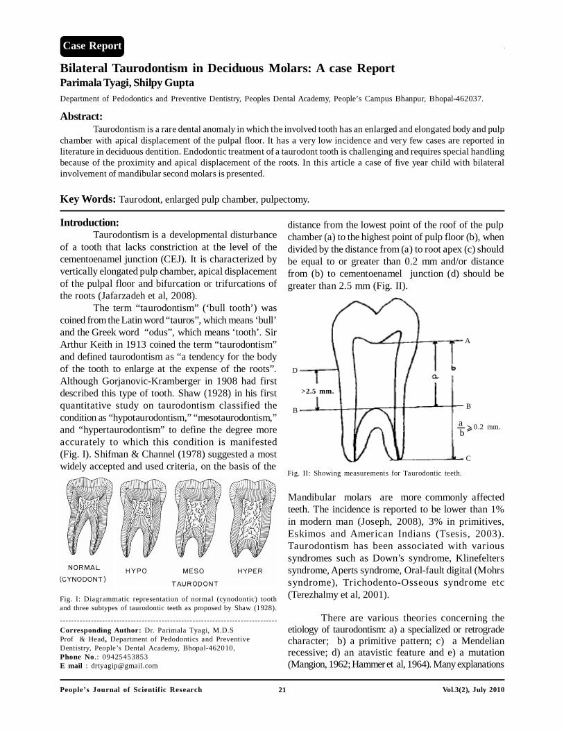
distance from the lowest point of the roof of the pulpchamber (a) to the highest point of pulp floor (b), whendivided by the distance from (a) to root apex (c) shouldbe equal to or greater than 0.2 mm and/or distancefrom (b) to cementoenamel junction (d) should begreater than 2.5 mm (Fig. II).
Fig. I: Diagrammatic representation of normal (cynodontic) toothand three subtypes of taurodontic teeth as proposed by Shaw (1928).
-----------------------------------------------------------------------------Corresponding Author: Dr. Parimala Tyagi, M.D.SProf & Head, Department of Pedodontics and PreventiveDentistry, People’s Dental Academy, Bhopal-462010,Phone No .: 09425453853E mail : [email protected]
Mandibular molars are more commonly affectedteeth. The incidence is reported to be lower than 1%in modern man (Joseph, 2008), 3% in primitives,Eskimos and American Indians (Tsesis, 2003).Taurodontism has been associated with varioussyndromes such as Down’s syndrome, Klinefelterssyndrome, Aperts syndrome, Oral-fault digital (Mohrssyndrome), Trichodento-Osseous syndrome etc(Terezhalmy et al, 2001).
There are various theories concerning theetiology of taurodontism: a) a specialized or retrogradecharacter; b) a primitive pattern; c) a Mendelianrecessive; d) an atavistic feature and e) a mutation(Mangion, 1962; Hammer et al, 1964). Many explanations
Fig. II: Showing measurements for Taurodontic teeth.
ab > 0.2 mm.
B
D
>2.5 mm.
B
A
C
22People’s Journal of Scientific Research Vol.3(2), July 2010
Case Report:A five year old male child was brought to
People’s Dental Academy, Bhopal with complain ofpain related to left posterior tooth region of mandiblefor last two weeks. There was no significant dental ormedical complaint in the past. His developmental
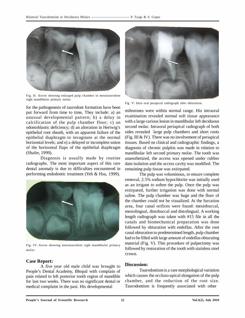
milestones were within normal range. His intraoralexamination revealed normal soft tissue appearancewith a large carious lesion in mandibular left deciduoussecond molar. Intraoral periapical radiograph of bothsides revealed large pulp chambers and short roots(Fig. III & IV). There was no involvement of periapicaltissues. Based on clinical and radiographic findings, adiagnosis of chronic pulpitis was made in relation tomandibular left second primary molar. The tooth wasanaesthetized, the access was opened under rubberdam isolation and the access cavity was modified. Theremaining pulp tissue was extirpated.
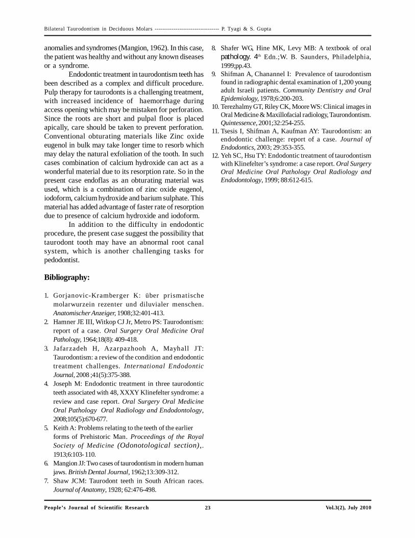
The pulp was voluminous, to ensure completeremoval, 2.5% sodium hypochlorite was initially usedas an irrigant to soften the pulp. Once the pulp wasextirpated, further irrigation was done with normalsaline. The pulp chamber was huge and the floor ofthe chamber could not be visualized. At the furcationarea, four canal orifices were found: mesiobuccal,mesiolingual, distobuccal and distolingual. A workinglength radiograph was taken with #15 file in all thecanals and biomechanical preparation was donefollowed by obturation with endoflas. After the rootcanal obturation to predetermined length, pulp chamberhad to be filled with large amount of endoflas obturatingmaterial (Fig. V). This procedure of pulpectomy wasfollowed by restoration of the tooth with stainless steelcrown.
Discussion:Taurodontism is a rare morphological variation
which causes the occluso-apical elongation of the pulpchamber, and the reduction of the root size.Taurodontism is frequently associated with other
Fig. V: Intra oral perapical radiograph after obturation.
Fig. IV: Arrow showing mesotaurodont right mandibular primarymolar.
Bilateral Taurodontism in Deciduous Molars ---------------------------------- P. Tyagi & S. Gupta
for the pathogenesis of taurodont formation have beenput forward from time to time, They include: a) anunusual developmental pattern; b) a delay incalcification of the pulp chamber floor; c) anodontoblastic deficiency; d) an alteration in Hertwig’sepithelial root sheath, with an apparent failure of theepithelial diaphragm to invaginate at the normalhorizontal levels; and e) a delayed or incomplete unionof the horizontal flaps of the epithelial diaphragm(Shafer, 1999).
Diagnosis is usually made by routineradiographs. The most important aspect of this raredental anomaly is due to difficulties encountered inperforming endodontic treatment (Yeh & Hsu, 1999).
Fig. II: Arrow showing enlarged pulp chamber in mesotaurodontright mandibular primary molar.
23People’s Journal of Scientific Research Vol.3(2), July 2010
anomalies and syndromes (Mangion, 1962). In this case,the patient was healthy and without any known diseasesor a syndrome.
Endodontic treatment in taurodontism teeth hasbeen described as a complex and difficult procedure.Pulp therapy for taurodonts is a challenging treatment,with increased incidence of haemorrhage duringaccess opening which may be mistaken for perforation.Since the roots are short and pulpal floor is placedapically, care should be taken to prevent perforation.Conventional obturating materials like Zinc oxideeugenol in bulk may take longer time to resorb whichmay delay the natural exfoliation of the tooth. In suchcases combination of calcium hydroxide can act as awonderful material due to its resorption rate. So in thepresent case endoflas as an obturating material wasused, which is a combination of zinc oxide eugenol,iodoform, calcium hydroxide and barium sulphate. Thismaterial has added advantage of faster rate of resorptiondue to presence of calcium hydroxide and iodoform.
In addition to the difficulty in endodonticprocedure, the present case suggest the possibility thattaurodont tooth may have an abnormal root canalsystem, which is another challenging tasks forpedodontist.
Bibliography:
1. Gorjanovic-Kramberger K: über prismatischemolarwurzein rezenter und diluvialer menschen.Anatomischer Anzeiger, 1908;32:401-413.
2. Hamner JE III, Witkop CJ Jr, Metro PS: Taurodontism:report of a case. Oral Surgery Oral Medicine OralPathology, 1964;18(8): 409-418.
3. Jafarzadeh H, Azarpazhooh A, Mayhall JT:Taurodontism: a review of the condition and endodontictreatment challenges. International EndodonticJournal, 2008 ;41(5):375-388.
4. Joseph M: Endodontic treatment in three taurodonticteeth associated with 48, XXXY Klinefelter syndrome: areview and case report. Oral Surgery Oral MedicineOral Pathology Oral Radiology and Endodontology,2008;105(5):670-677.
5. Keith A: Problems relating to the teeth of the earlierforms of Prehistoric Man. Proceedings of the RoyalSociety of Medicine (Odonotological section),.1913;6:103- 110.
6. Mangion JJ: Two cases of taurodontism in modern humanjaws. British Dental Journal, 1962;13:309-312.
7. Shaw JCM: Taurodont teeth in South African races.Journal of Anatomy, 1928; 62:476-498.
Bilateral Taurodontism in Deciduous Molars ---------------------------------- P. Tyagi & S. Gupta
8. Shafer WG, Hine MK, Levy MB: A textbook of oralpathology. 4th Edn.;W. B. Saunders, Philadelphia,1999;pp.43.
9. Shifman A, Chanannel I: Prevalence of taurodontismfound in radiographic dental examination of 1,200 youngadult Israeli patients. Community Dentistry and OralEpidemiology, 1978;6:200-203.
10. Terezhalmy GT, Riley CK, Moore WS: Clinical images inOral Medicine & Maxillofacial radiology, Taurondontism.Quintessence, 2001;32:254-255.
11. Tsesis I, Shifman A, Kaufman AY: Taurodontism: anendodontic challenge: report of a case. Journal ofEndodontics, 2003; 29:353-355.
12. Yeh SC, Hsu TY: Endodontic treatment of taurodontismwith Klinefelter’s syndrome: a case report. Oral SurgeryOral Medicine Oral Pathology Oral Radiology andEndodontology, 1999; 88:612-615.