Biglino, G., Caputo, M., Rajakaruna, C., Angelini, G., van ... · 5 1. Introduction: blooming...
49
Biglino, G., Caputo, M., Rajakaruna, C., Angelini, G., van Rooij, E., & Emanueli, C. (2017). Modulating MicroRNAs in Cardiac Surgery Patients: Novel Therapeutic Opportunities? Pharmacology and Therapeutics, 170, 192-204. https://doi.org/10.1016/j.pharmthera.2016.11.004 Peer reviewed version License (if available): CC BY-NC-ND Link to published version (if available): 10.1016/j.pharmthera.2016.11.004 Link to publication record in Explore Bristol Research PDF-document This is the author accepted manuscript (AAM). The final published version (version of record) is available online via Elsevier at https://www.sciencedirect.com/science/article/pii/S0163725816302303 . Please refer to any applicable terms of use of the publisher. University of Bristol - Explore Bristol Research General rights This document is made available in accordance with publisher policies. Please cite only the published version using the reference above. Full terms of use are available: http://www.bristol.ac.uk/pure/about/ebr-terms
Transcript of Biglino, G., Caputo, M., Rajakaruna, C., Angelini, G., van ... · 5 1. Introduction: blooming...

Biglino, G., Caputo, M., Rajakaruna, C., Angelini, G., van Rooij, E., &Emanueli, C. (2017). Modulating MicroRNAs in Cardiac Surgery Patients:Novel Therapeutic Opportunities? Pharmacology and Therapeutics, 170,192-204. https://doi.org/10.1016/j.pharmthera.2016.11.004
Peer reviewed version
License (if available):CC BY-NC-ND
Link to published version (if available):10.1016/j.pharmthera.2016.11.004
Link to publication record in Explore Bristol ResearchPDF-document
This is the author accepted manuscript (AAM). The final published version (version of record) is available onlinevia Elsevier at https://www.sciencedirect.com/science/article/pii/S0163725816302303 . Please refer to anyapplicable terms of use of the publisher.
University of Bristol - Explore Bristol ResearchGeneral rights
This document is made available in accordance with publisher policies. Please cite only the publishedversion using the reference above. Full terms of use are available:http://www.bristol.ac.uk/pure/about/ebr-terms

1
Modulating MicroRNAs in Cardiac Surgery Patients:
Novel Therapeutic Opportunities?
Giovanni Biglino1, Massimo Caputo1,2, Cha Rajakaruna1,
Gianni Angelini1, Eva van Rooij3, and Costanza Emanueli1,4
1Bristol Heart Institute, University of Bristol, Bristol, UK
2RUSH University Medical Center, Chicago, IL, USA
3Hubrecht Institute, Utrecht, the Netherlands
4NIHL, Imperial College London, London, UK
Corresponding author:
Professor Costanza Emanueli
Bristol Heart Institute
School of Clinical Sciences
University of Bristol
Bristol Royal Infirmary
Bristol BS2 8HW
United Kingdom
Tel/Fax: +44(0)117342-3512/3904
E-mail: [email protected]

2
Abstract
This review focuses on microRNAs (miRs) in cardiac surgery, where they are emerging as
potential targets for therapeutic intervention as well as novel clinical biomarkers.
Identification of the up/down-regulation of specific miRs in defined groups of cardiac surgery
patients can lead to the development of novel strategies for targeted treatment in order to
maximise therapeutic results and minimise acute, delayed or chronic complications. MiRs
could also be involved in determining the outcome independently of complications, for
example in relation to myocardial perfusion and fibrosis. Because of their relevance in
disease, their known sequence and pharmacological properties, miRs are attractive
candidates for therapeutic manipulation. Pharmacological inhibition of individual miRs can be
achieved by modified antisense oligonucleotides, referred to as antimiRs, while miR
replacement can be achieved by miR mimics to increase the level of a specific miR. MiR
mimics can restore the function of a lost or down-regulated miR, whilst antimiRs can inhibit
the levels of disease-driving or aberrantly expressed miRs, thus de-repressing the
expression of mRNAs targeted by the miR. The main delivery methods for miR therapeutics
involve lipid-based vehicles, viral systems, cationic polymers, and intravenous or local
injection of an antagomiR. Local delivery is particularly desirable for miR therapeutics and
options include the development of devices specific for local delivery, light-induced antimiR,
and vesicle-encapsulated miRs serving as therapeutic delivery agents able to improve
intracellular uptake.
Here, we discuss the potential therapeutic use of miRNAs in the context of cardiac surgery.
Keywords: MicroRNA, cardiac surgery, therapeutic, cardiac protection, angiogenesis,

3
Table of contents
1. Introduction: blooming microRNA research 2. Cardiovascular surgery 2.1. The ischemic heart and coronary artery bypass surgery 2.2. Surgical aortic valve replacement 2.3. Surgical repair of aneurysmal aorta 2.4. Surgical repair of congenital heart disease 3. MicroRNAs involved in mechanisms relevant for cardiac surgery patients 3.1. Myocardial protection 3.2. Therapeutic angiogenesis 3.3. Post-operative complications 4. MicroRNA therapeutics 4.1. Concepts of microRNA therapeutics 4.2. Delivering microRNA therapeutics 5. MicroRNAs as clinical biomarkers 6. Translational outlook and conclusions 7. Conflict of interest statement 8. Source of funding

4
Abbreviations
AAA = abdominal aortic aneurysm
AKI = acute kidney injury
AMI = acute myocardial infarction
BAV = bicuspid aortic valve
CABG = coronary artery bypass graft
CAD = coronary artery disease
CHD = congenital heart disease
CPB = cardiopulmonary bypass
HF = heart failure
HLHS = hypoplastic left heart syndrome
IHD = ischemic heart disease
LNA = locked nucleic acid
lncRNA = long non-coding RNA
LV = left ventricle
LVAD = left ventricular assist device
LVEF = left ventricular ejection fraction
MI = myocardial infarction
miR = microRNA
MMP = metalloproteinase
PAH = pulmonary arterial hypertension
RNA = ribonucleic acid
RV = right ventricle
RVOT = right ventricular outflow tract
SAVR = surgical aortic valve replacement
TAVR = transcatheter aortic valve replacement
TGA = transposition of the great arteries
ToF = tetralogy of Fallot
5
1. Introduction: blooming microRNA research
Since the human genome project mapped the first chromosome in 1999, it was observed
that only about 20,000 genes are protein coding (Pheasant and Mattick, 2007). In fact, the
majority of DNA is transcribed into non-coding ribonucleic acids (ncRNAs). NcRNAs are
functional RNA molecules that are not translated into proteins and work by regulating gene
expression at the transcriptional and/or post-transcriptional level. The discovery of ncRNAs
dates back 50 years, with the characterisation and sequencing of alanine transfer-RNA
(Holley et al., 1965). More recently, the ENCODE project (“The International Encyclopedia
of DNA Elements”) shed further light on the biochemical activity of the human genomic DNA
(ENCODE Project Consortium, 2012), revealing that only a fraction of the human genome
carries the codes for protein synthesis (Ponting and Hardison, 2011).
Depending on the number of nucleotides, ncRNAs have been classified as either ‘short’ (<
30 nucleotides) or ‘long’ (> 200 nucleotides). Amongst the short non-coding RNAs, further
distinctions can be made between different classes, including microRNAs (miRNAs, or
miRs), short interfering RNAs (siRNAs), and piwi-interacting RNAs (piRNAs). The other
class of long ncRNAs (lncRNAs) is currently being investigated for its function and
involvement in biological phenomena such as transcriptional enhancement, gene imprinting,
and dosage compensation of sex chromosomes (Mercer et al., 2009; Quinn and Chang,
2016).
MicroRNAs were first discovered in the 1990s (Lee et al., 1993; Wightman et al., 1993). The
miR biogenesis begins with a long 5’-capped and Poly A tailed primitive miRNA (pri-miR)
transcript derived from protein coding genes or an independent non-coding transcriptional
unit being configured into a hairpin structure. These miR-producing transcripts can contain
single miRs or form polycistronic miR clusters. The process of maturation of pri-miRs begins
in the cell nucleus, leading to the production of a precursor miR (pre-miR), which in turn is
transported into the cytosol or the endoplasmic reticulum to be processed into its
approximately 22-nucleotide long mature form by Dicer, a RNase III endonuclease. The
resulting structure is typically referred to as “hairpin” and presents itself as double stranded,
6
of which one is incorporated into the RNA-induced silencing complex (RISC) where the miR
and the mRNA target interact (Cai et al., 2004; Ha and Kim, 2014; Rodriguez et al., 2004).
The majority of miRs are located intracellularly, but a large number has also been identified
in the extracellular space (Cortez et al., 2011; Etheridge et al., 2011). Extracellular miRs
found circulating in the bloodstream were observed to be remarkably stable (Creemers et al.,
2012; Mitchell et al., 2008). In addition to bloodstream, cell-free miRs have found in other
body fluids, including the urine (Weber et al., 2010).
It has been suggested that miRs contribute to almost all developmental and pathological
processes in animals, including embryonic development and post-natal regeneration (Colas
et al., 2012; Ha and Kim, 2014; Porrello, 2013; Stepicheva and Song, 2016; Wang et al.,
2016; Yan and Jiao, 2016). Recently, miRs have been established as key players in the
regulation of several cellular processes and in the pathogenesis of different diseases.
Biomedical research in the field of miR has flourished, leading to important discoveries
implicating roles for miRs in several clinical scenarios, including cancer (Naidu and Garofalo,
2015; Ragusa et al., 2015; Thomas et al., 2015), liver (Lambrecht et al., 2015; Zarfeshani et
al., 2015), and skin conditions (Lai and Siu, 2014; Mancini et al., 2014). Of particular interest
here, miRs contribute to the embryonic development of the heart, normal cardiovascular
function and cardiac pathophysiology (Cai et al., 2010; Catalucci et al., 2008; Liu and Olson,
2010; Thum et al., 2008; van Rooij and Olson, 2007a), including stress response
mechanisms and tissue remodelling (van Rooij and Olson, 2007b; van Rooij, 2011). Since
their discovery and the earlier stages of cardiovascular research, deregulation of cardiac-
and vascular- expressed miRs has been functionally associated with the development of
heart and vascular diseases (Romaine et al., 2015). Consequently, therapeutic targeting of
miRs has been proposed as a novel approach to prevent and cure cardiovascular diseases
and cardiovascular complications of metabolic disease (van Rooij and Olson, 2007b).

7
This review focuses on translational efforts on miRs in the context of cardiac surgery, where
miRs represent potential biomarkers and therapeutic targets. Worldwide, there were over
17.5 million deaths from cardiovascular disease in 2012 (www.who.int). Cardiac surgery
broadly refers to surgical procedures on the heart or the great vessels that are performed to
treat ischemic, valvular, rheumatic heart disease of congenital or acquired nature, including
heart transplant. Thousands of heart surgeries are performed every day worldwide, and
more than 2,300 people receive a heart transplant each year in the USA alone
(http://www.cdc.gov, http://bluebook.scts.org, http://www.texasheart.org), with enormous
economic impact (Weiser et al., 2008). Furthermore, the number of surgical procedures was
estimated to increase by as much as 50% between 2006 and 2025, assuming stable
incidence rates, and even with significant decreases in treatment rates, the number of
procedures per year was still estimated to increase due to global population growth and
aging (Etzioni and Starnes, 2011).
In the context of cardiovascular pathology, identification of the mechanistic role of a single or
multiple groups of miRs will have a descriptive value and enhance our understanding of
pathophysiology. Perhaps even more exciting would be the identification of the deregulation
of specific miRs in specific classes of cardiac surgery patients. This could, potentially, lead to
the development of innovative therapeutic strategies for targeted treatment able to maximise
the therapeutic results by slowing or preventing recognised pathological processes, and
minimising acute or chronic complications. MiRs could also be involved in determining the
outcome independently of complications, for example in relation to angiogenesis (perfusion)
and fibrosis (heart, vasculature and graft remodelling). Moreover, miRs could translate to
new prognostic and predictive biomarkers in cardiac surgery, with potentially important
applications, such as to help predict optimal timing for surgical intervention before ventricular
dysfunction occurs, or to risk stratify patients for the development of complications after
surgery. This review will discuss the potential impact of miRs research in acquired and
congenital cardiovascular surgical pathologies.

8
2. Cardiovascular surgery
2.1. The ischemic heart and coronary artery bypass surgery
Coronary heart disease or ischemic heart disease (IHD) is the leading cause of death
worldwide, with an estimated 1/6 men and 1/10 women dying from IHD per year, and 2
million people in the UK alone presenting with angina, its most common symptom
(http://www.nhs.uk). Essentially, ischemia is caused by a reduction of blood flow to the
myocardium due to a pathological narrowing caused by atherosclerotic plaque in the
coronary arteries, which can lead to myocardial infarction (MI) and heart failure (HF).
Surgical treatment of ischemic heart disease typically involves coronary artery bypass graft
(CABG) (Diodato and Chedrawy, 2014), while support of the failing heart can necessitate the
use of different ventricular assist devices (VADs) or even heart transplant.
MiRs could contribute to biological processes underling heart ischemia and to post-surgery
heart adaptation, thus representing novel therapeutic targets. Different miRs have indeed
been shown to be involved in the process of atherosclerotic plaque formation, from
endothelial cell activation (miR-21) to plaque angiogenesis (miR-92a, miR-27) to fibrous cap
stabilisation (miR-143/145) (Caroli et al., 2013). MiR-15a and -15b have been particularly
indicated to be involved in mechanisms of ischemia and HF, and therefore represent
possible therapeutic target. Inhibition of miR-15-a/b reportedly prevents ischemia-induced
cardiomyocyte apoptosis (Small et al., 2010) and increases cardiomyocyte cell cycle with the
potential for cardiac repair (Porrello et al., 2011). Another miR that has been suggested to
play a crucial role in the possible treatment of IHD is miR-210. This is known as the
“hypoxia-miR” because it is induced by hypoxia in different cell types (Chan and Loscalzo,
2010; Fasanaro et al., 2008). MiR-210 is also expressed in endothelial cells, where it
induces angiogenesis, and cardiac myocytes, where it contrasts apoptosis (Mutharasan et
al., 2011). Vesicle-mediated miR-210 delivery to the ischemic mouse heart resulted in a
beneficial effect on global cardiac function, with pressure-volume loop measurements
revealing overall more favourable left ventricular remodelling (Hu et al., 2010).
9
Other animal studies reaffirm the role miRs are likely to play in IHD, which in animals is
usually modelled via left descending coronary ligation to induce MI. Based on murine MI
models, important effects were noted following treatment with inhibitors for miR-21 (Gu et al.,
2015), miR-150 (Liu et al., 2015), miR-99a (Li et al., 2014), miR-24 (Meloni et al., 2013), and
miR-130a (Lu et al., 2015). Beneficial changes post-MI included notable improvements in left
ventricular (LV) ejection fraction and fractional shortening, reduction of the infarcted area,
improved angiogenesis, decreased cellular apoptosis, and a reduction of collagen deposition
in the infarcted area, thereby suggesting therapeutic potential and a possible cardio-
protective role post-MI in terms of LV remodelling
Postoperative MI occurs in 2-15% of patients following cardiac surgery (Al-Attar, 2011).
Some miRs reportedly stimulate cardiomyocyte proliferation, with the possibility to promote
post-MI cardiac regeneration (Eulalio et al., 2012). In vivo work has shown improvements in
cardiac function and remodelling after adeno-associated virus (AAV)-mediated
administration of miR-199a and miR-590 (Eulalio et al., 2012). Also from a therapeutic
perspective, the possibility of improving/restoring the contractility of the myocardium is
appealing especially in patients following CABG, when myocardial stunning typically occurs
(Leung, 1993). Targeting miRs involved in controlling contractility and improving intracellular
calcium handling could represent a possible therapeutic target (Wahlquist et al., 2014). In
this case, miRs involved in suppressing intracellular calcium handling have been recognised
by means of high-throughput functional screening of the human miRNAome. In particular, it
was observed that miR-25 contributes to worsening heart performance post HF, thereby
representing a possible therapeutic target. This was confirmed by testing the effect of an
antisense oligonucleotide against miR-25 in a model of HF and consequently observing an
improvement in heart function in mice (Wahlquist et al., 2014). However, the role of miR-25
and the associated therapeutic potential is not conclusive at present. For instance, in
vivo inhibition of miR-25 has also been reported to lead to spontaneous cardiac dysfunction,
sensitizing the myocardium to HF (Dirkx et al., 2013). While further research is needed to
elucidate the role of miR-25, it has been suggested that this specific miR could have a

10
beneficial role in acute HF, however it may lead to maladaptive effects in the long term
(Bush and van Rooij, 2014).
The failing heart may also require support, as a therapy or bridge to transplant (Holley et al.,
2014) in the form of a LVAD. LVAD support has been shown to be associated with a
decrease in the expression of miR-1, miR-133a and miR-133b in the myocardium of patients
with dilated cardiomyopathy and an increase in expression in patients with IHD, irrespective
of the duration of mechanical support, despite a wide range of 57-557 days (Schipper et al.,
2008).
The role of miRs has also started to be explored in the context of heart transplantation, with
a study looking at a group of heart transplant receipts (n=113) and observing a differential
miR expression in patients rejecting allografts (Duong Van Huyen et al., 2014). In particular,
four circulating miRs (miR-10a, miR-31, miR-92a, and miR-155) were found to be good
discriminators of patients with and without allograft rejection. The importance of this
observation lies in the possibility of monitoring patients post-transplant and distinguishing
sub-groups of patients that could be at a higher risk for toxicity and/or adverse events,
thereby improving organ donor-recipient matching. Moreover, future studies could say
whether these miRs are also functionally involved in transplant rejection, thus representing
therapeutic targets for devising miR drugs that would reduce/prevent allograft rejection
(Batkai and Thum, 2014).
Another high impact therapeutic application for IHD patients that require CABG surgery is
represented by the possibility of targeting specific miRs to control vascular cells and stem
cells employed for tissue-engineering of seeded vascular conduits instead, or in addition to,
vein grafts, in order to improve graft patency (Caputo et al., 2015a). Tissue engineering
small vascular grafts is a challenging but clinically important field of research.
In summary, it is accepted that miRs play a role in the ischemic, infarcted and failing heart
and its surgical management, a role that could, potentially, be pivotal (Leite-Moreira et al.,
2013) for both diagnostic and therapeutic innovations.

11
2.2. Surgical aortic valve replacement
Aortic stenosis is the prevalent valvular disease in Western countries (Osnabrugge, 2013),
with an estimated 67,500 surgical aortic valve replacements (AVR) being carried out every
year in the USA alone (Clark et al., 2012). Following AVR, it has been shown that aortic
stenosis patients with impaired LV function exhibit a marked functional improvement
(Sharma et al., 2004). In the past ten years, a percutaneous alternative to AVR
(transcatheter aortic valve replacement, TAVR) has been shown to yield excellent results or
equivalent results to the surgical option (Hamm et al., 2015; Nagaraja et al., 2014). However,
TAVR is still typically offered to patients with high surgical risk. Additional knowledge and
possible innovative therapeutics for AVR are therefore still highly desirable and the role of
miRs has been explored also in this context.
One study discussed the role of miR-133a in the process of reverse LV remodelling following
pressure release in aortic stenosis patients (n=74), based on measurements from LV
biopsies and plasma (García et al., 2013). MiR-133 has been associated with cardiac
hypertrophy (Carè et al., 2007), playing a role in regulating apoptosis, fibrosis, and
potassium channel remodelling (Abdellatif, 2010). Another study (Yanagawa et al., 2012)
investigated the role of miR-141 as a regulator of aortic valve calcification, analysing tissue
from aortic valve leaflets collected at the time of surgery and comparing two groups of
patients, i.e. bicuspid aortic valve (BAV) vs. tricuspid aortic valve. A substantial and
statistically significant reduction (nearly 15 fold) in miR-141 was observed in the BAV
leaflets, suggesting that this miR could have therapeutic potential especially in BAV patients.
This is likely related to the in vitro observation that miR-141 represses valvular interstitial cell
response to osteogenic stimuli blocking BMP2-dependent calcification (Yanagawa et al.,
2012). A third study (Zhang et al., 2014) used a similar approach, comparing valve tissue
samples from AVR patients and heart donors with non-calcified valves (n=10 per group), and
concluded that miR-30b could be relevant in preventing – even – arresting aortic valve
calcification. The latter can indeed lead to AVR and valve failure after transplantation, and

12
thus the development of miR-based therapeutics for preventing bio-prosthetic calcification
could be a clinically important application.
2.3. Surgical repair of aneurysmal aorta
Aneurysms (i.e. dilatations) can form in the ascending and descending thoracic aorta and in
the abdominal aorta, with 80% of aortic aneurysms typically form in the infra renal abdominal
aorta (Aggarwal et al. 2011). The major risk of aortic aneurysms is related to their potential
complication of rupture or dissection, which is associated with a high mortality. Even
uncomplicated aneurysms would require clinical intervention or surveillance. Endovascular
stent graft treatments have revolutionised the treatment of abdominal aortic aneurysms
(AAA) (Schanzer and Messina, 2012), although associated with higher re-intervention rate
and more complications than open surgical repair (Wilt et al., 2006). The treatment carries
lower risk upfront with no difference in long-term mortality and cost (IMPROVE Trial
Investigators et al., 2014).
Regardless of treatment approach and location of the aortic aneurysm, miRs are likely to be
involved in different regulatory mechanisms that can lead to the dilatation of the thoracic and
abdominal aortic wall, in relation to proliferation, migration and phenotypic transformation of
vascular smooth muscle cells (Duggirala et al., 2015).
Aneurysm in the ascending thoracic aorta differ (etiologically) from AAA, with a considerable
proportion being linked to genetic diseases or hereditary causes and altered flow from the
aortic valve. Genetic alternations have been discussed in syndromes such as Marfan,
Loeys-Dietz, and Ehler-Danlos, or in association with BAV (Bisleri et al., 2013). Changes in
miR expression have been reported in thoracic aortic aneurysms (Ikonomidis et al., 2013).
This study compared ascending aortic tissue and plasma samples patients with BAV
(n = 21) and tricuspid aortic valve (n = 21) at the time of surgery, all patients presenting with
thoracic aortic aneurysm, as well as from controls without aortopathy (n=10). Levels of miR-
1 and miR-21 varied significantly between bicuspid and tricuspid valve samples, and
differences were observed between aneurysmal and normal aortas. These observations are

13
supported by another study (Jones et al., 2011) that found a significant inverse association
between miR expression levels for miR-1, -21, -29a, and -133a, and aortic diameter (i.e.
increasing aortic diameter related to decreased miR expression). A link was also established
between miR expression and metalloproteinases MMP-2 and MMP-9, which are proteins
involved in aneurysm development (Jones et al., 2011). Furthermore, expressional changes
in miRs belonging to either the miR-29 or miR-30 families were observed in thoracic aortic
dissection when comparing non-aneurysmal and non-dissected thoracic aorta, suggesting a
miR contribution not only in aneurysm development but also the threatening consequence of
aortic dissection (Liao et al., 2011).
Observations gathered from studies performed in AAA and descending thoracic aorta, which
are more likely to receive endovascular treatment, could be illuminating also for the case of
dilated ascending/thoracic aortas requiring surgical intervention. One miR likely to be
involved in aortic aneurysm formation is miR-29. It has been reported that miR-29 affects the
expression of extracellular matrix (collagens, elastin, fibrillins) and miR-29 inhibition by
means of anti-miRs mitigates aneurysm formation in an experimental mouse models (Boon
and Dimmeler, 2011; Zampetaki et al., 2014). The latter observation leads to envisage
possible clinical benefit of inhibition of miR-29 inhibition in patients with aortic aneurysm
(Boon and Dimmeler, 2011).
Although AAA is more frequent, more patients die from complications related to undetected
thoracic aortas. BAV patients are more at risk of developing ascending aortic aneurysms at a
younger age. These aneurysms seem to rupture at a lower diameter. This biological
behaviour pattern is inconstant in BAV suggesting an interplay between genetics regulation
and blood flow. It is in this class of patients in particular that miRs might represent new
biomarkers, by providing us with tools to predict behaviour and, most exciting of all,
potentially represent minimally invasive therapeutic solutions to slow or arrest the
development of aneurysms.
2.4. Surgical repair of congenital heart disease

14
Congenital heart disease (CHD) surgery represents a particularly relevant area with
substantial potential for future research in novel biomarkers and therapeutic solutions. CHD
accounts for the most common birth malformations, with estimates up to one third of all
major congenital anomalies (Hoffman and Kaplan, 2002; van der Linde et al., 2011). The
causes for CHD can be genetic, environmental or a combination of the two, and the role of
miRs in CHD development represents a fertile ground for new studies. Previous research
has shown miR-1 to be fundamental for embryonic heart development and that dysregulation
of miR-1 and other important miRs can have developmental consequences, resulting in CHD
(Bruneau, 2008). With the advent of high-throughput genomic technologies and a systems
biology approach it will be possible to gather further knowledge of isolated, non-syndromic
CHD, and this could have potential ramifications in three crucial areas –diagnosis,
monitoring and therapy (Vecoli et al., 2014).
An interesting study (Ma et al., 2015) investigated the potential role of miRs in pulmonary
arterial hypertension (PAH) secondary to CHD, albeit on a small group (n=12) of CHD
patients. Amongst the several miRs that differed significantly between the patients who
developed PAH and those who did not, the expression level of miR-27b correlated with
preoperative mean pulmonary arterial pressure. In vitro, overexpression of miR-27b
decreased the protein expression of NOTCH1. Despite its limitation of a small sample size,
this study indicates that miRs could also be involved in the regulation of PAHsecondary to
CHD.
With regards to surgical practice for repairing congenital defects, albeit this remains a largely
unexplored and exciting area of study, there has been preliminary research looking into
particularly relevant and challenging surgical scenarios. Firstly, BAV is the most common
congenital heart disease, accounting for 1.3% births (Hoffman and Kaplan, 2002; Michelena
et al., 2015). While linked with ascending aortic dilatation (see Section 7), it is worth
mentioning BAV in the context of CHD, as indeed the principal cause of aortic stenosis and
insufficiency. A small human study (Nigam et al., 2010), examining fused and unfused aortic
valve leaflets from surgical patients, set out to investigate possible underlying differences

15
between the aortic valve stenosis and aortic valve insufficiency patients. Whilst the study
was underpowered, it is interesting to observe it highlighting again the role of miRs in this
context, with miR-26a, miR-30b, and miR-195 being decreased in the valves of the aortic
stenosis patients in comparison to those requiring surgery due to insufficiency. The
mechanism underlying these differences might be partly related to the effect of specific miRs
on calcification, which should be explored in larger studies.
Secondly, tetralogy of Fallot (ToF) is the most common cyanotic congenital heart defect
(Downing and Kim, 2015). While aspects of the physiology are now well appreciated by the
congenital cardiac surgery community (Carminati et al., 2015), underlying mechanisms
specific to the development of the disease require further elucidation. MiR research remains
rather descriptive and only very few studies have tackled the genomic analysis of such CHD.
One study (O’Brien et al., 2012) examined miR expression in RV myocardium from ToF
infant patients (n=16, < 1 year of age), foetal samples (n=3, approximately 90 day gestation)
and tissue from normally developing infants (n=8, aged-matched, expired due to non-
cardiac-related causes). This study highlighted a striking shift in the miR expression,
suggesting fundamental differences in cell biology underlying the development of ToF.
Interestingly, some of the miRs associated with heart development such as miR-1 and miR-
133 were not expressed differently in the ToF samples. A second study (Zhang et al., 2013a)
also aimed to investigate possible abnormal miR expression in ToF, and analysed myectomy
right ventricular outflow tract (RVOT) tissues. Again, the study population is extremely small
(n=5 ToF patients vs. n=3 age-matched controls). Several miRs were expressionally
dysregulated in RVOT tissues from the ToF group, and this observation was corroborated by
in vitro experiments which specifically indicated miR-424/424* and miR-222, possibly
involved in cardiomyocyte proliferation and migration.
Thirdly, transposition of the great arteries (TGA) represents an important conotruncal
anomaly involving challenging surgical repairs, whether the historical Senning/Mustard
procedures (Love et al., 2008) or more recently the arterial switch operation (Villafañe et al.,
2014). One study targeted TGA patients (Tutarel et al., 2013) and specifically sought to

16
investigate the possible role of miR-423-5p, as an HF biomarker, with the rationale of
investigating a group of patients presenting with a systemic RV and reduced EF. While this
study analysed a slightly larger number of samples (n=41 TGA patients, n=10 age/sex-
matched controls), it was inconclusive, as levels of circulating miR-423-5p did not vary
between patients and controls.
Finally, hypoplastic left heart syndrome (HLHS) is a rare defect, requiring complex staged
surgical palliation (Feinstein et al., 2012). Newborns with HLHS present with a rudimentary
or absent LV and rely on a single systemic RV. One recent study investigated the miR profile
in HLHS (Sucharov et al., 2015). The analysis was based on RV tissue samples from HLHS
patients (n=15) and controls (n=6), and it should be noted the HLHS patients were at
different stages of palliation. A distinct miR profile was observed in HLHS, also including
miRs that have been shown to be regulated in HF, such as miR-29b, miR-130a and miR-
499. Interestingly miR expression was found to vary with stage of surgery (Norwood vs.
Fontan completion), and a possible explanation for such a difference could lie in volume
unloading and a response to such a physiological change. However, conclusions should be
drawn with caution given the small sample size.
Given the known association between neurodevelopmental complications and CHD
(Peyvandi et al., 2016) and the identified role of miRs in neurodevelopmental disorders (Sun
and Shi, 2015), it is possible that miR-based therapeutic solutions could also be devised to
reduce neurodevelopmental complications in CHD patients undergoing cardiac surgery.
Whilst CHD studies can suffer from small sample sizes, this area is likely to hold great
promise for future research, including possible targeting therapeutics, whether at the level of
myocardial function or tissue properties and changes in vessels distensibility, known to occur
in several CHDs, or post-operative neurological complications. Overall, miRs could certainly
represent “a new piece in the paediatric cardiovascular disease puzzle” (Omran et al., 2013).
3. MicroRNAs involved in mechanisms relevant for cardiac surgery patients
3.1. Myocardial protection
17
The identification of ways to protect the heart from ischaemia- and reperfusion-induced injury
is essential for surgery, and has been identified as one of the greatest challenges of
cardiology (Piper and Garcia-Dorado, 2009). A mechanism for protecting the myocardium
that has been extensively explored consists of alternating short (induced) episodes of
sublethal ischemia and reperfusion, either of the heart or at distance (in a leg/arm). This
manoeuvre results in the “ischemic preconditioning” (IPC) of the heart in preparation for
prolonged ischemia later (Hausenloy et al., 2015; Murry et al., 1986).
It has been suggested that miRs targeting could be a promising therapeutic strategy to
restore damaged myocardium and promote cardiac protection, and several specific miRs
have been studied in this context. For example, miR-1 has been suggested as an appealing
target in regulating cell apoptosis (Yang et al., 2007). MiR-29 has also been indicated as a
player in the ischaemia- and reperfusion-induced injury scenario, as a repressor of collagen
expression and negative regulation of anti-apoptotic genes, overall suggesting a protective
mechanism (Fan and Yang, 2015; Thum et al., 2007; van Rooij et al., 2008).
An interesting study was designed to investigate the hypothesis that miRs could pass the
“preconditioned phenotype” from heart to heart. MiRs were extracted from hearts of mice
that had received myocardial ischaemia- and reperfusion (I/R) and were incubated with
polyamine to form miRNA-amine or miRNA inhibitor-amine complexes, thereby facilitating
the entry of the miRNAs into cells after cardiac injection. The miRs were injected in vivo into
the left ventricular wall of mice 48 hours before subjecting their hearts to I/R. MiR-1, miR-21
and miR-24 increased in those mice that received “preconditioned miRs”. An observed
reduction in infarct size compared to untreated controls suggested that the injection of some
or all the preconditioning-derived miRs protected the hearts against further injury (Yin et al.,
2009).
One possible mechanism to explain the beneficial role of miRs in this context refers to their
role in the regulation of calcium signalling in ischemic/reperfusion injury (Choi et al., 2014),
as regulation of intracellular calcium is considered to be therapeutically relevant (Garcia-
Dorado et al., 2012). MiR-25 leads to an impairment in calcium uptake and aggravates HF

18
by interacting with SERCA2a mRNA, whereby inhibition of miR-25 could have therapeutic
potential in HF (Wahlquist et al., 2014). Furthermore, it has been indicated that miR-25 can
decrease mitochondrial calcium uptake through selective mitochondrial calcium uniporter
downregulation (Marchi and Pinton, 2013). Another study discussed the role of miR-145 in
the inhibition of calcium overload and calcium-related signalling, and how the overexpression
of miR-145 can be protective against cardiomyocyte apoptosis (Cha et al., 2013). Another
miR that might be implicated in this context is miR-214, which has been suggested to
regulate calcium homeostasis (Aurora et al., 2012). These miRs can ultimately play an
important role in regulating cardiac contractility, which is dependent on calcium signalling.
Indeed, miR-mediated changes in calcium handling and signalling, by
overexpression/knockdown of specific target miRs, could represent a new therapeutic
promise (Harada et al., 2014).
Another study examined miR expression in patients undergoing remote IPC for the purpose
of cardioprotection during surgery (Slagsvold et al., 2014a), randomising CABG patients to a
remote IPC (n=30) or a control group (n=30). Results from analysing right atrial appendage
samples indicated that miR-133a and miR-133b were increased in both groups after aortic
cross-clamping, miR-1 was upregulated in controls, and miR-338-3p was increased in the
IPC group. However, to the best of our knowledge, miR-338-3p has not, as yet, been
assigned a cardiovascular-relevant effect and is mainly known to regulate epithelial to
mesenchymal transition in cancer (Chen et al., 2016).
3.2. Therapeutic angiogenesis
The process of formation of new blood vessels is referred to as ‘angiogenesis’ and this is
crucial in the progression of post-ischemic blood flow recovery and wound healing,
facilitating revascularisation and vascular repair (Ferrara and Kerbel, 2005). In general,
therapies can impede or promote angiogenesis. While anti-angiogenic therapies are being
explored in scenarios such as tumour proliferation (Folkman, 1996), pro-angiogenic
therapies can prove beneficial for restoring compromised cardiovascular function, e.g. after

19
an acute ischemic event. MiRs might play an important therapeutic role, as indeed they can
control the expression of anti- or pro-angiogenic factors (Wang and Olson, 2009). To
investigate the potential beneficial effect of promoting angiogenesis in the diseases/infarcted
heart, several miRs have already been studied. For instance, miR-26a has been associated
with increased vascular smooth muscle cell proliferation (Leeper et al., 2011), miR-132 has
been associated with increased growth factor signalling (Anand et al., 2010), and miR-126
has been associated with increased Vascular Endothelial Growth Factor (VEGF) signalling in
endothelial cells (EC) (Wang et al., 2008).
An important study in this context focused on miR-92a, which was found to be up-regulated
in ischemic tissue in a mouse hind-limb ischemia model as well as after induction of acute MI
(Bonauer et al., 2009). The study showed that inhibition of miR-92a benefited the angiogenic
response, resulting in functional recovery of the damaged tissue and improved left
ventricular systolic and diastolic function.
A recent study focusing on miR-24 (Meloni et al., 2013) employed a local adenovirus-
mediated miR-24 decoy delivery to promote angiogenesis and restore blood perfusion in the
myocardium, and beneficial effects were noted in terms of reducing infarct size, inducing
fibroblast apoptosis, and an overall improvement in global cardiac function. Such
observations on the beneficial role of miR-24 are in agreement with an earlier study (Fiedler
et al., 2011) that also found that inhibition of miR-24 can increase vascularisation and
ameliorate global cardiac function post-MI, thus suggesting its therapeutic potential.
The involvement of different miRs in post-ischemic angiogenesis has been discussed not
only in the context of MI, but also in the context of peripheral artery disease leading to limb
ischemia (Caporali and Emanueli, 2012), further strengthening the case for their therapeutic
potential.
Therapeutic angiogenesis could indeed benefit from a targeted approach involving miRs,
and such a therapeutic approach could take two forms, i.e. either involving the increase of
one or more specific angiogenic miRs, or by inhibiting anti-angiogenic miRs (Kane et al.,
2014).

20
3.3. Post-operative complications
Following surgery on cardiopulmonary bypass (CPB), a series of acute complications can
occur. One of the most threatening and frequent is acute kidney injury (AKI), which is
initiated by renal ischemia and inflammation (Rosner and Okusa, 2006). AKI is associated
with an increase in morbidity and mortality (Patel and Angelini, 2014). AKI also affects the
paediatric population and it has been noted that repeated AKI can lead to chronic kidney
disease particularly in high-risk cases such as transplant patients (MacDonald et al., 2016;
Riley et al., 2015). Cardiac surgery-associated AKI is therefore considered a life threatening
post-operative complication and at present no pharmacological intervention is available for
successfully preventing it. In this light, further insight into AKI after cardiac surgery as well as
the identification of possible therapeutic targets would be highly desirable.
Research has started to address the prognostic and treatment potential of miRs. . In
particular, a study developed in urine and plasma samples of cardiac surgery patients
(n=120, of which n=39 with progressive AKI, n=41 with non-progressive AKI and n=40
controls) has identified miR-21 as an interesting target for AKI prediction/diagnosis (Du et al.,
2013). Up-regulated miR-21 levels both in urine and plasma in the AKI patients were
associated with AKI progression. MiR-21 has a pro-survival role in apoptosis, and has roles
in inflammatory and pro-fibrotic signalling pathways in AKI (Li et al., 2013). A recent animal
study (Lorenzen et al., 2011), in which MI was induced in a murine model to simulate AKI
onset post cardiac bypass surgery in humans, noted that deletion of miR-150 led to reduced
inflammation and reduced interstitial cell apoptosis. This suggested that inhibiting miR-150
could have a therapeutic potential in patients.
Aortic surgery patients are also at risk of neurological injury by prolonged deep hypothermia
circulatory arrest. Inhibition of miR-29c was suggested as protective for the neurological
function in a rat study (Wang et al., 2015). Albeit preliminary, these observation hold promise
for future exploration (Squiers et al., 2015) as neurological injury is a devastating
complication after cardiovascular surgery and few pharmacological agents or interventions

21
are available to effectively protect the brain and spinal cord in the postoperative period
(Torres and Ishida, 2015). In vivo studies have further suggested a neuroprotective role for
antagomiRs against miR-145, miR-497, miR-181a, and miR-1 (Maegdefessel, 2014).
Another possible complication after cardiac surgery is pulmonary injury, whereby the
deleterious effects of CPB combined with patient factors such as smoking or pneumonia
compromise lung function (Clark, 2006). Pulmonary dysfunction is a frequent complication
after cardiac surgery, often manifesting itself as pulmonary oedema, and in severe
manifestations it can lead to acute respiratory distress syndrome, which has a mortality rate
> 50% (Schlensak and Beyersdorf, 2005). A recent study (Yang et al., 2015a) performed
miR microarray experiments to determine miR levels in blood samples from patients with
acute lung injury induced by CPB, finding a significant correlation between the level of miR-
320 and levels of TNF-α, respiration index and oxygenation index. In A549 cells, up-
regulated miR-320 inhibited proliferation and increased apoptosis. Also, increased miR-320
levels resulted in lower expression of Silent mating type information regulation 2 homolog-1
(SIRT1) and the authors suggested miR-320 as a possible mediator for acute lung injury
after CPB.
4. MicroRNA therapeutics
4.1. Concepts of microRNA therapeutics
Because of their relevance in disease, their known sequence and pharmacological
properties, miRs are attractive candidates for therapeutic manipulation, including for different
cardiac surgery scenarios. Based on lessons learned from antisense technologies, it has
been relatively easy to develop and synthesize oligonucleotide chemistries to therapeutically
regulate miRs. In contrast to a classical drug approach, miR-therapeutics are designed
knowing that they will affect all genes that are under the control of the target miR. Since
individual miRs often target numerous related mRNA genes that encode multiple
components of complex intracellular networks, the regulation of a single miR can have a
profound impact on cellular phenotypes. Pharmacological inhibition of individual miRs can be

22
achieved by modified antisense oligonucleotides, referred to as antimiRs, while miR
replacement can be achieved by miR mimics to increase the level of a specific miR (van
Rooij and Kaupinnen, 2014).
Restoring the function of a lost or down-regulated miR can be realized by either a miR mimic
or by viral-mediated delivery of a miRNA. MiRNA mimics are synthetic RNA duplexes
harbouring chemical modifications that improve their stability and cellular uptake while
mimicking the endogenous functions of the miRNA of interest. Since these compounds need
to be recognized by the miR processing system as being a miR, the allowed chemical
modifications so far have been limited. Therefore high and repeated doses are currently
required to establish sufficient miR delivery (Montgomery et al., 2014). Further optimization
of the chemistry, local delivery or delivery vehicles will be required to translate these findings
into the clinic. Data from the first Phase 1 study of the liposome-formulated miR-34 mimic-
based drug (Bouchie, 2013) were announced in the end of 2015 and further in summer
2016, showing safety and dose-dependent effects on miR-34 target genes in white blood
cells of primary liver cancer or metastatic cancer with liver involvement (www.mirnarx.com).
Recently, the clinical trialling of a second mimic for miR-29b has shown decreased
expression of collagen and other proteins that are involved in fibrous scar formation and that
represent direct targets of miR-29 (van Rooij et al., 2008). After testing in healthy individuals,
the study will advance into patients with cutaneous scleroderma to investigate the safety and
tolerability of miR-29b mimic when injected into active fibrotic lesions (www.miRagenrx.com).
AntimiRs are modified antisense oligonucleotides that can inhibit the levels of disease-
driving or aberrantly expressed miRs. Because miRs typically act as inhibitors of gene
expression, antimiRs will result in a de-repression of the mRNAs that are normally targeted
by the miR. The use of anti-miRs involves using artificial miR target sites to inhibit
endogenous miR regulation (Brown and Naldini, 2009; van Rooij and Olson, 2012). Prior to
actually performing a clinical trial on an anti-miR, insight into the anti-miR drug is gathered
through the necessary steps of optimisation of suitable drug candidates, performing

23
pharmacokinetics, pharmacodynamics, and absorption, distribution, metabolism and
excretion studies, as described in detail elsewhere (van Rooij et al., 2012).
An efficacious antimiR requires for it to be stable in vivo, with a high specificity and binding
affinity to the miR of interest. This can be achieved with chemical modifications. The most
commonly used modifications are 2’ sugar modifications such as 2’-O-methyl (2’-OMe), 2’-O-
Methoxyethyl (2’-Moe), 2’-fluoro (2’-F), or locked nucleic acid (LNA). Additionally the balance
between phosphodiester (PO) and phosphorothioate (PS) linkages between the nucleotides
also influences stability by nuclease resistance and facilitation of cellular uptake. PS
backbone linkages, whereby sulfur replaces one of the non-bridging oxygen atoms in the
phosphate group, are more resistant to nucleases than PO, and thereby providing more
stability to the oligonucleotide. Moreover, increasing the abundance PS backbone
modifications promotes plasma protein binding, thereby reducing clearance by glomerular
filtration and urinary excretion, which facilitates tissue delivery of antimiRs in vivo.
While the first pre-clinical studies used cholesterol-conjugated oligonucleotides targeting the
full miR, currently the preferred chemistry are the shorter LNA-modified LNA/DNA mixmers.
Many in vivo pharmacokinetic studies have shown that these compounds can be delivered
subcutaneously and will distribute to all organs, including the cardiovascular system, with a
higher exposure to the kidney and liver (Hullinger et al., 2012). Also the first antimiR drugs
have entered the clinical arena. Santaris pharma showed both safety and efficacy of their
antimiR against miR-122, miravirsen, in humans (Janssen et al., 2013; van der Ree et al.,
2014). These data indicated that miravirsen given as a four-week monotherapy to HCV
patients provides long-lasting suppression of viremia and provides a high barrier to viral
infection. Additional clinical trials using GalNAc-conjugated antimiR-21 and antimiR-103/107
oligonucleotides were recently started with the aim of treating Alport syndrome, a genetic
kidney disease, or patients with metabolic diseases such as type 2 diabetes and NASH
(www.Regulusrx.com). Although there are currently no clinical trials for cardiovascular disease
on-going, the positive advancements in other areas of disease support the great enthusiasm
for exploring miR therapeutics as a new class of drugs for cardiovascular indications.

24
4.2. Delivering microRNA therapeutics
The main delivery methods for miR therapeutics discussed in the literature involve lipid-
based vehicles, viral systems, and cationic polymers (Di Pasquale et al., 2012; Giacca and
Zacchigna, 2012; Nouraee and Mowla, 2015). Adeno-associated viruses can be alternative
and possibly more efficient media, particularly for targeted miR therapeutics (Santulli et al.,
2014). Two main strategies can be used to inhibit miRs in the heart, namely using either 2′-
O-methyl group-modified oligonucleotides or LNA-modified oligonucleotides, with the
antimiRs injected either intravenously or subcutaneously (Bernardo et al., 2012). Beneficial
intravenous or subcutaneous injection of antagomirs has been discussed in the literature
(Bonauer et al., 2009; Montgomery et al., 2011). Nevertheless, using antimiR
oligonucleotides as a potential therapeutic tool can be associated with the risk of affecting
RNA species beyond the intended target (Stenvang et al., 2012) and using LNA-containing
anti-miRs can block the expression of miRNAs from the same family depending on the
specificity of the seed region (Li and Rana, 2014).
Local delivery is desirable for miR therapeutics, in order to target the myocardium, a specific
part of the vasculature, or heart valves. Moreover, protecting the organs damaged by
surgery is also desirable. Exciting concepts include the development of devices specific for
local delivery, as possibly novel biodegradable scaffolds (Hastings et al., 2015). Light-
induced antimiR activation has been discussed as another method, and this has been
presented as a viable option in the context of suppressing the anti-angiogenic miR-92a in
order to improve angiogenesis (Schäfer et al., 2013). This study in particular suggested that
the use of light-inducible antimiRs could facilitate local activation, overcoming a limitation of
systemic inhibition of miRs, i.e. possible side effects and oncogenic effects. This method of
delivery looks compatible with open-heart surgery; indeed, cardiac surgery might be the
ideal setting to test its validity at clinical level.
Another method for delivery could be represented by a new generation of minimally-invasive
(i.e. delivered via catheter) biodegradable scaffolds. Studies in the area of material

25
properties aimed at identifying the appropriate biochemical composition and structural
properties could then be coupled with innovative designs in order to create miR-releasing
devices that could be used also for treating aneurysms or calcifications. With regards to
controlled release of therapeutic agents (including miRs) with stent-based methods, i.e. gene
eluting stents, it has been suggested that delivery could involve the complexing of a
siRNA/miR molecule with cationic polymers, which is then loaded onto the stent surface
(Laçin et al., 2015; Santiago and Khachigian, 2001). The use of a pH-sensitive Upy hydrogel
has also been suggested for cardiac delivery of miR therapeutics (Hastings et al., 2015).
Another novel and appealing method for potentially delivering miRs for therapeutic purposes
involves the use of bioengineered exosomes of endogenous nature or “artificial” exosomes
(namely nanoparticles mimicking exosome composition). Exosomes are very small
extracellular vesicles which have the capacity to transport a cargo, including mRNAs, miRs,
actins, proteins, enzymes, molecular chaperones and signalling molecules (Emanueli et al.,
2015). So-called ‘exosome-encapsulated’ miRs play a role in the cardiovascular system
(Shantikumar et al., 2014) and could also serve as therapeutic delivery agents. Indeed, the
appealing idea of a “possible therapeutic Trojan Horse” to deliver biological therapeutics,
including short interfering RNAs and recombinant proteins” has been put forward (Emanueli
et al., 2015).
Advantages of miR therapeutics over molecular therapeutics include their small size,
preserved sequences and stability in fluids (Nouraee and Mowla, 2015). The use of
exosomes or nanovesicles for delivery therapeutics has the advantages of facilitating
transfer across biological barriers and avoid degradation that might occur if such
therapeutics were delivered in the circulation (Suntres et al., 2013). A schematic summary
possible beneficial effect of miR for cardiovascular therapeutics and associated delivery
methods is presented in Figure 4.
5. MicroRNAs as clinical biomarkers

26
Albeit beyond the main focus of this Review, the potential role of miRs as biomarkers in
cardiovascular disease and cardiac surgery patients deserves attention, and it is worth
mentioning it briefly as it applies to several of the scenarios that have been discussed so far.
The role of miRs as diagnostic biomarkers has been extensively explored in HF and MI
patients (Gao et al.,2015; Gidlöf et al., 2013; Goren et al., 2012; Lai et al., 2015; Vogel et
al., 2013) and a recent meta-analysis (Cheng et al., 2014) confirmed that “the correlation
between miRs and other diagnostic biomarkers of myocardial infarction [is] obvious”,
particularly so for miR-499 and miR-133a. With regards to open-heart surgery and CABG
surgery, recent work has demonstrated a significant increase in plasma concentrations of
exosomes and their cargo of cardiac miRs in CABG patients, suggesting trafficking of
exosomes from the heart into the circulation post CABG (Emanueli et al., 2016).
Measurements during open-heart surgery revealed variations in miR levels, including miR-1,
miR-208a and miR-499 during surgery, these levels in turn being associated with changes in
creatine kinase-muscle band and cardiac troponin (Emanueli et al., 2016; Yang et al.,
2015b). These results again suggest the potential of miRs as biomarkers, particularly for
ischemia and reperfusion injury during open-heart surgery. Observations in this context are,
however, not entirely in agreement. A slightly larger prospective study in surgical CAD
patients observed decreased miR-133 expression in right atrial myocardium samples is
associated with various characteristics of HF, but no significant changes in miR-1, thus
generally implying a role for miR-133 but not miR-1 in the ischemic heart remodelling
process (Danowski et al., 2013). A third study (Slagsvold et al., 2014b) used LV biopsies
from 60 CABG patients randomised in a remote IPC group and a control group, but found no
difference in miR expression between the two groups. Indeed, it has been pointed out that
larger studies on well-matched populations could shed further light on the role of miRs as
possible biomarkers specifically for I/R injury in cardiac surgery patients (Caputo et al.,
2015b). Moreover, circulating miR-499 has been proposed as possible biomarker for
perioperative MI in CABG patients operated “on pump” (i.e., using CPB) (Yao et al., 2014).
Higher miR-21 levels in atrial tissue samples have been linked to the presence of atrial

27
fibrosis and thus is a possible biomarker for atrial fibrillation in cardiac surgery patients (Nishi
et al., 2013). A smaller study also reported that variations in circulating miRs could convey
information about early myocardial injury post heart transplant, suggesting miR-133a, miR-
208a and in particular miR-133b as potential markers (Wang et al., 2013).
In the context of aortic stenosis and AVR, a recent study concluded that “circulating miR
profiling requires further refinement before translation into clinical use as a biomarker in
aortic stenosis” (Coffey et al., 2015). On the other hand, a blood test yielding information on
circulating miRs could possibly provide useful biomarkers for differentiating subtypes of
thoracic aortic aneurysm (Ikonomidis et al., 2013). In this case, both thoracic aortic
aneurysm and aortic dissection are associated with high mortality and high cost; from a
biomarker perspective, the benefit of identification of high-risk patients based on their miR
expression can be clinically valuable for risk stratification, hence following some patients
more frequently, or even performing prophylactic surgery for those at higher risk (Zhang et
al., 2013b). A previously mentioned study (García et al., 2013) identified miR-133a as a
predictor of LV hypertrophy and it reversibility following AVR, highlighting the possible role of
this miR for providing additional information on surgical timing in asymptomatic aortic
stenosis. In aortic aneurysm cases, miR-195 has been indicated as a potential biomarker, as
its plasma levels appeared to be reduced in patients with aneurysmal aortas, likely related to
its effect on the extracellular matrix (Zampetaki et al., 2014).
Another study observed up-regulation of miR-210 in AKI patients, and a significant reduction
in miR-320 and miR-16 (Lorenzen et al., 2011). Also, miR-210 was found to be an
independent and significant predictor of 28-day survival in AKI patients. The study looked at
circulating miRs and did not establish a role of such miRs in the progression of AKI; indeed,
as discussed elsewhere (Molitoris and Molitoris, 2011), it is difficult at this stage to argue the
specificity of such findings considering the systemic multi-organ involvement that could affect
observations in this population. Nevertheless, circulating biomarkers could have an
important diagnostic role, as the early diagnosis of AKI remains challenging (Bellinger et al.,
2014).

28
6. Translational outlook and conclusions
The field of miR research has touched upon several areas that are relevant to cardiac
surgeons, particularly for CABG and aortic aneurysms, but it has also explored the complex
area of congenital defects and their surgical repair. It is now considered that miRs play a
central role in cardiovascular disease (Olson, 2014) and, although the majority of the studies
present very insightful yet predominantly descriptive data, the possibility of targeting miRs for
therapeutic purposes could be revolutionary in the surgical field (Epstein, 2010). The very
nature of cardiac surgery affords a fortuitous opportunity for the delivery of miR therapeutics.
From a translational perspective, the development of suitable cardiac surgery animal models
would represent an important step toward testing novel miR therapeutics, particularly
involving large animal models. The latter can provide a more suitable setting to simulate
surgical practices and/or replicate scenarios, e.g. large animal models in AKI research
(Ghorbel et al., 2014), instead of murine models that are unlikely to mimic sufficiently the
pathology of the surgical scenarios of interest (Emanueli and Angelini, 2015).
On the other hand, the role of miRs as biomarkers is equally important and can be
tremendously insightful. In this context, the role of circulating miRs could be particularly
valuable as diagnostic or even prognostic biomarkers (Fichtlscherer et al., 2011). Another
exciting goal for future research is to improve our knowledge of pathophysiological
mechanisms underlying cardiovascular conditions, possibly by merging miR expression with
functional data from medical imaging, similar to a recent, elegant study that merged imaging
information with local tissue sample analysis (Guzzardi et al., 2015).
The development of miR-based therapeutics for cardiac surgery still requires more analyses
to be carried out directly on samples obtained from cardiac surgery patients across the
different populations described in this review, along with work on suitable large animal

29
models, and ultimately, investigations into efficacy and safety. This needs to be carried out in
parallel with pharmacokinetic and pharmacodynamic studies of potential novel miR-based
compounds.
7. Conflict of interest statement
The authors declare that there are no conflicts of interest. EvR is co-founder and scientific
advisor of miRagen Therapeutics, Inc.
8. Source of funding
Our microRNA research in Bristol and at Imperial College London is funded by the British
Heart Foundation (BHF) Programme grant “MicroRNAs from cardiac surgery to basic
science –and back?” (to CE), the BHF Chair in Cardiovascular Science Research
programme (CE), the Leducq transatlantic network in vascular microRNAs (MIRVAD) (CE),
the BHF Regenerative Medicine Centres (CE) and the National Institute of Health Research
(NIHR) Bristol Cardiovascular Biomedical Research Unit (BRU) (GDA). The views expressed
are those of the Authors and not necessarily those of the NHS, the NIHR or the Department
of Health.
We thank Dr. Andrew Shearn (University of Bristol) for English proofing of the manuscript.
30
References
Abdellatif, M. (2010). The role of microRNA-133 in cardiac hypertrophy uncovered.
Circulation research, 106, 16-8
Aggarwal, S., Qamar, A., Sharma, V., Sharma, A. (2011). Abdominal aortic aneurysm: A comprehensive review. Experimental and clinical cardiology, 16, 11-5 Al-Attar N. (2011). Postoperative myocardial infarction. The e-journal of the ESC Council for
Cardiology Practice, 10, available at: https://www.escardio.org/Guidelines-&-
Education/Journals-and-publications/ESC-journals-family/E-journal-of-Cardiology-
Practice/Volume-10/Postoperative-myocardial-infarction
Anand, S., Majeti, B.K., Acevedo, L.M., Murphy, E.A., Mukthavaram, R., Scheppke, L., et al. (2010). MicroRNA-132-mediated loss of p120RasGAP activates the endothelium to facilitate pathological angiogenesis. Nature Medicine, 16, 909-14 Aurora, A.B., Mahmoud. A.I., Luo, X., Johnson, B.A., van Rooij, E., Matsuzaki, S., et al.
(2012). MicroRNA-214 protects the mouse heart from ischemic injury by controlling Ca²⁺ overload and cell death. The journal of clinical investigation, 122, 1222-32
Batkai, S., Thum, T. (2014). Towards novel theranostic approaches in cardiac transplantation medicine. European Heart Journal, 35, 3152-4 Bellinger, M.A., Bean, J.S., Rader, M.A., Heinz-Taheny, K.M., Nunes, J.S., Haas, J.V., et al.
(2014). Concordant changes of plasma and kidney microRNA in the early stages of acute
kidney injury: time course in a mouse model of bilateral renal ischemia-reperfusion. PLoS
One, 9, e93297
Bernardo, B.C., Charchar, F.J., Lin, R.C., McMullen, J.R. (2012). A microRNA guide for
clinicians and basic scientists: background and experimental techniques. Heart, Lung and
Circulation, 21, 131-42
Bisleri, G., Bagozzi, L., Muneretto, C. (2013). Current evidence and insights about genetics
in thoracic aorta disease. TheScientificWorldJournal, 2013, 962097
Bonauer, A., Carmona, G., Iwasaki, M., Mione, M., Koyanagi, M., Fischer, A., et al. (2009).
MicroRNA-92a controls angiogenesis and functional recovery of ischemic tissues in mice.
Science, 324, 1710-3
Boon, R.A., Dimmeler, S. (2011). MicroRNAs and aneurysm formation. Trends in
cardiovascular medicine, 21, 172-7
Bouchie, A. (2013). First microRNA mimic enters clinic. Nature Biotechnology, 31, 577
Brown, B.D., Naldini, L. (2009). Exploiting and antagonizing microRNA regulation for
therapeutic and experimental applications. Nature Reviews Genetics, 10, 578-85
Bruneau, B.G. (2008). The developmental genetics of congenital heart disease. Nature, 451,
943-8
Bush, E.W., van Rooij, E. (2014). miR-25 in heart failure. Circulation Research, 115, 610-2
Cai, X., Hagedorn, C.H., Cullen, B.R. (2004). Human microRNAs are process from capped,
polyadenylated transcripts that can also function as mRNAs. RNA, 10, 1957-66
http://www.ncbi.nlm.nih.gov/pubmed/?term=Matsuzaki%20S%5BAuthor%5D&cauthor=true&cauthor_uid=22426211

31
Cai, B., Pan, Z., Lu, Y. (2010). The roles of microRNAs in heart diseases: a novel important
regulator. Current medicinal chemistry, 2010, 17, 407-11
Caporali, A., Emanueli, C. (2012). MicroRNAs in Postischemic Vascular Repair. Cardiology
Research and Practice, 2012, 486702
Caputo, M., Saif, J., Rajakaruna, C., Brooks, M., Angelini, G.D., Emanueli, C. (2015a). MicroRNAs in vascular tissue engineering and post-ischemic neovascularization. Advanced drug delivery reviews, 88, 78-91 Caputo, M., Skerritt, C., Emanueli, C. (2015b). Circulating MicroRNAs as new biomarkers of
ischaemia/reperfusion injury during cardiac surgery. Cardiology, 130, 234-6
Carè, A., Catalucci, D., Felicetti, F., Bonci, D., Addario, A., Gallo, P., et al. (2007).
MicroRNA-133 controls cardiac hypertrophy. Nature Medicine, 13, 613-8
Carminati, M., Pluchinotta, F.R., Piazza, L., Micheletti, A., Negura, D., Chessa, M., et al.
(2015). Echocardiographic assessment after surgical repair of tetralogy of fallot. Frontiers in
pediatrics, 3, 3
Caroli, A., Cardillo, M.T., Galea, R., Biasucci, L.M. (2013). Potential therapeutic role of
microRNAs in ischemic heart disease. Journal of Cardiology, 61, 315-20
Catalucci, D., Latronico, M.V., Condorelli, G. (2008). MicroRNAs control gene expression:
importance for cardiac development and pathophysiology. Annals of the New York Academy
of Science, 1123, 20-9
Cha, M.J., Jang, J.K., Ham, O., Song, B.W., Lee, S.Y., Lee, C.Y., et al. (2013). MicroRNA-
145 suppresses ROS-induced Ca2+ overload of cardiomyocytes by targeting CaMKIIδ.
Biochemical and biophysical research communications, 435, 720-6
Chan, S.Y., Loscalzo, J. (2010). MicroRNA-210: a unique and pleiotropic hypoxamir. Cell
Cycle, 9, 1072-83
Chen, J.S., Liang, L.L., Xu, H.X., Chen, F., Shen, S.L., Chen, W., et al. (2016). miR-338-3p
inhibits epithelial-mesenchymal transition and metastasis in hepatocellular carcinoma cells.
Oncotarget (epub ahead of print) doi: 10.18632/oncotarget.10138
Cheng, C., Wang, Q., You, W., Chen, M., Xia, J. (2014). MiRNAs as biomarkers of
myocardial infarction: a meta-analysis. PLoS One, 9, e88566
Choi, E., Cha, M.J., Hwang, K.C. (2014). Roles of Calcium Regulating MicroRNAs in Cardiac
Ischemia-Reperfusion Injury. Cells, 3, 899-913
Clark, M.A., Duhay, F.G., Thompson, A.K., Keyes, M.J., Svensson, L.G., Bonow, R.O., et al.
(2012). Clinical and economic outcomes after surgical aortic valve replacement in Medicare
patients. Risk management and healthcare policy, 5, 117-26
Clark, S.C. (2006). Lung injury after cardiopulmonary bypass. Perfusion, 21, 225-8
Coffey, S., Williams, M.J., Phillips, L.V., Jones, G.T. (2015). Circulating microRNA Profiling
Needs Further Refinement Before Clinical Use in Patients With Aortic Stenosis. Journal of
the American Heart Association, 4, e002150
Colas, A.R., McKeithan, W.L., Cunningham, T.J., Bushway, P.J., Garmire, L.X., Duester, G.,
et al. (2012). Whole-genome microRNA screening identified let-7 and mir-18 as regulators of
germ layer formation during early embryogenesis. Genes & Development, 26, 2567-79
http://www.ncbi.nlm.nih.gov/pubmed/?term=Angelini%20GD%5BAuthor%5D&cauthor=true&cauthor_uid=25980937
http://www.ncbi.nlm.nih.gov/pubmed/?term=Carminati%20M%5BAuthor%5D&cauthor=true&cauthor_uid=25699243
http://www.ncbi.nlm.nih.gov/pubmed/?term=Cardillo%20MT%5BAuthor%5D&cauthor=true&cauthor_uid=23490563
http://www.ncbi.nlm.nih.gov/pubmed/?term=Biasucci%20LM%5BAuthor%5D&cauthor=true&cauthor_uid=23490563
http://www.ncbi.nlm.nih.gov/pubmed/?term=Catalucci%20D%5BAuthor%5D&cauthor=true&cauthor_uid=18375574
http://www.ncbi.nlm.nih.gov/pubmed/?term=Thompson%20AK%5BAuthor%5D&cauthor=true&cauthor_uid=23152716
http://www.ncbi.nlm.nih.gov/pubmed/?term=Svensson%20LG%5BAuthor%5D&cauthor=true&cauthor_uid=23152716
http://www.ncbi.nlm.nih.gov/pubmed/?term=Williams%20MJ%5BAuthor%5D&cauthor=true&cauthor_uid=26304936

32
Cortez, M.A., Bueso-Ramos, C., Ferdin, J., Lopez-Berestein, G., Sood, A.K., Calin, G.A.
(2011). MicroRNAs in body fluids-the mix of hormones and biomarkers. Nature Reviews
Clinical Oncology, 8, 467-77
Creemers, E.E., Tijsen, A.J., Pinto, Y.M. (2012). Circulating microRNAs: novel biomarkers
and extracellular communicators in cardiovascular disease? Circulation research, 110, 483-
95
Danowski, N., Manthey, I., Jakob, H.G., Siffert, W., Peters, J., Frey, U.H. (2013). Decreased
expression of miR-133a but not of miR-1 is associated with signs of heart failure in patients
undergoing coronary bypass surgery. Cardiology, 125, 125-30
Diodato, M., Chedrawy, E.G. (2014). Coronary artery bypass graft surgery: the past, present,
and future of myocardial revascularisation. Surgery research and practice, 2014, 726158
Di Pasquale, E., Latronico, M.V., Jotti, G.S., Condorelli, G. (2012). Lentiviral vectors and
cardiovascular diseases: a genetic tool for manipulating cardiomyocyte differentiation and
function. Gene Therapy, 19, 642-8
Dirkx, E., Gladka, M.M., Philippen, L.E., Armand, A.S., Kinet, V., Leptidis, S., et al. (2013).
Nfat and miR-25 cooperate to reactivate the transcription factor Hand2 in heart failure.
Nature cell biology, 15, 1282-93
Downing, T.E., Kim, Y.Y. (2015). Tetralogy of Fallot: General Principles of Management.
Cardiology clinics, 33, 531-41
Du, J., Cao, X., Zou, L., Chen, Y., Guo, J., Chen, Z., et al. (2013) MicroRNA-21 and risk of
severe acute kidney injury and poor outcomes after adult cardiac surgery. PLoS One, 8,
e63390
Duggirala, A., Delogu, F., Angelini, T.G., Smith, T., Caputo, M., Rajakaruna, C., et al. (2015).
Non coding RNAs in aortic aneurysmal disease. Frontiers in genetics, 6, 125
Duong Van Huyen, J.P., Tible, M., Gay, A., Guillemain, R., Aubert, O., Varnous, S., et al.
(2014). MicroRNAs as non-invasive biomarkers of heart transplant rejection. European Heart
Journal, 35, 3194-202
Emanueli, C., Angelini, G.D. (2015). Bridging Basic Science with Cardiac Surgery: The
Bristol Heart Institute Experience. Frontiers in surgery, 2, 31
Emanueli, C., Shearn, A.I., Angelini, G.D., Sahoo, S. (2015). Exosomes and exosomal
miRNAs in cardiovascular protection and repair. Vascular pharmacology, 71, 24-30
Emanueli, C., Shearn, A.I., Laftah, A., Fiorentino, F., Reeves, B.C., Beltrami, C., et al.
(2016). Coronary artery-bypass-graft surgery increases the plasma concentration of
exosomes carrying a cargo of cardiac microRNAs: an example of exosome trafficking out of
the human heart with potential for cardiac biomarker discovery. PLoS One, 11, e0154274
ENCODE Project Consortium (2012). An integrated encyclopedia of DNA elements in the
human genome. Nature, 489, 57-74
Epstein, J.A. (2010). Franklin H. Epstein Lecture. Cardiac development and implications for
heart disease. The New England Journal of Medicine, 363, 1638-47
Etheridge, A., Lee, I., Hood, L., Galas, D., Wang, K. (2011). Extracellular microRNA: a new
source of biomarkers. Mutation research, 717, 85-90
http://www.ncbi.nlm.nih.gov/pubmed/?term=Chedrawy%20EG%5BAuthor%5D&cauthor=true&cauthor_uid=25374960
http://www.ncbi.nlm.nih.gov/pubmed/?term=Duggirala%20A%5BAuthor%5D&cauthor=true&cauthor_uid=25883602
http://www.ncbi.nlm.nih.gov/pubmed/?term=Angelini%20TG%5BAuthor%5D&cauthor=true&cauthor_uid=25883602
http://www.ncbi.nlm.nih.gov/pubmed/?term=Angelini%20GD%5BAuthor%5D&cauthor=true&cauthor_uid=26258127

33
Etzioni, D.A., & Starnes, V.A. (2011). The Epidemiology and Economics of Cardiothoracic Surgery in the Elderly. In Katlic M.R. (Ed.), Cardiothoracic Surgery in the Elderly (pp. 5-24). Heidelberg: Springer
Eulalio, A., Mano, M., Dal Ferro, M., Zentilin, L., Sinagra, G., Zacchigna, S., Giacca, M. (2012). Functional screening identifies miRNAs inducing cardiac regeneration. Nature, 492, 376-81
Fan, Z.X., Yang, J. (2015). The role of microRNAs in regulating myocardial ischemia reperfusion injury. Saudi Medical Journal, 36, 787-93 Fasanaro, P., D’Alessandra, Y., Di Stefano, V., Melchionna, R., Romani, S., Pompilio, G., Capogrossi, M.C., Martelli, F. (2008). MicroRNA-210 modulates endothelial cell response to hypoxia and inhibits the receptor tyrosine kinase ligand Ephrin-A3. Journal of Biological Chemistry, 283, 15878-83 Feinstein, J.A., Benson, D.W., Dubin, A.M., Cohen, M.S., Maxey, D.M., Mahle, W.T., et al.
(2012). Hypoplastic left heart syndrome: current considerations and expectations. Journal of
the American College of Cardiology, 59 (1Suppl), S1-42
Ferrara, N., Kerbel, R.S. (2005). Angiogenesis as a therapeutic target. Nature, 438, 967-74 Fichtlscherer, S., Zeiher, A.M., Dimmeler, S. (2011). Circulating microRNAs: biomarkers or
mediators of cardiovascular diseases? Arteriosclerosis, thrombosis, and vascular biology,
31, 2383-90
Fiedler, J., Jazbutyte, V., Kirchmaier, B.C., Gupta, S.K., Lorenzen, J., Hartmann, D., et al.
(2011). MicroRNA-24 regulates vascularity after myocardial infarction. Circulation, 124, 720-
30
Folkman, J. (1996). Fighting cancer by attacking its blood supply. Scientific American, 275,
150-4
Gao, H., Guddeti, R.R., Matsuzawa, Y., Liu, L.P., Su, L.X., Guo, D., et al. (2015). Plasma
Levels of microRNA-145 Are Associated with Severity of Coronary Artery Disease. PLoS
One, 10, e0123477
Garcia-Dorado, D., Ruiz-Meana, M., Inserte, J., Rodriguez-Sinovas, A., Piper, H.M. (2012).
Calcium-mediated cell death during myocardial reperfusion. Cardiovascular research, 94,
168-80
García, R., Villar, A.V., Cobo, M., Llano, M., Martín-Durán, R., Hurlé, M.A., et al. (2013).
Circulating levels of miR-133a predict the regression potential of left ventricular hypertrophy
after valve replacement surgery in patients with aortic stenosis. Journal of the American
Heart Association, 2, e000211
Ghorbel, M.T., Patel, N.N., Sheikh, M., Angelini, G.D., Caputo, M., Murphy, G.J. (2014).
Changes in renal medulla gene expression in a pre-clinical model of post cardiopulmonary
bypass acute kidney injury. BMC genomics, 15, 916
Giacca, M., Zacchigna, S. (2012). Virus-mediated gene delivery for human gene therapy.
Journal of Controlled Release, 161, 377-88
Gidlöf O1, Smith JG, Miyazu K, Gilje P, Spencer A, Blomquist S, et al. (2013). Circulating
cardio-enriched microRNAs are associated with long-term prognosis following myocardial
infarction. BMC cardiovascular disorders, 13, 12
http://www.ncbi.nlm.nih.gov/pubmed/?term=Jazbutyte%20V%5BAuthor%5D&cauthor=true&cauthor_uid=21788589
http://www.ncbi.nlm.nih.gov/pubmed/?term=Matsuzawa%20Y%5BAuthor%5D&cauthor=true&cauthor_uid=25938589

34
Goren, Y., Kushnir, M., Zafrir, B., Tabak, S., Lewis, B.S., Amir, O. (2012). Serum levels of
microRNAs in patients with heart failure. European journal of heart failure, 14, 147-54
Gu, G.L., Xu, X.L., Sun, X.T., Zhang, J., Guo, C.F., Wang, C.S., et al. (2015).
Cardioprotective Effect of MicroRNA-21 in Murine Myocardial Infarction. Cardiovascular
therapeutics, 33, 109-17
Guzzardi, D.G., Barker, A.J., van Ooij, P., Malaisrie, S.C., Puthumana, J.J., Belke, D.D., et
al. (2015). Valve-Related Hemodynamics Mediate Human Bicuspid Aortopathy: Insights
From Wall Shear Stress Mapping. Journal of the American College of Cardiology, 66, 892-
900
Ha, M., Kim, V.N. (2014). Regulation of microRNA biogenesis. Nature Reviews Molecular
Cell Biology, 15, 509-24
Hamm, C.W., Arsalan, M., Mack, M.J. (2015). The future of transcatheter aortic valve
implantation. European Heart Journal, pii, ehv574 [Epub ahead of print]
Harada, M., Luo, X., Murohara, T., Yang, B., Dobrev, D., Nattel, S. (2014). MicroRNA regulation and cardiac calcium signaling: role in cardiac disease and therapeutic potential. Circulation research, 114, 689-705 Hastings, C.L., Roche, E.T., Ruiz-Hernandez, E., Schenke-Layland, K., Walsh, C.J., Duffy,
G.P. (2015). Drug and cell delivery for cardiac regeneration. Advanced drug delivery
reviews, 84, 85-106
Hausenloy, D.J., Candilio, L., Evans, R., Ariti, C., Jenkins, D.P., Kolvekar, S., et al. (2015). Remote Ischemic Preconditioning and Outcomes of Cardiac Surgery. The New England Journal of Medicine, 373, 1408-17 Hoffman, J.I., Kaplan, S. (2002). The incidence of congenital heart disease. Journal of the
American College of Cardiology, 39, 1890-900
Holley, C.T., Harvey, L., John, R. (2014). Left ventricular assist devices as a bridge to cardiac transplantation. Journal of thoracic disease, 6, 1110-9 Holley, R.W., Apgar, J., Everett, G.A., Madison, J.T., Marquisee, M., Merrill, S.H., et al.
(1965). Structure of a ribonucleic acid. Science, 147, 1462-5
http://bluebook.scts.org/#ActivityRates, last accessed February 2016
http://www.cdc.gov/nchs/data/nhds/4procedures/2010pro4_numberprocedureage.pdf, last
accessed February 2016
http://www.nhs.uk/conditions/coronary-heart-disease/pages/introduction.aspx, last accessed
February 2016
http://www.texasheart.org/HIC/Topics/Proced/
http://www.who.int/mediacentre/factsheets/fs317/en/
Hu, S., Huang, M., Li, Z., Jia, F., Ghosh, Z., Lijkwan, M.A., et al. (2010). MicroRNA-210 as a
novel therapy for treatment of ischemic heart disease. Circulation, 122, S124-31
Hullinger, T.G., Montgomery, R.L., Seto, A.G., Dickinson, B.A., Semus, H.M., Lynch, J.M., et
al. (2012). Inhinition of miR-15 protects against cardiac ischemic injury. Circulation
Research, 110, 71-81
http://www.ncbi.nlm.nih.gov/pubmed/?term=Guzzardi%20DG%5BAuthor%5D&cauthor=true&cauthor_uid=26293758

35
Ikonomidis, J.S., Ivey, C.R., Wheeler, J.B., Akerman, A.W., Rice, A., Patel, R.K., et al.
(2013). Plasma biomarkers for distinguishing etiologic subtypes of thoracic aortic aneurysm
disease. Journal of thoracic and cardiovascular surgery, 145, 1326-33
IMPROVE Trial Investigators, Powell, J.T., Sweeting, M.J., Thompson, M.M., Ashleigh, R.,
Bell, R., et al. (2014). Endovascular or open repair strategy for ruptured abdominal aortic
aneurysm: 30 day outcomes from IMPROVE randomised trial. BMJ, 348, f7661
Janssen, H.L., Reesink, H.W., Lawitz, E.J., Zeuzem, S., Rodriguez-Torres, M., Patel, K., et
al. (2013). Treatment of HCV infection by targeting microRNA. The New England Journal of
Medicine, 368, 1685-94
Jones, J.A., Stroud, R.E., O'Quinn, E.C., Black, L.E., Barth, J.L., Elefteriades, J.A., et al.
(2011). Selective microRNA suppression in human thoracic aneurysms: relationship of miR-
29a to aortic size and proteolytic induction. Circulation Cardiovascular Genetics, 4, 605-13
Kane, N.M., Thrasher, A.J., Angelini, G.D., Emanueli, C. (2014). Concise review: MicroRNAs
as modulators of stem cells and angiogenesis. Stem cells, 32, 1059-66
Laçin, N.T., Kızılbey, K., Mansuroğlu, B. (2015). Use of Biomaterials and Biomolecules for
the Prevention of Restenosis. In Sakuma K. (Ed.), Muscle Cell and Tissue. Rijeka: InTech
Lai, K.B., Sanderson, J.E., Izzat, M.B., Yu, C.M. (2015). Micro-RNA and mRNA myocardial
tissue expression in biopsy specimen from patients with heart failure. International journal of
cardiology, 199, 79-83
Lai, W.F., Siu, P.M. (2014). MicroRNAs as regulators of cutaneous wound healing. Journal
of biosciences, 39, 519-24
Lambrecht, J., Mannaerts, I., van Grunsven, L.A. (2015). The role of miRNAs in stress-
responsive hepatic stellate cells during liver fibrosis. Frontiers in physiology, 6, 209
Lee, R.C., Feinbaum, R.L., Ambros, V. (1993). The C. elegans heterochronic gene lin-4
encodes small RNAs with antisense complementarity to lin-14. Cell, 75, 843-54
Leeper, N.J., Raiesdana, A., Kojima, Y., Chun, H.J., Azuma, J., Maegdefessel, L., et al.
(2011). MicroRNA-26a is a novel regulator of vascular smooth muscle cell function. Journal
of cellular physiology, 226, 1035-43
Leite-Moreira, A.M., Lourenço, A.P., Falcão-Pires, I., Leite-Moreira, A.F. (2013). Pivotal role
of microRNAs in cardiac physiology and heart failure. Drug discovery today, 18, 1243-9
Leung, J.M. (1993). Clinical evidence of myocardial stunning in patients undergoing CABG
surgery. Journal of Cardiac Surgery, 8, 220-3
Li, Q., Xie, J., Li, R., Shi, J., Sun, J., Gu, R., et al. (2014). Overexpression of microRNA-99a
attenuates heart remodelling and improves cardiac performance after myocardial infarction.
Journal of cellular and molecular medicine, 18, 919-28
Li, Y.F., Jing, Y., Hao, J., Frankfort, N.C., Zhou, X., Shen, B., et al. (2013) MicroRNA-21 in
the pathogenesis of acute kidney injury. Protein & Cell, 4, 813-9
Li, Z., Rana, T.M. (2014). Therapeutic targeting of microRNAs: current status and future
challenges. Nature Reviews Drug Discovery, 13, 622-38
Liao M1, Zou S, Weng J, Hou L, Yang L, Zhao Z, et al. (2011). A microRNA profile
comparison between thoracic aortic dissection and normal thoracic aorta indicates the
http://www.ncbi.nlm.nih.gov/pubmed/?term=Sweeting%20MJ%5BAuthor%5D&cauthor=true&cauthor_uid=24418950
http://www.ncbi.nlm.nih.gov/pubmed/?term=Thompson%20MM%5BAuthor%5D&cauthor=true&cauthor_uid=24418950
http://www.ncbi.nlm.nih.gov/pubmed/?term=Thrasher%20AJ%5BAuthor%5D&cauthor=true&cauthor_uid=24449004
http://www.ncbi.nlm.nih.gov/pubmed/?term=Angelini%20GD%5BAuthor%5D&cauthor=true&cauthor_uid=24449004
http://www.ncbi.nlm.nih.gov/pubmed/?term=Lambrecht%20J%5BAuthor%5D&cauthor=true&cauthor_uid=26283969
http://www.ncbi.nlm.nih.gov/pubmed/?term=Mannaerts%20I%5BAuthor%5D&cauthor=true&cauthor_uid=26283969

36
potential role of microRNAs in contributing to thoracic aortic dissection pathogenesis.
Journal of vascular surgery, 53, 1341-9
Liu, N., Olson, E.N. (2010). MicroRNA regulatory networks in cardiovascular development.
Developmental cell, 18, 510-25
Liu, Z., Ye, P., Wang, S., Wu, J., Sun, Y., Zhang, A., et al. (2015). MicroRNA-150 protects
the heart from injury by inhibiting monocyte accumulation in a mouse model of acute
myocardial infarction. Circulation cardiovascular genetics, 8, 11-20
Lorenzen, J.M., Kielstein, J.T., Hafer, C., Gupta, S.K., Kümpers, P., Faulhaber-Walter, R., et
al. (2011). Circulating miR-210 predicts survival in critically ill patients with acute kidney
injury. Clinical Journal of the American Society of Nephrology, 6, 1540-6
Love, B.A., Mehta, D., Fuster, V.F. (2008). Evaluation and management of the adult patient
with transposition of the great arteries following atrial-level (Senning or Mustard) repair.
Nature clinical practice cardiovascular medicine, 5, 454-67
Lu, C., Wang, X., Ha, T., Hu, Y., Liu, L., Zhang, X., et al. (2015). Attenuation of cardiac
dysfunction and remodeling of myocardial infarction by microRNA-130a are mediated by
suppression of PTEN and activation of PI3K dependent signaling. Journal of molecular and
cellular cardiology, 89, 87-97
Ma, K., Zhao, Q., Chen, W., Zhang, H., Li, S., Pan, X., et al. (2015). Human lung microRNA
profiling in pulmonary arterial hypertension secondary to congenital heart defect. Pediatric
pulmonology, 50, 1214-23
MacDonald, C., Norris, C., Alton, G.Y., Urschel, S., Joffe, A.R., Morgan, C.J. et al. (2016).
Acute kidney injury after heart transplant in young children: risk factors and outcomes.
Pediatric nephrology, 31, 671-8
Maegdefessel, L. (2014). The emerging role of microRNAs in cardiovascular disease.
Journal of Internal Medicine, 276, 633-44
Mancini, M., Lena, A.M., Saintigny, G., Mahé, C., Di Daniele, N., Melino, G., et al. (2014).
MicroRNAs in human skin ageing. Ageing research reviews, 17, 9-15
Marchi, S., Pinton, P. (2013). Mitochondrial calcium uniporter, MiRNA and cancer: Live and
let die. Communicative & Integrative Biology, 6, e23818
Meloni, M., Marchetti, M., Garner, K., Littlejohns, B., Sala-Newby, G., Xenophontos, N., et al. (2013). Local inhibition of microRNA-24 improves reparative angiogenesis and left ventricle remodeling and function in mice with myocardial infarction. Molecular therapy, 21, 1390-402 Mercer, T.R., Dinger, M.E., Mattick, J.S. (2009). Long non-coding RNAs: insights into functions. Nature Reviews Genetics, 10, 155-9 Michelena, H.I., Della Corte, A., Prakash, S.K., Milewicz, D.M., Evangelista, A., Enriquez-
Sarano, M. (2015). Bicuspid aortic valve aortopathy in adults: Incidence, etiology, and
clinical significance. International journal of cardiology, 201, 400-7
Mitchell, P.S., Parkin, R.K., Kroh, E.M., Fritz, B.R., Wyman, S.K., Pogosova-Agadjanyan,
E.L., et al. (2008). Circulating microRNAs as stable blood-based markers for cancer
detection. Proceedings of the National Academy of Science of the United States of America,
105, 10513-8
http://www.ncbi.nlm.nih.gov/pubmed/?term=Lorenzen%20JM%5BAuthor%5D&cauthor=true&cauthor_uid=21700819
http://www.ncbi.nlm.nih.gov/pubmed/?term=MacDonald%20C%5BAuthor%5D&cauthor=true&cauthor_uid=26559064
http://www.ncbi.nlm.nih.gov/pubmed/?term=Saintigny%20G%5BAuthor%5D&cauthor=true&cauthor_uid=24784027
http://www.ncbi.nlm.nih.gov/pubmed/?term=Mah%C3%A9%20C%5BAuthor%5D&cauthor=true&cauthor_uid=24784027
http://www.ncbi.nlm.nih.gov/pubmed/?term=Marchetti%20M%5BAuthor%5D&cauthor=true&cauthor_uid=23774796
http://www.ncbi.nlm.nih.gov/pubmed/?term=Milewicz%20DM%5BAuthor%5D&cauthor=true&cauthor_uid=26310986

37
Molitoris, J.K., Molitoris, B.A. (2011). Circulating micro-RNAs in acute kidney injury: early
observations. Clinical Journal of the American Society of Nephrology, 6, 1517-9
Montgomery, R.L., Yu, G., Latimer, P.A., Stack, C., Robinson, K., Dalby, C.M., et al. (2014)
MicroRNA mimicry blocks pulmonary fibrosis. EMBO molecular medicine, 6, 1347-56
Montgomery, R.L., Hullinger, T.G., Semus, H.M., Dickinson, B.A., Seto, A.G., Lynch, J.M., et
al. (2011). Therapeutic inhibition of miR-208a improves cardiac function and survival during
heart failure. Circulation, 124, 1537-47
Mutharasan, R.K., Nagpal, V., Ichikawa, Y., Ardehali, H. (2011). MicroRNA-210 is
upregulated in hypoxic cardiomyocytes through Akt- and p53-dependent pathways and
exerts cytoprotective effects. American Journal of Physiology Heart and Circulatory
Physiology, 301, H1519-30
Murry, C.E., Jennings, R.B., Reimer, K.A. (1986). Preconditioning with ischemia: a delay of lethal cell injury in ischemic myocardium. Circulation, 74, 1124-36 Nagaraja, V., Raval, J., Eslick, G.D., Ong, A.T. (2014). Transcatheter versus surgical aortic
valve replacement: a systematic review and meta-analysis of randomised and non-
randomised trials. Open Heart, 1, e000013
Naidu, S., Garofalo, M. (2015) microRNAs: An Emerging Paradigm in Lung Cancer
Chemoresistance. Frontiers in medicine, 2, 77
Nigam, V., Sievers, H.H., Jensen, B.C., Sier, H.A., Simpson, P.C., Srivastava, D., et al.
(2010). Altered microRNAs in bicuspid aortic valve: a comparison between stenotic and
insufficient valves. The journal of heart valve disease, 19, 459-65
Nishi, H., Sakaguchi, T., Miyagawa, S., Yoshikawa, Y., Fukushima, S., Saito, S., et al.
(2013). Impact of microRNA expression in human atrial tissue in patients with atrial
fibrillation undergoing cardiac surgery. PLoS One, 8, e73397
Nouraee, N., Mowla, S.J. (2015). miRNA therapeutics in cardiovascular diseases: promises
and problems. Frontiers in genetics, 6, 232
O'Brien, J.E. Jr, Kibiryeva, N., Zhou, X.G., Marshall, J..A, Lofland, G.K., Artman, M., et al.
(2012). Noncoding RNA expression in myocardium from infants with tetralogy of Fallot.
Circulation Cardiovascular Genetics, 5, 279-86
Olson, E.N. (2014). MicroRNAs as therapeutic targets and biomarkers of cardiovascular
disease. Science translational medicine, 6, 239ps3
Omran, A., Elimam, D., Webster, K.A., Shehadeh, L.A., Yin, F. (2013). MicroRNAs: a new
piece in the paediatric cardiovascular disease puzzle. Cardiology in the young, 23, 642-55
Osnabrugge, R.L., Mylotte, D., Head, S.J., Van Mieghem, N.M., Nkomo, V.T., LeReun, C.M.,
et al. (2013). Aortic stenosis in the elderly: disease prevalence and number of candidates for
transcatheter aortic valve replacement: a meta-analysis and modeling study. Journal of the
Americal College of Cardiology, 62, 1002-12
Patel, N.N., Angelini, G.D. (2014). Pharmacological strategies for the prevention of acute
kidney injury following cardiac surgery: an overview of systematic reviews. Current
pharmaceutical design, 20, 5484-8
Peyvandi, S., De Santiago, V., Chakkarapani, E., Chau, V., Campbell, A., Poskitt, K.J., et al.
(2016). Association of Prenatal Diagnosis of Critical Congenital Heart Disease With
http://www.ncbi.nlm.nih.gov/pubmed/?term=Sakaguchi%20T%5BAuthor%5D&cauthor=true&cauthor_uid=24069193
http://www.ncbi.nlm.nih.gov/pubmed/?term=Yoshikawa%20Y%5BAuthor%5D&cauthor=true&cauthor_uid=24069193
http://www.ncbi.nlm.nih.gov/pubmed/?term=Fukushima%20S%5BAuthor%5D&cauthor=true&cauthor_uid=24069193
http://www.ncbi.nlm.nih.gov/pubmed/?term=Kibiryeva%20N%5BAuthor%5D&cauthor=true&cauthor_uid=22528145
http://www.ncbi.nlm.nih.gov/pubmed/?term=Marshall%20JA%5BAuthor%5D&cauthor=true&cauthor_uid=22528145
http://www.ncbi.nlm.nih.gov/pubmed/?term=Shehadeh%20LA%5BAuthor%5D&cauthor=true&cauthor_uid=23443043

38
Postnatal Brain Development and the Risk of Brain Injury. JAMA Pediatrics, Epub ahead of
Pheasant, M., Mattick, J.S. (2007). Raising the estimate of functional human sequences.
Genome Research, 17, 1245-53
Piper, H.M., Garcia-Dorado, D. (2009). Cardiac protection takes off. Cardiovascular Research, 83, 163-4 Ponting, C.P., Hardison, R.C. (2011). What fraction of the human genome is functional? Genome Research, 21, 1769-76 Porrello, E.R. (2013). MicroRNAs in cardiac development and regeneration. Clinical Science, 125, 151-66 Porrello, E.R., Johnson, B.A., Aurora, A.B., Simpson, E., Nam, Y.J., Matkovich, S.J., et al.
(2011). MiR-15 family regulates postnatal mitotic arrest of cardiomyocytes. Circulation
Research, 109, 670-9
Quinn, J.J., Chang, H.Y. (2016). Unique features of long non-coding RNA biogenesis and function. Nature Reviews Genetics, 17, 47–62
Ragusa, M., Barbagallo, C., Statello, L., Condorelli, A.G., Battaglia, R., Tamburello, L., et al.
(2015). Non-coding landscapes of colorectal cancer. World journal of gastroenterology, 21,
11709-39
Riley, A., Gebhard, D.J., Arikan, A.A. (2015). Acute Kidney Injury in Pediatric Heart Failure. Current cardiology reviews, Epub ahead of print Rodriguez, A., Griffiths-Jones, S., Ashurst, J.L., Bradley, A. (2004). Identification of
mammalian microRNA host genes and transcription units. Genome research, 14, 1902-10
Romaine, S.P., Tomaszewski, M., Condorelli, G., Saman, N.J. (2015). MicroRNAs in
cardiovascular disease: an introduction for clinicians. Heart, 101, 921-8
Rosner, M.H., Okusa, M.D. (2006). Acute kidney injury associated with cardiac surgery.
Clinical Journal of the American Society of Nephrology, 1, 19-32
Santiago F.S., Khachigian L.M. (2001). Nucleic acid based strategies as potential
therapeutic tools: mechanistic considerations and implications to restenosis. Journal of
Molecular Medicine, 79, 695-706
Santulli, G., Wronska, A., Uryu, K., Diacovo, T.G., Gao, M., Marx, S.O., et al. (2014). A
selective microRNA-based strategy inhibits restenosis while preserving endothelial function.
The journal of clinical investigation, 124, 4102-14
Schäfer, F., Wagner, J., Knau, A., Dimmeler, S., Heckel, A. (2013). Regulating angiogenesis
with light-inducible AntimiRs. Angewandte Chemie (International Ed. English), 52, 13558-61
Schanzer, A., Messina, L. (2012). Two decades of endovascular abdominal aortic aneurysm repair: enormous progress with serious lessons learned. Journal of the American Heart Association, 1, e000075 Schipper, M.E., van Kuik, J., de Jonge, N., Dullens, H.F., de Weger, R.A. (2008). Changes in regulatory microRNA expression in myocardium of heart failure patients on left ventricular assist device support. The journal of heart and lung transplantation, 27, 1282-5
http://www.ncbi.nlm.nih.gov/pubmed/?term=Porrello%20ER%5BAuthor%5D&cauthor=true&cauthor_uid=21778430
http://www.ncbi.nlm.nih.gov/pubmed/?term=Battaglia%20R%5BAuthor%5D&cauthor=true&cauthor_uid=26556998
39
Schlensak, C., Beyersdorf, F. (2005). Lung injury during CPB: pathomechanisms and clinical
relevance. Interactive cardiovascular and thoracic surgery, 4, 381-2
Shantikumar, S., Angelini, G.D., Emanueli, C. (2014). Diabetes, microRNAs and exosomes:
Les liaisons dangereuses. Journal of molecular and cellular cardiology, 74, 196-8
Sharma, U.C., Barenbrug, P., Pokharel, S., Dassen, W.R., Pinto, Y.M., Maessen, J.G.
(2004). Systematic review of the outcome of aortic valve replacement in patients with aortic
stenosis. The annals of thoracic surgery, 78, 90-
Slagsvold, K.H., Rognmo, O., Høydal, M., Wisløff, U., Wahba, A. (2014a). Remote ischemic preconditioning preserves mitochondrial function and influences myocardial microRNA expression in atrial myocardium during coronary bypass surgery. Circulation research, 114, 851-9 Slagsvold, K.H., Moreira, J.B., Rognmo, O., Høydal, M., Bye, A., Wisløff, U., et al. (2014b).
Remote ischemic preconditioning preserves mitochondrial function and activates pro-survival
protein kinase Akt in the left ventricle during cardiac surgery: a randomized trial. International
journal of cardiology, 177, 409-17
Small, E.M., Frost, R.J., Olson, E.N. (2010). MicroRNAs add a new dimension to
cardiovascular disease. Circulation, 121, 1022-32
Squiers, J.J., Arsalan, M., Lima, B., DiMaio, J.M. (2015). Cerebral protection during deep
hypothermic circulatory arrest: Can a molecular approach via microRNA inhibition improve
on a millennia-old strategy? Journal of thoracic and cardiovascular surgery, 150, 684-6
Stenvang, J., Petri, A., Lindow, M., Obad, S., Kauppinen, S. (2012). Inhibition of microRNA
function by antimiR oligonucleotides. Silence, 3, 1
Stepicheva, N.A., Song, J.L. (2016). Function and regulation of microRNA-31 in
development and disease. Molecular Reproduction and Development, 83, 654-74
Sucharov, C.C., Sucharov, J., Karimpour-Fard, A., Nunley, K., Stauffer, B.L., Miyamoto, S.D.
(2015). Micro-RNA expression in hypoplastic left heart syndrome. Journal of cardiac failure,
21, 83-8
Sun, E., Shi, Y. (2015). MicroRNAs: Small molecules with big roles in neurodevelopment
and diseases. Experimental neurology, 268, 46-53
Suntres, Z.E., Smith, M.G., Momen-Heravi, F., Hu, J., Zhang, X., Wu, Y., et al. (2013).
Therapeutic uses of exosomes. Exosomes Microvesicles, 1, 5
Thomas, J., Ohtsuka, M., Pichler, M., Ling, H. (2015). MicroRNAs: Clinical Relevance in
Colorectal Cancer. International Journal of Molecular Science, 16, 28063-76
Thum, T., Galuppo, P., Wolf, C., Fiedler, J., Kneitz, S., van Laake, L.W., et al. (2007). MicroRNAs in the human heart: a clue to fetal gene reprogramming in heart failure. Circulation, 116, 258-67 Thum, T., Catalucci, D., Bauersachs, J. (2008). MicroRNAs: novel regulators in cardiac
development and disease. Cardiovascular research, 79, 562-70
Torres, J., Ishida, K. (2015). Neuroprotection after major cardiovascular surgery. Current
treatment options in neurology, 17, 357
http://www.ncbi.nlm.nih.gov/pubmed/?term=Schlensak%20C%5BAuthor%5D&cauthor=true&cauthor_uid=17670436
http://www.ncbi.nlm.nih.gov/pubmed/?term=Angelini%20GD%5BAuthor%5D&cauthor=true&cauthor_uid=24874423
http://www.ncbi.nlm.nih.gov/pubmed/?term=Barenbrug%20P%5BAuthor%5D&cauthor=true&cauthor_uid=15223410
http://www.ncbi.nlm.nih.gov/pubmed/?term=Sucharov%20CC%5BAuthor%5D&cauthor=true&cauthor_uid=25291457
http://www.ncbi.nlm.nih.gov/pubmed/?term=Stauffer%20BL%5BAuthor%5D&cauthor=true&cauthor_uid=25291457
http://www.ncbi.nlm.nih.gov/pubmed/?term=Miyamoto%20SD%5BAuthor%5D&cauthor=true&cauthor_uid=25291457

40
Tutarel, O., Dangwal, S., Bretthauer, J., Westhoff-Bleck, M., Roentgen, P., Anker, S.D., et al.
(2013). Circulating miR-423_5p fails as a biomarker for systemic ventricular function in
adults after atrial repair for transposition of the great arteries. International journal of
cardiology, 167, 63-6
van der Linde, D., Konings, E.E., Slager, M.A., Witsenburg, M., Helbing, W.A., Takkenberg,
J..J., et al. (2011). Birth prevalence of congenital heart disease worldwide: a systematic
review and meta-analysis. Journal of the American College of Cardiology, 58, 2241-7
van der Ree, M.H., van der Meer, A.J., de Bruijne, J., Maan, R., van Vliet, A., Welzel, T.M.,
et al., (2014). Long-term safety and efficacy of microRNA-targeted therapy in chronic
hepatitis C patients. Antiviral Research, 111, 53-9
van Rooij E. (2011) The art of microRNA research. Circulation research, 108, 219-34
van Rooij E., Kauppinen, S. (2014) Development of microRNA therapeutics is coming of
age. EMBO molecular medicine, 6, 851-64
van Rooij, E., Olson, E.N. (2007a) microRNAs put their signatures on the heart.
Physiological genomics, 31, 365-6
van Rooij, E., Olson, E.N. (2007b). MicroRNAs: powerful new regulators of heart disease
and provocative therapeutic targets. The journal of clinical investigation, 117, 2369
van Rooij, E., Olson, E.N. (2012). MicroRNA therapeutics for cardiovascular disease:
opportunities and obstacles. Nature Reviews Drug Discovery, 11, 860-72
van Rooij, E., Sutherland, L.B., Thatcher, J.E., DiMaio, J.M., Naseem, R.H., Marshall, W.S.,
et al. (2008). Dysregulation of microRNAs after myocardial infarction reveals a role of miR-
29 in cardiac fibrosis. Proceedings of the National Academy of Science of the United States
of America, 105, 13027-32
van Rooij, E., Purcell, A.L., Levin, A.A. (2012). Developing microRNA therapeutics.
Circulation Research, 110, 496-507
Vecoli, C., Pulignani, S., Foffa, I., Andreassi, M.G. (2014). Congenital heart disease: the
crossroads of genetics, epigenetics and environment. Current genomics, 15, 390-9
Villafañe, J., Lantin-Hermoso, M.R., Bhatt, A.B., Tweddell, J.S., Geva, T., Nathan, M., et al.
(2014). D-transposition of the great arteries: the current era of the arterial switch operation.
Journal of the American College of Cardiology, 64, 498-511
Vogel B1, Keller A, Frese KS, Leidinger P, Sedaghat-Hamedani F, Kayvanpour E, et al.
(2013). Multivariate miRNA signatures as biomarkers for non-ischaemic systolic heart failure.
European Heart Journal, 34, 2812-22
Wahlquist, C., Jeong, D., Rojas-Muñoz, A., Kho, C., Lee, A., Mitsuyama, S., et al. (2014).
Inhibition of miR-25 improves cardiac contractility in the failing heart. Nature, 508, 531-5
Wang, S., Aurora, A.B., Johnson, B.A., Qi, X., McAnally, J., Hill, J.A., et al. (2008). The
endothelial-specific microRNA miR-126 governs vascular integrity and angiogenesis.
Developmental cell, 15, 261-71
Wang, S., Olson, E.N. (2009). AngiomiRs--key regulators of angiogenesis. Current opinion in
genetics & development, 19, 205-11
http://www.ncbi.nlm.nih.gov/pubmed/?term=Thatcher%20JE%5BAuthor%5D&cauthor=true&cauthor_uid=18723672
http://www.ncbi.nlm.nih.gov/pubmed/?term=Marshall%20WS%5BAuthor%5D&cauthor=true&cauthor_uid=18723672
http://www.ncbi.nlm.nih.gov/pubmed/?term=Pulignani%20S%5BAuthor%5D&cauthor=true&cauthor_uid=25435801
http://www.ncbi.nlm.nih.gov/pubmed/?term=Tweddell%20JS%5BAuthor%5D&cauthor=true&cauthor_uid=25082585
http://www.ncbi.nlm.nih.gov/pubmed/?term=Leidinger%20P%5BAuthor%5D&cauthor=true&cauthor_uid=23864135
http://www.ncbi.nlm.nih.gov/pubmed/?term=Wahlquist%20C%5BAuthor%5D&cauthor=true&cauthor_uid=24670661
41
Wang, E., Nie, Y., Zhao, Q., Wang, W., Huang, J., Liao, Z., et al. (2013). Circulating miRNAs
reflect early myocardial injury and recovery after heart transplantation. Journal of
cardiothoracic surgery, 8, 165
Wang, Y., Gu, T., Shi, E., Yu, L., Wang, C., Zhang, Y., et al. (2015). Inhibition of microRNA-
29c protects the brain in a rat model of prolonged hypothermic circulatory arrest. Journal of
thoracic and cardiovascular surgery, 150, 675-84
Wang, Y., Zhou, T., Wan, J., Yang, Y., Chen, X., Wang J., et al. (2016). Comparative
transcriptome analysis reveals a regulatory network of microRNA-29b during mouse early
embryonic development. Oncotarget [epub ahead print]
Weber, J.A., Baxter, D.H., Zhang, S., Huang, D.Y., Huang, K.H., Lee, M.J., et al. (2010). The
microRNA spectrum in 12 body fluids. Clinical Chemistry, 56, 1733-41
Weiser, T.G., Regenbogen, S.E., Thompson, K.D., Haynes, A.B., Lipsitz, S.R., Berry, W.R.,
et al. (2008). An estimation of the global volume of surgery: a modelling strategy based on
available data. Lancet, 372, 139-44
Wightman, B., Ha, I., Ruvkun, G. (1993). Posttranscriptional regulation of the heterochronic
gene lin-14 by lin-4 mediates temporal pattern formation in C. elegans. Cell, 75, 855-62
Wilt, T.J., Lederle, F.A., Macdonald, R., Jonk, Y.C., Rector, T.S., Kane, R.L. (2006).
Comparison of endovascular and open surgical repairs for abdominal aortic aneurysm.
Evidence report/technology assessment, 144, 1-113
Yan, S., Jiao, K. (2016). Functions of miRNAs during mammalian heart development.
International Journal of Molecular Sciences, 17, E789
Yanagawa, B., Lovren, F., Pan, Y., Garg, V., Quan, A., Tang, G., et al. (2012). miRNA-141 is
a novel regulator of BMP-2-mediated calcification in aortic stenosis. Journal of thoracic and
cardiovascular surgery, 144, 256-62
Yang, B., Lin, H., Xiao, J., Lu, Y., Luo, X., Li, B., et al. (2007). The muscle-specific microRNA miR-1 regulates cardiac arrhythmogenic potential by targeting GJA1 and KCNJ2. Nature Medicine, 13, 486-91 Yang, K., Gao, B., Wei, W., Li, Z., Pan, L., Zhang, J., et al. (2015a). Changed profile of
microRNAs in acute lung injury induced by cardio-pulmonary bypass and its mechanism
involved with SIRT1. International journal of clinical and experimental pathology, 8, 1104-15
Yang, W., Shao, J., Bai, X., Zhang, G. (2015b). Expression of Plasma microRNA-1/21/
208a/499 in myocardial ischemic reperfusion injury. Cardiology, 130, 237-41
Yao, Y., Du, J., Cao, X., Wang, Y., Huang, Y., Hu, S., et al. (2014). Plasma levels of
microRNA-499 provide an early indication of perioperative myocardial infarction in coronary
artery bypass graft patients. PLoS One, 9, e104618
Yin, C., Salloum, F.N., Kukreja, R.C. (2009). A novel role of microRNA in late preconditioning: upregulation of endothelial nitric oxide synthase and heat shock protein 70. Circulation Research, 104, 572-5 Zampetaki, A., Attia, R., Mayr, U., Gomes, R.S., Phinikaridou, A., Yin, X., et al. (2014). Role
of miR-195 in aortic aneurysmal disease. Circulation Research, 115, 857-66
Zarfeshani, A., Ngo, S., Sheppard, A.M. (2015). MicroRNA Expression Relating to Dietary-
Induced Liver Steatosis and NASH. Journal of clinical medicine, 4, 1938-50
http://www.ncbi.nlm.nih.gov/pubmed/?term=Thompson%20KD%5BAuthor%5D&cauthor=true&cauthor_uid=18582931
http://www.ncbi.nlm.nih.gov/pubmed/?term=Macdonald%20R%5BAuthor%5D&cauthor=true&cauthor_uid=17764213

42
Zhang, M., Liu, X., Zhang, X., Song, Z., Han, L., He, Y., et al. (2014). MicroRNA-30b is a
multifunctional regulator of aortic valve interstitial cells. Journal of thoracic and
cardiovascular surgery, 147, 1073-80
Zhang, J., Chang, J.J., Xu, F., Ma, X.J., Wu, Y., Li, W.C., et al. (2013a). MicroRNA
deregulation in right ventricular outflow tract myocardium in nonsyndromic tetralogy of fallot.
The Canadian journal of cardiology, 29, 1695-703
Zhang, P., Zhang, E., Fan, J., Gu, J. (2013b). Non-syndromic thoracic aortic aneurysms and
dissections--a genetic review. Frontiers in bioscience (Landmark ed.), 18, 305-11

43
Table 1: Summary of relevant miRNAs in cardiac surgery patients
Surgical context Relevant process Involved miRNA(s) Reference
The ischemic heart
Atherosclerotic plaque formation miR-21, -27, -92a, -143/145 Caroli 2013 Ischemia and cardiac repair miR-15 Small 2010; Porrello 2011 Improved angiogenesis and inhibition of cardiac apoptosis
miR-210 Hu 2010
Stimulation of cardiomyocyte proliferation miR-199a, -590 Eulalio 2012
LVAD support Ventricular remodelling miR-1, -133a, -133b Schipper 2008
Aortic valve replacement
Improved LV remodelling (regulating apoptosis, fibrosis, and potassium channels remodelling)
miR-133 Garcia 2013; Care’ 2007
Aortic valve calcification miR-141 Yanagawa 2012
miR-30b Zhang 2014
Aneurysmal aorta Aneurysm development miR-1, -21, -29a, and -133a Ikonomidis 2013; Jones 2011 Aortic dissection miR-29, -30 Liao 2011
Congenital heart disease Embryonic development miR-1 Bruneau 2008
Acute kidney injury
Pro-survival role in apoptosis, and inflammatory and pro-fibrotic signalling pathways in AKI
miR-21 Li 2013
Inflammation and apoptosis miR-150 Lorenzen 2011

44
Figure legends
Figure 1: Schematic summary of different microRNAs (miRs) playing a role in cardiac
surgery patients. CABG = coronary artery bypass graft; AVR = aortic valve replacement.
Figure 2: Schematic summary of different microRNAs (miRs) playing a role in congenital
heart disease. PAH = pulmonary arterial hypertension secondary to congenital heart
disease; BAV = bicuspid aortic valve; ToF = tetralogy of Fallot; TGA = transposition of the
great arteries; HLHS = hypoplastic left heart syndrome. Note: the role of miR-423-5p in TGA
was investigated but no conclusive evidence was found. It is, nevertheless, included in the
figure for completeness.
Figure 3: Schematic summary of different microRNAs (miRs) playing a role in mechanisms
relevant in cardiac surgery patients, including post-operative complications, angiogenesis
and myocardial protection.
Figure 4: Schematic summary of possible beneficial effect of microRNAs for cardiovascular
therapeutics, including delivery methods.
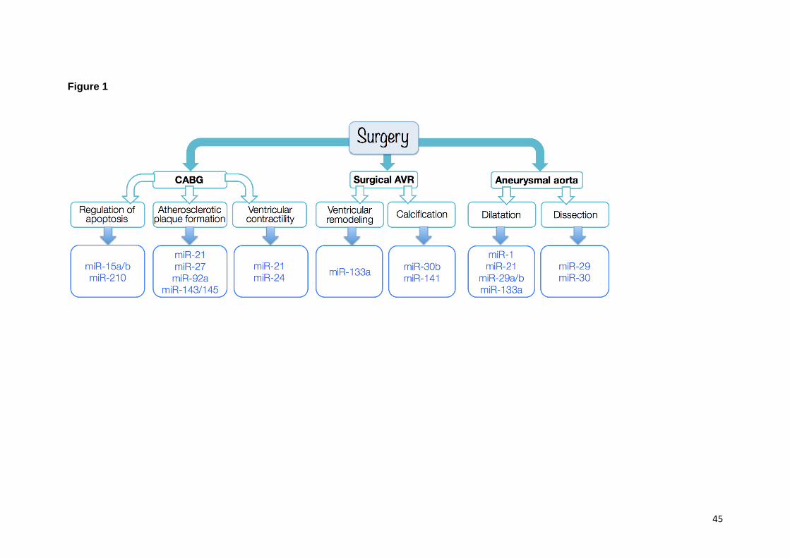
45
Figure 1

46
Figure 2
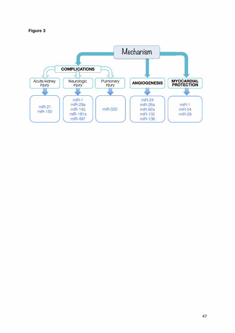
47
Figure 3

48
Figure 4