Bidirectional Notch Signaling and Osteocyte- Derived...
13
Molecular and Cellular Pathobiology Bidirectional Notch Signaling and Osteocyte- Derived Factors in the Bone Marrow Microenvironment Promote Tumor Cell Proliferation and Bone Destruction in Multiple Myeloma Jesus Delgado-Calle 1,2 , Judith Anderson 3 , Meloney D. Cregor 1 , Masahiro Hiasa 3 , John M. Chirgwin 2,4 , Nadia Carlesso 5 , Toshiyuki Yoneda 3 , Khalid S. Mohammad 1,4 , Lilian I. Plotkin 1,2 , G. David Roodman 2,3 , and Teresita Bellido 1,2,4 Abstract In multiple myeloma, an overabundance of monoclonal plasma cells in the bone marrow induces localized osteolytic lesions that rarely heal due to increased bone resorption and suppressed bone formation. Matrix-embedded osteocytes comprise more than 95% of bone cells and are major reg- ulators of osteoclast and osteoblast activity, but their con- tribution to multiple myeloma growth and bone disease is unknown. Here, we report that osteocytes in a mouse model of human MM physically interact with multiple myeloma cells in vivo, undergo caspase-3–dependent apoptosis, and express higher RANKL (TNFSF11) and sclerostin levels than osteocytes in control mice. Mechanistic studies reveal- ed that osteocyte apoptosis was initiated by multiple mye- loma cell-mediated activation of Notch signaling and was further amplified by multiple myeloma cell-secreted TNF. The induction of apoptosis increased osteocytic Rankl expres- sion, the osteocytic Rankl/Opg (TNFRSF11B) ratio, and the ability of osteocytes to attract osteoclast precursors to induce local bone resorption. Furthermore, osteocytes in contact with multiple myeloma cells expressed high levels of Sost/ sclerostin, leading to a reduction in Wnt signaling and subsequent inhibition of osteoblast differentiation. Impor- tantly, direct contact between osteocytes and multiple mye- loma cells reciprocally activated Notch signaling and increased Notch receptor expression, particularly Notch3 and 4, stimulating multiple myeloma cell growth. These studies reveal a previously unknown role for bidirectional Notch signaling that enhances MM growth and bone disease, sug- gesting that targeting osteocyte-multiple myeloma cell inter- actions through specific Notch receptor blockade may repre- sent a promising treatment strategy in multiple myeloma. Cancer Res; 76(5); 1089–100. Ó2016 AACR. Introduction Multiple myeloma is characterized by expansion of monoclo- nal plasma cells in the bone marrow (BM) that induce marked bone destruction in the majority of patients (1). Multiple mye- loma patients present with severe bone pain caused by osteolytic lesions that rarely heal (2). The osteolytic lesions result from increased bone resorption and concomitant long-term suppres- sion of bone formation. The bone/BM microenvironment is a major contributor to tumor growth and bone destruction in multiple myeloma (3). The bone remodeling compartment is disrupted in multiple myeloma, allowing exchange of soluble factors and direct cell-to-cell contact between multiple myelo- ma cells and bone cells (4). Previous studies demonstrated that multiple cytokines are secreted or induced by the interaction of multiple myeloma cells with the different cell types in the bone microenvironment to increase bone destruction, including the receptor of activator nuclear factor-kappa B ligand (RANKL), the chemokine (C-C motif) ligand 3 (MIP1a), and IL3. Further, multiple myeloma–derived IL7, dickkopf WNT signaling path- way inhibitor 1 (DKK1), and IL3, as well as direct contact by multiple myeloma cells with osteoblasts inhibit osteoblast differentiation (1, 5). Although osteoclasts and osteoblasts remodel bone by resorbing or forming bone respectively, osteocytes, which con- stitute >95% of all bone cells, are the central regulators of these processes (6, 7). Osteocytes, although buried within bone mineral, extensively communicate with each other and with cells on the bone surface and in the marrow via cytoplasmic projections that run along canaliculi and form the osteocyte network. This network allows cell-to-cell communication and 1 Department of Anatomy and Cell Biology, Indiana University School of Medicine, Indianapolis, Indiana. 2 Roudebush Veterans Administra- tion Medical Center, Indianapolis, Indiana. 3 Division of Hematology/ Oncology, Department of Medicine, Indiana University School of Med- icine, Indianapolis, Indiana. 4 Division of Endocrinology, Department of Medicine, Indiana University School of Medicine, Indianapolis, Indiana. 5 Department of Pediatrics Indiana, University School of Medicine, Indianapolis, Indiana. Note: Supplementary data for this article are available at Cancer Research Online (http://cancerres.aacrjournals.org/). Corresponding Authors: Teresita Bellido, Indiana University School of Medicine, 635 Barnhill Drive Medical Science Building, Room 5045A, Indianapolis, IN 46202. Phone: 317-274-7410; Fax: 317-278-2040; E-mail: [email protected]; or G. David Roodman, Indiana University School of Medicine, 980 W. Walnut Street, Suite C312, Indianapolis, IN 46202. Phone: 317-278-6255; Fax: 317-274- 0396; E-mail: [email protected] doi: 10.1158/0008-5472.CAN-15-1703 Ó2016 American Association for Cancer Research. Cancer Research www.aacrjournals.org 1089 on June 5, 2018. © 2016 American Association for Cancer Research. cancerres.aacrjournals.org Downloaded from Published OnlineFirst February 1, 2016; DOI: 10.1158/0008-5472.CAN-15-1703
Transcript of Bidirectional Notch Signaling and Osteocyte- Derived...

Molecular and Cellular Pathobiology
Bidirectional Notch Signaling and Osteocyte-Derived Factors in the Bone MarrowMicroenvironment Promote Tumor CellProliferation and Bone Destruction in MultipleMyelomaJesus Delgado-Calle1,2, Judith Anderson3, Meloney D. Cregor1, Masahiro Hiasa3,John M. Chirgwin2,4, Nadia Carlesso5, Toshiyuki Yoneda3, Khalid S. Mohammad1,4,Lilian I. Plotkin1,2, G. David Roodman2,3, and Teresita Bellido1,2,4
Abstract
In multiple myeloma, an overabundance of monoclonalplasma cells in the bone marrow induces localized osteolyticlesions that rarely heal due to increased bone resorption andsuppressed bone formation. Matrix-embedded osteocytescomprise more than 95% of bone cells and are major reg-ulators of osteoclast and osteoblast activity, but their con-tribution to multiple myeloma growth and bone disease isunknown. Here, we report that osteocytes in a mouse modelof human MM physically interact with multiple myelomacells in vivo, undergo caspase-3–dependent apoptosis, andexpress higher RANKL (TNFSF11) and sclerostin levelsthan osteocytes in control mice. Mechanistic studies reveal-ed that osteocyte apoptosis was initiated by multiple mye-loma cell-mediated activation of Notch signaling and wasfurther amplified by multiple myeloma cell-secreted TNF.The induction of apoptosis increased osteocytic Rankl expres-
sion, the osteocytic Rankl/Opg (TNFRSF11B) ratio, and theability of osteocytes to attract osteoclast precursors to inducelocal bone resorption. Furthermore, osteocytes in contactwith multiple myeloma cells expressed high levels of Sost/sclerostin, leading to a reduction in Wnt signaling andsubsequent inhibition of osteoblast differentiation. Impor-tantly, direct contact between osteocytes and multiple mye-loma cells reciprocally activated Notch signaling andincreased Notch receptor expression, particularly Notch3 and4, stimulating multiple myeloma cell growth. These studiesreveal a previously unknown role for bidirectional Notchsignaling that enhances MM growth and bone disease, sug-gesting that targeting osteocyte-multiple myeloma cell inter-actions through specific Notch receptor blockade may repre-sent a promising treatment strategy in multiple myeloma.Cancer Res; 76(5); 1089–100. �2016 AACR.
IntroductionMultiple myeloma is characterized by expansion of monoclo-
nal plasma cells in the bone marrow (BM) that induce markedbone destruction in the majority of patients (1). Multiple mye-loma patients present with severe bone pain caused by osteolyticlesions that rarely heal (2). The osteolytic lesions result from
increased bone resorption and concomitant long-term suppres-sion of bone formation. The bone/BM microenvironment is amajor contributor to tumor growth and bone destruction inmultiple myeloma (3). The bone remodeling compartment isdisrupted in multiple myeloma, allowing exchange of solublefactors and direct cell-to-cell contact between multiple myelo-ma cells and bone cells (4). Previous studies demonstrated thatmultiple cytokines are secreted or induced by the interaction ofmultiple myeloma cells with the different cell types in the bonemicroenvironment to increase bone destruction, including thereceptor of activator nuclear factor-kappa B ligand (RANKL),the chemokine (C-C motif) ligand 3 (MIP1a), and IL3. Further,multiple myeloma–derived IL7, dickkopf WNT signaling path-way inhibitor 1 (DKK1), and IL3, as well as direct contact bymultiple myeloma cells with osteoblasts inhibit osteoblastdifferentiation (1, 5).
Although osteoclasts and osteoblasts remodel bone byresorbing or forming bone respectively, osteocytes, which con-stitute >95% of all bone cells, are the central regulators of theseprocesses (6, 7). Osteocytes, although buried within bonemineral, extensively communicate with each other and withcells on the bone surface and in the marrow via cytoplasmicprojections that run along canaliculi and form the osteocytenetwork. This network allows cell-to-cell communication and
1Department of Anatomy and Cell Biology, Indiana University Schoolof Medicine, Indianapolis, Indiana. 2Roudebush Veterans Administra-tion Medical Center, Indianapolis, Indiana. 3Division of Hematology/Oncology, Department ofMedicine, IndianaUniversity School ofMed-icine, Indianapolis, Indiana. 4Division of Endocrinology,DepartmentofMedicine, IndianaUniversity School of Medicine, Indianapolis, Indiana.5Department of Pediatrics Indiana, University School of Medicine,Indianapolis, Indiana.
Note: Supplementary data for this article are available at Cancer ResearchOnline (http://cancerres.aacrjournals.org/).
CorrespondingAuthors: Teresita Bellido, Indiana University School of Medicine,635 Barnhill Drive Medical Science Building, Room 5045A, Indianapolis, IN46202. Phone: 317-274-7410; Fax: 317-278-2040; E-mail: [email protected];or G. David Roodman, Indiana University School of Medicine, 980 W. WalnutStreet, Suite C312, Indianapolis, IN 46202. Phone: 317-278-6255; Fax: 317-274-0396; E-mail: [email protected]
doi: 10.1158/0008-5472.CAN-15-1703
�2016 American Association for Cancer Research.
CancerResearch
www.aacrjournals.org 1089
on June 5, 2018. © 2016 American Association for Cancer Research. cancerres.aacrjournals.org Downloaded from
Published OnlineFirst February 1, 2016; DOI: 10.1158/0008-5472.CAN-15-1703
distributes osteocyte-secreted molecules that regulate osteo-blast and osteoclast function. Osteocytes are the primary pro-ducers of sclerostin, the product of the SOST gene, a potentinhibitor of bone formation, and are a major source of RANKL,the central osteoclastogenic factor (8, 9). Lack of sclerostinincreases osteoblast number and activity, and deletion ofosteocytic RANKL inhibits osteoclast formation and boneresorption, demonstrating that osteocytes are key regulatorsof osteoblast and osteoclast activity (10–12). Moreover, apo-ptotic osteocytes, which accumulate with skeletal disuse, glu-cocorticoid excess, or estrogen deficiency, increase local boneresorption by attracting osteoclast precursors to particularareas of bone (6, 13). Although knowledge of the role ofosteocytes and the osteocytic network in bone homeostasisand common skeletal diseases has greatly increased, the con-tribution of osteocytes to the development and progression ofcancer involving bone is just beginning to be defined.
In the current study, we determined if reciprocal communica-tion between multiple myeloma cells and osteocytes occurs andexplored themechanisms involved and the consequences of theseinteractions for the progression of multiple myeloma bonedisease.
Materials and MethodsReagents
Reagents used in this study can be found in SupplementaryMethods.
Cells and culture conditionsL. Bonewald (University of Missouri–Kansas City, Kansas
City, MO) provided the murine MLO-A5 in 1997 and MLO-Y4in 2001 osteocyte-like cells (14, 15). JJN3, 5TGM1, and MM1.Smultiple myeloma cell lines were provided by N. Giuliani(University of Parma, Parma, Italy) in 2006, B. Oyajobi (Uni-versity of Texas at San Antonio, San Antonio, TX) in 2007, andS. Rosen (Northwestern University, Evanston, IL) in 2003 (16–18). R. Jilka (University of Arkansas for Medical Sciences, LittleRock, AR) provided the OB-6 osteoblast–like cells in 1997 (19).Nonadherent osteoclast precursors were collected as describedbefore (20). After informed consent, CD138þ cells frommultiplemyeloma patients were prepared as previously detailed (16).Studies were approved by the Indiana University School ofMedicine Institutional Review Board. Cell lines were authenti-cated by morphology, gene expression profile, and tumorigeniccapacity (multiple myeloma cells). Cocultures were establishedby (i) adding multiple myeloma cells on top of osteocyte-likecells, (ii) adding multiple myeloma cells in transwell chambersin a 1:5 ratio (osteocytic:multiple myeloma), or (iii) adding 50%conditioned media (CM) from 48-hour culture of multiplemyeloma cells to osteocytes. DEVD (50 nmol/L), anti-TNFa(0.3 mg/mL), or GSIXX (2.5–10 mmol/L) were added 1 hourbefore addition of multiple myeloma cells or CM. MLO-A5 cellswere treated with 0.01 ng/mL TNFa, 5 ng/mL TGFb, or 10 ng/mLIL6 for 4 to 24 hours. For Notch activation, MLO-A5 cells werecultured on delta-like 1 ligand (DLL1)-IgG2 or control IgG2-coated plates for 24 hours. For OB-6 osteoblast–like cell dif-ferentiation, cells were cultured with osteogenic media (OM;0.2 mmol/L ascorbic acid, 10 mmol/L b-glycerophosphate) orOM containing 50% of CM fromMLO-A5 or JJN3 cells culturedalone, or from JJN3 directly cocultured with MLO-A5 cells.
Ex vivo bone organ culturesThese assays were performed as previously described (21).
Detailed description of the assay can be found in SupplementaryMethods.
Cell viability and apoptosisMultiple myeloma cells were separated from adherent osteo-
cyte-like cells using EDTA. Cell death was quantified by trypanblue uptake and apoptosis by chromatin condensation andnuclear fragmentation of cells transfected with nuclear greenfluorescent protein (nGFP; refs. 22, 23). At least 100 cells in fivedifferent fields selected by systematic random sampling wereexamined for each experimental condition. Representative photo-micrographs were taken with an EVOS FLCell Imaging System(Life Technologies).
Cell proliferation analysisViable cells were enumerated by trypan blue exclusion. Fluo-
rescence emitted by 5TGM1-GFP cells was measured in a Spec-traMaxi3 microplate reader (Molecular Devices), set at 485nm/520nm. 5TGM1-GFP cell numbers linearly correlated with rela-tive fluorescent units (RFU; Supplementary Fig. S1A).
TransfectionsMLO-A5 cells were transiently transfected using Lipofectamine-
Plus (Invitrogen; refs. 23, 24).
Gene expression analysismRNA expression was quantified as previously described (25).
Murine soluble RANKL,OPG, or human TNFawere quantified byELISA (R&D Systems) of culture supernatants.
Western blot analysisThe assays were performed as described previously (25).
Detailed descriptions of antibodies can be found in Supplemen-tary Methods.
Osteoclast precursor migrationForty-eight–hour CM from JJN3, MLO-A5, or from JJN3 and
MLO-A5 cocultured in direct contact was placed in the bottom of8-mmpore chambers to attractmurine nonadherent BMcells (26).Detailed description of the assay can be found in SupplementaryMethods.
Mouse model of human multiple myelomaSix-week-old female SCID mice B6.CB17-Prkdcscid/SzJ (The
Jackson laboratories) and NIH-LystbgFoxn1nuBtkxid (Charles Riv-erWilmington) were injected intratibially with JJN3 cells or salineand sacrificed 4 weeks later (16). Detailed information regardingimaging and micro-CT analysis can be found in SupplementaryMethods. Studies were approved by the Institutional Animal CareandUseCommittee of the IndianaUniversity School ofMedicine.The sample size was calculated based on a previous study (16).
ImmunohistochemistryAntigen detection was performed on paraffin-embedded tibiae
as described before (25). Detailed descriptions of antibodies canbe found in Supplementary Methods.
Delgado-Calle et al.
Cancer Res; 76(5) March 1, 2016 Cancer Research1090
on June 5, 2018. © 2016 American Association for Cancer Research. cancerres.aacrjournals.org Downloaded from
Published OnlineFirst February 1, 2016; DOI: 10.1158/0008-5472.CAN-15-1703

Acid etching scanning electron microscopyThe assays were performed as described previously (27).
Detailed description of the assay can be found in SupplementaryMethods.
Statistical analysisData were analyzed using SigmaStat (SPSS Science). Differ-
ences between mean were evaluated using unpaired t test, one-way, or two-way ANOVA, followed by pairwise multiple compar-isons using the Student–Newman–Keuls method. Mean� SD arereported. P values <0.05 were considered significant.
ResultsMultiple myeloma cells increase osteocyte apoptosis andosteocytic RANKL and sclerostin production in an in vivomodelof multiple myeloma bone disease
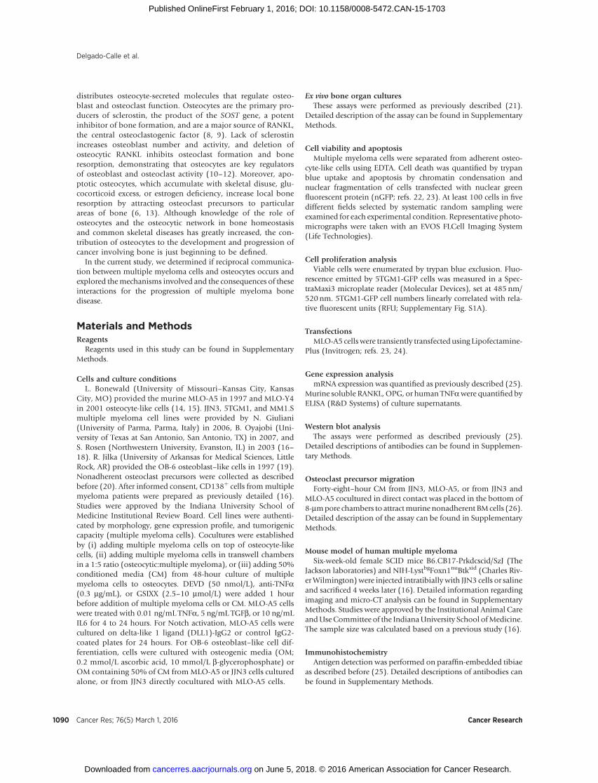
Our murine model of human multiple myeloma is wellestablished and reproduces all features of human multiple mye-loma–induced bone disease (16). Osteolytic lesions were firstdetected radiographically 2 weeks after JJN3 cell injection (Sup-plementary Fig. S1B), and their size progressively increased up to4 weeks (Fig. 1A). Micro-CT analysis of JJN3-involved tibiaperformed at 4 weeks confirmed the presence of osteolytic lesionsand demonstrated decreased trabecular bone volume (BV/TV),trabecular number (Tb.N.), trabecular thickness (Tb.Th.), andincreased trabecular separation (Tb.Sp.) compared with controls(Fig. 1B). The percentage of apoptotic osteocytes, stained with anantiactive caspase-3 (CASP3) antibody, was increased 2-fold inbones injected with JJN3 cells compared with controls (Fig. 1C).Moreover, the percentage of osteocytes expressing RANKL andsclerostin was significantly higher in JJN3-injected mice com-pared with controls. These findings suggest that in addition tosurvival, osteocyte gene expression is altered in multiple myelo-ma–bearing bones. Histologic analysis of multiple myeloma–bearing bones showed that multiple myeloma cells were adjacentto the bone surface, and acid etching-scanning electron micros-copy (SEM) images revealed that osteocytic dendritic processeswere in direct contact with multiple myeloma cells in the marrow(Fig. 1D and E). Thus, osteocytes are in close contact withmultiple myeloma cells, suggesting that multiple myeloma–induced changes in osteocytes may result from cell-to-cell contactand/or exchange of soluble factors between osteocytes and mul-tiple myeloma cells.
Osteocyte apoptosis is initiated by multiple myeloma cell–mediated activation of Notch signaling in osteocytes andamplified by multiple myeloma cell–derived TNFa
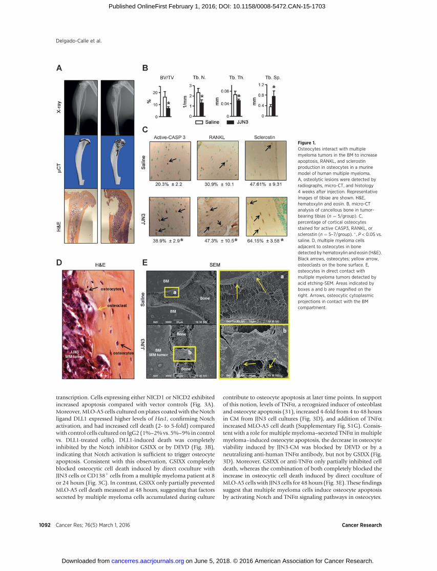
In vitro cocultures between osteocyte-like MLO-A5 cells andmultiple myeloma cells were performed to determine themechanisms responsible for the increased osteocyte apoptosisand upregulation of RANKL and sclerostin observed in osteocytesin multiple myeloma–bearing bones. Communication betweenmultiple myeloma cells and bone cells, including osteocytes, canoccur in different manners: (i) via secretion of soluble factors, (ii)via direct cell-to-cell contact, or (iii) a combination of both. First,direct cell-to-cell cultures were established to determine the effectof physical interactions withmultiplemyeloma cells on osteocyteapoptosis. Coculture of MLO-A5 cells in direct contact withhuman JJN3 multiple myeloma cells induced a 2- to 3-foldincrease in MLO-A5 cell death (1%–4% vs. 6%–15% in MLO-
A5alone vs.MLO-A5 cells coculturedwith JJN3 cells; Fig. 2A).Celldeath was detected at 8 hours and progressively increased at 24and 48 hours. To elucidate the effect of factors released bymultiple myeloma cells on osteocyte apoptosis, the cell typeswere separated by a porous membrane that prevents physicalinteractions and only allows exchange of soluble factors. Cocul-ture of JJN3 and MLO-A5 cells separated by transwell chambersdid not increase cell death at 8 or 24 hours, but it did at 48 hours(1%–3%vs. 8%–13%; Fig. 2B). These results suggest that differentmechanisms mediate the early and late increases in osteocyte celldeath induced by multiple myeloma cells. Coculture of MLO-A5cells with JJN3 cells or JJN3-CM increased the percentage ofMLO-A5 cells exhibiting chromatin condensation and nuclear fragmen-tation, sine qua non features of apoptosis (Fig. 2A and B). More-over, osteocyte death was inhibited by the CASP3 inhibitor,DEVD, at all time points confirming that it was due to apoptosis(Fig. 2A and B; Supplementary Fig. S1C). Coculture of MLO-A5cells with CD138þ cells from 6 different multiple myelomapatients (Fig. 2A) or CM from CD138þ cells from a multiplemyeloma patient (Fig. 2B) also increased MLO-A5 cell apoptosis.Similar results were obtainedwhenMLO-A5were coculturedwithhuman MM.1s or murine 5TGM1 multiple myeloma cells, orwhen MLO-Y4 cells, another murine osteocytic cell line, werecocultured with JJN3 cells (Supplementary Fig. S1D).
Notch signaling is activated by cell-to-cell contact and reg-ulates cell proliferation and apoptosis in multiple cell types(28). Further, Notch signaling is aberrantly activated in mul-tiple myeloma cells due to the overexpression of Notch recep-tors and ligands (29, 30), demonstrating that multiple myelo-ma cells are potential signalers as well as targets of Notch. Wetherefore assessed if direct interactions between multiple mye-loma cells and osteocytes activate Notch in these cells and thepotential biologic consequences. Direct contact of MLO-A5cells with JJN3 multiple myeloma cells activated Notch signal-ing in MLO-A5 cells, as shown by the increased expression ofthe Notch target gene hairy/enhancer-of-split related withYRPW motif 1 (Hey1; Fig. 2C) and hairy and enhancer of split1 (Hes1), which persisted up to 48 hours (Supplementary Fig.S1E). JJN3 cell activation of osteocytic Notch signaling wassuppressed by the Notch inhibitor GSIXX, confirming thespecificity of the effect (Fig. 2C). Hey1 expression was alsoincreased in MLO-A5 cells cocultured with CD138þ cells from 3of 5 multiple myeloma patients (Fig. 2C). As expected, Notchactivation (Hey1 and Hes1) in osteocytes did not occur whenMLO-A5 cells were cocultured with multiple myeloma cells intranswell chambers (Supplementary Fig. S1F). MM1.s or5TGM1 multiple myeloma cells also increased the Notch targetgene expression in osteocyte-like MLO-A5 or MLO-Y4 cells(Supplementary Fig. S1F). In addition, interactions with JJN3cells rapidly upregulated the expression of receptorsNotch3 andNotch4 in MLO-A5 cells, whereas Notch1 or Notch2 receptorexpression was unchanged (Fig. 2D). Moreover, the levels ofNOTCH3 and of Notch intracelular domain 3 (NICD3), theactive form and downstream effector of the receptor NOTCH3,were elevated in tumor-bearing bones compared with controlbones, demonstrating that Notch signaling activation alsooccurs in vivo (Fig. 2E and F).
To evaluate the potential impact of gain-of-function of Notchsignaling on osteocyte life span, MLO-A5 cells were transfectedwith nGFP and the Notch intracellular domains (NICD) 1 or 2,which translocate to the nucleus and activate Notch target gene
Role of Osteocytes in Multiple Myeloma
www.aacrjournals.org Cancer Res; 76(5) March 1, 2016 1091
on June 5, 2018. © 2016 American Association for Cancer Research. cancerres.aacrjournals.org Downloaded from
Published OnlineFirst February 1, 2016; DOI: 10.1158/0008-5472.CAN-15-1703
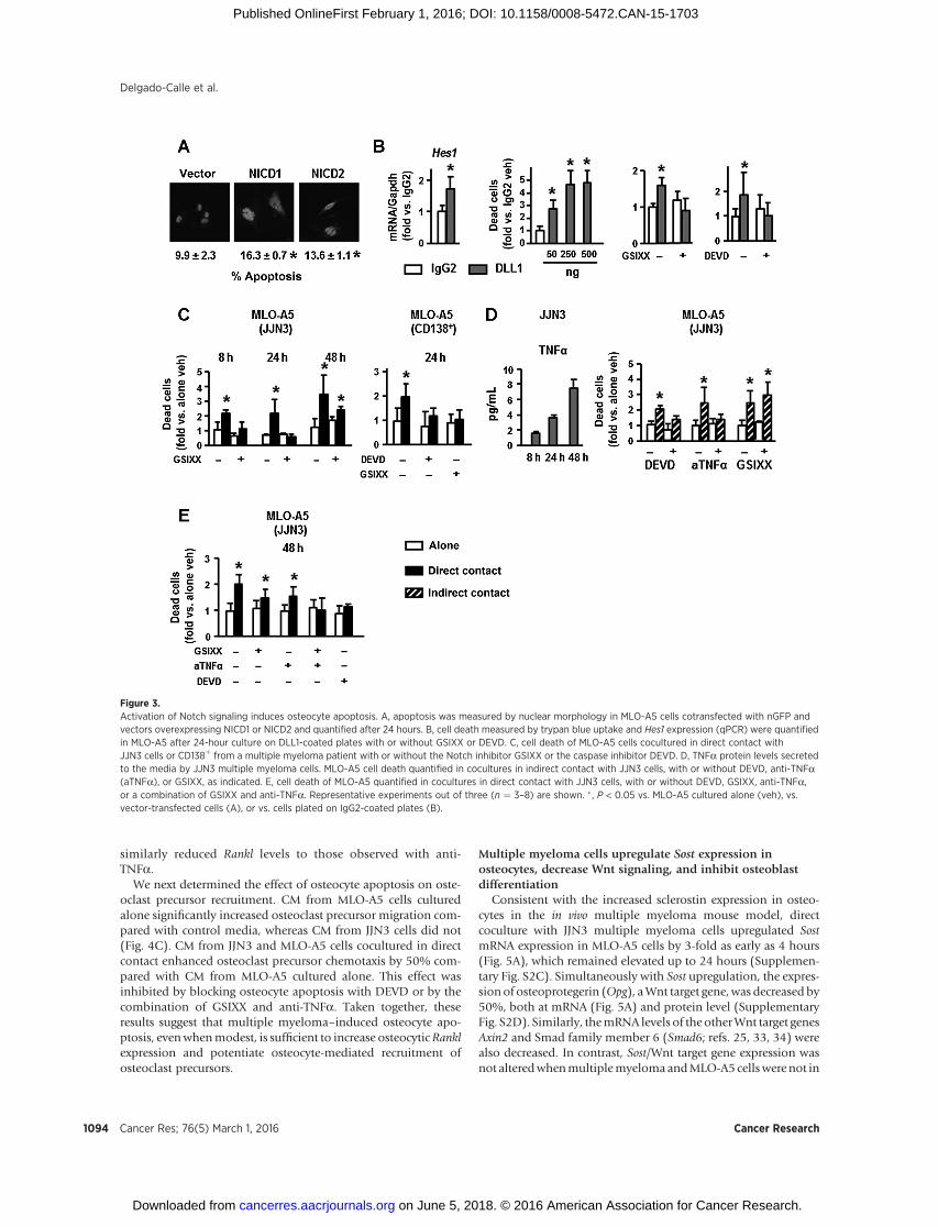
transcription. Cells expressing either NICD1 or NICD2 exhibitedincreased apoptosis compared with vector controls (Fig. 3A).Moreover, MLO-A5 cells cultured on plates coated with the Notchligand DLL1 expressed higher levels of Hes1, confirming Notchactivation, and had increased cell death (2- to 5-fold) comparedwith control cells culturedon IgG2 (1%–2%vs. 5%–9%in controlvs. DLL1-treated cells). DLL1-induced death was completelyinhibited by the Notch inhibitor GSIXX or by DEVD (Fig. 3B),indicating that Notch activation is sufficient to trigger osteocyteapoptosis. Consistent with this observation, GSIXX completelyblocked osteocytic cell death induced by direct coculture withJJN3 cells or CD138þ cells from a multiple myeloma patient at 8or 24 hours (Fig. 3C). In contrast, GSIXX only partially preventedMLO-A5 cell death measured at 48 hours, suggesting that factorssecreted by multiple myeloma cells accumulated during culture
contribute to osteocyte apoptosis at later time points. In supportof this notion, levels of TNFa, a recognized inducer of osteoblastand osteocyte apoptosis (31), increased 4-fold from 4 to 48 hoursin CM from JJN3 cell cultures (Fig. 3D), and addition of TNFaincreased MLO-A5 cell death (Supplementary Fig. S1G). Consis-tent with a role for multiple myeloma–secreted TNFa in multiplemyeloma–induced osteocyte apoptosis, the decrease in osteocyteviability induced by JJN3-CM was blocked by DEVD or by aneutralizing anti-human TNFa antibody, but not by GSIXX (Fig.3D). Moreover, GSIXX or anti-TNFa only partially inhibited celldeath, whereas the combination of both completely blocked theincrease in osteocytic cell death induced by direct coculture ofMLO-A5 cells with JJN3 cells for 48 hours (Fig. 3E). These findingssuggest that multiple myeloma cells induce osteocyte apoptosisby activating Notch and TNFa signaling pathways in osteocytes.
Figure 1.Osteocytes interact with multiplemyeloma tumors in the BM to increaseapoptosis, RANKL, and sclerostinproduction in osteocytes in a murinemodel of human multiple myeloma.A, osteolytic lesions were detected byradiographs, micro-CT, and histology4 weeks after injection. Representativeimages of tibiae are shown. H&E,hematoxylin and eosin. B, micro-CTanalysis of cancellous bone in tumor-bearing tibias (n ¼ 5/group). C,percentage of cortical osteocytesstained for active CASP3, RANKL, orsclerostin (n ¼ 5–7/group). � , P < 0.05 vs.saline. D, multiple myeloma cellsadjacent to osteocytes in bonedetected by hematoxylin and eosin (H&E).Black arrows, osteocytes; yellow arrow,osteoclasts on the bone surface. E,osteocytes in direct contact withmultiple myeloma tumors detected byacid etching-SEM. Areas indicated byboxes a and b are magnified on theright. Arrows, osteocytic cytoplasmicprojections in contact with the BMcompartment.
Delgado-Calle et al.
Cancer Res; 76(5) March 1, 2016 Cancer Research1092
on June 5, 2018. © 2016 American Association for Cancer Research. cancerres.aacrjournals.org Downloaded from
Published OnlineFirst February 1, 2016; DOI: 10.1158/0008-5472.CAN-15-1703
Osteocyte apoptosis increases the osteoclastogenic potential ofosteocytes and stimulates osteoclast precursor recruitment
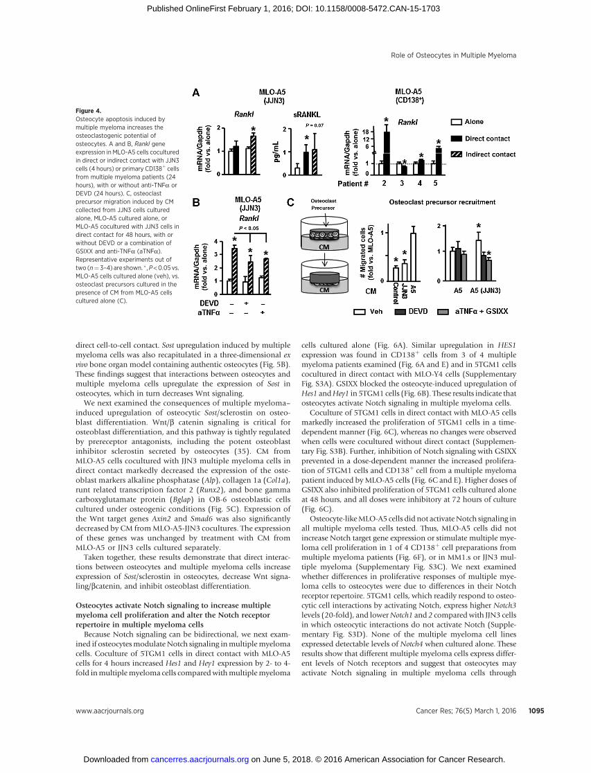
We then investigated the consequences of osteocyte apo-ptosis on the osteoclastogenic potential of osteocytes. MLO-A5 cells cocultured with JJN3 multiple myeloma cells exhib-ited increased Rankl expression at the mRNA and proteinlevels (Fig. 4A). Further, CD138þ cells from 3 of 4 multiplemyeloma patients also increased Rankl transcripts in MLO-A5cells. Like apoptosis, Rankl expression in osteocytes wasincreased by either direct or indirect contact with multiple
myeloma cells, suggesting that soluble factors secreted bymultiple myeloma cells were responsible. Similar results wereobtained with other multiple myeloma and osteocyte-like celllines (Supplementary Fig. S2A). Among several cytokinessecreted by multiple myeloma cells known to upregulateRankl (32), only TNFa increased Rankl mRNA in MLO-A5cells after 4 hours (Supplementary Fig. S2B) or 24 hours.Moreover, anti-TNFa significantly blunted the increased RanklmRNA levels in MLO-A5 cells treated with CM from JJN3cells (Fig. 4B). Inhibition of osteocyte apoptosis by DEVD
Figure 2.Multiple myeloma–induced osteocyte apoptosis is triggered by activation of Notch signaling in osteocytes and is sustained by multiple myeloma–derivedTNFa. A, cell death, measured by trypan blue uptake, and apoptosis (24 hours), measured by nuclear morphology, were quantified in MLO-A5 cells cocultured indirect contact with JJN3 multiple myeloma cells or CD138þ cells isolated from six multiple myeloma patients, with or without the caspase inhibitor DEVD for24 hours. B, MLO-A5 cell death or apoptosis (24 hours) was quantified in cocultures in indirect contact with JJN3 cells, cultured with 48-hour CM collected fromJJN3 cultured alone, or 48-hour CM from primary CD138þ cells from a multiple myeloma patient with or without the caspase inhibitor DEVD. Representativeexperiments out of three (n ¼ 3–8) are shown. C and D, Hey1, Notch1, 2, 3, and 4 expression in MLO-A5 cells cocultured for 4 hours in direct contact with JJN3multiple myeloma cells or CD138þ cells from multiple myeloma patients. Data from one-well per condition are reported for patient #1; 3 wells per condition arereported for the other patients. E, percentage of cortical osteocytes stained for NOTCH3 (n ¼ 3–7/group). � , P < 0.05 vs. saline. F, protein levels ofNICD3, the activated form of the NOTCH3 receptor, in bone lysates obtained from saline- and JJN3-injected tibias.
Role of Osteocytes in Multiple Myeloma
www.aacrjournals.org Cancer Res; 76(5) March 1, 2016 1093
on June 5, 2018. © 2016 American Association for Cancer Research. cancerres.aacrjournals.org Downloaded from
Published OnlineFirst February 1, 2016; DOI: 10.1158/0008-5472.CAN-15-1703
similarly reduced Rankl levels to those observed with anti-TNFa.
We next determined the effect of osteocyte apoptosis on oste-oclast precursor recruitment. CM from MLO-A5 cells culturedalone significantly increased osteoclast precursor migration com-pared with control media, whereas CM from JJN3 cells did not(Fig. 4C). CM from JJN3 and MLO-A5 cells cocultured in directcontact enhanced osteoclast precursor chemotaxis by 50% com-pared with CM from MLO-A5 cultured alone. This effect wasinhibited by blocking osteocyte apoptosis with DEVD or by thecombination of GSIXX and anti-TNFa. Taken together, theseresults suggest that multiple myeloma–induced osteocyte apo-ptosis, evenwhenmodest, is sufficient to increase osteocyticRanklexpression and potentiate osteocyte-mediated recruitment ofosteoclast precursors.
Multiple myeloma cells upregulate Sost expression inosteocytes, decrease Wnt signaling, and inhibit osteoblastdifferentiation
Consistent with the increased sclerostin expression in osteo-cytes in the in vivo multiple myeloma mouse model, directcoculture with JJN3 multiple myeloma cells upregulated SostmRNA expression in MLO-A5 cells by 3-fold as early as 4 hours(Fig. 5A), which remained elevated up to 24 hours (Supplemen-tary Fig. S2C). Simultaneously with Sost upregulation, the expres-sion of osteoprotegerin (Opg), aWnt target gene,was decreased by50%, both at mRNA (Fig. 5A) and protein level (SupplementaryFig. S2D). Similarly, themRNA levels of the otherWnt target genesAxin2 and Smad family member 6 (Smad6; refs. 25, 33, 34) werealso decreased. In contrast, Sost/Wnt target gene expression wasnot alteredwhenmultiplemyelomaandMLO-A5 cellswerenot in
Figure 3.Activation of Notch signaling induces osteocyte apoptosis. A, apoptosis was measured by nuclear morphology in MLO-A5 cells cotransfected with nGFP andvectors overexpressing NICD1 or NICD2 and quantified after 24 hours. B, cell death measured by trypan blue uptake and Hes1 expression (qPCR) were quantifiedin MLO-A5 after 24-hour culture on DLL1-coated plates with or without GSIXX or DEVD. C, cell death of MLO-A5 cells cocultured in direct contact withJJN3 cells or CD138þ from a multiple myeloma patient with or without the Notch inhibitor GSIXX or the caspase inhibitor DEVD. D, TNFa protein levels secretedto the media by JJN3 multiple myeloma cells. MLO-A5 cell death quantified in cocultures in indirect contact with JJN3 cells, with or without DEVD, anti-TNFa(aTNFa), or GSIXX, as indicated. E, cell death of MLO-A5 quantified in cocultures in direct contact with JJN3 cells, with or without DEVD, GSIXX, anti-TNFa,or a combination of GSIXX and anti-TNFa. Representative experiments out of three (n ¼ 3–8) are shown. � , P < 0.05 vs. MLO-A5 cultured alone (veh), vs.vector-transfected cells (A), or vs. cells plated on IgG2-coated plates (B).
Delgado-Calle et al.
Cancer Res; 76(5) March 1, 2016 Cancer Research1094
on June 5, 2018. © 2016 American Association for Cancer Research. cancerres.aacrjournals.org Downloaded from
Published OnlineFirst February 1, 2016; DOI: 10.1158/0008-5472.CAN-15-1703
direct cell-to-cell contact. Sost upregulation induced by multiplemyeloma cells was also recapitulated in a three-dimensional exvivo bone organ model containing authentic osteocytes (Fig. 5B).These findings suggest that interactions between osteocytes andmultiple myeloma cells upregulate the expression of Sost inosteocytes, which in turn decreases Wnt signaling.
We next examined the consequences of multiple myeloma–induced upregulation of osteocytic Sost/sclerostin on osteo-blast differentiation. Wnt/b catenin signaling is critical forosteoblast differentiation, and this pathway is tightly regulatedby prereceptor antagonists, including the potent osteoblastinhibitor sclerostin secreted by osteocytes (35). CM fromMLO-A5 cells cocultured with JJN3 multiple myeloma cells indirect contact markedly decreased the expression of the oste-oblast markers alkaline phosphatase (Alp), collagen 1a (Col1a),runt related transcription factor 2 (Runx2), and bone gammacarboxyglutamate protein (Bglap) in OB-6 osteoblastic cellscultured under osteogenic conditions (Fig. 5C). Expression ofthe Wnt target genes Axin2 and Smad6 was also significantlydecreased by CM fromMLO-A5-JJN3 cocultures. The expressionof these genes was unchanged by treatment with CM fromMLO-A5 or JJN3 cells cultured separately.
Taken together, these results demonstrate that direct interac-tions between osteocytes and multiple myeloma cells increaseexpression of Sost/sclerostin in osteocytes, decrease Wnt signa-ling/bcatenin, and inhibit osteoblast differentiation.
Osteocytes activate Notch signaling to increase multiplemyeloma cell proliferation and alter the Notch receptorrepertoire in multiple myeloma cells
Because Notch signaling can be bidirectional, we next exam-ined if osteocytesmodulate Notch signaling inmultiplemyelomacells. Coculture of 5TGM1 cells in direct contact with MLO-A5cells for 4 hours increased Hes1 and Hey1 expression by 2- to 4-fold inmultiplemyeloma cells comparedwithmultiplemyeloma
cells cultured alone (Fig. 6A). Similar upregulation in HES1expression was found in CD138þ cells from 3 of 4 multiplemyeloma patients examined (Fig. 6A and E) and in 5TGM1 cellscocultured in direct contact with MLO-Y4 cells (SupplementaryFig. S3A). GSIXX blocked the osteocyte-induced upregulation ofHes1 andHey1 in 5TGM1 cells (Fig. 6B). These results indicate thatosteocytes activate Notch signaling in multiple myeloma cells.
Coculture of 5TGM1 cells in direct contact with MLO-A5 cellsmarkedly increased the proliferation of 5TGM1 cells in a time-dependent manner (Fig. 6C), whereas no changes were observedwhen cells were cocultured without direct contact (Supplemen-tary Fig. S3B). Further, inhibition of Notch signaling with GSIXXprevented in a dose-dependent manner the increased prolifera-tion of 5TGM1 cells and CD138þ cell from a multiple myelomapatient induced byMLO-A5 cells (Fig. 6C and E). Higher doses ofGSIXX also inhibited proliferation of 5TGM1 cells cultured aloneat 48 hours, and all doses were inhibitory at 72 hours of culture(Fig. 6C).
Osteocyte-likeMLO-A5 cells did not activateNotch signaling inall multiple myeloma cells tested. Thus, MLO-A5 cells did notincrease Notch target gene expression or stimulate multiple mye-loma cell proliferation in 1 of 4 CD138þ cell preparations frommultiple myeloma patients (Fig. 6F), or in MM1.s or JJN3 mul-tiple myeloma (Supplementary Fig. S3C). We next examinedwhether differences in proliferative responses of multiple mye-loma cells to osteocytes were due to differences in their Notchreceptor repertoire. 5TGM1 cells, which readily respond to osteo-cytic cell interactions by activating Notch, express higher Notch3levels (20-fold), and lowerNotch1 and 2 compared with JJN3 cellsin which osteocytic interactions do not activate Notch (Supple-mentary Fig. S3D). None of the multiple myeloma cell linesexpressed detectable levels of Notch4 when cultured alone. Theseresults show that different multiple myeloma cells express differ-ent levels of Notch receptors and suggest that osteocytes mayactivate Notch signaling in multiple myeloma cells through
Figure 4.Osteocyte apoptosis induced bymultiple myeloma increases theosteoclastogenic potential ofosteocytes. A and B, Rankl geneexpression in MLO-A5 cells coculturedin direct or indirect contact with JJN3cells (4 hours) or primary CD138þ cellsfrom multiple myeloma patients (24hours), with or without anti-TNFa orDEVD (24 hours). C, osteoclastprecursor migration induced by CMcollected from JJN3 cells culturedalone, MLO-A5 cultured alone, orMLO-A5 cocultured with JJN3 cells indirect contact for 48 hours, with orwithout DEVD or a combination ofGSIXX and anti-TNFa (aTNFa).Representative experiments out oftwo (n¼ 3–4) are shown. � ,P<0.05 vs.MLO-A5 cells cultured alone (veh), vs.osteoclast precursors cultured in thepresence of CM from MLO-A5 cellscultured alone (C).
Role of Osteocytes in Multiple Myeloma
www.aacrjournals.org Cancer Res; 76(5) March 1, 2016 1095
on June 5, 2018. © 2016 American Association for Cancer Research. cancerres.aacrjournals.org Downloaded from
Published OnlineFirst February 1, 2016; DOI: 10.1158/0008-5472.CAN-15-1703

Notch3, leading to increased multiple myeloma cell proliferation.Moreover, osteocytic interactions rapidly upregulated Notch3expression in 5TGM1 cells and induced the expression ofNotch4,which is undetectable in multiple myeloma cells cultured alone(Fig. 6D). Further, culture of 5TGM1multiplemyeloma cells withauthentic osteocytes in ex vivobone organ cultures activatedNotchsignaling and increased the expression of Notch3 and 4 withoutaltering Notch1 or 2 mRNA levels (Fig. 6G). Similarly, CD138þ
cells from a multiple myeloma patient that exhibit Notch acti-vation induced by osteocytes also displayed upregulation ofNOTCH3 (Fig. 6E), whereas no changes were found in otherNotch receptors. In contrast, no changes in the expression ofNOTCH3 were observed in CD138þ cells from a multiple mye-loma patient that did not show activation of Notch signaling aftercontact with MLO-A5 cells (Fig. 6F). MLO-A5 cells also upregu-lated Notch1 and 2 in 5TGM1 cells at later time points (Fig. 6D).These results demonstrate that osteocytes change the Notchreceptor repertoire expressed by multiple myeloma cells. Thus,the capacity of osteocytes to alter the Notch receptor repertoire onmultiple myeloma cells may regulate osteocyte enhancement ofmultiple myeloma cell growth.
DiscussionOsteocytes play a central role in bone homeostasis by regulat-
ing osteoclast and osteoblast activity, and premature death ofosteocytes leads to targeted bone resorption during skeletal disuseand estrogen loss (6). We report that osteocytes also contribute toa microenvironment favorable for the progression of multiplemyeloma and its associated bone disease. First, we show that
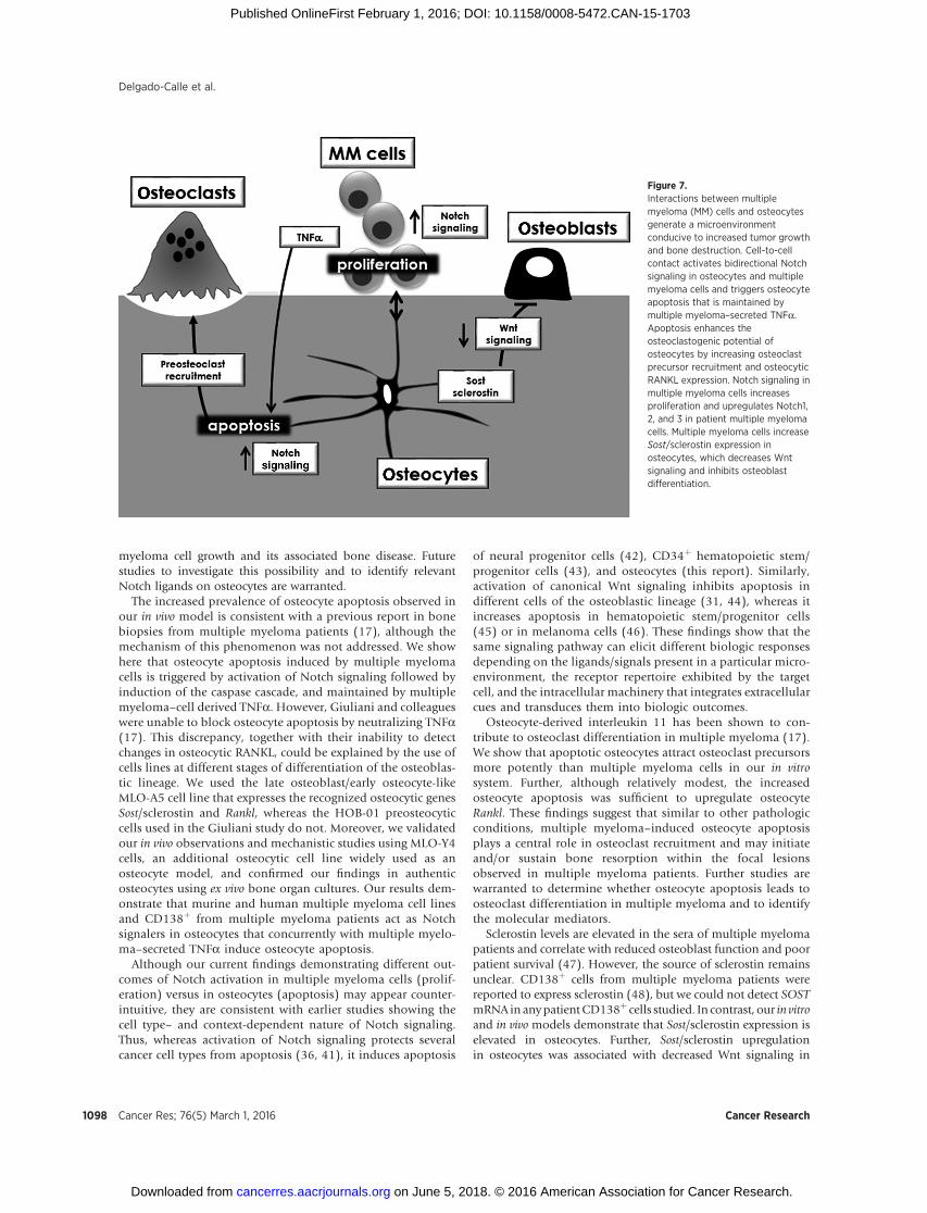
osteocytes physically interact with multiple myeloma tumors invivo, which increases sclerostin and RANKL production by osteo-cytes and reduces osteocyte viability. Second, the reduced viabilityof osteocytes is due to apoptosis triggered by multiple myelomacell–mediated activation of Notch signaling and sustained bymultiple myeloma–derived TNFa. Third, elevated osteocyte apo-ptosis increases osteocytic Rankl expression and enhances theability of osteocytes to attract osteoclast precursors. Fourth, theincrease in Sost/sclerostin decreases Wnt signaling and inhibitsosteoblast differentiation. Fifth, osteocytes induce reciprocal acti-vation of Notch signaling in multiple myeloma cells, which inturn enhances multiple myeloma cell proliferation and alters theNotch receptor repertoire on multiple myeloma cells. Important-ly, these results were validated in in vivo and in vitro, as well as in anovel ex vivomodel, using severalmultiplemyeloma cell lines andCD138þ cells from multiple myeloma patients. Taken together,these findings demonstrate that interactions between multiplemyeloma cells and osteocytes within the bone/BM microenvi-ronment generate a permissive niche for multiple myeloma cellgrowth and bone destruction (Fig. 7). Thus, targeting osteocytesand their derived factorsmight represent a novel approach to treatmultiple myeloma bone disease.
Consistent with our findings showing that osteocytes inducemultiple myeloma cell growth by activating Notch signaling,several studies have shown that dysregulation of Notch signalingincreases multiple myeloma growth (36–38), and paracrineNotch activationmediated by BM stromal cells increasesmultiplemyeloma cell proliferation, through NOTCH1 and 2 signaling(30, 39, 40). However, NOTCH3 and 4 appear to mediate theosteocytic effects, suggesting that regulation ofNotch activation in
Figure 5.Multiple myeloma increases Sost expression in osteocytes, which decreases Wnt signaling and osteoblasts differentiation. A, Sost, Opg, and Wnt targetgenes mRNA levels in MLO-A5 cells cocultured in direct or indirect contact with JJN3 cells (4 hours). B, Sost expression in authentic osteocytes coculturedwith 5TGM1 multiple myeloma cells in ex vivo bone organ cultures (48 hours). C, osteoblast marker expression (Alp, Col1a, Runx2, and Bglap) andWnt target genes(Axin2 and Smad6) in osteoblasts cultured in OM with or without 50% CM from JJN3 cells cultured alone, MLO-A5 cultured alone, or MLO-A5 coculturedwith JJN3 cells in direct contact for 48 hours. Representative experiments out of two (n ¼ 3–6) are shown. � , P < 0.05 vs. MLO-A5 cells cultured alone (A), vs.authentic osteocytes (bone; B), vs. osteoblast cultured with OM and without CM (C).
Delgado-Calle et al.
Cancer Res; 76(5) March 1, 2016 Cancer Research1096
on June 5, 2018. © 2016 American Association for Cancer Research. cancerres.aacrjournals.org Downloaded from
Published OnlineFirst February 1, 2016; DOI: 10.1158/0008-5472.CAN-15-1703

multiple myeloma is mediated by complex paracrine interac-tions with different cell types within the bone/BMmicroenviron-ment. Further, the changes in the Notch receptor repertoire onmultiple myeloma cells induced by osteocytic interactions could
impact both the homotypic and heterotypic cell-to-cell interac-tions of multiple myeloma cells. Although the specific roles ofNOTCH3 and 4 in multiple myeloma are not fully understood,selective Notch receptor targeting may allow control of multiple
Figure 6.Osteocytes activate Notch signaling, regulate Notch receptor expression, and increase proliferation in multiple myeloma cells. A and B, Hes1 and Hey1 geneexpression in 5TGM1 multiple myeloma cells or CD138þ cells from three different multiple myeloma patients cocultured in direct contact with MLO-A5 cells, with orwithout GSIXX. Data from one well per condition are reported for patient #1; 3 wells per condition are reported for the other patients. C, cell growthof 5TGM1 cells measured as RFU, cultured alone or cocultured in direct contact with MLO-A5 cells, with or without GSIXX. D, time course of Notch receptorsexpression in 5TGM1 cells cocultured with MLO-A5 cells for 4, 24, 48, and 72 hours. For Notch1, 2, and 3 expression, fold changes were calculated vs.5TGM1 cells cultured alone. For Notch4 expression, fold change was calculated vs. Notch4 expression in 5TGM1 cells cocultured with MLO-A5 for 4 hours. Cellproliferation (number of viable cells), Hey1, and Notch1, 2, 3, and 4 gene expression in CD138þ cells from a multiple myeloma patient in which MLO-A5 cells induce(E) or not (F) Notch activation. G, Hey1 and Notch1, 2, 3, and 4 gene expression in 5TGM1 cells cocultured with authentic osteocytes in ex vivo bone organcultures. A representative experiment of two (n ¼ 4) is shown. � , P < 0.05 vs. multiple myeloma cells cultured alone (veh), vs. multiple myeloma culturedwith MLO-A5 for 4 hours (D; Notch4).
Role of Osteocytes in Multiple Myeloma
www.aacrjournals.org Cancer Res; 76(5) March 1, 2016 1097
on June 5, 2018. © 2016 American Association for Cancer Research. cancerres.aacrjournals.org Downloaded from
Published OnlineFirst February 1, 2016; DOI: 10.1158/0008-5472.CAN-15-1703
myeloma cell growth and its associated bone disease. Futurestudies to investigate this possibility and to identify relevantNotch ligands on osteocytes are warranted.
The increased prevalence of osteocyte apoptosis observed inour in vivo model is consistent with a previous report in bonebiopsies from multiple myeloma patients (17), although themechanism of this phenomenon was not addressed. We showhere that osteocyte apoptosis induced by multiple myelomacells is triggered by activation of Notch signaling followed byinduction of the caspase cascade, and maintained by multiplemyeloma–cell derived TNFa. However, Giuliani and colleagueswere unable to block osteocyte apoptosis by neutralizing TNFa(17). This discrepancy, together with their inability to detectchanges in osteocytic RANKL, could be explained by the use ofcells lines at different stages of differentiation of the osteoblas-tic lineage. We used the late osteoblast/early osteocyte-likeMLO-A5 cell line that expresses the recognized osteocytic genesSost/sclerostin and Rankl, whereas the HOB-01 preosteocyticcells used in the Giuliani study do not. Moreover, we validatedour in vivo observations and mechanistic studies using MLO-Y4cells, an additional osteocytic cell line widely used as anosteocyte model, and confirmed our findings in authenticosteocytes using ex vivo bone organ cultures. Our results dem-onstrate that murine and human multiple myeloma cell linesand CD138þ from multiple myeloma patients act as Notchsignalers in osteocytes that concurrently with multiple myelo-ma–secreted TNFa induce osteocyte apoptosis.
Although our current findings demonstrating different out-comes of Notch activation in multiple myeloma cells (prolif-eration) versus in osteocytes (apoptosis) may appear counter-intuitive, they are consistent with earlier studies showing thecell type– and context-dependent nature of Notch signaling.Thus, whereas activation of Notch signaling protects severalcancer cell types from apoptosis (36, 41), it induces apoptosis
of neural progenitor cells (42), CD34þ hematopoietic stem/progenitor cells (43), and osteocytes (this report). Similarly,activation of canonical Wnt signaling inhibits apoptosis indifferent cells of the osteoblastic lineage (31, 44), whereas itincreases apoptosis in hematopoietic stem/progenitor cells(45) or in melanoma cells (46). These findings show that thesame signaling pathway can elicit different biologic responsesdepending on the ligands/signals present in a particular micro-environment, the receptor repertoire exhibited by the targetcell, and the intracellular machinery that integrates extracellularcues and transduces them into biologic outcomes.
Osteocyte-derived interleukin 11 has been shown to con-tribute to osteoclast differentiation in multiple myeloma (17).We show that apoptotic osteocytes attract osteoclast precursorsmore potently than multiple myeloma cells in our in vitrosystem. Further, although relatively modest, the increasedosteocyte apoptosis was sufficient to upregulate osteocyteRankl. These findings suggest that similar to other pathologicconditions, multiple myeloma–induced osteocyte apoptosisplays a central role in osteoclast recruitment and may initiateand/or sustain bone resorption within the focal lesionsobserved in multiple myeloma patients. Further studies arewarranted to determine whether osteocyte apoptosis leads toosteoclast differentiation in multiple myeloma and to identifythe molecular mediators.
Sclerostin levels are elevated in the sera of multiple myelomapatients and correlate with reduced osteoblast function and poorpatient survival (47). However, the source of sclerostin remainsunclear. CD138þ cells from multiple myeloma patients werereported to express sclerostin (48), but we could not detect SOSTmRNA in anypatientCD138þ cells studied. In contrast, our in vitroand in vivo models demonstrate that Sost/sclerostin expression iselevated in osteocytes. Further, Sost/sclerostin upregulationin osteocytes was associated with decreased Wnt signaling in
Figure 7.Interactions between multiplemyeloma (MM) cells and osteocytesgenerate a microenvironmentconducive to increased tumor growthand bone destruction. Cell-to-cellcontact activates bidirectional Notchsignaling in osteocytes and multiplemyeloma cells and triggers osteocyteapoptosis that is maintained bymultiple myeloma–secreted TNFa.Apoptosis enhances theosteoclastogenic potential ofosteocytes by increasing osteoclastprecursor recruitment and osteocyticRANKL expression. Notch signaling inmultiple myeloma cells increasesproliferation and upregulates Notch1,2, and 3 in patient multiple myelomacells. Multiple myeloma cells increaseSost/sclerostin expression inosteocytes, which decreases Wntsignaling and inhibits osteoblastdifferentiation.
Delgado-Calle et al.
Cancer Res; 76(5) March 1, 2016 Cancer Research1098
on June 5, 2018. © 2016 American Association for Cancer Research. cancerres.aacrjournals.org Downloaded from
Published OnlineFirst February 1, 2016; DOI: 10.1158/0008-5472.CAN-15-1703
osteoblasts and reduced osteoblast differentiation. In addition,the expression of Wnt target genes, including OPG, was reduced,thereby further increasing the Rankl/Opg ratio. These results sug-gest that osteocytes contribute to the generation of a multiplemyelomamicroenvironment with high sclerostin concentrations,affecting both bone formation and bone resorption.
Physical interactions between multiple myeloma cellsand different cells in the bone microenvironment, includingstromal cells, osteoclasts, and immune cells, were shown to beimportant for tumor proliferation and survival (49). Ourresults suggest that osteocytes also contribute to multiplemyeloma cell growth and multiple myeloma–induced bonedisease by stimulating osteoclast recruitment and inhibitingbone formation via direct and indirect contact with multiplemyeloma cells. Because osteocytes comprise more than 95% ofthe bone cells, they should be major contributors to generatinga microenvironment favorable for multiple myeloma progres-sion. However, the relative contribution of osteocytes versusother cell types in the bone/BM microenvironment to multiplemyeloma disease remains to be determined.
Our study identified pathways activated by interactionsbetween multiple myeloma cells and osteocytes that providepotential new ways to inhibit multiple myeloma tumor growthand bone disease. Prevention of osteocyte apoptosis might sup-press initiation of bone resorption by inhibiting osteoclast recruit-ment as well as improving the bone fragility syndrome associatedwith multiple myeloma and with some of the current antitumordrugs (e.g., glucocorticoids that also increase osteocyte apopto-sis). Pharmacologic inhibition of Notch signaling can inhibitmultiple myeloma cell growth (50, 51) and could provide addi-tional benefits by inhibiting osteocyte apoptosis, either alone orin combination with TNFa signaling blockade. Our findingsdemonstrate that osteocytes regulate the expression and repertoireof Notch receptors on multiple myeloma cells, supporting devel-opment of therapeutic approaches that block specific Notchreceptors. This approach might be an effective alternative toovercome the side effects associated with generalized Notchinhibition (36).
Disclosure of Potential Conflicts of InterestNo potential conflicts of interest were disclosed.
Authors' ContributionsConception and design: J. Delgado-Calle, K.S. Mohammad, L.I. Plotkin,G.D. Roodman, T. BellidoDevelopment of methodology: J. Delgado-Calle, J.M. Chirgwin, L.I. Plotkin,T. BellidoAcquisition of data (provided animals, acquired and managed patients,provided facilities, etc.): J. Delgado-Calle, J. Anderson, M.D. Cregor, M. Hiasa,T. Yoneda, K.S. Mohammad, T. BellidoAnalysis and interpretation of data (e.g., statistical analysis, biostatistics,computational analysis): J. Delgado-Calle, J. Anderson, M. Hiasa, J.M. Chirg-win, L.I. Plotkin, T. BellidoWriting, review, and/or revision of the manuscript: J. Delgado-Calle,J. Anderson,M.Hiasa, J.M. Chirgwin, N. Carlesso, K.S.Mohammad, L.I. Plotkin,T. BellidoAdministrative, technical, or material support (i.e., reporting or organizingdata, constructing databases): J. Delgado-Calle, L.I. Plotkin, T. BellidoStudy supervision: G.D. Roodman, T. BellidoOther (provided advises and expertise on the experimental design of theexperiments addressing Notch signaling): N. Carlesso
AcknowledgmentsThe authors thank KevinMcAndrews, HannahM.Davis, Keith Condon, Amy
Sato,DanZhou,DougTompkins, andNingMa for assistance in tissue collectionand Caroline Miller (Electron Microscopy Center, Indiana University School ofMedicine) for assistance with SEM.
Grant SupportThis work was supported by the NIH grants (Indiana-CTSI P30,
1R21CA179017-02, and R01AR059679 to G.D. Roodman; R01AR059357,R01DK076007, and S10-RR023710 to T. Bellido), the Veteran's Administration(Merit Review to T. Bellido andG.D.Roodman), and the IBMSGideon and SevgiRodan Fellowship (J. Delgado-Calle), and funds from the CTSI at IndianaUniversity.
The costs of publication of this articlewere defrayed inpart by the payment ofpage charges. This article must therefore be hereby marked advertisement inaccordance with 18 U.S.C. Section 1734 solely to indicate this fact.
Received June 23, 2015; revised November 18, 2015; accepted December 14,2015; published OnlineFirst February 1, 2016.
References1. Roodman GD. Pathogenesis of myeloma bone disease. Leukemia 2009;
23:435–41.2. Greenberg AJ, Rajkumar SV, Therneau TM, Singh PP, Dispenzieri A, Kumar
SK. Relationship between initial clinical presentation and the molecularcytogenetic classification of myeloma. Leukemia 2014;28:398–403.
3. Roodman GD. Targeting the bone microenvironment in multiple myelo-ma. J Bone Miner Metab 2010;28:244–50.
4. Andersen TL, Soe K, Sondergaard TE, Plesner T, Delaisse JM. Myeloma cell-induced disruption of bone remodelling compartments leads to osteolyticlesions and generation of osteoclast-myeloma hybrid cells. Br J Haematol2010;148:551–61.
5. Giuliani N, Colla S, Morandi F, Lazzaretti M, Sala R, Bonomini S, et al.Myeloma cells block RUNX2/CBFA1 activity in human bone marrowosteoblast progenitors and inhibit osteoblast formation and differentia-tion. Blood 2005;106:2472–83.
6. Bellido T. Osteocyte-driven bone remodeling. Calcif Tissue Int 2013;94:25–34.
7. Ke HZ, Richards WG, Li X, Ominsky MS. Sclerostin and dickkopf-1 astherapeutic targets in bone diseases. Endocr Rev 2012;33:747–83.
8. Van Bezooijen RL, Roelen BA, Visser A, Wee-Pals L, de Wilt E, Karperien M,et al. Sclerostin is an osteocyte-expressed negative regulator of boneformation, but not a classical BMP antagonist. J Exp Med 2004;199:805–14.
9. Bonewald LF. The amazing osteocyte. J Bone Miner Res 2011;26:229–38.10. Li X,OminskyMS,NiuQT, SunN,Daugherty B,D'AgostinD, et al. Targeted
deletion of the sclerostin gene in mice results in increased bone formationand bone strength. J Bone Miner Res 2008;23:860–9.
11. O'Brien CA, Nakashima T, Takayanagi H. Osteocyte control of osteoclas-togenesis. Bone 2013;54:258–63.
12. Balemans W, Ebeling M, Patel N, Van Hul E, Olson P, Dioszegi M, et al.Increased bone density in sclerosteosis is due to the deficiency of a novelsecreted protein (SOST). Hum Mol Genet 2001;10:537–43.
13. Bellido T. Osteocyte apoptosis induces bone resorption and impairs theskeletal response to weightlessness. BoneKEy-osteovision 2007;4:252–6.
14. Kato Y, Boskey A, Spevak L, Dallas M, HoriM, Bonewald LF. Establishmentof an osteoid preosteocyte-like cell MLO-A5 that spontaneously miner-alizes in culture. J Bone Miner Res 2001;16:1622–33.
15. Kato Y, Windle JJ, Koop BA, Mundy GR, Bonewald LF. Establishmentof an osteocyte-like cell line, MLO-Y4. J Bone Miner Res 1997;12:2014–23.
16. D'Souza S, del PD, Jin S, SunQ, Huston AJ, Kostov FE, et al. Gfi1 expressedin bone marrow stromal cells is a novel osteoblast suppressor in patientswith multiple myeloma bone disease. Blood 2011;118:6871–80.
17. Giuliani N, Ferretti M, Bolzoni M, Storti P, Lazzaretti M, Dalla PB, et al.Increased osteocyte death inmultiplemyeloma patients: role inmyeloma-induced osteoclast formation. Leukemia 2012;26:1391–401.
Role of Osteocytes in Multiple Myeloma
www.aacrjournals.org Cancer Res; 76(5) March 1, 2016 1099
on June 5, 2018. © 2016 American Association for Cancer Research. cancerres.aacrjournals.org Downloaded from
Published OnlineFirst February 1, 2016; DOI: 10.1158/0008-5472.CAN-15-1703
18. Greenstein S, Krett NL, Kurosawa Y, Ma C, Chauhan D, Hideshima T,et al. Characterization of the MM.1 human multiple myeloma (MM)cell lines: a model system to elucidate the characteristics, behavior, andsignaling of steroid-sensitive and -resistant MM cells. Exp Hematol2003;31:271–82.
19. Lecka-Czernik B, Gubrij I, Moerman EA, Kajkenova O, Lipschitz DA,Manolagas SC, et al. Inhibition of Osf2/Cbfa1 expression and terminal osteo-blast differentiation by PPAR-gamma 2. J Cell Biochem 1999;74:357–71.
20. Pacheco-Costa R, Hassan I, Reginato RD, Davis HM, Bruzzaniti A, AllenMR, et al. High bonemass in mice lacking Cx37 due to defective osteoclastdifferentiation. J Biol Chem 2014;289:8508–20.
21. Suvannasankha A, Tompkins DR, Edwards DF, Petyaykina KV, Crean CD,Fournier PG, et al. FGF23 is elevated in multiple myeloma and increasesheparanase expression by tumor cells. Oncotarget 2015;6:19647–60.
22. Bellido T, Plotkin LI. Detection of apoptosis of bone cells in vitro. In:Westendorf JJ, editor.Osteoporosis. Clifton,NJ,USA:HumanaPress; 2007.p. 51–75.
23. Kousteni S, Bellido T, Plotkin LI, O'Brien CA, Bodenner DL, Han L, et al.Nongenotropic, sex-nonspecific signaling through the estrogen or andro-gen receptors: dissociation from transcriptional activity. Cell 2001;104:719–30.
24. Bivi N, Lezcano V, Romanello M, Bellido T, Plotkin LI. Connexin43interacts with barrestin: a pre-requisite for osteoblast survival induced byparathyroid hormone. J Cell Biochem 2011;112:2920–30.
25. Tu X, Delgado-Calle J, Condon KW, Maycas M, Zhang H, Carlesso N, et al.Osteocytes mediate the anabolic actions of canonica Wnt/b-catenin sig-naling in bone. Proc Natl Acad Sci U S A 2015;112:E478–86.
26. Al-Dujaili SA, Lau E, Al-Dujaili H, Tsang K, Guenther A, You L. Apoptoticosteocytes regulate osteoclast precursor recruitment and differentiation invitro. J Cell Biochem 2011;112:2412–23.
27. Kubek DJ, Gattone VH, Allen MR. Methodological assessment of acid-etching for visualizing the osteocyte lacunar-canalicular networks usingscanning electron microscopy. Microsc Res Tech 2010;73:182–6.
28. Bray SJ.Notch signalling: a simple pathway becomes complex.Nat RevMolCell Biol 2006;7:678–89.
29. Houde C, Li Y, Song L, Barton K, Zhang Q, Godwin J, et al. Overexpressionof the NOTCH ligand JAG2 in malignant plasma cells from multiplemyeloma patients and cell lines. Blood 2004;104:3697–704.
30. Jundt F, Probsting KS, Anagnostopoulos I, Muehlinghaus G, Chatterjee M,Mathas S, et al. Jagged1-induced Notch signaling drives proliferation ofmultiple myeloma cells. Blood 2004;103:3511–5.
31. Jilka RL, Bellido T, Almeida M, Plotkin LI, O'Brien CA, Weinstein RS, et al.Apoptosis in bone cells. In:Bilezikian JP, Raisz LG, Martin TJ, editors.Principles of bone biology. 3rd ed. San Diego, San Francisco, New York,London, Sydney, Tokyo: Academic Press; 2008. p. 237–61.
32. Aggarwal R, Ghobrial IM, Roodman GD. Chemokines in multiple mye-loma. Exp Hematol 2006;34:1289–95.
33. YochumGS,McWeeney S, RajaramanV,ClelandR, Peters S,GoodmanRH.Serial analysis of chromatin occupancy identifies beta-catenin target genesin colorectal carcinoma cells. Proc Natl Acad Sci U S A 2007;104:3324–9.
34. O'Brien CA, Plotkin LI, Galli C, Goellner J, Gortazar AR, Allen MR, et al.Control of bone mass and remodeling by PTH receptor signaling inosteocytes. PLoS ONE 2008;3:e2942.
35. BaronR, KneisselM.WNT signaling in bone homeostasis anddisease: fromhuman mutations to treatments. Nat Med 2013;19:179–92.
36. Colombo M, Mirandola L, Platonova N, Apicella L, Basile A, Figueroa AJ,et al. Notch-directed microenvironment reprogramming in myeloma: asingle path to multiple outcomes. Leukemia 2013;27:1009–18.
37. Nefedova Y, Sullivan DM, Bolick SC, Dalton WS, Gabrilovich DI. Inhibi-tion of Notch signaling induces apoptosis of myeloma cells and enhancessensitivity to chemotherapy. Blood 2008;111:2220–9.
38. Guo D, Li C, Teng Q, Sun Z, Li Y, Zhang C. Notch1 overexpressionpromotes cell growth and tumor angiogenesis in myeloma. Neoplasma2013;60:33–40.
39. Chiron D, Maiga S, Descamps G, Moreau P, Le GS, Marionneau S, et al.Critical role of the NOTCH ligand JAG2 in self-renewal of myeloma cells.Blood Cells Mol Dis 2012;48:247–53.
40. Xu D, Hu J, Xu S, De BE, Menu E, Van CB, et al. Dll1/Notch activationaccelerates multiple myeloma disease development by promotingCD138þ MM-cell proliferation. Leukemia 2012;26:1402–5.
41. Mungamuri SK, Yang X, Thor AD, Somasundaram K. Survival signaling byNotch1: mammalian target of rapamycin (mTOR)-dependent inhibitionof p53. Cancer Res 2006;66:4715–24.
42. Yang X, Klein R, Tian X, Cheng HT, Kopan R, Shen J. Notch activationinduces apoptosis in neural progenitor cells through a p53-dependentpathway. Dev Biol 2004;269:81–94.
43. Chadwick N, NostroMC, BaronM,MottramR, Brady G, Buckle AM. Notchsignaling induces apoptosis in primary human CD34þ hematopoieticprogenitor cells. Stem Cells 2007;25:203–10.
44. Almeida M, Han L, Bellido T, Manolagas SC, Kousteni S. Wnt proteinsprevent apoptosis of both uncommitted osteoblast progenitors and dif-ferentiated osteoblasts by beta-catenin-dependent and -independent sig-naling cascades involving Src/ERKandphosphatidylinositol 3-kinase/AKT.J Biol Chem 2005;280:41342–51.
45. Ming M, Wang S, Wu W, Senyuk V, Le Beau MM, Nucifora G, et al.Activation of Wnt/beta-catenin protein signaling induces mitochondria-mediated apoptosis in hematopoietic progenitor cells. J Biol Chem2012;287:22683–90.
46. Zimmerman ZF, Kulikauskas RM, Bomsztyk K, Moon RT, Chien AJ.Activation ofWnt/beta-catenin signaling increases apoptosis inmelanomacells treated with trail. PLoS ONE 2013;8:e69593.
47. Terpos E, Christoulas D, Katodritou E, Bratengeier C, Gkotzamanidou M,Michalis E, et al. Elevated circulating sclerostin correlates with advanceddisease features and abnormalbone remodeling in symptomaticmyeloma:reductionpost-bortezomibmonotherapy. Int JCancer 2012;131:1466–71.
48. Brunetti G, Oranger A, Mori G, Specchia G, Rinaldi E, Curci P, et al.Sclerostin is overexpressed by plasma cells from multiple myelomapatients. Ann N Y Acad Sci 2011;1237:19–23.
49. Bianchi G, Munshi NC. Pathogenesis beyond the cancer clone(s) inmultiple myeloma. Blood 2015;125:3049–58.
50. Li M, Chen F, Clifton N, Sullivan DM, Dalton WS, Gabrilovich DI, et al.Combined inhibition of Notch signaling and Bcl-2/Bcl-xL results in syn-ergistic antimyeloma effect. Mol Cancer Ther 2010;9:3200–9.
51. Schwarzer R, Nickel N, Godau J, Willie BM, Duda GN, Schwarzer R, et al.Notch pathway inhibition controls myeloma bone disease in the murineMOPC315.BM model. Blood Cancer J 2014;4:e217.
Cancer Res; 76(5) March 1, 2016 Cancer Research1100
Delgado-Calle et al.
on June 5, 2018. © 2016 American Association for Cancer Research. cancerres.aacrjournals.org Downloaded from
Published OnlineFirst February 1, 2016; DOI: 10.1158/0008-5472.CAN-15-1703
2016;76:1089-1100. Published OnlineFirst February 1, 2016.Cancer Res Jesus Delgado-Calle, Judith Anderson, Meloney D. Cregor, et al. and Bone Destruction in Multiple MyelomaBone Marrow Microenvironment Promote Tumor Cell Proliferation Bidirectional Notch Signaling and Osteocyte-Derived Factors in the
Updated version
10.1158/0008-5472.CAN-15-1703doi:
Access the most recent version of this article at:
Material
Supplementary
http://cancerres.aacrjournals.org/content/suppl/2016/07/20/0008-5472.CAN-15-1703.DC1
Access the most recent supplemental material at:
Cited articles
http://cancerres.aacrjournals.org/content/76/5/1089.full#ref-list-1
This article cites 49 articles, 14 of which you can access for free at:
Citing articles
http://cancerres.aacrjournals.org/content/76/5/1089.full#related-urls
This article has been cited by 1 HighWire-hosted articles. Access the articles at:
E-mail alerts related to this article or journal.Sign up to receive free email-alerts
Subscriptions
Reprints and
To order reprints of this article or to subscribe to the journal, contact the AACR Publications Department at
Permissions
Rightslink site. Click on "Request Permissions" which will take you to the Copyright Clearance Center's (CCC)
.http://cancerres.aacrjournals.org/content/76/5/1089To request permission to re-use all or part of this article, use this link
on June 5, 2018. © 2016 American Association for Cancer Research. cancerres.aacrjournals.org Downloaded from
Published OnlineFirst February 1, 2016; DOI: 10.1158/0008-5472.CAN-15-1703