Beyond Blood Can Be Very Bad: Brain Imaging 2018
55
Ben Huang MD MPH University of North Carolina School of Medicine Department of Radiology Beyond Blood Can Be Very Bad: Brain Imaging 2018
Transcript of Beyond Blood Can Be Very Bad: Brain Imaging 2018

Ben Huang MD MPHUniversity of North Carolina School of Medicine
Department of Radiology
Beyond Blood Can Be Very Bad: Brain Imaging 2018

➢ Current neuroimaging modalities
➢ Brain anatomy
➢ How to approach a head CT
➢ Wrap up

➢ Computed tomography (CT)
➢ Magnetic resonance (MR)DWI ‒ SWIDSC PWI ‒ DTI1H MRS ‒ DCEfMRI
➢ Ultrasound
➢ Angiography to include noninvasive

Pros
▪ Fast
▪ Excellent screening tool
▪ Clinician comfort Cons
▪ Poorer soft tissue contrast
▪ Ionizing radiation
▪ Acute contrast nephropathy

Pros
▪ Better soft tissue contrast
▪ Multiplanar acquisition
▪ Gives more information
▪ No ionizing radiation
Cons
▪ Takes a long time
▪ ↑ susceptibility to motion
▪ More difficult to interpret
▪ Certain implants/FB contraindicated
▪ NSF
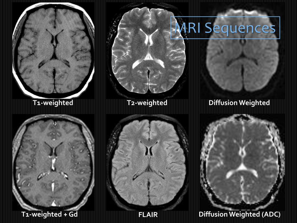
T1-weighted
T1-weighted + Gd
T2-weighted
FLAIR
Diffusion Weighted
Diffusion Weighted (ADC)
MRI Sequences

➢ Current neuroimaging modalities
➢ Brain anatomy
➢ How to approach a head CT
➢ Wrap up


SMG AngGPreCS
PostCSCS
SF


Superior frontal gyrus
Middle frontal gyrus
Precentral gyrus(Frontal lobe)
Central sulcus
Postcentral gyrus(Parietal lobe)
Precentral sulcus
Superior frontal sulcus
Postcentral sulcus
Pars marginalis Falx cerebri
Vertex to Tonsils:CT Anatomy

Central sulcus
Centrum semiovale
Precentral sulcus
Postcentral sulcus
Precentral gyrus(Frontal lobe)
Intraparietal sulcus
Superior frontal gyrus
Middle frontal gyrus
Parieto-occipital fissure
Postcentral gyrus(Parietal lobe)

Lateral (Sylvian) fissure, posterior segment
Caudate body
Central sulcus
Corona radiata
Lateral ventricle
Superior temporal gyrus
Parieto-occipital fissure
Intra-occipital sulcus
Superior temporal sulcus
Superior sagittal sinus

Lateral (Sylvian) fissure
Thalamus
Internal capsule, posterior limb
3rd ventricle
Basal ganglia, lentiform nuclei (GP & putamen)
Caudate, head
Lateral ventricle, occipital horn
Lateral ventricle, frontal horn
Internal capsule, anterior limb
Insula
Parieto-occipital fissure
Calcarine sulcus
Superior temporal sulcus

Lateral (Sylvian) fissure
Quadrigeminalcistern
MidbrainCerebral aqueduct (of Sylvius)
Superior temporal gyrus

Olfactory sulcus
Gyrus rectus
Suprasellar cistern
Amygdala
Hippocampus
Cerebellum
Interpeduncular cistern
Ambient cistern
Cerebral peduncle

Sella turcica
Prepontine cistern
Temporal lobe
Pons
4th ventricle
Vermis
Middle cerebellar peduncle (Brachium pontis)
Cerebellar hemisphere
Cerebellopontine angle cistern
Sigmoid sinus

Mandibular condyle
Medulla
Cerebellar tonsil
Mastoids
Nasopharynx
Foramen magnum
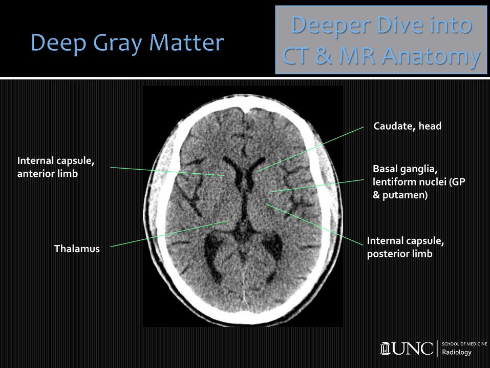
ThalamusInternal capsule, posterior limb
Basal ganglia, lentiform nuclei (GP & putamen)
Caudate, head
Internal capsule, anterior limb
Deeper Dive intoCT & MR Anatomy

PGP
C
Th
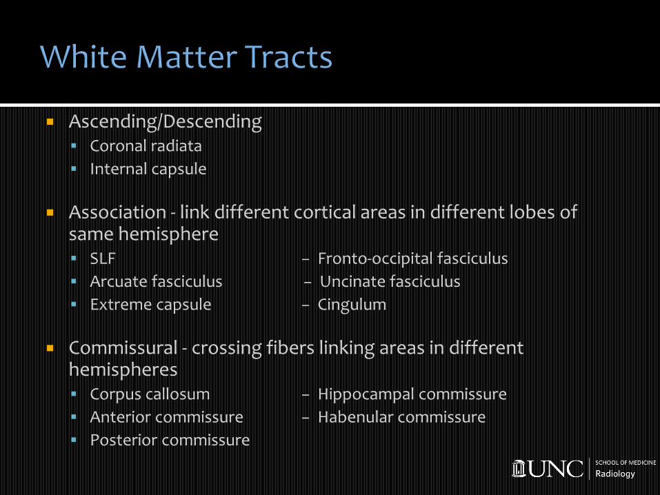
Ascending/Descending▪ Coronal radiata
▪ Internal capsule
Association - link different cortical areas in different lobes of same hemisphere▪ SLF − Fronto-occipital fasciculus
▪ Arcuate fasciculus − Uncinate fasciculus
▪ Extreme capsule − Cingulum
Commissural - crossing fibers linking areas in different hemispheres▪ Corpus callosum − Hippocampal commissure
▪ Anterior commissure − Habenular commissure
▪ Posterior commissure

Centrum semiovale Corona radiata Internal capsule

ACPC
PinealOC
Pit MB

Associated with memory and emotional, fight/flight & sexual responses
Consists of:▪ Amygdala
▪ Hippocampus
▪ Parahippocampalgyrus
▪ Cingulate gyrus
▪ Subcallosal Gyri
Uncus

Associated with memory and emotional, fight/flight & sexual responses
Consists of:▪ Amygdala
▪ Hippocampus
▪ Parahippocampalgyrus
▪ Cingulate gyrus
▪ Subcallosal Gyri
Uncus

1 = Optic chiasm2 = Pituitary infundibulum (stalk)3 = Tuber cinereum4= Mammillary body

Network of ependymal lined CSF filled spaces
▪ Derivative of neural tube cavity
4 ventricles:
▪ Paired lateral ventricles
▪ 3rd ventricle
▪ 4th ventricle

Body, lateral ventricle
Frontal horn, lateral ventricle
Occipital horn, lateral ventricle
3rd ventricle
Foramen of Monro
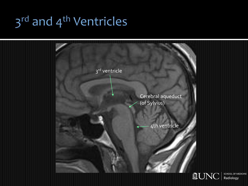
3rd ventricle
4th ventricle
Cerebral aqueduct(of Sylvius)


Sylvian Fissure
Ambient Cistern
Quadrigeminal Plate Cistern
Interpeduncular Cistern
SuprasellarCistern
CP AngleCistern
Prepontine Cistern

Cerebellum
▪ Paired hemispheres joined by midline vermis
▪ Coordination & muscle tone
Brainstem
▪ Midbrain, pons, medulla

Cerebellum
▪ Paired hemispheres joined by midline vermis
▪ Coordination & muscle tone
Brainstem
▪ Midbrain, pons, medulla
MCP
BrainstemMidbrain Pons Medulla
Cerebral Peduncle
Substantia Nigra
Cerebral Aqueduct
Red Nucleus
MCPICP
4th VentricleICP
Olive
Pyramid

C1 – Cervical C5 – ClinoidC2 – Petrous C6 – OpthalmicC3 – Lacerum C7 – Communicating (Terminal)C4 - Cavernous

C1 – Cervical C5 – ClinoidC2 – Petrous C6 – OpthalmicC3 – Lacerum C7 – Communicating (Terminal)C4 - Cavernous


➢ Current neuroimaging modalities
➢ Brain anatomy
➢ How to approach a head CT
➢ Wrap up

➢ Blood (intra- vs extraaxial)➢ Gray/white differentiation➢ Edema➢ Masses/Mass effect (midline shift, cistern
effacement, herniation ➢ Ventricles (hydrocephalus)➢ Bones (fx, lysis, sclerosis)➢ Paranasal sinuses and mastoids➢ Extracranial soft tissues
Blood Can Be
Very Bad!*
~~~~~~~~~~~~
Blood
Cisterns
Brain
Ventricles
Bones
*Dr Andy Perron

➢ Blood (Intra- vs extraaxial)➢ Gray/white differentiation➢ Edema➢ Masses/Mass effect (midline shift, cistern
effacement, herniation ➢ Ventricles (hydrocephalus)➢ Bones (fx, lysis, sclerosis)➢ Paranasal sinuses and mastoids➢ Extracranial soft tissues
Blood Can Be
Very Bad!
~~~~~~~~~~~~
Blood
Cisterns
Brain
Ventricles
Bones
Hypertensive intraaxialhemorrhage
Posttraumatic extraaxialhemorrhage - epidural

Epidural Hemorrhage Epidural hemorrhage forms between inner table of calvarium and outer layer of the dura, results from middle meningeal arterial tear.
>90% are assoc with skull fx usutemporoparietal bones, also frontal and parieto-occipital regions.
On CT: hyperdense biconvex lens shape, mass effect. Early identification critical to guide evacuation vs early reevaluation

➢ Blood (Intra- vs extraaxial)➢ Gray/white differentiation➢ Edema➢ Masses/Mass effect (midline shift, cistern
effacement, herniation ➢ Ventricles (hydrocephalus)➢ Bones (fx, lysis, sclerosis)➢ Paranasal sinuses and mastoids➢ Extracranial soft tissues
Blood Can Be
Very Bad!
~~~~~~~~~~~~
Blood
Cisterns
Brain
Ventricles
Bones
Stroke CT windowing W 30 L 30 –note left parietal lobe loss of gray-
white matter differentiation compared to right
Complete loss of gray-white matter differentiation bilateral
hemispheres

➢ Blood (Intra- vs extraaxial)➢ Gray/white differentiation➢ Edema➢ Masses/Mass effect (midline shift, cistern
effacement, herniation ➢ Ventricles (hydrocephalus)➢ Bones (fx, lysis, sclerosis)➢ Paranasal sinuses and mastoids➢ Extracranial soft tissues
Blood Can Be
Very Bad!
~~~~~~~~~~~~
Blood
Cisterns
Brain
Ventricles
Bones
Stroke CT windowing W 30 L 30 –note left parietal lobe loss of gray-
white matter differentiation compared to right

➢ Is there midline shift?➢ Is brain symmetry preserved?➢ Are the cisterns patent
(smile and pentagon)?➢ Is the 4th ventricle patent
and symmetric?➢ Are the ventricles enlarged
with sulcal effacement?
Blood Can Be
Very Bad!
~~~~~~~~~~~~
Blood
Cisterns
Brain
Ventricles
Bones
+midline shift+hemispheric asymmetry
+cistern effacement+lateral ventriculomegaly and
sulcal effacement

Subdural Hemorrhage Subdural hemorrhage (SDH) collects in the potential space between the inner dura and the arachnoid layer
Result from bridging vein tear in a MVA or fall
On CT: hyperdense crescentic shape when acute, then isodense to hypodense when subacute and chronic. Mass effect -> altered mental status

+SDHCT Checklist:
+ Midline shift
+ Loss of symmetry
+ Cisternal effacementSubfalcine and uncalherniation
Needs NSU !

➢ Blood (Intra- vs extraaxial)➢ Gray/white differentiation➢ Edema➢ Masses/Mass effect (midline shift, cistern
effacement, herniation ➢ Ventricles (hydrocephalus)➢ Bones (fx, lysis, sclerosis)➢ Paranasal sinuses and mastoids➢ Extracranial soft tissues
Blood Can Be
Very Bad!
~~~~~~~~~~~~
Blood
Cisterns
Brain
Ventricles
Bones
Stroke CT windowing W 30 L 30 –note left parietal lobe loss of gray-
white matter differentiation compared to right

➢ Blood (Intra- vs extraaxial)➢ Gray/white differentiation➢ Edema➢ Masses/Mass effect (midline shift, cistern
effacement, herniation ➢ Ventricles (hydrocephalus)➢ Bones (fx, lysis, sclerosis)➢ Paranasal sinuses and mastoids➢ Extracranial soft tissues
Blood Can Be
Very Bad!
~~~~~~~~~~~~
Blood
Cisterns
Brain
Ventricles
Bones
Note bilateral lateral ventriculomegaly. Distinguish
hydrocephalus from atrophy via diffuse sulci prominence

➢ Blood (Intra- vs extraaxial)➢ Gray/white differentiation➢ Edema➢ Masses/Mass effect (midline shift, cistern
effacement, herniation ➢ Ventricles (hydrocephalus)➢ Bones (fx, lysis, sclerosis)➢ Paranasal sinuses and mastoids➢ Extracranial soft tissues
Blood Can Be
Very Bad!
~~~~~~~~~~~~
Blood
Cisterns
Brain
Ventricles
BonesDepressed skull fx left parietooccipital bone from
penetrating trauma

➢ Blood (Intra- vs extraaxial)➢ Gray/white differentiation➢ Edema➢ Masses/Mass effect (midline shift, cistern
effacement, herniation ➢ Ventricles (hydrocephalus)➢ Bones (fx, lysis, sclerosis)➢ Paranasal sinuses and mastoids➢ Extracranial soft tissues
Blood Can Be
Very Bad!
~~~~~~~~~~~~
Blood
Cisterns
Brain
Ventricles
Bones
Expansile soft tissue massleft frontal sinus

➢ Blood - intra- and extraaxial➢ Cisterns and extraaxial CSF - effacement or
asymmetry➢ Brain parenchyma - gray/white differentiation
symmetry, shift, hyper- hypodensity➢ Ventricles - too large or too small➢ Bones - skull fx, paranasal sinuses, mastoids
and extracranial soft tissues
Blood Can Be
Very Bad!
~~~~~~~~~~~~
Blood
Cisterns
Brain
Ventricles
Bones

➢ CT vs MRICT is a great screening tool
MR is good for problem solving & assessing established pathology
➢ Brain anatomy
➢ How to approach a head CTBlood Can Be Very Bad
➢ Emergency checklist Shift, cisternal effacement, or hydrocephalus

Thank you for participating!
http://guides.lib.unc.edu/radiology/booksNeuroradiology references