Bevacizumab-Induced Alterations in Vascular Permeability and
13
Cancer Therapy: Preclinical Bevacizumab-Induced Alterations in Vascular Permeability and Drug Delivery: A Novel Approach to Augment Regional Chemotherapy for In-Transit Melanoma Ryan S. Turley 1,3 , Andrew N. Fontanella 2 , James C. Padussis 1,3 , Hiroaki Toshimitsu 1,3 , Yoshihiro Tokuhisa 1,3 , Eugenia H. Cho 1 , Gabi Hanna 2 , Georgia M. Beasley 1 , Christina K. Augustine 1,3 , Mark W. Dewhirst 2 , and Douglas S. Tyler 1,3 Abstract Purpose: To investigate whether the systemically administered anti-VEGF monoclonal antibody bev- acizumab could improve regional chemotherapy treatment of advanced extremity melanoma by enhancing delivery and tumor uptake of regionally infused melphalan (LPAM). Experimental Design: After treatment with systemic bevacizumab or saline, changes in vascular permeability were determined by spectrophotometric analysis of tumors infused with Evan’s blue dye. Changes in vascular structure and tumor hemoglobin-oxygen saturation HbO 2 were determined by intravital microscopy and diffuse reflectance spectroscopy, respectively. Rats bearing the low-VEGF secreting DM738 and the high-VEGF secreting DM443 melanoma xenografts underwent isolated limb infusion (ILI) with melphalan (LPAM) or saline via the femoral vessels. The effect of bevacizumab on terminal drug delivery was determined by immunohistochemical analysis of LPAM-DNA adducts in tumor tissues. Results: Single-dose bevacizumab given three days before ILI with LPAM significantly decreased vascular permeability (50.3% in DM443, P < 0.01 and 35% in DM738, P < 0.01) and interstitial fluid pressure (57% in DM443, P < 0.01 and 50% in DM738, P ¼ 0.01). HbO 2 decreased from baseline in mice following treatment with bevacizumab. Systemic bevacizumab significantly enhanced tumor response to ILI with LPAM in two melanoma xenografts, DM443 and DM738, increasing quadrupling time 37% and 113%, respectively (P ¼ 0.03). Immunohistochemical analyses of tumor specimens showed that pretreatment with systemic bevacizumab markedly increased LPAM-DNA adduct formation. Conclusions: Systemic treatment with bevacizumab before regional chemotherapy increases delivery of LPAM to tumor cells and represents a novel way to augment response to regional therapy for advanced extremity melanoma. Clin Cancer Res; 1–12. Ó2012 AACR. Introduction While the incidence of several other cancers declines, the incidence of melanoma continues to rise and is now the most common fatal malignancy of young adults and the sixth most common cancer among Americans. In 2009, there were an estimated 68,720 people newly diagnosed with invasive melanoma, and more than 8,650 people died of melanoma in the United States (1). Unfortunately, mortality rates for metastatic melanoma have remained high in part because of its high resistance to standard cytotoxic agents (2). A major breakthrough for the treat- ment of metastatic melanoma has been the recent U.S. Food and Drug Administration (FDA)-approval for the anti- CTLA4 antibody (ipilimumab) and a specific inhibitor targeting the V600E-activating BRAF mutation (vemurafe- nib) found in approximately 50% of patients with mela- noma (3). For patients who recur after treatment with these new therapies, response to other systemic therapy is poor, with standard agents such as dacarbazine, temozolomide, and paclitaxel having complete response rates of <15% (2). A significant fraction of patients with melanoma will recur with in-transit disease which represents multifocal metastases that have spread through the lymphatic system occurring between the site of the primary lesion and the regional draining lymph node basin (4). This pattern of recurrence is associated with an unfavorable prognosis, having 5-year survival rates ranging from 25% to 30% (5). Historically, standard systemic chemotherapy or immu- notherapy has provided little benefit for in-transit disease (6). However, for patients with in-transit disease confined to the extremities, regional chemotherapy delivered by Authors' Affiliations: Departments of 1 Surgery and 2 Radiation Oncology, Duke University; and 3 Durham VA Medical Center, Durham, North Carolina Note: Supplementary data for this article are available at Clinical Cancer Research Online (http://clincancerres.aacrjournals.org/). Corresponding Author: Ryan S. Turley, Duke University, 2301 Erwin Rd., DUMC Box 3443, Durham, NC 27710. Phone: 919-668-2768; Fax: 919- 684-6044; E-mail: [email protected] doi: 10.1158/1078-0432.CCR-11-3000 Ó2012 American Association for Cancer Research. Clinical Cancer Research www.aacrjournals.org OF1 Research. on January 3, 2019. © 2012 American Association for Cancer clincancerres.aacrjournals.org Downloaded from Published OnlineFirst April 10, 2012; DOI: 10.1158/1078-0432.CCR-11-3000
Transcript of Bevacizumab-Induced Alterations in Vascular Permeability and

Cancer Therapy: Preclinical
Bevacizumab-Induced Alterations in Vascular Permeabilityand Drug Delivery: A Novel Approach to Augment RegionalChemotherapy for In-Transit Melanoma
Ryan S. Turley1,3, Andrew N. Fontanella2, James C. Padussis1,3, Hiroaki Toshimitsu1,3, Yoshihiro Tokuhisa1,3,Eugenia H. Cho1, Gabi Hanna2, Georgia M. Beasley1, Christina K. Augustine1,3, Mark W. Dewhirst2, andDouglas S. Tyler1,3
AbstractPurpose: To investigate whether the systemically administered anti-VEGF monoclonal antibody bev-
acizumab could improve regional chemotherapy treatment of advanced extremitymelanoma by enhancing
delivery and tumor uptake of regionally infused melphalan (LPAM).
Experimental Design: After treatment with systemic bevacizumab or saline, changes in vascular
permeability were determined by spectrophotometric analysis of tumors infused with Evan’s blue dye.
Changes in vascular structure and tumor hemoglobin-oxygen saturation HbO2 were determined by
intravitalmicroscopy and diffuse reflectance spectroscopy, respectively. Rats bearing the low-VEGF secreting
DM738 and the high-VEGF secreting DM443melanoma xenografts underwent isolated limb infusion (ILI)
with melphalan (LPAM) or saline via the femoral vessels. The effect of bevacizumab on terminal drug
delivery was determined by immunohistochemical analysis of LPAM-DNA adducts in tumor tissues.
Results: Single-dose bevacizumab given three days before ILI with LPAM significantly decreased vascular
permeability (50.3% inDM443, P < 0.01 and 35% inDM738, P < 0.01) and interstitial fluid pressure (57%
in DM443, P < 0.01 and 50% in DM738, P ¼ 0.01). HbO2 decreased from baseline in mice following
treatment with bevacizumab. Systemic bevacizumab significantly enhanced tumor response to ILI with
LPAM in two melanoma xenografts, DM443 and DM738, increasing quadrupling time 37% and 113%,
respectively (P¼ 0.03). Immunohistochemical analyses of tumor specimens showed that pretreatmentwith
systemic bevacizumab markedly increased LPAM-DNA adduct formation.
Conclusions: Systemic treatment with bevacizumab before regional chemotherapy increases delivery of
LPAM to tumor cells and represents a novel way to augment response to regional therapy for advanced
extremity melanoma. Clin Cancer Res; 1–12. �2012 AACR.
IntroductionWhile the incidence of several other cancers declines, the
incidence of melanoma continues to rise and is nowthe most common fatal malignancy of young adults andthe sixth most common cancer among Americans. In 2009,there were an estimated 68,720 people newly diagnosedwith invasive melanoma, andmore than 8,650 people diedof melanoma in the United States (1). Unfortunately,mortality rates for metastatic melanoma have remainedhigh in part because of its high resistance to standard
cytotoxic agents (2). A major breakthrough for the treat-ment ofmetastaticmelanomahas been the recentU.S. Foodand Drug Administration (FDA)-approval for the anti-CTLA4 antibody (ipilimumab) and a specific inhibitortargeting the V600E-activating BRAF mutation (vemurafe-nib) found in approximately 50% of patients with mela-noma (3). For patients who recur after treatment with thesenew therapies, response to other systemic therapy is poor,with standard agents such as dacarbazine, temozolomide,and paclitaxel having complete response rates of <15% (2).
A significant fraction of patients with melanoma willrecur with in-transit disease which represents multifocalmetastases that have spread through the lymphatic systemoccurring between the site of the primary lesion and theregional draining lymph node basin (4). This pattern ofrecurrence is associated with an unfavorable prognosis,having 5-year survival rates ranging from 25% to 30%(5).Historically, standard systemic chemotherapyor immu-notherapy has provided little benefit for in-transit disease(6). However, for patients with in-transit disease confinedto the extremities, regional chemotherapy delivered by
Authors' Affiliations: Departments of 1Surgery and 2Radiation Oncology,Duke University; and 3Durham VAMedical Center, Durham, North Carolina
Note: Supplementary data for this article are available at Clinical CancerResearch Online (http://clincancerres.aacrjournals.org/).
Corresponding Author: Ryan S. Turley, Duke University, 2301 Erwin Rd.,DUMC Box 3443, Durham, NC 27710. Phone: 919-668-2768; Fax: 919-684-6044; E-mail: [email protected]
doi: 10.1158/1078-0432.CCR-11-3000
�2012 American Association for Cancer Research.
ClinicalCancer
Research
www.aacrjournals.org OF1
Research. on January 3, 2019. © 2012 American Association for Cancerclincancerres.aacrjournals.org Downloaded from
Published OnlineFirst April 10, 2012; DOI: 10.1158/1078-0432.CCR-11-3000
isolated limb perfusion (ILP) or isolated limb infusion (ILI)is a viable treatment option.
Briefly, ILP is conducted by surgically exposing the fem-oral or subclavian vessels and then cannulating them at theroot of the limb. Next, an esmarch tourniquet is placedproximal to the cannulated vessels and the limb is perfusedwith a high-flow, melphalan-based perfusate using a mem-brane oxygenator tomaintain physiologic oxygenation andpH(5). ILI is a generally less-invasive surgical technique andis conducted by percutaneous catheterization of theinvolved limb followed by infusion of melphalan in alow-flow circuit without an oxygenator such that tissuebecomes hypoxic and acidotic (7).
Using these techniques, complete response rates rangingfrom 30% to 60% have been reported in larger series (5).Although these initial response rates represent an improve-ment over systemic therapy, at least 40%of patients with in-transit melanoma will eventually recur after regional che-motherapy (8). Thus, novel strategies areneeded to improvedurable responses rates of locally advance melanoma toregional therapy.
Strategies to optimize delivery of cytotoxic agents tomelanoma during regional chemotherapy may enhancenot only the initial response to treatment but also thedurability of this response.Melanoma induces angiogenesisthat results in tumor vasculature which is anatomically andfunctionally distinct from that seen in normal tissue (9, 10).The vasculature of solid tumors has been characterized astortuous, dilated, saccular, and irregular in its pattern ofinterconnection (11). This erratic tumor vasculature canpose a barrier to optimal drug delivery thus limiting theefficacy of cytotoxic therapy. An important mediator oftumor angiogenesis is VEGF, a cytokine which stimulatesangiogenesis and is normally seen during embryonic devel-opment aswell as in postinjury angiogenesis (12). VEGF is amultifunctional cytokine capable of stimulating endothe-lial cell proliferation, migration, and survival (13). VEGF isalso a potent stimulator of vessel permeability (14). Com-monly overexpressed in metastatic melanoma, high serumlevels of VEGF have been correlated with adverse prognosisand implicated as an important mediator in creating a
dysfunctional vascular network antagonizing optimal drugpenetration (15).
Bevacizumab is an FDA-approved monoclonal antibodyto VEGF that neutralizes all isoforms of VEGF. It has beenused in combination with standard chemotherapies inpatients with metastatic colorectal, brain, and lung cancersand is being investigated in combination with other che-motherapy agents for melanoma in multiple clinical trialsacross the United States (16–18). Bevacizumab also offers aunique tool to temporally restore the balance betweenangiogenic stimulators and angiogenic inhibitors (11).Emerging evidence shows that vascular targeting agentssuch as bevacizumab can transiently "normalize" tumormicrovasculature toward that seen innormal tissues therebycreating an optimal window for chemotherapy delivery(19). By effectively neutralizing VEGF, bevacizumab istheorized toprune immature and leaky vessels characteristicof solid tumors, leading to functional changes that includeincreased tumor oxygenation and drug penetration (11).Although beneficial when used in a well circumscribedwindow, sustained antiangiogenic treatment may lead tosevere tumor vessel regression that blocks penetration oftherapeutic drugs into the tumor antagonizing, rather thanaugmenting, cytotoxic therapy. Thus, systemic therapies,which are often administered in multiple doses, may ini-tially benefit from the antiangiogenic therapy but will behindered at later time points as tumor vessels wither andbecome inadequate for drug delivery (19).
Because regional chemotherapy is administrated as asingle infusion or perfusion of high-dose cytotoxic therapy(5), it is an ideal modality to test whether bevacizumab canenhance drug delivery and subsequently improve tumorresponse. In this report, we provide preclinical evidence thatsystemically administered bevacizumab decreases tumorvascular permeability and interstitial fluid pressure (IFP)leading to increased drug delivery and improved efficacy ofregionally infused melphalan. Pretreatment with systemicbevacizumab before arterial infusion of melphalanimproves drug penetration and tumor responses, support-ing bevacizumab as a beneficial adjunct to regional therapyfor melanoma.
Materials and MethodsMelanoma cell lines and reagents
Tumor cell lines derived from human melanomas (DMlines) were courtesy of Dr. H. Seigler (Duke UniversityMedical Center, Durham, NC). Cells were cultured in Iso-cove’s Modified Dulbecco’s Medium with 10% FBS,2 mmol/L glutamine, 1,000 IU/mL penicillin, and100 mg/mL streptomycin and grown at 37�C and 5% CO2.Bevacizumab (Avastin by Genentech) was purchased fromthe Duke University Pharmacy.
Quantification ofmelanoma cell VEGF secretion and itsmodulation with bevacizumab
Human VEGF Quantikine ELISA Kits (R&D Systems)were used according to the directions of the manufacturer.
Translational RelevanceThis is the first study to evaluate the use of bevacizu-
mab as a part of a regional therapy strategy for thetreatment of advanced extremity melanoma with thespecific intent of augmenting delivery of chemotherapyto a tumor. We show that a single dose of bevacizumabbefore isolated limb infusion improves the delivery ofchemotherapy and enhances melanoma tumorresponses by decreasing tumor vascular permeabilityand interstitial fluid pressure. On the basis of theseresults, a phase I clinical study examining the safety ofsingle-dose bevacizumab before regional infusion ofmelphalan has been proposed.
Turley et al.
Clin Cancer Res; 2012 Clinical Cancer ResearchOF2
Research. on January 3, 2019. © 2012 American Association for Cancerclincancerres.aacrjournals.org Downloaded from
Published OnlineFirst April 10, 2012; DOI: 10.1158/1078-0432.CCR-11-3000
Two-hundred microliters of conditioned media were col-lected from triplicate samples.
VEGF neutralization and its effect on melphalansensitivity of melanoma cellsUsing cell survival as an endpoint, DM443 and DM738
cells were plated at 1 � 103 per well in a 96-well microtiterplate and after 24 hours, treated with 0 to 2 mg/mL bev-acizumab and refed with freshmedium containing 0 to 150mmol/L melphalan. Cell survival was measured after 72hours, using the Cell Titer-Blue assay (Promega), as per themanufacturer’s instructions, with excitation and emissionwavelengths of 560 and 590 nm, respectively. Survivingfractions, SF, were computed using the equation: SF¼ Ft/Fc,where Ft and Fc are the fluorescence of treated and untreatedcells, respectively, normalized against backgroundfluorescence.
Hyperspectral analysis of tumor vasculature aftertreatment with bevacizumabThe preparation of the dorsal skin-fold window chamber
has been described elsewhere (20). All procedures used inthis study were approved by the Duke University Institu-tional Animal Care and Use Committee. Briefly, a 12-mmdiameter flap of skin was dissected away from opposingsurfaces of the dorsal skin flap of anesthetized nude mice,leaving a fascial plane with associated vasculature. The holewas held vertically away from the body with a titaniumsaddle that was sutured to both sides of the flap. A total of 3� 106 DM443 or DM738 cells suspended in 30 mL normalgrowth medium were injected in the middle of the fascialplane. Glass windows were attached to the center of thesaddle to cover the surgical site. BALB/cmice were obtainedfrom Duke Cancer Center Isolation Facility.For imaging of tumor microvasculature, animals were
anesthetized with isoflurane (1.5%with oxygen) andmain-tained at body temperature. Imaging began when tumorsreached 1 to 2 mm in diameter, approximately 5 days afterinjection. All images were collected with a 2.5� objective.Hyperspectral (hemoglobin-oxygen saturation) imagingwas collected using an upright microscope (Carl ZeissAxiovert 200). Hyperspectral images were collected with a100-W halogen lamp for transillumination, and detectionwas achieved with a 10-nm bandwidth liquid crystal tun-able filter (LCTF; CRI Incorporated) placed in front of aDVC1412CCDcamera (DVCCompany). Custom softwarewas used to tune the filter and acquire images at 10-nmoptical wavelength increments between 500 and 620 nm.The spectral characteristics of the illumination source itselfwere corrected for by measuring the transmission of thesource light through a neutral density filter at each wave-length. After the system response was compensated for,hemoglobin saturationwas calculated at each pixel locationby applying the principles of the Beer–Lambert law in awavelength-dependent manner. Assuming oxygenated anddeoxygenated hemoglobin as the primary absorbers, alongwith a tissue scattering parameter, these factorswere decom-posed from the total absorption spectrum using linear
nonnegative least-squares regression, yielding hemoglobinsaturation values at every observable pixel location. Vascu-lar maps derived from total hemoglobin measurementswere used to measure vascular tortuosity using a user-guided algorithm for vessel identification. Tortuosity isrepresented as the ratio of the vessel centerline distance tothe straight line distance between branch points. Vascularlength density (VLD) was calculated by dividing the totaltumor-associated vessel length by the tumor area (mm/mm2). The hyperspectral system, software, and analysistechniques have previously been validated on liquid phan-tomswith an accuracy of approximately 1%and in vivo (21).
Diffuse reflectance spectroscopy for quantitativetumor hemoglobin concentration and oxygensaturation
A total of 20 nude mice were inoculated subcutaneouslyin the right flank with 3 � 106 DM443 melanoma cellssuspended in 100mLof normal growthmedium. Threemicedid not grow tumors.Once tumor diameters reached 8 to 10mm (�2–3 weeks after inoculation), mice (n ¼ 8) wereinjected with intraperitoneal bevacizumab (5 mg/kg) orequivalent volumes of intraperitoneal normal saline (n ¼9). All tumors were monitored optically before treatmentand then daily for 6 days after treatment. Briefly, animalswere anesthetized with inhaled 1.5% isoflurane gas mixedwith oxygen. A bifurcated fiber optic probe using separateillumination and collection paths was noninvasivelyapplied in direct contact with DM443 xenografts andsecured with a clamp to prevent movement. Diffuse reflec-tance was themeasured in the spectral range of 480 and 650nm. The acquired diffuse reflectance spectra were calibratedand analyzed using an inverse scalable Monte Carlo modelto obtain wavelength-dependent scattering and absorptionspectra (22). The concentration of oxygenated hemoglobin(HbO2) and deoxygenated hemoglobin (dHb) was derivedusing the Beer–Lambert equation. Total hemoglobin (Hb)was calculated by the sumof HbO2 and dHb. Differences in%D Hb and %D HbO2 were compared between treatmentsgroupswith aMann–WhitneyU test using the commerciallyavailable software GraphPad Prism.
Rat xenograft studiesFemale nude rats (6-week-old; Charles River Laboratories
International, Inc.) were injected subcutaneously in theright hind limb with 5 � 106 DM738 or 7 � 106 DM443cells. Tumor volume was calculated as: 1/2 � [(length) �(width)2]. Treatment began when tumor volume was 100mm3. The rat protocolwas approved by theDukeUniversityand Durham VA Medical Center Institutional Animal Careand Use Committees.
ILI was conducted as described previously (23). Thefemoral artery and vein were cannulated. The arterial cath-eter was attached to a peristaltic pump while venous drain-age flowed by gravity into a reservoir. A tourniquet wasplaced around the thigh. For survival studies, a 15-minuteinfusion (1.5 mL/min) of melphalan (90 mg/kg) or salinewas followed by a 1-minute saline washout (3.0 mL/min).
Bevacizumab Augments Regional Chemotherapy for Melanoma
www.aacrjournals.org Clin Cancer Res; 2012 OF3
Research. on January 3, 2019. © 2012 American Association for Cancerclincancerres.aacrjournals.org Downloaded from
Published OnlineFirst April 10, 2012; DOI: 10.1158/1078-0432.CCR-11-3000

Tumor vascular permeabilityILI was conducted as described earlier. Rats received an
injection of bevacizumab5mg/kg intraperitoneally (i.p.) orsaline 3 days before a 15-minute infusion of Evan’s Blue dyesolution (50 mg/kg dissolved in normal saline and infusedat 1.5 mL/min) followed by a 2-minute saline washout(3.0 mL/min). Animals were euthanized and tumors wereexcised followed by incubation in formamide solution for72 hours at 37�C to extract the Evan’s blue dye. To quantifythe Evan’s blue, absorbance of the formamide solution wasmeasured at 595 and 655 nm and normalized to tumorvolume.
Interstitial fluid pressureTumor IFPmeasurements weremadewith a needle probe
pressure monitor (Intra-Compartmental Pressure MonitorSystem, Stryker), fitted with an 18-gauge side-ported needle(Stryker) and connected to a syringe filled with 0.9% saline.Using isoflurane anesthesia, the needle probe was insertedinto the center of the tumor on the hind limb of the rat. IFPwas recorded in mmHg when the measurement stabilized.
Immunohistochemistry of tumor vessel densityTen DM738 (n ¼ 5, saline; n ¼ 5, 5 mg/kg i.p. bevaci-
zumab) and 13 DM443 (n ¼ 6, saline; 7 5 mg/kg bevaci-zumab) xenografts were flash-frozen using liquid nitrogen.They were then cut into 10 mm cross-sections using a LEICACM 1850 cryotome (Meyer Instruments Incorporated) andmaintained at�24�C to�28�C. Sections were then fixed inacetone for 30 minutes, allowed to dry for 1 hour, and thenblocked for 30 minutes at room temperature with 5%donkey serum (Jackson ImmunoResearch) in PBS. Sectionswere incubated with primary antibody (1:200 mouse anti-rat CD31, BD bioscience) at room temperature for 1 hour,followed by a 30-minute incubation with a fluorescent-conjugated secondary antibody (1:100; Jackson ImmunoR-esearch) at room temperature. Samples were rinsed withPBS 3 times for 5 minutes each between every consecutivestep.
Immunohistochemical staining for CD31 was assessedwith a Zeiss AxioScop II microscope with a scanning stage.Images of whole tumors were recorded at�5magnificationand subsequently segmented and evaluated in ImageJ.Distinct immunoreactive endothelial cells were counted asindividual microvessels, and nonspecific staining of none-ndothelial structureswas disregarded inmicrovessel counts.Meanmicrovessel density for CD31 was calculated over thetotal area of the tumor.
Immunohistochemistry of DNA-melphalan adductRats were treated with bevacizumab (5 mg/kg i.p.) or
saline 3 days before ILI. Rats were then euthanized 24 hoursafter LPAM- or saline-ILI. Tumors were then fixed withformalin, paraffin-embedded, and stained using immuno-histochemistry as previously described (23, 24). Treatedand nontreated DM443 cells were used as positive andnegative controls for the MP5/73 antibody which waskindly provided by Dr. M.J. Tilby, Northern Institute for
Cancer Research, Newcastle University, Newcastle uponTyne, United Kingdom (25).
Sections were viewed and digitally photographedwith anOlympus Vanox inverted light microscope coupled to anOlympusDT-70 camera. Imageswere taken at�2,�10, and�60magnification and saved as JPEGor TIFFfiles for furtherprocessing in Adobe Photoshop (Adobe Systems, Inc.).Positive staining was quantified as the ratio of brown tototal pixels using the commercially available softwareMATLAB (Mathworks). Differences in staining among treat-ment groups were compared using a one-way ANOVA.
In vivo tumor growth studiesAnimals were treated with a single dose of bevacizumab
(5mg/kg i.p.) 3 days before ILI, as based onprevious studiesshowing peak changes tumor vasculature occurred 3 daysafter bevacizumab (19, 26). Response to drug treatmentwas followed until tumor volume reached 5,000 mm3 or60 days for regional studies. Response was evaluated by thepercentage of increase in tumor volume and tumor quadru-pling time.
ResultsMelanoma cell lines secrete VEGF
To evaluate the therapeutic potential of anti-VEGF ther-apy for the treatment of in-transit melanoma, we measuredVEGF secretion across 11 melanoma cell lines capable offorming tumor xenografts in our animalmodel of extremitymelanoma. VEGF secretion was variable across these celllines, with A2058, DM792, DM751, andDM738, an LPAM-resistant cell line, showing little to no VEFG secretionwhereas DM443, a moderately LPAM-sensitive cell line,showing high levels of VEGF secretion (Supplementary Fig.S1A). To confirm bevacizumab can effectively neutralizemelanoma VEGF secretion in vitro, we treated DM443 andDM738 cells, grown to 80% confluence, with 0 to 2.0 mg/mL bevacizumab for 24 to 72 hours. Bevacizumab at allconcentrations tested effectively prevented detection ofVEGF by ELISA (Supplementary Fig. S1B and S1C).
Bevacizumab does not alter in vitro LPAMchemosensitivity
VEGF possesses multiple cytokine functions and can actas a potentmitogen for endothelial cell proliferation. Giventhe possibility that VEGF might act as an autocrine growthfactor for melanoma (27), we tested whether VEGF neu-tralization in itself could augment chemosensitivity ofmelanoma cells to melphalan in an endothelial cell-freesystem using a low-VEGF secreting, melanoma cell line(LPAM-resistant DM738) and a high-VEGF secreting, mel-anoma cell line (LPAM-sensitive DM443). VEGF neutrali-zation in an endothelial cell-free systemhadno effect on theLPAM chemosensitivity for either DM443 or DM738 (Sup-plementary Fig. S2). On the basis of these data and others(19, 28), we focused our studies on evaluating 2 potentialmechanisms by which bevacizumab could augment tumorcytotoxicity to regionally delivered chemotherapy through
Turley et al.
Clin Cancer Res; 2012 Clinical Cancer ResearchOF4
Research. on January 3, 2019. © 2012 American Association for Cancerclincancerres.aacrjournals.org Downloaded from
Published OnlineFirst April 10, 2012; DOI: 10.1158/1078-0432.CCR-11-3000

2 separate temporal effects: (i) neutralization of VEGFwould lead to normalization of tumor microvasculatureresulting in amore optimal vascular anatomy for deliveringcytotoxic therapy and (ii) impairment of the requisiteangiogenesis needed for further tumor growth.
Bevacizumab decreases vascular permeability andinterstitial pressure of melanoma xenograftsPrevious studies have shown bevacizumab to significant-
ly decrease tumor vascular permeability and the peak func-tional effects of bevacizumab to occur 3 days after treatment(14, 19, 26, 29). On the basis of this evidence, we hypoth-esized that a single dose of bevacizumab, given3days beforeILI would decrease vessel permeability. To test this, ILI wasconducted on tumor-bearing rats using Evan’s blue dye asthe infusate. Absorbance of Evan’s blue dye was measuredand used as a surrogate of vascular permeability in thexenograft. As shown in Fig. 1A and B, bevacizumab (5mg/kg) given 3 days before ILI significantly decreasedvascular permeability of Evan’s blue dye by 70% in DM443xenografts and 30% in DM738 xenografts.Increased vascular permeability seen in tumor vascula-
ture may lead to increased IFP which can hinder effectivedrug delivery to tumor cells (30). Thus, we also measuredIFP in melanoma xenografts 3 days after treatment with i.p.bevacizumab (5 mg/kg). Mean IFP measurements in bev-
acizumab-treated animals were 57% (14.9 � 0.9 vs. 8.3 �0.8mmHg, P < 0.01) and 50%(18.0� 2.9 vs. 9.1� 1.2mmHg, P ¼ 0.01) lower than saline-treated controls forboth DM443 and DM738 xenografts, respectively (Fig.1C and D).
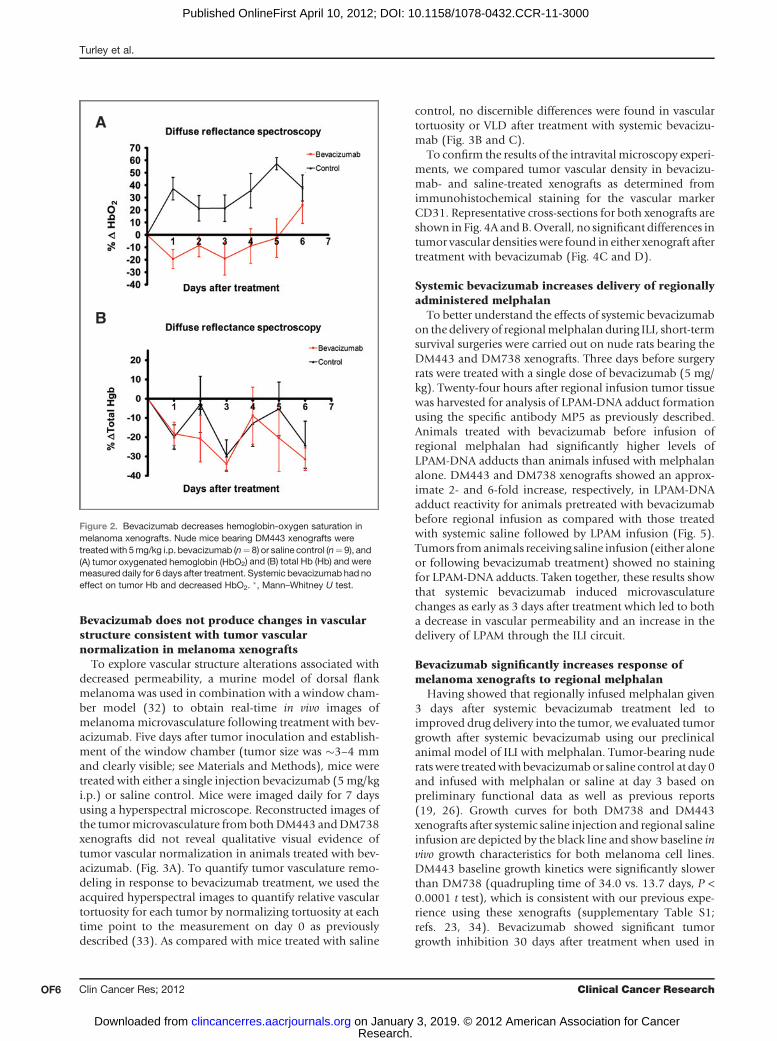
Bevacizumab decreases hemoglobin oxygen saturationin melanoma tumors
Tumor vascular normalization is hypothesized to induce atransient increase in tumor tissue oxygenation after treat-ments with antiangiogenic agents (11). To test the effect ofbevacizumab on tumor tissue oxygenation, we measuredtumor hemoglobin saturation HbO2 using diffuse reflec-tance spectroscopy; a method which has been previouslyvalidated as a surrogate for tissue oxygenation (31). Nudemice bearing DM443 melanoma xenografts were treatedwith 5 mg/kg of i.p. bevacizumab (n ¼ 8) or saline (n ¼9). The effects of systemic bevacizumab on tumor totalhemoglobin (Hb) and HbO2 levels, Hb and HbO2 weremeasured at baseline before treatment and daily for 6 daysusing diffuse reflectance spectroscopy. Daily HbO2 levelswere shown to be consistently lower than baseline measure-ments in thebevacizumab-treatedmice than in saline-treatedcontrols (P ¼ 0.004). Hb levels in both groups decreased asmuch as 30% from baseline measurements without discern-ible differences between treatment groups (P¼ 0.38; Fig. 2).
Figure 1. Bevacizumab decreasespermeability and IFP of melanomaxenografts. Three days after i.p.injection of 5mg/kg of bevacizumab,ILI was carried out on tumor-bearingrats using Evan's blue dye as theinfusate. After infusion, animals weresacrificed and tumors were excisedand incubated in formamide for 72hours at 37�C to extract the Evan'sblue dye into solution. Theabsorbance was of Evan's blue dyewas thenmeasured in the formamideat 595 and 655 nmand normalized totumor volume. Error bars representthe SEM of 4 to 6 rats. A, DM443tumor permeability decreased byapproximately 70% from salinecontrol in DM443 xenografts aftertreatment with bevacizumab. B,DM738 tumor permeabilitydecreased approximately 30% aftertreatment with bevacizumab. C,mean IFP measured approximately43% lower (8.3 � 0.8 vs. 14.9 � 0.9mm Hg) in DM443 tumors treatedwith bevacizumab than with salinecontrols. D, mean IFP was alsoapproximately 50% lower (9.1 � 1.2vs. 18.0 � 2.9 mm Hg) inbevacizumab-treated DM738xenografts. �, 2-tailed t test.
0
10
20
30
40
50
60
70
80
90
100
595 nm 655 nm
Per
cen
t ab
sorb
ance
Wavelength
DM443A B
C D
Saline
Bevacizumab
0
10
20
30
40
50
60
70
80
90
100
595 nm 655 nm
Per
cen
t ab
sorb
ance
Wavelength
DM738
Saline
Bevacizumab
P = 0.02*P < 0.01*
P = 0.01*P = 0.02*
Bevacizumab Augments Regional Chemotherapy for Melanoma
www.aacrjournals.org Clin Cancer Res; 2012 OF5
Research. on January 3, 2019. © 2012 American Association for Cancerclincancerres.aacrjournals.org Downloaded from
Published OnlineFirst April 10, 2012; DOI: 10.1158/1078-0432.CCR-11-3000
Bevacizumab does not produce changes in vascularstructure consistent with tumor vascularnormalization in melanoma xenografts
To explore vascular structure alterations associated withdecreased permeability, a murine model of dorsal flankmelanoma was used in combination with a window cham-ber model (32) to obtain real-time in vivo images ofmelanomamicrovasculature following treatment with bev-acizumab. Five days after tumor inoculation and establish-ment of the window chamber (tumor size was �3–4 mmand clearly visible; see Materials and Methods), mice weretreated with either a single injection bevacizumab (5mg/kgi.p.) or saline control. Mice were imaged daily for 7 daysusing a hyperspectral microscope. Reconstructed images ofthe tumormicrovasculature frombothDM443 andDM738xenografts did not reveal qualitative visual evidence oftumor vascular normalization in animals treated with bev-acizumab. (Fig. 3A). To quantify tumor vasculature remo-deling in response to bevacizumab treatment, we used theacquired hyperspectral images to quantify relative vasculartortuosity for each tumor by normalizing tortuosity at eachtime point to the measurement on day 0 as previouslydescribed (33). As compared with mice treated with saline
control, no discernible differences were found in vasculartortuosity or VLD after treatment with systemic bevacizu-mab (Fig. 3B and C).
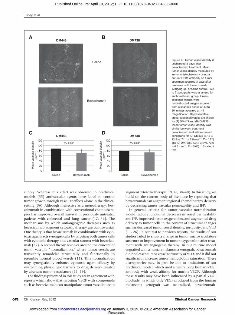
To confirm the results of the intravitalmicroscopy experi-ments, we compared tumor vascular density in bevacizu-mab- and saline-treated xenografts as determined fromimmunohistochemical staining for the vascular markerCD31. Representative cross-sections for both xenografts areshown in Fig. 4A andB.Overall, no significant differences intumor vascular densitieswere found in either xenograft aftertreatment with bevacizumab (Fig. 4C and D).
Systemic bevacizumab increases delivery of regionallyadministered melphalan
To better understand the effects of systemic bevacizumabon the delivery of regionalmelphalan during ILI, short-termsurvival surgeries were carried out on nude rats bearing theDM443 and DM738 xenografts. Three days before surgeryrats were treated with a single dose of bevacizumab (5 mg/kg). Twenty-four hours after regional infusion tumor tissuewas harvested for analysis of LPAM-DNA adduct formationusing the specific antibody MP5 as previously described.Animals treated with bevacizumab before infusion ofregional melphalan had significantly higher levels ofLPAM-DNA adducts than animals infused with melphalanalone. DM443 and DM738 xenografts showed an approx-imate 2- and 6-fold increase, respectively, in LPAM-DNAadduct reactivity for animals pretreated with bevacizumabbefore regional infusion as compared with those treatedwith systemic saline followed by LPAM infusion (Fig. 5).Tumors fromanimals receiving saline infusion (either aloneor following bevacizumab treatment) showed no stainingfor LPAM-DNA adducts. Taken together, these results showthat systemic bevacizumab induced microvasculaturechanges as early as 3 days after treatment which led to botha decrease in vascular permeability and an increase in thedelivery of LPAM through the ILI circuit.
Bevacizumab significantly increases response ofmelanoma xenografts to regional melphalan
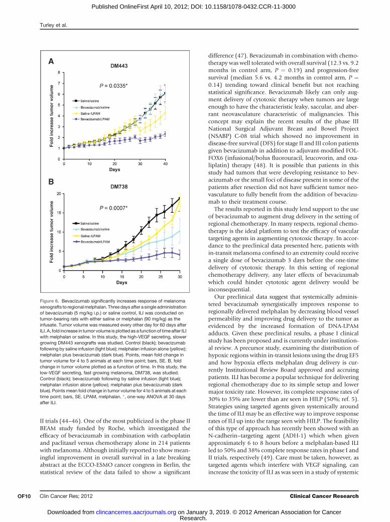
Having showed that regionally infused melphalan given3 days after systemic bevacizumab treatment led toimproved drug delivery into the tumor, we evaluated tumorgrowth after systemic bevacizumab using our preclinicalanimal model of ILI with melphalan. Tumor-bearing nuderatswere treatedwithbevacizumabor saline control at day 0and infused with melphalan or saline at day 3 based onpreliminary functional data as well as previous reports(19, 26). Growth curves for both DM738 and DM443xenografts after systemic saline injection and regional salineinfusion are depicted by the black line and show baseline invivo growth characteristics for both melanoma cell lines.DM443 baseline growth kinetics were significantly slowerthan DM738 (quadrupling time of 34.0 vs. 13.7 days, P <0.0001 t test), which is consistent with our previous expe-rience using these xenografts (supplementary Table S1;refs. 23, 34). Bevacizumab showed significant tumorgrowth inhibition 30 days after treatment when used in
Figure 2. Bevacizumab decreases hemoglobin-oxygen saturation inmelanoma xenografts. Nude mice bearing DM443 xenografts weretreatedwith 5mg/kg i.p. bevacizumab (n¼ 8) or saline control (n¼ 9), and(A) tumor oxygenated hemoglobin (HbO2) and (B) total Hb (Hb) and weremeasured daily for 6 days after treatment. Systemic bevacizumab had noeffect on tumor Hb and decreased HbO2. �, Mann–Whitney U test.
Turley et al.
Clin Cancer Res; 2012 Clinical Cancer ResearchOF6
Research. on January 3, 2019. © 2012 American Association for Cancerclincancerres.aacrjournals.org Downloaded from
Published OnlineFirst April 10, 2012; DOI: 10.1158/1078-0432.CCR-11-3000

combination with regionally delivered melphalan in boththe low-VEGF secreting, fast growingDM738 xenograft (P¼0.0007, one-way ANOVA) and the high-VEGF secreting,slower growing DM443 xenograft (P ¼ 0.0335, one-wayANOVA). Systemically administered bevacizumab aloneshowed similar efficacy to regional melphalan infusionalone for the low-VEGF secreting DM738 xenograft but notfor the high-VEGF secreting DM443 xenograft (Fig. 6A andB). Combination therapy of systemic bevacizumab fol-lowed by regional melphalan infusion had the most robusteffect on tumor growth inhibition for both xenografts, witha 37% and 113% increase in quadrupling time for DM443and DM738, respectively, as compared with melphalaninfusion alone (Supplementary Table S1).
DiscussionThis study shows that systemic bevacizumab given before
ILI with melphalan augments response by increasing deliv-
ery of melphalan to the tumor cells. This is first study toevaluate the use of bevacizumab as a part of a regionaltherapy strategy with the specific intent of augmenting thedelivery of chemotherapy to a tumor. Our results, in thecontext ofmelanoma, clearly support the emerging conceptthat the efficacy of antiangiogenic therapy in combinationwith chemotherapy is not simply an additive effect of long-term hypoxia resulting from tumor vasculature destructionand cytotoxicity from the chemotherapy. Rather it is asynergistic effect resulting frommodulation of tumor bloodsupply leading to improved delivery and penetration ofconventional chemotherapeutic agents (11). Rats undergo-ing limb infusions of high-dose melphalan 3 days afteradministration of bevacizumab had a robust and signifi-cantly greater response than either chemotherapy or bev-acizumab alone.
Antiangiogenic therapies such as bevacizumab weredeveloped with the goal of starving tumors of their blood
Figure 3. Bevacizumab (Bev.) doesnot significantly alter tumormicrovasculature structure orhemoglobin saturation in melanomaxenografts. The effect ofbevacizumab in altering tumormicrovasculature was determinedusing a murine model of dorsal flankmelanoma in combination with awindow chamber model (seeMaterials and Methods). Aftersurgical excision of a 12-mmdiameter flap of skin, mice wereinoculated in the exposed fasciawith3 � 106 DM443 or DM738 cells. Theexposed fascia was surgicallysecured with a window chambersaddle providing access forsubsequent imaging. Five days afterinoculation, mice were injected with5 mg/kg i.p. bevacizumab or salinecontrol.Melanoma tumorswere thenimaged daily using hyperspectralmicroscopy to observe changes intumor microvasculature. Two to 4mice were used for each treatmentarm for both cell lines.Representative images are shownearlier. A, in general, mice harboringDM443 and DM738 melanomaxenografts showed no significantalterations in tumormicrovasculature after treatmentwith bevacizumab. Usinghyperspectral images acquired fromthe window chamber model,vascular tortuosity (B) and VLD (C)were calculated as surrogate markerfor tumor vascular normalization. Inboth cell lines, no significantdifferences in tortuosity or VLD wereshowed.
DM443
Day 0 Day 3
DM738
Day 0 Day 3
Saline
A
B
C
Bev.
Bevacizumab Augments Regional Chemotherapy for Melanoma
www.aacrjournals.org Clin Cancer Res; 2012 OF7
Research. on January 3, 2019. © 2012 American Association for Cancerclincancerres.aacrjournals.org Downloaded from
Published OnlineFirst April 10, 2012; DOI: 10.1158/1078-0432.CCR-11-3000
supply. Whereas this effect was observed in preclinicalmodels (35) antivascular agents have failed to controltumor growth through vascular effects alone in the clinicalsetting (36). Although ineffective as a monotherapy, bev-acizumab in combination with conventional chemothera-pies has improved overall survival in previously untreatedpatients with colorectal and lung cancer (17, 36). Themechanisms by which antiangiogenic therapies such asbevacizumab augment cytotoxic therapy are controversial.One theory is that bevacizumab in combination with cyto-toxic agents acts synergistically by targeting both tumor cellswith cytotoxic therapy and vascular stroma with bevacizu-mab (37). A second theory revolves around the concept oftumor vascular "normalization," where tumor vessels aretransiently remodeled structurally and functionally toresemble normal blood vessels (11). This normalizationmay synergistically enhance cytotoxic agent efficacy byovercoming physiologic barriers to drug delivery createdby aberrant tumor vasculature (11, 19).
The findings presented in this study are in agreementwithreports which show that targeting VEGF with compoundssuch as bevacizumab can manipulate tumor vasculature to
augment cytotoxic therapy (19, 26, 38–40). In this study,webuild on the current body of literature by reporting thatbevacizumab can augment regional chemotherapy deliveryby decreasing tumor vascular permeability and IFP.
In general, criteria for tumor vascular normalizationwould include functional decreases in vessel permeabilityand IFP, improved tissue oxygenation, and augmented drugdelivery to tumor cells in the context of structural changessuch as decreased tumor vessel density, tortuosity, and VLD(11, 26). In contrast to previous reports, the results of ourstudies failed to show a change in tumor microvasculaturestructure or improvement in tumor oxygenation after treat-ment with antiangiogenic therapy. In our murine modelengraftedwith ahumanmelanoma xenograft, bevacizumabdid not lessen tumor vessel tortuosity or VLD, and it did notsignificantly increase tumor hemoglobin saturation. Thesediscrepancies may, in part, be due to limitations of ourpreclinical model, which used a neutralizing human-VEGFantibody with weak affinity for murine-VEGF. Althoughthese results may have been influenced by a partial VEGFblockade, in which only VEGF produced from the humanmelanoma xenograft was neutralized, bevacizumab-
DM443 DM738
DM443
A B
C D120
100
80
60
40
20
0
120
100
80
60
40
20
0
P = 0.26* P = 0.64*
Mea
n va
scul
ar d
ensi
ty(m
m–2
)
Mea
n va
scul
ar d
ensi
ty(m
m–2
)
DM738
Saline
Saline
Bevacizumab
Bevacizumab Saline Bevacizumab
Figure 4. Tumor vessel density isunchanged 3 days afterbevacizumab treatment. Meantumor vessel density measured byimmunohistochemistry using ananti-rat CD31 antibody on tumorspecimen acquired 3 days aftertreatment with bevacizumab(5mg/kg i.p.) or saline control. Fiveto 7 xenografts were analyzed foreach treatment group. Cross-sectional images werereconstructed images acquiredfrom a scanned series of 40 to80 images acquired at �5magnification. Representativecross-sectional images are shownfor (A) DM443 and (B) DM738.Mean tumor vessel density wassimilar between treatmentbevacizumab and saline-treatedxenografts for (C) DM433 (87.6 �12.8 vs. 71.1�7.8mm�2,P¼0.26)and (D) DM738 (77.9� 9.4 vs. 73.0� 6.3 mm�2, P¼ 0.63). �, 2-tailed ttest.
Turley et al.
Clin Cancer Res; 2012 Clinical Cancer ResearchOF8
Research. on January 3, 2019. © 2012 American Association for Cancerclincancerres.aacrjournals.org Downloaded from
Published OnlineFirst April 10, 2012; DOI: 10.1158/1078-0432.CCR-11-3000

induced changes in tumor oxygenation and microvascula-ture have been previously reported in the syngeneic B16mousemelanomamodels (26). It is also possible that otherstructural alterations such as increases in interendothelialcell gaps or pericyte coverage could have occurred but notbeen measured in this report. Finally, the results of ourexperimental models may have been measured during amore persistent window of functional vascular normaliza-tion, which could have occurred after a more transientwindow of structural normalization had past (41). Takentogether, we cannot conclude with certainty that structuralvascular normalization did not occur in bevacizumab-trea-ted animals.Despite a lack of conclusive evidence supporting struc-
tural vascular normalization, our results are in support offunctional vascular normalization as evidence by enhanceddrug delivery in the setting of by decreased tumor vessel
permeability and IFP. These results support an importantrole for IFP in the dynamics of blood supply to the tumor. Ithas long been recognized that VEGF acts by increasingblood vessel permeability (14, 29). This, in turn, can leadto a loss of normal oncotic and hydrostatic pressure gra-dients (42)which eventuallywill cause IFP levels to rise. Thedecreased permeability seen with antiangiogenic agentssuch as bevacizumab, therefore, could lead to decreasedIFP levels creating a pressure gradient across blood vesselwalls and tumor interstitium that is conducive to drugpenetration into tumor cells (30).Overcoming this pressuregradient is critically important for delivery of small mole-cules such as LPAM, for which 60% to 90% of the drug isbound to plasma proteins (43).
The efficacy of bevacizumab in augmenting systemicallyadministered chemotherapy for treatment of melanomahas been investigated recently inmultiple phase I and phase
Figure 5. Systemic bevacizumab (Bev.) increases delivery of regionally administered melphalan. Immunohistochemistry of tissue slices taken from saline-,melphalan (LPAM)-, bevacizumab-, and melphalan plus bevacizumab–treated samples. LPAM-DNA adduct formation were measured using the antibodyMP5/73.Representative images are shown from rats used in each treatment arm for bothDM443andDM738 tumors. For bothDM443 andDM738 xenografts,LPAM-DNA adduct formation was higher (brown stain) in the bevacizumab plus melphalan–treated samples compared with the melphalan alone–treatedsamples. As expected, no LPAM-DNA adduct staining was seen in samples that were not exposed to LPAM. Average staining intensity for each treatmentgroup for both xenografts is plotted with error bars representing SEM. �, one-way ANOVA. Abbreviations: intra-arterial.
Bevacizumab Augments Regional Chemotherapy for Melanoma
www.aacrjournals.org Clin Cancer Res; 2012 OF9
Research. on January 3, 2019. © 2012 American Association for Cancerclincancerres.aacrjournals.org Downloaded from
Published OnlineFirst April 10, 2012; DOI: 10.1158/1078-0432.CCR-11-3000
II trials (44–46). One of the most publicized is the phase IIBEAM study funded by Roche, which investigated theefficacy of bevacizumab in combination with carboplatinand paclitaxel versus chemotherapy alone in 214 patientswithmelanoma. Although initially reported to showmean-ingful improvement in overall survival in a late breakingabstract at the ECCO-ESMO cancer congress in Berlin, thestatistical review of the data failed to show a significant
difference (47). Bevacizumab in combination with chemo-therapy was well tolerated with overall survival (12.3 vs. 9.2months in control arm, P ¼ 0.19) and progression-freesurvival (median 5.6 vs. 4.2 months in control arm, P ¼0.14) trending toward clinical benefit but not reachingstatistical significance. Bevacizumab likely can only aug-ment delivery of cytotoxic therapy when tumors are largeenough to have the characteristic leaky, saccular, and aber-rant neovasculature characteristic of malignancies. Thisconcept may explain the recent results of the phase IIINational Surgical Adjuvant Breast and Bowel Project(NSABP) C-08 trial which showed no improvement indisease-free survival (DFS) for stage II and III colon patientsgiven bevacizumab in addition to adjuvant-modified FOL-FOX6 (infusional/bolus fluorouracil, leucovorin, and oxa-liplatin) therapy (48). It is possible that patients in thisstudy had tumors that were developing resistance to bev-acizumab or the small foci of disease present in some of thepatients after resection did not have sufficient tumor neo-vasculature to fully benefit from the addition of bevacizu-mab to their treatment course.
The results reported in this study lend support to the useof bevacizumab to augment drug delivery in the setting ofregional chemotherapy. In many respects, regional chemo-therapy is the ideal platform to test the efficacy of vasculartargeting agents in augmenting cytotoxic therapy. In accor-dance to the preclinical data presented here, patients within-transit melanoma confined to an extremity could receivea single dose of bevacizumab 3 days before the one-timedelivery of cytotoxic therapy. In this setting of regionalchemotherapy delivery, any later effects of bevacizumabwhich could hinder cytotoxic agent delivery would beinconsequential.
Our preclinical data suggest that systemically adminis-tered bevacizumab synergistically improves response toregionally delivered melphalan by decreasing blood vesselpermeability and improving drug delivery to the tumor asevidenced by the increased formation of DNA-LPAMadducts. Given these preclinical results, a phase I clinicalstudy has been proposed and is currently under institution-al review. A precursor study, examining the distribution ofhypoxic regions within in-transit lesions using the drug EF5and how hypoxia effects melphalan drug delivery is cur-rently Institutional Review Board approved and accruingpatients. ILI has become a popular technique for deliveringregional chemotherapy due to its simple setup and lowermajor toxicity rate. However, its complete response rates of30% to 35% are lower than are seen in HILP (50%; ref. 5).Strategies using targeted agents given systemically aroundthe time of ILI may be an effective way to improve responserates of ILI up into the range seen with HILP. The feasibilityof this type of approach has recently been showed with anN-cadherin–targeting agent (ADH-1) which when givenapproximately 6 to 8 hours before a melphalan-based ILIled to 50% and 38% complete response rates in phase I andII trials, respectively (49). Care must be taken, however, astargeted agents which interfere with VEGF signaling, canincrease the toxicity of ILI as was seen in a study of systemic
Figure 6. Bevacizumab significantly increases response of melanomaxenografts to regionalmelphalan. Three days after a single administrationof bevacizumab (5 mg/kg i.p.) or saline control, ILI was conducted ontumor-bearing rats with either saline or melphalan (90 mg/kg) as theinfusate. Tumor volume was measured every other day for 60 days afterILI. A, fold increase in tumor volume is plotted as a function of time after ILIwith melphalan or saline. In this study, the high-VEGF secreting, slowergrowing DM443 xenografts was studied. Control (black); bevacizumabfollowing by saline infusion (light blue); melphalan infusion alone (yellow);melphalan plus bevacizumab (dark blue). Points, mean fold change intumor volume for 4 to 5 animals at each time point; bars, SE. B, foldchange in tumor volume plotted as a function of time. In this study, thelow-VEGF secreting, fast growing melanoma, DM738, was studied.Control (black); bevacizumab following by saline infusion (light blue);melphalan infusion alone (yellow); melphalan plus bevacizumab (darkblue). Points mean fold change in tumor volume for 4 to 5 animals at eachtime point; bars, SE. LPAM, melphalan. �, one-way ANOVA at 30 daysafter ILI.
Turley et al.
Clin Cancer Res; 2012 Clinical Cancer ResearchOF10
Research. on January 3, 2019. © 2012 American Association for Cancerclincancerres.aacrjournals.org Downloaded from
Published OnlineFirst April 10, 2012; DOI: 10.1158/1078-0432.CCR-11-3000
sorafenib given for 1 week before and after melphalan-based ILI in patients with advanced extremity melanoma(50).Clinical trials in the setting of in-transit disease of the
extremity while providing important tools toward improv-ing regional response rates can, in addition, provide tre-mendous insight into how to optimally use targeted agentsin conjunction with chemotherapeutic agents because ofthe ease of obtaining sequential tissue biopsies to evaluatethe effect of the targeted agent on the tumor concurrentlywith the ability to obtain drug pharmacokinetic data. Stud-ies like the one proposed in this article using systemicbevacizumab and regional melphalan can have applicationto other tumor types where regional therapy is widely usedsuch as peritoneal perfusion for abdominal carcinomatosisor liver infusion/perfusion for isolated liver metastasis.Furthermore, they can also provide important insight onhow to develop rational strategies of incorporating targetedagents into systemic chemotherapeutic protocols for met-
astatic melanoma for which few effective treatment optionscurrently exist.
Disclosure of Potential Conflicts of InterestD.S. Tyler has commercial research grant from Adherex Technologies and
Roche/Schering; material transfer agreements with Bayer, Schering, andGenta pharmaceuticals; honoraria from speakers bureau for Novartis; andis the consultant/advisory board member for Genentech. No potentialconflicts of interest were disclosed by other authors.
Grant SupportThe study was supported by Duke University Melanoma Research Fund
andVAMerit Review grant (toD.S. Tyler), NIH grant 5T32CA093245 (to R.S.Turley and J.C. Padussis), CA40355 (to M.W. Dewhirst), Department ofDefense grant BC083195 (to A.N. Fontanella),NIH Loan Repayment (to R.S.Turley).
The costs of publication of this article were defrayed in part by thepayment of page charges. This article must therefore be hereby markedadvertisement in accordance with 18 U.S.C. Section 1734 solely to indicatethis fact.
Received November 23, 2011; revised March 22, 2012; accepted April 2,2012; published OnlineFirst April 10, 2012.
References1. Jemal A, Siegel R, Ward E, Hao Y, Xu J, Thun MJ. Cancer statistics,
2009. CA Cancer J Clin 2009;59:225–49.2. de Vries E, Bray FI, Coebergh JW, Parkin DM. Changing epidemiol-
ogy of malignant cutaneous melanoma in Europe 1953–1997: risingtrends in incidence and mortality but recent stabilizations in westernEurope and decreases in Scandinavia. Int J Cancer 2003;107:119–26.
3. Hodi FS, O'Day SJ, McDermott DF, Weber RW, Sosman JA, HaanenJB, et al. Improved survival with ipilimumab in patients with metastaticmelanoma. N Engl J Med 2010;363:711–23.
4. Pawlik TM, Ross MI, Johnson MM, Schacherer CW, McClain DM,Mansfield PF, et al. Predictors and natural history of in-transit mela-noma after sentinel lymphadenectomy. Ann Surg Oncol 2005;12:587–96.
5. Turley RS,RaymondAK, Tyler DS. Regional treatment strategies for in-transit melanoma metastasis. Surg Oncol Clin N Am 2011;20:79–103.
6. Bhatia S, Tykodi SS, Thompson JA. Treatment of metastatic melano-ma: an overview. Oncology (Williston Park) 2009;23:488–96.
7. Thompson JF, KamPC,WaughRC, HarmanCR. Isolated limb infusionwith cytotoxic agents: a simple alternative to isolated limb perfusion.Semin Surg Oncol 1998;14:238–47.
8. Sharma K, Beasley GM, Turley RS, Broadwater G, Petersen B, MoscaP, et al. Patterns of recurrence following complete response to regionalchemotherapy for in-transit melanoma. Ann Surg Oncol. 2012 Apr 3.[Epub ahead of print].
9. Rofstad EK, Mathiesen B. Metastasis in melanoma xenografts isassociated with tumor microvascular density rather than extent ofhypoxia. Neoplasia 2010;12:889–98.
10. Mahabeleshwar GH, Byzova TV. Angiogenesis in melanoma. SeminOncol 2007;34:555–65.
11. Jain RK. Normalization of tumor vasculature: an emerging concept inantiangiogenic therapy. Science 2005;307:58–62.
12. Dvorak HF. Vascular permeability factor/vascular endothelial growthfactor: a critical cytokine in tumor angiogenesis and a potential targetfor diagnosis and therapy. J Clin Oncol 2002;20:4368–80.
13. Olsson AK, Dimberg A, Kreuger J, Claesson-Welsh L. VEGF receptorsignalling - in control of vascular function. Nat Rev Mol Cell Biol2006;7:359–71.
14. Senger DR, Galli SJ, Dvorak AM, Perruzzi CA, Harvey VS, Dvorak HF.Tumor cells secrete a vascular permeability factor that promotesaccumulation of ascites fluid. Science 1983;219:983–5.
15. Sabatino M, Kim-Schulze S, Panelli MC, Stroncek D, Wang E, TabackB, et al. Serum vascular endothelial growth factor and fibronectin
predict clinical response to high-dose interleukin-2 therapy. J ClinOncol 2009;27:2645–52.
16. Friedman HS, Prados MD, Wen PY, Mikkelsen T, Schiff D, Abrey LE,et al. Bevacizumab alone and in combination with irinotecan in recur-rent glioblastoma. J Clin Oncol 2009;27:4733–40.
17. Hurwitz H, Fehrenbacher L, Novotny W, Cartwright T, Hainsworth J,Heim W, et al. Bevacizumab plus irinotecan, fluorouracil, and leucov-orin formetastatic colorectal cancer. N Engl JMed 2004;350:2335–42.
18. Pallis AG, Serfass L, Dziadziusko R, van Meerbeeck JP, Fennell D,Lacombe D, et al. Targeted therapies in the treatment of advanced/metastatic NSCLC. Eur J Cancer 2009;45:2473–87.
19. Dickson PV, Hamner JB, Sims TL, Fraga CH, Ng CY, Rajasekeran S,et al. Bevacizumab-induced transient remodeling of the vasculature inneuroblastoma xenografts results in improved delivery and efficacy ofsystemically administered chemotherapy. Clin Cancer Res2007;13:3942–50.
20. PalmerGM,FontanellaAN,ShanS,HannaG,ZhangG,FraserCL, et al.In vivo optical molecular imaging and analysis in mice using dorsalwindow chamber models applied to hypoxia, vasculature and fluo-rescent reporters. Nat Protoc 2011;6:1355–66.
21. Skala MC, Fontanella A, Lan L, Izatt JA, Dewhirst MW. Longitudinaloptical imaging of tumor metabolism and hemodynamics. J BiomedOpt 2010;15:011112.
22. Dewhirst MW, Chitneni SK, Palmer GM, Zalutsky MR. Molecularimaging of hypoxia. J Nucl Med 2011;52:165–8.
23. Augustine CK, Yoshimoto Y, Gupta M, Zipfel PA, Selim MA, Febbo P,et al. Targeting N-cadherin enhances antitumor activity of cytotoxictherapies in melanoma treatment. Cancer Res 2008;68:3777–84.
24. Attis MG, Burchette JL, Selim MA, Pham T, Soler AP. Differentialexpression of N-cadherin distinguishes a subset of metastasizingdesmoplastic melanomas. Hum Pathol 2006;37:899–905.
25. Tilby MJ, Styles JM, Dean CJ. Immunological detection of DNAdamage caused by melphalan using monoclonal antibodies. CancerRes 1987;47:1542–6.
26. Dings RP, Loren M, Heun H, McNiel E, Griffioen AW, Mayo KH, et al.Scheduling of radiation with angiogenesis inhibitors anginex andAvastin improves therapeutic outcome via vessel normalization. ClinCancer Res 2007;13:3395–402.
27. Erhard H, Rietveld FJ, van Altena MC, Brocker EB, Ruiter DJ, deWaal RM. Transition of horizontal to vertical growth phase mela-noma is accompanied by induction of vascular endothelial growthfactor expression and angiogenesis. Melanoma Res 1997;7 Suppl2:S19–26.
Bevacizumab Augments Regional Chemotherapy for Melanoma
www.aacrjournals.org Clin Cancer Res; 2012 OF11
Research. on January 3, 2019. © 2012 American Association for Cancerclincancerres.aacrjournals.org Downloaded from
Published OnlineFirst April 10, 2012; DOI: 10.1158/1078-0432.CCR-11-3000
28. Yuan F, Chen Y, Dellian M, Safabakhsh N, Ferrara N, Jain RK. Time-dependent vascular regression and permeability changes in estab-lished human tumor xenografts induced by an anti-vascular endothe-lial growth factor/vascular permeability factor antibody.ProcNatl AcadSci U S A 1996;93:14765–70.
29. Senger DR, Perruzzi CA, Feder J, Dvorak HF. A highly conservedvascular permeability factor secreted by a variety of human and rodenttumor cell lines. Cancer Res 1986;46:5629–32.
30. TongRT, Boucher Y, Kozin SV,Winkler F, Hicklin DJ, Jain RK. Vascularnormalization by vascular endothelial growth factor receptor 2 block-ade induces a pressure gradient across the vasculature and improvesdrug penetration in tumors. Cancer Res 2004;64:3731–6.
31. Palmer GM, Viola RJ, Schroeder T, Yarmolenko PS, Dewhirst MW,Ramanujam N. Quantitative diffuse reflectance and fluorescencespectroscopy: tool to monitor tumor physiology in vivo. J Biomed Opt2009;14:024010.
32. Papenfuss HD, Gross JF, Intaglietta M, Treese FA. A transparentaccess chamber for the rat dorsal skin fold. Microvasc Res 1979;18:311–8.
33. Cao Y, Sonveaux P, Liu S, Zhao Y, Mi J, Clary BM, et al. Systemicoverexpression of angiopoietin-2 promotes tumormicrovessel regres-sion and inhibits angiogenesis and tumor growth. Cancer Res2007;67:3835–44.
34. Augustine CK, Toshimitsu H, Jung SH, Zipfel PA, Yoo JS, YoshimotoY, et al. Sorafenib, a multikinase inhibitor, enhances the response ofmelanoma to regional chemotherapy. Mol Cancer Ther 2010;9:2090–101.
35. Fukumura D, Duda DG, Munn LL, Jain RK. Tumor microvasculatureandmicroenvironment: novel insights through intravital imaging in pre-clinical models. Microcirculation 2010;17:206–25.
36. JainRK,DudaDG,Clark JW, Loeffler JS. Lessons fromphase III clinicaltrials on anti-VEGF therapy for cancer. Nat Clin Pract Oncol 2006;3:24–40.
37. Teicher BA. A systems approach to cancer therapy. (Antioncogenicsþstandard cytotoxics–>mechanism(s) of interaction). Cancer Metasta-sis Rev 1996;15:247–72.
38. EichhornME, Strieth S, Luedemann S, Kleespies A, Noth U, Passon A,et al. Contrast enhanced MRI and intravital fluorescence microscopyindicate improved tumor microcirculation in highly vascularized mel-anomas upon short-term anti-VEGFR treatment. Cancer Biol Ther2008;7:1006–13.
39. OhtaM, Kawabata T, YamamotoM, Tanaka T, Kikuchi H, Hiramatsu Y,et al. TSU68, an antiangiogenic receptor tyrosine kinase inhibitor,induces tumor vascular normalization in a human cancer xenograftnude mouse model. Surg Today 2009;39:1046–53.
40. Shrimali RK, Yu Z, TheoretMR, ChinnasamyD, Restifo NP, RosenbergSA. Antiangiogenic agents can increase lymphocyte infiltration intotumor and enhance the effectiveness of adoptive immunotherapy ofcancer. Cancer Res 2010;70:6171–80.
41. Batchelor TT, SorensenAG, di Tomaso E, ZhangWT, DudaDG,CohenKS, et al. AZD2171, a pan-VEGF receptor tyrosine kinase inhibitor,normalizes tumor vasculature and alleviates edema in glioblastomapatients. Cancer Cell 2007;11:83–95.
42. Netti PA,HambergLM,Babich JW,KiersteadD,GrahamW,HunterGJ,et al. Enhancement of fluid filtration across tumor vessels: implicationfor delivery of macromolecules. Proc Natl Acad Sci U S A 1999;96:3137–42.
43. Greig NH, Sweeney DJ, Rapoport SI. Melphalan concentration depen-dent plasma protein binding in healthy humans and rats. Eur J ClinPharmacol 1987;32:179–85.
44. Hainsworth JD, Infante JR, Spigel DR, Peyton JD, ThompsonDS, LaneCM, et al. Bevacizumab and everolimus in the treatment of patientswith metastatic melanoma: a phase 2 trial of the Sarah CannonOncology Research Consortium. Cancer 2010;116:4122–9.
45. Vihinen PP, Hernberg M, Vuoristo MS, Tyynela K, Laukka M, Lundin J,et al. A phase II trial of bevacizumab with dacarbazine and daily low-dose interferon-alpha2a as first line treatment inmetastaticmelanoma.Melanoma Res 2010;20:318–25.
46. PerezDG,SumanVJ,FitchTR,AmatrudaT III,MortonRF,JilaniSZ,etal.Phase 2 trial of carboplatin, weekly paclitaxel, and biweekly bevacizu-mab in patients with unresectable stage IV melanoma: a North CentralCancer Treatment Group study, N047A. Cancer 2009;115:119–27.
47. O'Day SJ, Kim KB, Sosman JA, Peterson AC, Feng S, Minor DR, et al.BEAM: a randomized phase II study evaluating the activity of Bev-acizumab in combination with carboplatin plus placitaxel in patientswith previously untreated advanced melanoma. Eur J Cancer2009;7:13.
48. Allegra CJ, Yothers G, O'Connell MJ, Sharif S, Petrelli NJ, ColangeloLH, et al. Phase III trial assessing bevacizumab in stages II and IIIcarcinoma of the colon: results of NSABP protocol C-08. J Clin Oncol2011;29:11–6.
49. Beasley GM, Riboh JC, Augustine CK, Zager JS, Hochwald SN,Grobmyer SR, et al. Prospective multicenter phase II trial of systemicADH-1 in combination with melphalan via isolated limb infusion inpatients with advanced extremity melanoma. J Clin Oncol2011;29:1210–5.
50. McMahon NGB, Sanders G, Augustine C, Tyler DS. A phase I study ofsystemic sorafenib in combination with isolated limb infusion withmelphalan (ILI-M) in patients (pts) with locally advanced in-transitmelanoma. J Clin Oncol 2009;27:Abstr 9065.
Turley et al.
Clin Cancer Res; 2012 Clinical Cancer ResearchOF12
Research. on January 3, 2019. © 2012 American Association for Cancerclincancerres.aacrjournals.org Downloaded from
Published OnlineFirst April 10, 2012; DOI: 10.1158/1078-0432.CCR-11-3000
Published OnlineFirst April 10, 2012.Clin Cancer Res Ryan S. Turley, Andrew N. Fontanella, James C. Padussis, et al. Chemotherapy for In-Transit Melanomaand Drug Delivery: A Novel Approach to Augment Regional Bevacizumab-Induced Alterations in Vascular Permeability
Updated version
10.1158/1078-0432.CCR-11-3000doi:
Access the most recent version of this article at:
Material
Supplementary
http://clincancerres.aacrjournals.org/content/suppl/2012/04/10/1078-0432.CCR-11-3000.DC1Access the most recent supplemental material at:
E-mail alerts related to this article or journal.Sign up to receive free email-alerts
Subscriptions
Reprints and
To order reprints of this article or to subscribe to the journal, contact the AACR Publications
Permissions
Rightslink site. (CCC)Click on "Request Permissions" which will take you to the Copyright Clearance Center's
.http://clincancerres.aacrjournals.org/content/early/2012/05/11/1078-0432.CCR-11-3000To request permission to re-use all or part of this article, use this link
Research. on January 3, 2019. © 2012 American Association for Cancerclincancerres.aacrjournals.org Downloaded from
Published OnlineFirst April 10, 2012; DOI: 10.1158/1078-0432.CCR-11-3000