Benjamin S Abella, MD MPhil Department of Emergency Medicine University of Pennsylvania...
64
Benjamin S Abella, MD MPhil Department of Emergency Medicine University of Pennsylvania [email protected] u
-
Upload
camron-smith -
Category
Documents
-
view
219 -
download
0
Transcript of Benjamin S Abella, MD MPhil Department of Emergency Medicine University of Pennsylvania...

Benjamin S Abella, MD MPhilDepartment of Emergency MedicineUniversity of [email protected]

CC: Chest pain

vs.

1. ABC’s/Stabilization/Resuscitation IV, O2, monitor, pulse oximeter
2. ECG3. +/- CXR
CC: Chest pain

64 year old man presents with 5 hours of SS chest pressure associated with SOB, nausea and diaphoresis. Gradual onset while shoveling the snow. Improved with rest.
PMH: HTN, DM
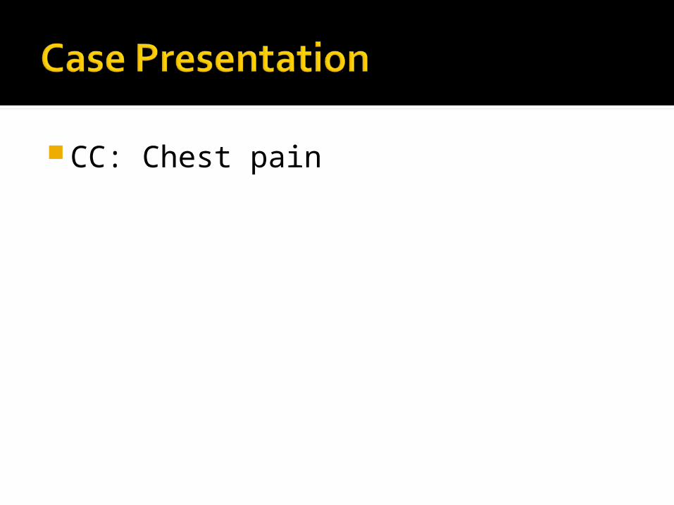
Gen: Nontoxic appearing, apprehensive, mildly diaphoretic
Vitals: 37.5ºC, RR16, HR 100, BP 160/95
CVS: RRR, Normal S1, S2, no M/R/GResp: CTAB, easy respirationsAbd: Soft, NTNDExt: No calf tenderness or swelling,
no edema, strong distal pulses


MedPixTM


Epidemiology >6,000,000 Americans have CAD 500,000 deaths/yr. in the U.S. from CAD 250,000 pts. with prehospital cardiac arrest▪ 6% survive to hospital discharge
>4,000,000 E.D. visits per year for acute chest pain
$100 billion per year Etiology
Ischemia: imbalance between oxygen demand and oxygen supply
Fixed atherosclerotic lesion vs. plaque disruption with platelet/thrombi aggregation vs. spasm

Stable angina: transient, episodic chest discomfort that is predictable and reproducible.
Unstable angina: angina that is new in onset, occurs at rest or is similar but somewhat “different” than previous episodes.
Acute myocardial infarction

Substernal chest discomfort > 15 minutes associated with dyspnea, diaphoresis, light-headedness, palpitations, nausea and/or vomiting NSTEMI STEMI
12.5% of acute MI’s are clinically “silent”

Smoking Hypertension Diabetes mellitis Hypercholesterolemia Family history of CAD prior to age 55 in
a first-degree relative Previous history of CAD or peripheral
vascular disease Cardiac risk factors are very poor
predictors of risk for ACS in the E.D
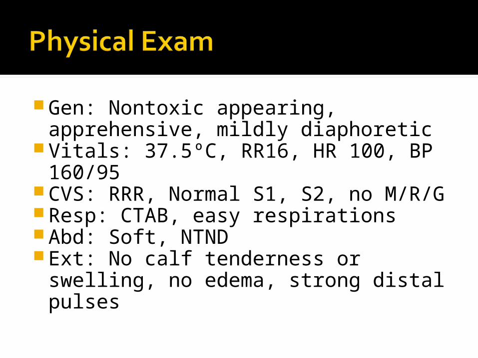
Not helpful in distinguishing patients with ACS from those with noncardiac chest pain unless an alternative diagnosis is clear e.g. pneumothorax
Normal cardiopulmonary exam is most common
S3 in 15-20% with AMI Chest wall tenderness to palpation in
~15% with ACS

The standard ECG is the single best test to identify patients with an AMI upon E.D. presentation
But sensitivity is still far from ideal ST elevation in 50% of AMI’s 1-5% of AMI’s have a normal initial ECG 4 - 23% of pts. with unstable angina
have a normal ECG

CK-MB Sensitivity > 90% for MI 5-6h after symptom onset 50% sensitive shortly after presentation
Troponin Tn-I: similar sensitivity and specificity to CK-MB for
AMI in E.D. patients Tn-T: less sensitive for myocardial injury▪ Independent marker of cardiovascular risk
Marker Elevation Peak Duration
CK-MB 3-12 h 18-24 h 2 days
Troponin-I 3-12 h 18 h 5-10 d
Troponin-T 3-12 h 12 h 5-14 d

Aspirin Inhibits thromboxane A2 … decreases
platelet aggregation Nitrates
Decreases preload and afterload; increases coronary perfusion in obstructed vessels
Beta blockers Decreases infarct size, cardiovascular
complications, and mortality Consider heparin (or enoxaparin),
antiplatelet agents, GIIb/IIIa inhibitors Fibrinolysis or PCI

ST elevation > 0.1 mV in two or more continguous leads or new LBBB
Time to therapy < 12 hours (Class I), 12-24 hours (Class IIb)
Door to balloon time
Epidemiology: 6% of pts. with cocaine-associated
chest pain have an AMI 20-60% have transient myocardial
ischemia Often atypical chest pain Can be delayed for hours to days after
the most recent use
Spasm Increased myocardial oxygen
demandClot formationAccelerated atherosclerosis and
LVH

Diagnosis: ECG less sensitive and specific for MI CK-MB less sensitive Troponin I may be more useful
Prognosis: Favorable short-term prognosis 1 year mortality primarily due to
comorbidities and/or continued cocaine use Treatment:
Benzodiazepines Avoid Beta blockers

>4 million E.D. visits per year for acute chest pain
<15% with ACS 2-6% sent home from ED ultimately with
ACS Chest pain units/Obs

CC: Stroke

51 year old woman brought in by EMS with acute onset of right sided weakness and aphasia.
PMH: HTN

Gen: Somnolent, diaphoretic Vitals: 37.5ºC, RR16, HR 110, BP 180/110 CVS: Tachy, Normal S1, S2, no M/R/G Resp: CTAB, easy respirations Abd: Soft, NTND Ext: No calf tenderness or swelling, no
edema, weak distal pulses Neuro: Right leg/arm 4/5 strength,
+expressive aphasia


Definition: Intimal tear with entry of blood into the media “dissects” between the intima and adventitia
#1 site: ascending aorta at the ligamentum arteriosum
Stanford Classification: A: involves Ascending aorta (w/ or w/o
descending)▪ 80% of dissections
B: descending aorta only DeBakey Classification

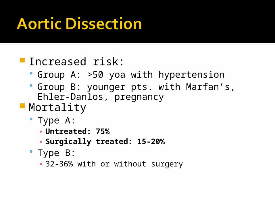
Increased risk: Group A: >50 yoa with hypertension Group B: younger pts. with Marfan’s, Ehler-
Danlos, pregnancy Mortality
Type A:▪ Untreated: 75%▪ Surgically treated: 15-20%
Type B:▪ 32-36% with or without surgery

History: >90% with abrupt and severe pain
in the chest or between the scapulae▪ “tearing” or “ripping”▪ Can be dull or pressure-like▪ Anterior chest ~ ascending aorta; Back ~
descending Nausea, vomiting, diaphoresis
common

Associated symptoms based on progression of dissection:▪ Carotid arteries: stroke▪ Spinal arteries: paraplegia▪ Abdominal aorta/renal arteries/iliacs:
Abdominal/flank pain▪ Coronary arteries: aortic insufficiency; pericardial
effusion/tamponade▪ Laryngeal nerve compression: hoarseness▪ Tracheal compression: dyspnea/stridor/wheezing ▪ Esophageal compression: dysphagia

Physical Exam: Symptoms/signs as above Most commonly: normal heart and lungs▪ Aortic insufficiency murmur in 16-20%
Unequal, decreased, or absent peripheral pulses only found in 50%

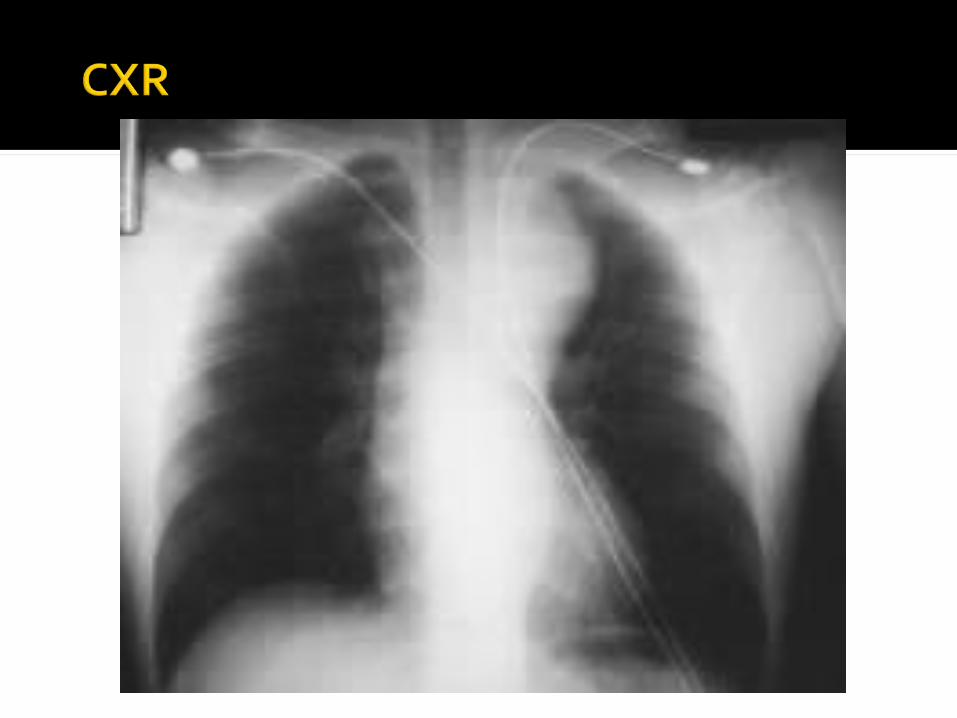
CXR 85% with some abnormality▪ widened mediastinum most common▪ left pleural effusion; indistinct aortic knob;
displaced, calcified intima > 6mm from outer aortic wall
CT vs. TEE vs. aortogram

Considering it? 2 large bore IV’s, monitor, T&C, ECG
Blood pressure: Decrease the shear force on the intima
to minimize progression▪ Lower arterial blood pressure▪ Decrease LV contractility

Goal: SBP 100-110 mm Hg; HR 60-80
Options:A. Nitroprusside + esmololB. Labetalol
Early CT surgery involvement

CC: Chest pain

40 year old woman brought in by EMS with acute onset pleuritic right sided chest sharpness associated with SOB.
PMH: HTN
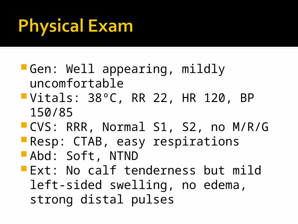
Gen: Well appearing, mildly uncomfortable
Vitals: 38ºC, RR 22, HR 120, BP 150/85 CVS: RRR, Normal S1, S2, no M/R/G Resp: CTAB, easy respirations Abd: Soft, NTND Ext: No calf tenderness but mild left-
sided swelling, no edema, strong distal pulses

MedPixTM


650,000 cases/year in the U.S. Mortality
2-10% if diagnosed and treated 30% if undiagnosed #3 cause of death overall #1 cause of nonsurgical maternal death in the
peripartum period The source is lower extremity DVT in 80-
90% of cases Upper extremity DVT in 10-15% Others: pelvic vein thrombosis; fat emboli; septic
emboli; right heart thrombosis
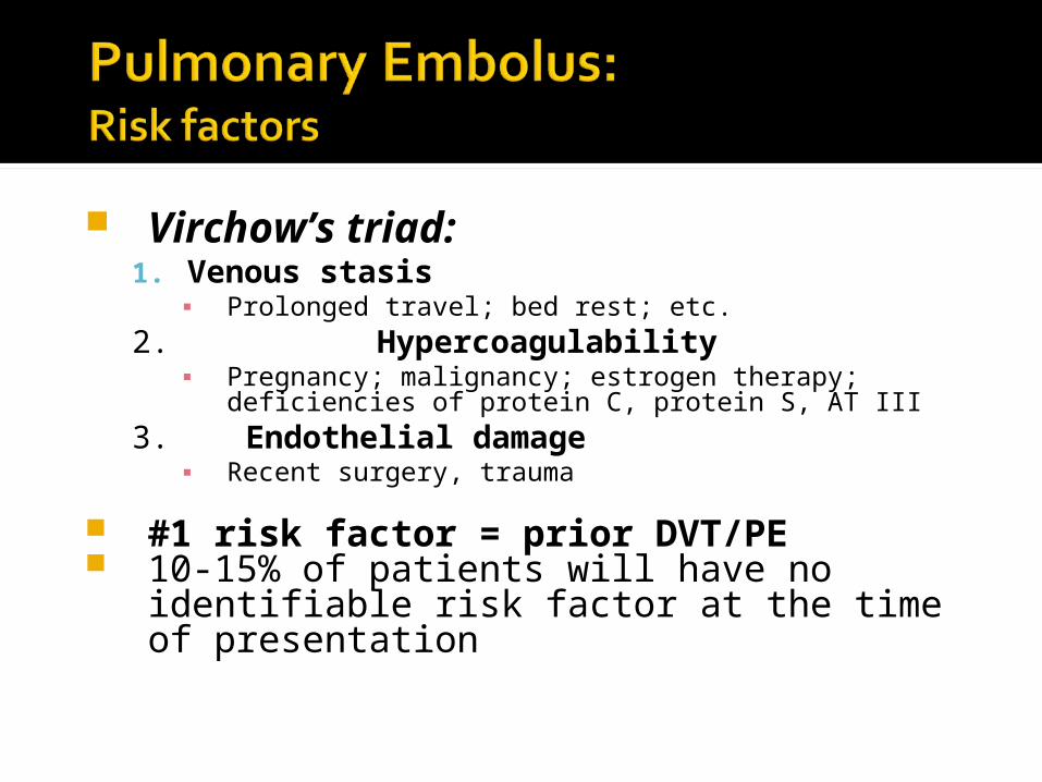
Virchow’s triad:1. Venous stasis
▪ Prolonged travel; bed rest; etc.2. Hypercoagulability
▪ Pregnancy; malignancy; estrogen therapy; deficiencies of protein C, protein S, AT III
3. Endothelial damage▪ Recent surgery, trauma
#1 risk factor = prior DVT/PE 10-15% of patients will have no
identifiable risk factor at the time of presentation

“Classic Triad”: Dyspnea, hemoptysis, pleuritic CP in
only 20% of pts.Three notable findings:
Pleuritic chest pain in 74% Dyspnea in 84% Respiratory rate > 16 in 92%
Heart rate > 100 in only 44%

Kline, 2003

Often normal> 40% with nonspecific ST and T
wave abnormalitiesSinus tachycardia is the most
common rhythm disturbance.S1Q3T3 is seen in only 6% of
patients with PE.

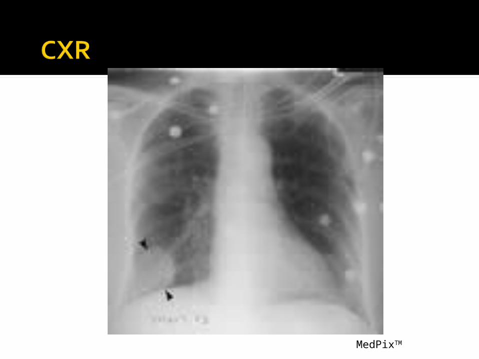
Normal in ~30% and a concerning finding in the setting of
dyspnea and hypoxemia w/o RAD Atelectasis in ~50% Elevated hemidiaphragm in 40% Hampton’s Hump: Pleural based wedge
shaped infiltrate Westermark sign: Proximally dilated
pulmonary artery with abrupt cut-off Needed for subsequent interpretation of
the V/Q scan
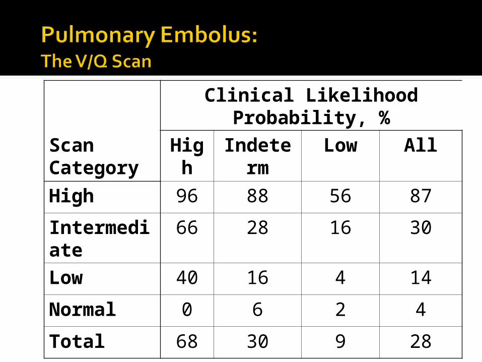
Clinical Likelihood Probability, %
Scan Category
High Indeterm Low All
High 96 88 56 87
Intermediate 66 28 16 30
Low 40 16 4 14
Normal 0 6 2 4
Total 68 30 9 28

Normal Ventilation
Abnormal Perfusion

LE Doppler USPulmonary angiographySpiral CTMRI
Garg, 1999

Considering it: IV, O2 prn, monitor, pulse ox.
High pretest probability: Anticoagulate 1st, then order your study Heparin 80 U/kg i.v. bolus; 18 U/kg/hr i.v.
drip Low (+/- intermediate) pretest
probability: Study 1st, then anticoagulate if appropriate
Consider thrombolytics if unstable

CC: “can’t catch my breath”

40 year old man presents with acute onset of SOB and pleuritic right sided chest sharpness.
PMH: none

Gen: Mildly uncomfortableVitals: 37ºC, RR 22, HR 120, BP
145/95CVS: Tacky, Normal S1, S2, no M/R/GResp: Decreased bs on the right,
tachypeicAbd: Soft, NTNDExt: No calf tenderness or swelling,
no edema, strong distal pulses
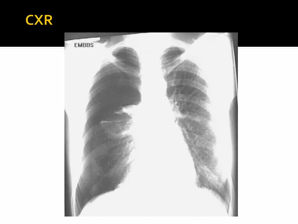

Especially tall, thin male smokersOnly 10 – 20% occur with exertionMost thought to result from
rupture of a subpleural blebSymptoms vary with size and rate
of progression of pneumothorax
Acute pleuritic CP in 95%Dyspnea in 80%Decreased breath sounds over the
affected lung in 85%Tachypnea > 24 in only 5%Hyperresonance in <1/3
Tension pneumothorax: Clinical presentation of pneumothorax
with hemodynamic compromise Treatment is immediate needle
decompression“Non-tension” spontaneous
pneumothorax Upright PA CXR is 83% sensitive

Tube thoracostomy Minicatheter or standard chest tube
Catheter aspiration Single or sequential
Observation x 6 hrs. with repeat CXR:o Stable; minimal/no symptoms; <25% ptxo No significant comorbidities
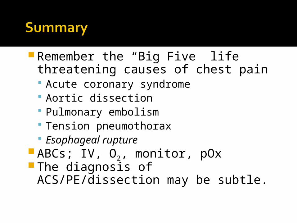
Remember the “Big Five” life threatening causes of chest pain Acute coronary syndrome Aortic dissection Pulmonary embolism Tension pneumothorax Esophageal rupture
ABCs; IV, O2, monitor, pOxThe diagnosis of ACS/PE/dissection
may be subtle.