Bellevue Hospital in NYC, First “Official” Morgue in the U.S.
82
vue Hospital in NYC, First “Official” Morgue in the
-
Upload
damian-atkinson -
Category
Documents
-
view
224 -
download
0
Transcript of Bellevue Hospital in NYC, First “Official” Morgue in the U.S.

Bellevue Hospital in NYC, First “Official” Morgue in the U.S.

What is an Autopsy?
• An autopsy is a post mortem examination preformed on a corpse to determine the cause and manner of death.
• The prefix 'auto-' means 'self', and so autopsy means 'to see for oneself‘.

Why is a Forensic Autopsy preformed?
• Forensics autopsies are preformed when the cause of death of a victim may be a criminal case, often involving foul play.
• A forensic autopsy applies science to legal law.

Classification
• In a forensic autopsy, death is placed into five different categories.• Natural• Accident• Homicide• Suicide• Unknown
• Following an in-depth examination of all the evidence, a medical examiner or coroner will assign a manner of death as one of the five listed above; and detail the evidence on the mechanism of the death.

Natural Death
• Death by natural cause is a term used by coroners to describe the death of someone by occurring disease process, or is not apparent given medical history or circumstances.
• The majority of natural death is caused by old age. • Other causes of natural death are heart disease,
stroke, gentic disorders, etc.

Accidental Death
• Accidental death is a death that is often caused by mistake or in a freak occurrence. These deaths are not planned yet can be explained by surrounding circumstances.
• In the City of Philadelphia, any death as a result of a medical procedure can warrant an autopsy by the Medical Examiners Office.

Homicidal Death
• The term ‘homicide’ refers to the act of killing another person.
• Homicide is often the most investigated death, therefore making it the most autopsied.

Suicidal Death
• The act of ending ones own life.
• These autopsies often easily identify source, cause, and other factors of the death.
• Suicide is often identified in the forensic autopsy as a cause of toxic, firearms, blunt force trauma, etc.

Unknown Death
• In some jurisdictions, the Undetermined category may include deaths in absentia, such as deaths at sea and missing persons declared dead in a court of law; in others, such deaths are classified under "Other".
Experts Who Perform Forensic Autopsies
• A forensic autopsy is usually preformed by a specialized medical doctor called a forensics pathologist or medical examiner.
• To be a pathologist, the doctor must have completed a four-year undergraduate program, four years of medical school training, and three to four years of postgraduate training in the form of a pathology residency.
What Warrants an Autopsy (Post-Mortem) by The ME’s Office
• Questionable Death-as determined by the coroner• Homicide• Drowning• Auto Accidents• Death of a child
***Most teaching hospitals will offer a post-mortem to any family of a patient that dies.

Benefits of Autopsy
Discover cause of death for:• Criminal cases• Family peace of mind• Genetic disease or deformity• Insurance claims• Medical advancement
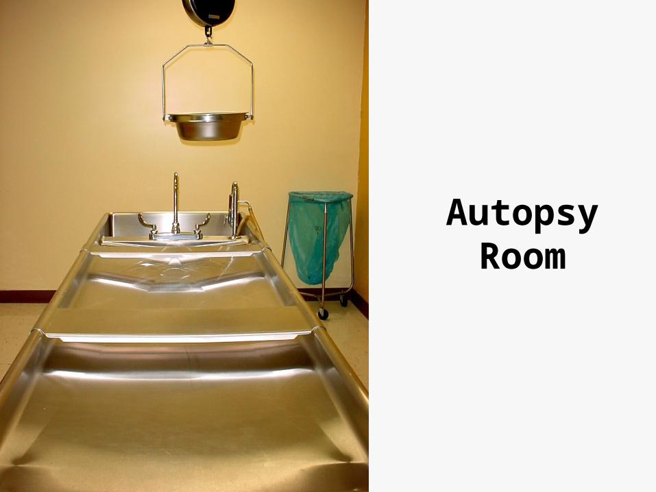
Autopsy Room
Physical Examination
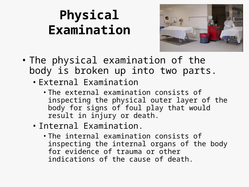
• The physical examination of the body is broken up into two parts.• External Examination
• The external examination consists of inspecting the physical outer layer of the body for signs of foul play that would result in injury or death.
• Internal Examination.• The internal examination consists of inspecting the
internal organs of the body for evidence of trauma or other indications of the cause of death.

External Examination
Steps of an external examination.
1. Photographed.
2. Physical evidence collected off body.
3. Samples of hair, nails, etc. are collected.
4. Undressed, examined for wounds.1. Lacerations, abrasions, bruises.
5. Measured, weighed, cleaned.

External Examination
• Trace Evidence
• Sign of injury or mistreatment
• Sign of illness, disease, or abnormalities


Livor Mortis
• Defined as ‘Color of Death’.• Coloration of the skin.
• At death, the heart stops working. When the heart stops working, the blood stops pumping. The blood stops pumping, the red blood cells and plasma gather on the bottom part of the body, closet to the floor.
• A line forms after 8 hours if the body hasn’t been moved. If moved, a new line starts to form. It is impossible to tell which was first. The thinker the line, the longer the position the body was in.

Algor Mortis
• Defined as ‘Coolness of Death’.• Temperature of body.
• In a controlled environment, stating at 98.6 degrees, the body will drop one degree per hour.
• This happens because at death, the respiratory system stops working, the body stops functioning, it is no longer moving.
• When taking the temperature of a corpse, you can’t take it in the mouth because the muscles will be relaxed and the tongue wont stay on top of the thermometer.
• Thinner people cool faster then fat people.

Rigor Mortis
• Defined as ‘Stiffness of Death’.• Flexibility of the body.
• Shows up 2 hours after death
• Peaks 12 hours after death.
• Takes 12-24 hours for entire rigor mortis effect to take place.
• At approximately 0 hours after death, the body is at its stiffest.
• The eyelids are affected first, the the jaw, face, trunk, arms, legs.
• Ends after 24-36 hours.
Pallor Mortis
• Defined as ‘Paleness of Death’.• Tone of the body.
• Happens 15-20 minutes after death.
• Happens due to lack of capillary circulation in the body.
• Can not be used to determine time of death except if body is found still with color.

Body Block
bsapp.combsapp.com
Internal Examination
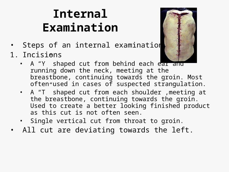
• Steps of an internal examination.1. Incisions
• A “Y” shaped cut from behind each ear and running down the neck, meeting at the breastbone, continuing towards the groin. Most often used in cases of suspected strangulation.
• A “T” shaped cut from each shoulder ,meeting at the breastbone, continuing towards the groin. Used to create a better looking finished product as this cut is not often seen.
• Single vertical cut from throat to groin.
• All cut are deviating towards the left.

Internal Examination (cont’d)
2. Cuts1. The chest cavity is cut open using shears.2. The ribs are sawed away, letting them be lifted off the
body, exposing the heart and lungs.
3. Removal1. En masse technique of Letulle – All organs removed at
once.2. En bloc method of Ghon – organs divided into four groups
and removed in sections.
4. All removed organs are now weighed and examined for unusual markings or signs.
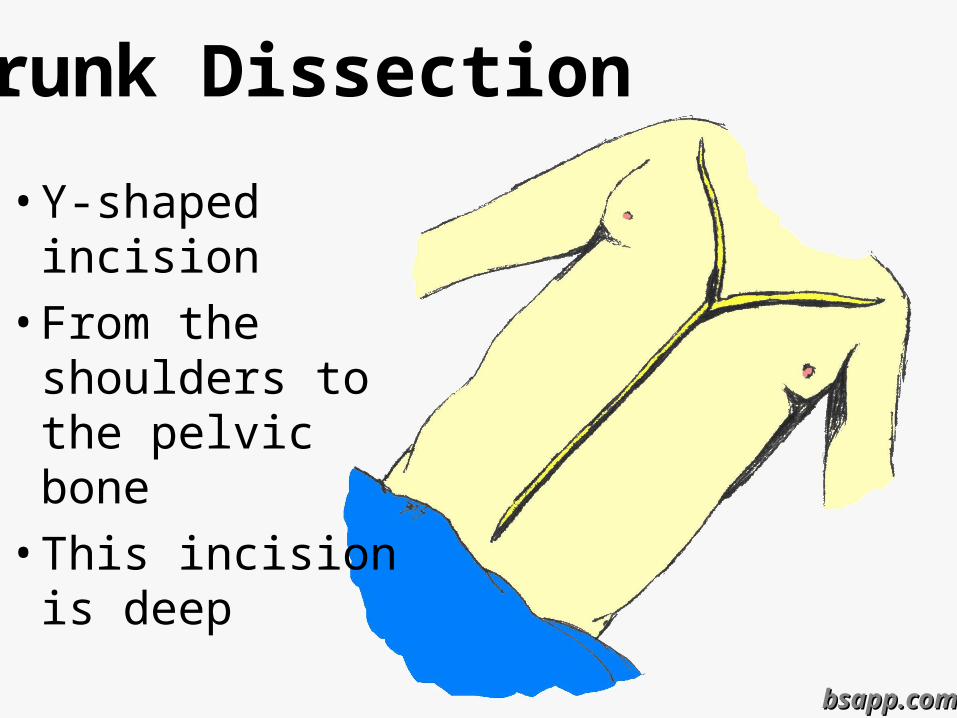
Trunk Dissection
• Y-shaped incision
• From the shoulders to the pelvic bone
• This incision is deep
bsapp.combsapp.com

Opening the Chest
• Skin & muscle, are pulled from the chest wall
• Chest Plate is extracted
• Heart is extracted

Opening the Chest

Removal and Dissection of the Organs
• One organ at a time
• All body organs at once - ”Rokitansky Method” • Upon removal each organ is:
• Weighed & measured
• Examined
• Sliced in cross sections
• Sampled for microscopic & chemical analysis

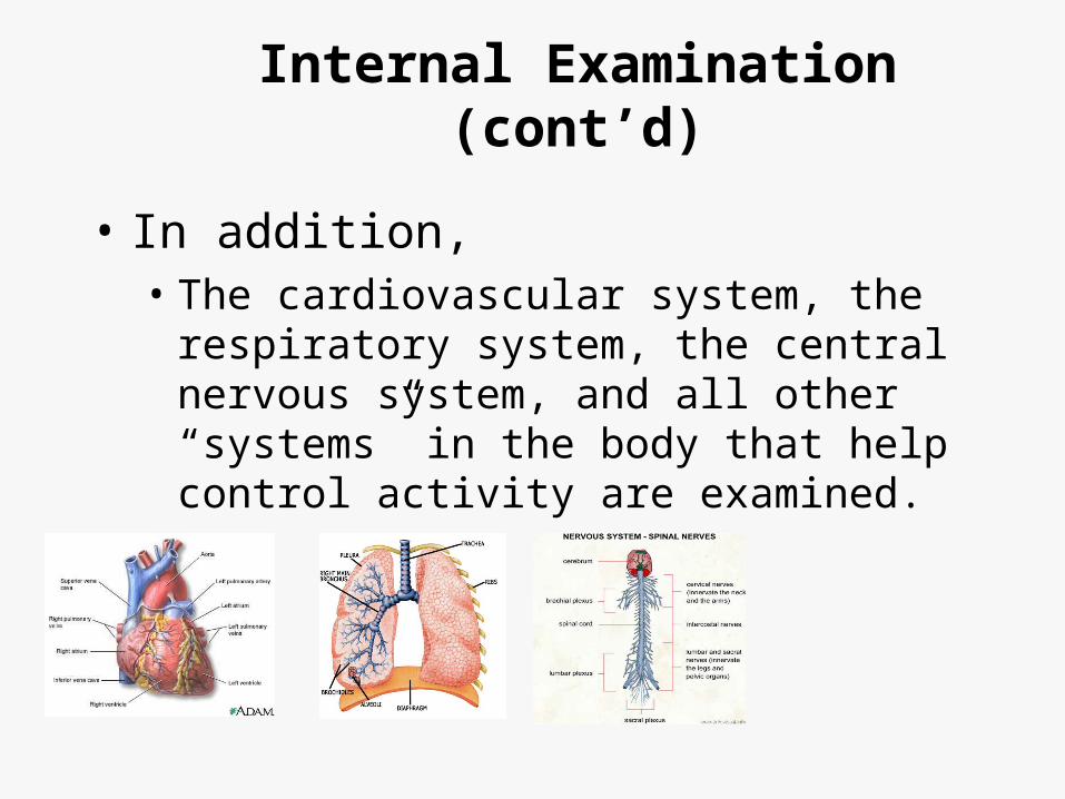
• In addition,• The cardiovascular system, the respiratory
system, the central nervous system, and all other “systems” in the body that help control activity are examined.
Internal Examination (cont’d)
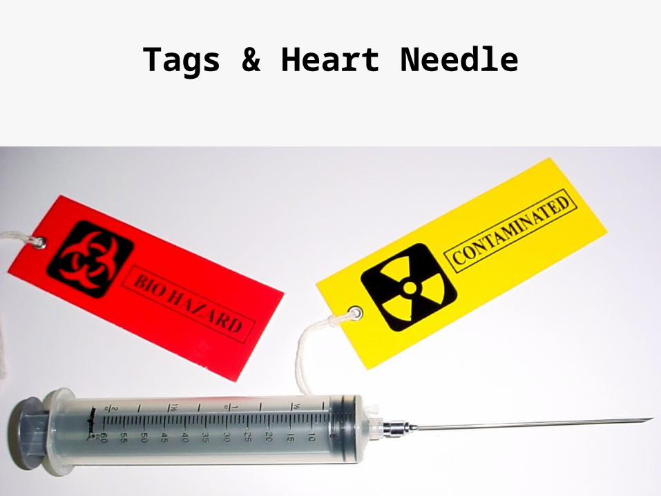
Tags & Heart Needle

Samples

“Running the Gut”
The contents of the stomach, intestines, and bowels must be inspected as well

Removing the Brain
The Scalp is cut ear to ear across the crown of the
head

Exposing the Skull
Next the scalp is pulled forward and back to expose the skull

Exposing the BrainCutting the skull cap

Removal of the Brain• Spinal Cord is cut• The soft brain is
removed• Brain is so soft it
must be placed in formaldehyde for about a week before an in depth examination

Close Up• Skull cap is replace
• Skin pulled back in place
• Body Organs may or may not be replaced
• Incisions are sown up with the use of a baseball stitch

Close Up

High Risk Autopsy
As Recommended by CDC

Source Material

High Risk Autopsies
• Case Selection
• Permissions• Local• NPDSU (required for genetic testing)
• Precautions• Autopsy - BSL 2• Tissue - BSL 3
• Equipment
• Procedure
• Tissue Handling
• Shipping
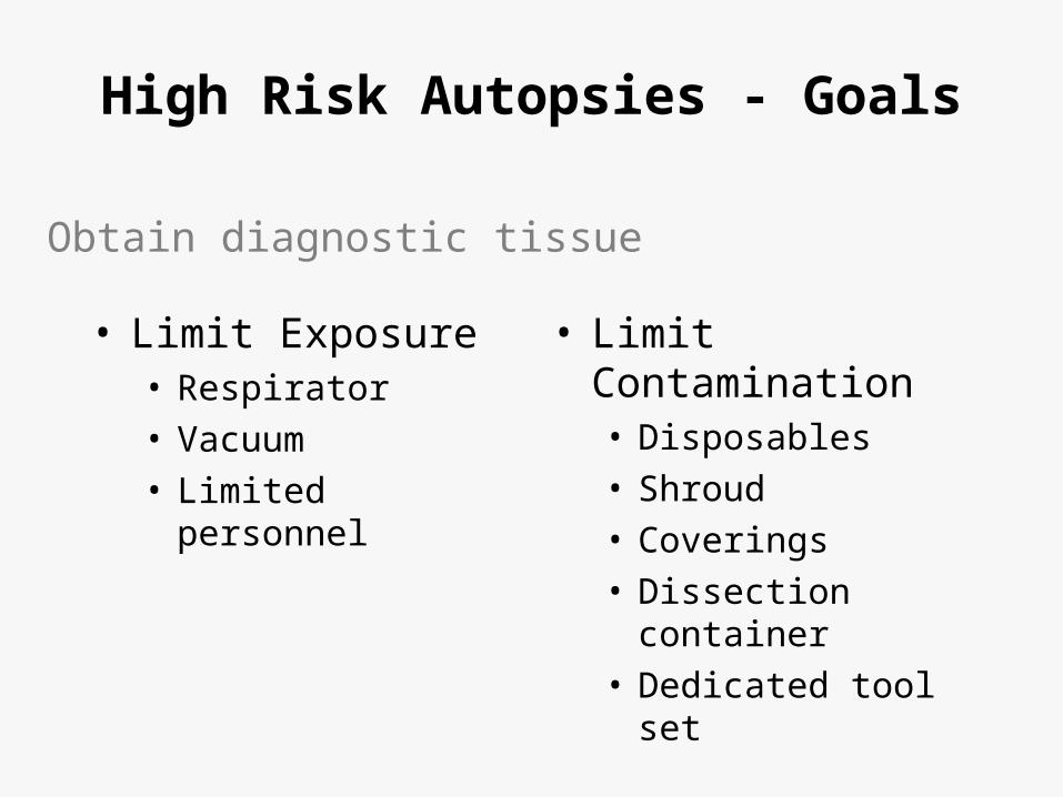
High Risk Autopsies - Goals
• Limit Exposure• Respirator
• Vacuum
• Limited personnel
• Limit Contamination• Disposables
• Shroud
• Coverings
• Dissection container
• Dedicated tool set
. Obtain diagnostic tissue


High Risk Autopsies - Decontamination
• Stainless steel• Autoclavable
• 1M NaOH• 30’
• Non-autoclavable• Immersible
– 1-2M NaOH, 1hr, RT
• Non-Immersible– 1-2M
• Aluminum• Sodium hypochlorite
(2% free chlorine) DO NOT AUTOCLAVE
• Liquid waste• 1M NaOH• Autoclave 132°, 4.5hr
• Dry waste• Incineration• Autoclave 132°, 4.5hr

Tissue Processing
• Post-fixation Formic Acid• >95%, 1hr, RT with post-treatment formalin
48hr.
• Process as usual• Discard waste as biohazardous waste

Post-Mortem Forensic Toxicology
Postmortem Forensic Toxicology
• Qualitative and quantitative analysis of drugs or poisons in biological specimens collected at autopsy
• Interpretation of findings in terms of:• Physiological effect at time of death• Behavioural effect at time of death

Quantitative vs. Qualitative
• Qualitative analysis – determines the presence or absence of a drug or poison in a submitted sample
• Quantitative analysis – determines the amount of drug or poison that is present in the submitted sample

Postmortem Forensic Toxicology
Types of cases:
• Suspected drug intoxication cases
• Fire deaths
• Homicides
• Driver and pilot fatalities
• Therapeutic drug monitoring
• Sudden infant death (SIDS)

Issues in Specimen Collection
• Selection• Multiple, varied sites of collection
• Collection• Appropriate method of collection• Adequate volumes for analysis
• Storage and handling
Important to ensure analytical results are accurate and interpretations are sound

Typical autopsy specimens for toxicology
• Blood
• Urine
• Stomach contents
• Bile
• Liver
• Hair
• Vitreous humor

Blood
• Antemortem ideal blood sample• “just before death”
• Postmortem blood is not truly “blood”
• Anatomical site of collection at autopsy should be noted

• Central sites• Heart
• Peripheral sites• Femoral• Iliac• Subclavian
• Other sites• Head blood• Hematoma blood
Sites for Blood Collection

Hematoma
• Extravascular blood clot
• Protected from metabolism
• Analysis will indicate what drugs were present in the blood at the time of formation

Hematoma case example
• A 26 year old man was found dead at the bottom of a staircase. Death was due to physical injuries.
• Question as to alcohol use prior to fall down stairs• No urine available at autopsy
• Alcohol not detected in femoral blood
• Alcohol in hematoma blood 150 mg/100 mL
• The deceased had been drinking prior to receiving the head trauma.
• The deceased had survived for several hours after the injury.

Hematoma
• Caution: There may be a delay between the incident which resulted in hematoma and the actual formation of the hematoma
• Therefore, this alcohol concentration does not necessarily indicate the BAC at the time of the fall down the stairs.

Urine
• Produced by the kidneys
• Blood filtered by the kidneys
• Stored in the bladder until voided
• Qualitative - the presence of a drug in the urine of an individual indicates that some time prior to death the drug or poison was present in the blood of the individual

Stomach contents
• Visual examination may reveal tablets• Drugs that have been orally ingested may be detected in
stomach contents• Caution: drugs administered by other routes may also
diffuse into stomach contents from the blood• Generally qualitative:
• Stomach contents are not homogeneous
• Only a portion of stomach contents collected (unmixed?)
• Useful for directing further analysis

Case Example
• A 26 year old woman is found dead in bed• Numerous medications in her home:
• Amitriptyline, Oxycodone, Morphine, Paroxetine, Diphenhydramine, Pseudoephedrine, Phenobarbital, Codeine, Temazepam, Diazepam
• Only 3 mL of blood collected at autopsy• Qualitative analysis of stomach contents:
• Amitriptyline: detected• Nortriptyline: detected
• Quantitation can now be performed in blood
Liver
• Drug metabolism occurs in the liver
• Both parent compounds and metabolites may be present in higher concentrations in the liver than in the blood ease of detection
• Limitation is that drugs are not uniformly distributed throughout the liver confounds interpretation

Bile
• Digestive secretion
• Continuously produced by the liver
• Stored in the gallbladder
• Qualitative - the presence of a drug in the bile of an individual indicates that sometime prior to death, the individual was exposed to the drug

Vitreous humor
• Fluid that occupies the space between the lens and the retina of the eye.
• Sequestered from putrefaction, charring and trauma, microorganisms.
• Useful in cases where decomposition is advanced, body is exhumed or in fire deaths
• Limitation is blood:vitreous ratio may not be known

Hair
• Recent specimen of interest
• Metabolism does not occur in hair
• Can provide a historical record of drug or poison exposure
• Pros and cons of hair analysis still being uncovered racial variability?

Non-biological submissions
• Used to direct analysis of biologicals
• May indicate the nature of substances that may have been ingested, inhaled or injected
• Examples:
• Containers found at the scene • Syringes• Unidentified tablets or liquids
Proper specimen handling
• Identification of samples• Continuity• Contents
• Specimens delivered to lab without delay
• Specimens should be analyzed as soon as possible
• Storage areas should be secure

Storage and Handling
• Not feasible to analyze specimens immediately
• Sample should be in well-sealed container
• Sample containers must be sterile
• Use of preservatives and anti-coagulants
• Refrigeration vs. Freezing• Both inhibit bacterial action; esp. freezing• Freezing results in prep time• Freeze-thaw cycle may promote breakdown

Storage of Samples
• Preservative• Sodium fluoride
• Anti-coagulants• Sodium citrate• Potassium oxalate• EDTA• Heparin• Not imperative for postmortem blood samples

Determining analyses
• Case history• Medical history• Autopsy findings• Symptomatology
• Experience of the toxicologist
• Amount of specimen available
• Nature of specimens available
• Policies of the organization

Pitfalls in Postmortem Forensic Toxicology

Decomposition
• Autolysis• The breakdown of cellular material by enzymes
• Putrefaction• A septic/infectious process• The destruction of soft tissues by the action of
bacteria and enzymes• Traumatic deaths may demonstrate putrefaction

Decomposition
• Fewer samples available for collection
• Quality of samples is diminished
• Putrefaction produces alcohols• Ethanol• Isopropanol• Acetaldehyde• n-propanol

Postmortem redistribution
• A phenomenon whereby increased concentrations of some drugs are observed in postmortem samples and/or site dependent differences in drug concentrations may be observed
• Typically central blood samples are more prone to postmortem changes (will have greater drug concentrations than peripheral blood samples)

Drug Stability
• Knowledge of a drug’s stability is necessary to facilitate interpretation of concentrations
• Breakdown of drugs may occur after death and during storage via non-enzymatic mechanisms
• Cocaine Benzoylecgonine (Hydrolysis)• LSD degradation due to light sensitivity• Others ?

Evaporation of volatiles
• Ethanol
• Carbon monoxide
• Cyanide
• Toluene
• Other alcohols

Interpretation
Therapeutic, toxic or fatal? How do you know?
• Compare measured blood concentrations with concentrations reported in the literature:• Clinical pharmacology studies• Incidental drug findings• Plasma blood
• Consider case history: • Symptoms observed by witnesses?• Tolerance of the individual to the drug