Behaviour of the GermCell Specific Lamin Through ...repository.ias.ac.in/42548/1/113_pub.pdf ·...
12
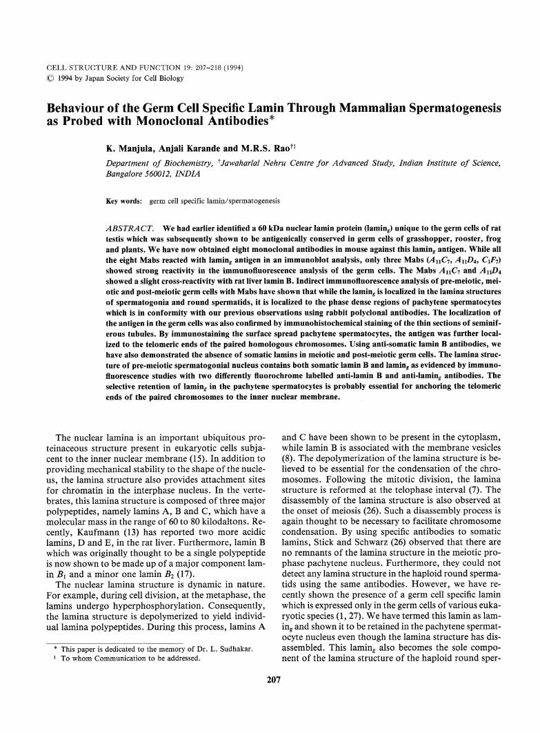
CELL STRUCTURE AND FUNCTION 19: 207-218 (1994) © 1994 by Japan Society for Cell Biology Behaviour of the Germ Cell Specific Lamin Through MammalianSpermatogenesis as Probed with Monoclonal Antibodies* K. Manjula, Anjali Karande and M.R.S. Raon Department of Biochemistry, ^Jawaharlal Nehru Centre for Advanced Study, Indian Institute of Science, Bangalore 560012, INDIA Keywords: germ cell specific lamin/spermatogenesis ABSTRACT. We had earlier identified a 60 kDa nuclear lamin protein (laming unique to the germ cells of rat testis which was subsequently shown to be antigenically conserved in germ cells of grasshopper, rooster, frog and plants. Wehave now obtained eight monoclonal antibodies in mouse against this lamin? antigen. While all the eight Mabs reacted with laming antigen in an immunoblot analysis, only three Mabs (AnCi, A\\D^ C\F-j) showed strong reactivity in the immunofluorescence analysis of the germ cells. The Mabs A\\Cn and A11D4 showed a slight cross-reactivity with rat liver lamin B. Indirect immunofluorescence analysis of pre-meiotic, mei- otic and post-meiotic germ cells with Mabs have shown that while the lamin? is localized in the lamina structures of spermatogonia and round spermatids, it is localized to the phase dense regions of pachytene spermatocytes which is in conformity with our previous observations using rabbit polyclonal antibodies. The localization of the antigen in the germ cells was also confirmed by immunohistochemical staining of the thin sections of seminif- erous tubules. By immunostaining the surface spread pachytene spermatocytes, the antigen was further local- ized to the telomeric ends of the paired homologous chromosomes. Using anti-somatic lamin B antibodies, we have also demonstrated the absence of somatic lamins in meiotic and post-meiotic germ cells. The lamina struc- ture of pre-meiotic spermatogonial nucleus contains both somatic lamin B and lamin? as evidenced by immuno- fluorescence studies with two differently fluorochrome labelled anti-lamin B and anti-lamin? antibodies. The selective retention of lamin? in the pachytene spermatocytes is probably essential for anchoring the telomeric ends of the paired chromosomesto the inner nuclear membrane. The nuclear lamina is an important ubiquitous pro- teinaceous structure present in eukaryotic cells subja- cent to the inner nuclear membrane (15). In addition to providing mechanical stability to the shape of the nucle- us, the lamina structure also provides attachment sites for chromatin in the interphase nucleus. In the verte- brates, this lamina structure is composedof three major polypeptides, namely lamins A, B and C, which have a molecular mass in the range of 60 to 80 kilodaltons. Re- cently, Kaufmann (13) has reported two more acidic lamins, D and E, in the rat liver. Furthermore, lamin B which was originally thought to be a single polypeptide is nowshownto be madeup ofa major componentlam- in B\ and a minor one lamin B2 (17). The nuclear lamina structure is dynamic in nature. For example, during cell division, at the metaphase, the lamins undergo hyperphosphorylation. Consequently, the lamina structure is depolymerized to yield individ- ual lamina polypeptides. During this process, lamins A and C have been shown to be present in the cytoplasm, while lamin B is associated with the membranevesicles (8). The depolymerization of the lamina structure is be- lieved to be essential for the condensation of the chro- mosomes. Following the mitotic division, the lamina structure is reformed at the telophase interval (7). The disassembly of the lamina structure is also observed at the onset of meiosis (26). Such a disassembly process is again thought to be necessary to facilitate chromosome condensation. By using specific antibodies to somatic lamins, Stick and Schwarz (26) observed that there are no remnants of the lamina structure in the meiotic pro- phase pachytene nucleus. Furthermore, they could not detect any lamina structure in the haploid round sperma- tids using the same antibodies. However, we have re- cently shown the presence of a germ cell specific lamin which is expressed only in the germ cells of various euka- ryotic species (1 , 27). We have termed this lamin as lam- ing and shown it to be retained in the pachytene spermat- ocyte nucleus even though the lamina structure has dis- assembled. This laming also becomes the sole compo- nent of the lamina structure of the haploid round sper- This paper is dedicated to the memory of Dr. L. Sudhakar. To whomCommunicationto be addressed. 207
Transcript of Behaviour of the GermCell Specific Lamin Through ...repository.ias.ac.in/42548/1/113_pub.pdf ·...
CELL STRUCTURE AND FUNCTION 19: 207-218 (1994)
© 1994 by Japan Society for Cell Biology
Behaviour of the Germ Cell Specific Lamin Through MammalianSpermatogenesisas Probed with Monoclonal Antibodies*
K. Manjula, Anjali Karande and M.R.S. RaonDepartment of Biochemistry, ^Jawaharlal Nehru Centre for Advanced Study, Indian Institute of Science,Bangalore 560012, INDIA
Keywords: germcell specific lamin/spermatogenesis
ABSTRACT.Wehad earlier identified a 60 kDa nuclear lamin protein (laming unique to the germ cells of rattestis which was subsequently shown to be antigenically conserved in germ cells of grasshopper, rooster, frogand plants. Wehave now obtained eight monoclonal antibodies in mouse against this lamin? antigen. While allthe eight Mabs reacted with laming antigen in an immunoblot analysis, only three Mabs (AnCi, A\\D^ C\F-j)showed strong reactivity in the immunofluorescence analysis of the germ cells. The Mabs A\\Cn and A11D4showed a slight cross-reactivity with rat liver lamin B. Indirect immunofluorescence analysis of pre-meiotic, mei-otic and post-meiotic germ cells with Mabs have shown that while the lamin? is localized in the lamina structuresof spermatogonia and round spermatids, it is localized to the phase dense regions of pachytene spermatocyteswhich is in conformity with our previous observations using rabbit polyclonal antibodies. The localization ofthe antigen in the germ cells was also confirmed by immunohistochemical staining of the thin sections of seminif-erous tubules. By immunostaining the surface spread pachytene spermatocytes, the antigen was further local-ized to the telomeric ends of the paired homologous chromosomes. Using anti-somatic lamin B antibodies, wehave also demonstrated the absence of somatic lamins in meiotic and post-meiotic germ cells. The lamina struc-ture of pre-meiotic spermatogonial nucleus contains both somatic lamin B and lamin? as evidenced by immuno-fluorescence studies with two differently fluorochrome labelled anti-lamin B and anti-lamin? antibodies. Theselective retention of lamin? in the pachytene spermatocytes is probably essential for anchoring the telomericends of the paired chromosomesto the inner nuclear membrane.
The nuclear lamina is an important ubiquitous pro-teinaceous structure present in eukaryotic cells subja-cent to the inner nuclear membrane (15). In addition toproviding mechanical stability to the shape of the nucle-us, the lamina structure also provides attachment sitesfor chromatin in the interphase nucleus. In the verte-brates, this lamina structure is composedof three majorpolypeptides, namely lamins A, B and C, which have amolecular mass in the range of 60 to 80 kilodaltons. Re-cently, Kaufmann (13) has reported two more acidiclamins, D and E, in the rat liver. Furthermore, lamin Bwhich was originally thought to be a single polypeptideis nowshownto be madeup ofa major componentlam-in B\ and a minor one lamin B2 (17).The nuclear lamina structure is dynamic in nature.For example, during cell division, at the metaphase, thelamins undergo hyperphosphorylation. Consequently,the lamina structure is depolymerized to yield individ-ual lamina polypeptides. During this process, lamins A
and C have been shown to be present in the cytoplasm,while lamin B is associated with the membranevesicles(8). The depolymerization of the lamina structure is be-lieved to be essential for the condensation of the chro-mosomes. Following the mitotic division, the laminastructure is reformed at the telophase interval (7). Thedisassembly of the lamina structure is also observed atthe onset of meiosis (26). Such a disassembly process isagain thought to be necessary to facilitate chromosomecondensation. By using specific antibodies to somaticlamins, Stick and Schwarz (26) observed that there areno remnantsof the lamina structure in the meiotic pro-phase pachytene nucleus. Furthermore, they could notdetect any lamina structure in the haploid round sperma-tids using the same antibodies. However, we have re-
cently shown the presence of a germ cell specific laminwhich is expressed only in the germ cells of various euka-ryotic species (1 , 27). Wehave termed this lamin as lam-ing and shown it to be retained in the pachytene spermat-ocyte nucleus even though the lamina structure has dis-assembled. This laming also becomes the sole compo-nent of the lamina structure of the haploid round sper-
This paper is dedicated to the memory of Dr. L. Sudhakar.To whomCommunication to be addressed.
207

K. Manjula, A. Karande and M.R.S. Rao
matid nucleus. The rabbit polyclonal antibodies againstthe lamin^ which was used in most of these studiesshowed very little cross-reactivity with the somaticlamins A, B and C of rat (28). These observationsprompted us to generate specific monoclonal antibodiesagainst lamin^ in order to study the epitopes that are ex-clusively present in laming, but not in other somatic lam-ins. Moreover, these monoclonal antibodies would bevaluable tools for studies on the expression of this anti-gen during the spermatogenic process. Wehave beensuccessful in generating a set of monoclonal antibodiesagainst lamin^ in mouse and we describe here ourstudies on the nature and the extent of cross-reactivitythese monoclonal antibodies exhibit with somatic lam-ins. Weare also describing the results of the immunolo-calization of this antigen in pre-meiotic, meiotic pro-phase and post-meiotic germ cells using these monoclon-al antibodies.
MATERIALS AND METHODS
Materials. All the biochemicals used, unless specified werefrom Sigma Chemical Company, St. Louis, MO, U.S.A.
FITC labelled goat anti-rabbit IgG, from Cappel Labora-tories Inc., Cochranville, PA, U.S.A., HRP-conjugated goatanti-rabbit IgG, rabbit anti-mouse IgG and FITC labelled
goat anti-mouse IgG were purchased from Bangalore Genei,Bangalore, India. Nitrocellulose sheets were obtained fromSchleicher & Schuell, Dassel, Germany.All plasticware used for raising monoclonal antibodies werepurchased from Becton and Dickinson Labware. Polyethyl-ene glycol 3500, Nystatin, glutamine, hypoxanthine, amino-
pterin, thymidine and p-nitrophenyl phosphate were obtainedfrom Sigma Chemical Company, U.S.A. Iscove's modifiedDulbecco's medium (IMDM) was purchased from Gibco Lab-oratories, U.S.A. Fetal calf serum was the product of SERAlabs, U.K. Pencillin, streptomycin sulfate and gentamycinwere obtained from Alembic Chemicals, Sarabhai Chemicalsand Pharmaceutical company of India, Bombay, respectively.All other reagents used were of analytical reagent grade.Animals. Malealbino rats of Wistar strain wereused inall the studies. Splenocytes from immunized BALB/cmicewere used for establishing hybridoma.Purification of nuclei. Rats were sacrificed by cervical dis-location, testes removed and decapsulated. Livers were per-fused with ice-cold physiological saline before they were ex-cised. The tissues were homogenized in 9 volumes of buffer A(10mM Tris-HCl, pH 7.5/25 mM KC1/5 mM MgCl2/l mM
CaCl2) containing 0.34 M sucrose in a Teflon Potter-Elvejhemhomogenizer. Testicular tissue was homogenized by handwhile livers were homogenized with a motor-driven pestle. Nu-clei were isolated from the homogenate as described by Rao etal. (22).
Preparation of sonication-resistant spermatid nuclei.Sonication-resistant nuclei from rat testes represent nuclei
from elongating and elongated spermatids. They were isolatedfrom the testes of 60 day old rats as described by Singh andRao (24). Briefly, testes were homogenized in buffer B (10 mMTris-HCl, pH 7.4/0.1 mMphenyl methyl sulfonyl fluoride/0.1% Triton X-100) containing 0.34M sucrose, filtered
through cheese cloth and centrifuged at 1,000 x g for 10 min-utes. The pellet was suspended in buffer B and sonicated for12 bursts at setting 6 for 15 seconds (each duration) with 45seconds intervals in a Branson (Model B-30) sonifier. The soni-cate was then layered on a 10ml cushion of 1.5 Msucrose inbuffer B and centrifuged at 1,000 x g for 30 minutes. The puri-ty of the pelleted sonication resistant nuclei was checked un-der a microscope.Preparation of liver nuclear lamina and the DNase 1 andhigh salt resistant fraction ofsonication resistant nuclei. Nu-clear lamina fraction was isolated from purified rat liver nu-clei essentially according to the method of Dwyer and Blobel(5). Thesonication resistant nuclei of rat testes were also sub-jected to a similar procedure as described by Sudhakar et al.(28) to obtain in DNase 1 and high salt resistant fraction.SDS-PAGE, Electroelution and Immunoblotting. Anal-ysis of the polypeptides was done on SDS5-15% gradientpolyacrylamide gels essentially according to the method ofLaemmli (16). The polypeptides were visualized by stainingwith 0.1% coomassie brilliant blue. In some cases, the gel wasstained with AgNC>3as described by Merril et al. (19).For electroelution of lami%, the polypeptides of the sonica-tion resistant nuclei were separated on a gradient gel as de-scribed above and stained with KC1 as described by Hager andBurgess (10). The 60 kDa polypeptide bands, reacting withpolyclonal anti-laming antibodies in a parallel immunoblot,were excised out from several lanes. They were then placed inthe large well of the sample cup of the ISCO sample concentra-tor (Model 1750). The sample cup and the inner compartmentbuffer composition was 0.057 MTris-acetate, pH 8.6/0.002 MEDTA/0.001% SDS. The composition of the electrode bufferwas 0.1 M Tris-acetate, pH8.6/0.002M EDTA/0.001%SDS. Electroelution was carried out for 12 hr. at 4°C follow-ing which all the fractions of the sample cup were collectedand lyophilized. The residue was dissolved in double distilledwater, dialyzed against \% acetic acid and relyophilized.For immunoblotting studies, proteins were transferred elec-trophoretically onto a nitrocellulose sheet according to themethod of Towbin et al. (29) for 2 hr. at 1 Amp current usinga Hoeffer Transphor electrophoresis unit. On the nitrocellu-lose sheet laming antigen was detected by using HRP-conju-gated goat anti-rabbit IgG (for polyclonal antibodies) orHRP-conjugated rabbit anti-mouse IgG (for monoclonal anti-bodies) as secondary antibodies at a dilution ranging from 1 :500to 1 :2000.
Generation of monoclonal antibodies. For immunizationof BALB/cmice, the laming bands were cut out from a co-omassie blue stained gel of sonication resistant nuclear associ-ated proteins. A gel piece containing approximately 10 fig oflaming was inserted subdermally on the dorsolateral side of
208
GermCell Specific Lamin during Rat Spermatogenesis
the torso of each mouse through an incision which was su-tured later. The mice were subjected to this protocol threetimes at 21 day intervals. Test serum was obtained from the or-bital plexus 9th day after the third administration. Three daysbefore the scheduled fusion, mice were injected intraperitone-ally with approximately 20 fig of the electroeluted antigen insaline. Splenocytes were fused with Sp2/O mouse myelomacells to generate hybridoma using standard procedures (32).The screening procedures used at various stages of generatingmonoclonal antibodies included ELISA (1 1), Dot-binding as-say (12), solid phase radioimmuno assay (ll) and finally theimmunoblotting technique. One month after fusion, subclon-ing was Carried out by the method of limiting dilution to ob-tain monoclonals.
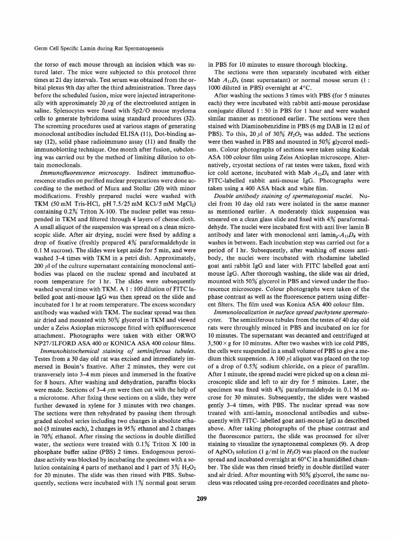
Immunofluorescencemicroscopy. Indirect immunofluo-rescence studies on purified nuclear preparations were done ac-cording to the method of Mura and Stollar (20) with minormodifications. Freshly prepared nuclei were washed withTKM (50mM Tris-HCl, pH 7.5/25 mM KC1/5 mM MgCl2)containing 0.2% Triton X-100. The nuclear pellet was resus-pended in TKMand filtered through 4 layers of cheese cloth.A small aliquot of the suspension was spread on a clean micro-scopic slide. After air drying, nuclei were fixed by adding adrop of fixative (freshly prepared 4%paraformaldehyde in0.1 Msucrose). The slides were kept aside for 5 min, and werewashed 3-4 times with TKMin a petri dish. Approximately,200 fA of the culture supernatant containing monoclonal anti-bodies was placed on the nuclear spread and incubated atroomtemperature for 1 hr. The slides were subsequentlywashed several times with TKM. A 1 : 100 dilution ofFITC la-belled goat anti-mouse IgG was then spread on the slide andincubated for 1 hr at room temperature. The excess secondaryantibody was washed with TKM.The nuclear spread was thenair dried and mounted with 50% glycerol in TKMand viewedunder a Zeiss Axioplan microscope fitted with epifluorescenceattachment. Photographs were taken with either ORWONP27/ILFORD ASA 400 or KONICA ASA 400 colour films.Immunohistochemical staining of seminiferous tubules.Testes from a 30 day old rat was excised and immediately im-mersed in Bouin's fixative. After 2 minutes, they were cuttransversely into 3-4 mmpieces and immersed in the fixativefor 8 hours. After washing and dehydration, paraffin blockswere made. Sections of 3-4 //m were then cut with the help ofa microtome.After fixing these sections on a slide, they werefurther dewaxed in xylene for 3 minutes with two changes.The sections were then rehydrated by passing them throughgraded alcohol series including two changes in absolute etha-nol (3 minutes each), 2 changes in 95% ethanol and 2 changesin 10%ethanol. After rinsing the sections in double distilledwater, the sections were treated with 0.1% Triton X 100 inphosphate buffer saline (PBS) 2 times. Endogenous peroxi-dase activity was blocked by incubating the specimen with a so-lution containing 4 parts of methanol and 1 part of 3% H2O2for 20 minutes. The slide was then rinsed with PBS. Subse-quently, sections were incubated with \% normal goat serum
in PBSfor 10 minutes to ensure thorough blocking.The sections were then separately incubated with eitherMabAuD^(neat supernatant) or normal mouse serum (1 :1000 diluted in PBS) overnight at 4°C.After washing the sections 3 times with PBS(for 5 minuteseach) they were incubated with rabbit anti-mouse peroxidaseconjugate diluted 1 : 50 in PBS for 1 hour and were washedsimilar manneras mentioned earlier. The sections were thenstained with Diaminobenzidine in PBS (6 mg DABin 12 ml ofPBS). To this, 20[A of 30% H2O2 was added. The sectionswere then washed in PBS and mounted in 50% glyeerol medi-um. Colour photographs of sections were taken using KodakASA100 colour film using Zeiss Axioplan microscope. Alter-natively, cryostat sections of rat testes were taken, fixed withice cold acetone, incubated with MabA\\D^ and later withFITC-labelled rabbit anti-mouse IgG. Photographs weretaken using a 400 ASAblack and white film.Double antibody staining of spermatogonial nuclei. Nu-clei from 10 day old rats were isolated in the same manneras mentioned earlier. A moderately thick suspension wassmeared on a clean glass slide and fixed with 4%paraformal-dehyde. The nuclei were incubated first with anti liver lamin Bantibody and later with monoclonal anti laming-^nZ^ withwashes in between. Each incubation step was carried out for aperiod of 1 hr. Subsequently, after washing off excess anti-body, the nuclei were incubated with rhodamine labelledgoat anti rabbit IgG and later with FITC labelled goat antimouse IgG. After thorough washing, the slide was air dried,mounted with 50% glyeerol in PBS and viewed under the fluo-rescence microscope. Colour photographs were taken of thephase contrast as well as the fluorescence pattern using differ-ent filters. The film used was Konica ASA400 colour film.Immunolocalization in surface spread pachytene spermato-cytes. The seminiferous tubules from the testes of 40 day oldrats were throughly minced in PBS and incubated on ice for10 minutes. The supernatant was decanted and centrifuged at3,500 x g for 10 minutes. After two washes with ice cold PBS,the cells were suspended in a small volume of PBSto give a me-dium thick suspension. A 100 {A aliquot was placed on the topof a drop of 0.5% sodium chloride, on a piece of parafilm.After 1 minute, the spread nuclei were picked up on a clean mi-croscopic slide and left to air dry for 5 minutes. Later, thespecimen was fixed with 4% paraformaldehyde in 0.1 M su-crose for 30 minutes. Subsequently, the slides were washedgently 3-4 times, with PBS. The nuclear spread was nowtreated with anti-laming monoclonal antibodies and subse-quently with FITC- labelled goat anti-mouse IgG as describedabove. After taking photographs of the phase contrast andthe fluorescence pattern, the slide was processed for silverstaining to visualize the synaptonemal complexes (9). A dropof AgNC>3solution (1 g/ml in H2O) was placed on the nuclearspread and incubated overnight at 60°C in a humidified cham-ber. The slide was then rinsed briefly in double distilled waterand air dried. After mounting with 50% glyeerol, the same nu-cleus was relocated using pre-recorded coordinates and photo-
209
K. Manjula, A. Karande and M.R.S. Rao
graphed.Analytical procedures. Protein estimations were done
either by the method of Lowry et al. (18) or turbidometry(21).
RESULTS
Monoclonal antibodies against laming and theircross-reactivity with somatic lamins. A previous com-munication from our laboratory had shown that thelaming which is the sole component of the lamina struc-ture of round spermatids, persists through spermiogene-sis process and is even present in the epididymal spermnucleus (28). Laming was a component of the DNase 1and high salt resistant fraction of sonication resistantnuclei (representing elongating and elongated spermatidnuclei). Although we used the same lamina isolationprocedure to obtain this fraction, we have called it asthe 'DNase 1 and high salt resistant fraction' since it isnot clear as to what type of structure the lamina is trans-
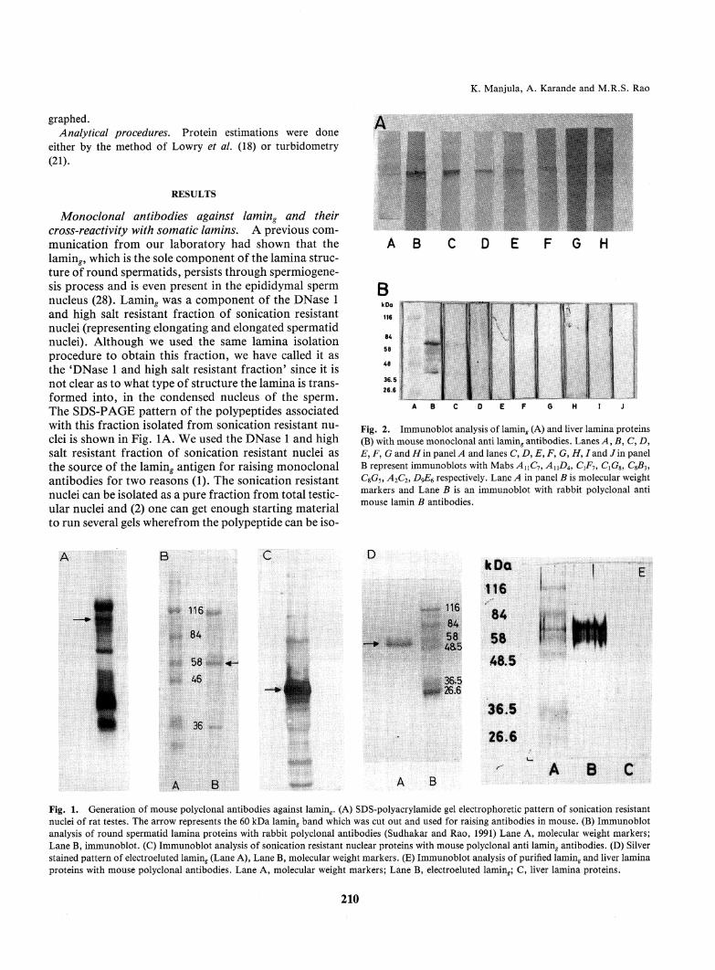
formed into, in the condensed nucleus of the sperm.The SDS-PAGEpattern of the polypeptides associatedwith this fraction isolated from sonication resistant nu-clei is shown in Fig. 1A. Weused the DNase 1 and high
salt resistant fraction of sonication resistant nuclei asthe source of the laming antigen for raising monoclonalantibodies for two reasons (1). The sonication resistantnuclei can be isolated as a pure fraction from total testic-ular nuclei and (2) one can get enough starting materialto run several gels where from the polypeptide can be iso-
Fig. 2. Immunoblot analysis of lamin^ (A) and liver lamina proteins(B) with mouse monoclonal anti laming antibodies. Lanes A, B, C, D>E, F, G andH'm panelA and lanes C, D, E, F, G, H, /and Jin panelB represent immunoblots with Mabs AUC7, AnD4, CiF7, CXG%,CSB3,CSG5, A2C2, D^E6 respectively. Lane A in panel B is molecular weightmarkers and Lane B is an immunoblot with rabbit polyclonal antimouse lamin B antibodies.
Fig. 1. Generation of mousepolyclonal antibodies against laming. (A) SDS-polyacrylamide gel electrophoretic pattern of sonication resistantnuclei of rat testes. The arrow represents the 60 kDa laminf band which was cut out and used for raising antibodies in mouse. (B) Immunoblot
analysis of round spermatid lamina proteins with rabbit polyclonal antibodies (Sudhakar and Rao, 1991) Lane A, molecular weight markers;Lane B, immunoblot. (C) Immunoblot analysis of sonication resistant nuclear proteins with mouse polyclonal anti lamin^ antibodies. (D) Silverstained pattern of electroeluted laming (Lane A), Lane B, molecular weight markers. (E) Immunoblot analysis of purified laming and liver laminaproteins with mouse polyclonal antibodies. Lane A, molecular weight markers; Lane B, electroeluted laming C, liver lamina proteins.
210

GermCell Specific Lamin during Rat Spermatogenesis
lated by electroelution after staining with KC1.Wehave tried several protocols for immunizing the
mouse and found that subdermal placement of the gelband gave the best immune response. Fig. 1C shows theresults of an immunoblot analysis of mouse polyclonalanti-lamin^ antibodies in comparison with rabbit poly-clonal anti-lamin^ antibodies, shown in Fig. IB, whichclearly indicates the efficiency of the immunization pro-tocol we have used. The specificity of antigen recogni-tion of the mousepolyclonal antibodies was then com-pared between (a) purified lamin^ (isolated by electroelu-tion from KC1stained bands, the purity of which isshown in Fig. ID) and (b) liver lamina proteins. The im-munoblot pattern shown in Fig. IE clearly shows thatthe antibodies are highly reactive with lamin^. Two veryfaint bands lighted up with the liver lamina proteins.
The upper band corresponds to lamin B while the bot-tom band may be either lamin C or a partially degradedlamin B itself. It may be worth mentioning here thateven the rabbit anti-lamin^ polyclonal antibodies also
showed a very faint cross reactivity with liver lamin B(27). In fact, we had inferred that laming may be a vari-ant form of lamin B based on the iodinated tryptic mapanalysis.
After confirming the presence of anti-lamin^ anti-
bodies in the immunizedmouse serum, we then pro-ceeded to construct hybridoma cell lines and generatespecific monoclonal antibodies. Eight monoclonal anti-bodies were obtained after various screening proceduresand each of them was then finally checked for its reactiv-ity with laming by immunoblotting. The results of suchan analysis is shown in Fig. 2A. It is clear that all the
Fig. 3A. Indirect immunofluorescence analysis of pachytene spermatocyte nuclei with anti lamin^ Mabs, AB(AnC7), CD(AnD^), EFiC^j),GH(CXG%),IJ(C8B3), KL(CSC5), MN(A2C2)and OPiD^). ACEGIKMOrepresent phase contrast micrographs while BDFHJLNPrepresent thefluorescence pattern, x 1,000.
211

K. Manjula, A. Karande and M.R.S. Rao
eight monoclonal antibodies reacted specifically withthe laming polypeptide, among with AnD4, CXF1 and
QG8 showed strong reactions. The differences in the in-tensity of reaction observed in the immunoblot analysismay reflect differences in the affinities of the monoclon-al antibodies with the antigen. Occasionally, there wasalso a reaction with a band just below the 60 kDa lamin^band. Since this was not reproducible in all the immuno-blots, it may probably represent partially degraded lam-in^. All the monoclonal antibodies were then checked
for their cross-reactivity with the liver lamina proteins.The results of these immunoblotting studies are shownin Fig. 2B. As a positive control, we have used rabbit an-ti-mouse lamin B polyclonal antibodies (courtesy of Dr.Veena Parnaik, CCMB,Hyderabad) which is shown inlane B. Amongthe monoclonals, only AxXDAand ^4nC7showed a very faint reaction with somatic lamin B. Ithas been observed that the strongest epitope in lamin B
Sg
Sg
RS
RS
Fig. 3B. Indirect immunofluorescence analysis of spermatogonia
and round spermatid nuclei withAnC7 (A, B, E and F) andAnD4 (C,D, G and H) and laming Mabs. A, C, E and G represent phase con-trast micrographs while B, D, F and H represent the fluorescence pat-tern, x 1,000.
lies in a 46 kDa chymotryptic fragment of lamin B (3).Since the two monoclonals AnD4and AnC7showedonly a faint reaction with lamin B, it is possible that thisepitope is extensively modified in laming. The observa-tion that the other six monoclonals did not react withlamin B at all, indicates probably that they might recog-nize unique epitope(s) in lamin^ which are absent in lam-inB.
Immunofluorescencestudies on the localization pat-tern of laming in germ cells using Mabs. Earlier, wehad made a detailed study on the localization of lamin^in spermatogonia, spermatocytes and round spermatidsby the indirect immunofluorescence technique using rab-bit polyclonal antibodies. Now, since we have obtained8 different monoclonals recognizing the lamin^ antigen,we have repeated the indirect immunofluorescencestudies with each of the monoclonals to get more insighton the pattern of localization of this antigen in the sper-matogenic cells. Fig. 3A shows the fluorescence patternobtained with pachytene spermatocyte nuclei. Amongthe 8 Mabs, AnCl9 AnD4 and CiG8 showed the strong-est fluorescence pattern. The fluorescence pattern isvery similar to the ones we have reported earlier usingpolyclonal antibodies in that it is patchy in appearancewith a discontinuous fluorescence pattern around thenuclear periphery. Furthermore, the strongest fluores-cence within the nucleus was localized to the phasedense regions. The fluorescence pattern observed withMabsCiF7 and A2C2was much less intense as comparedto the ones observed with AUC7,AnD4and CiG8. Onthe other hand, there was negligible fluorescence ob-served with Mabs C$B3, C%C5and D^E^. It is interestingto note here that although all the 8 Mabs showed reac-tion with the lamin^ antigen in the immunoblot analysis
A B
C DFig. 4. Indirect immunofluorescence pattern of liver nuclei with
anti laminf Mabs, AnC7(A, B) and AnDA(C, D). A and C representphase contrast micrographs while B and D represent the fluorescence
pattern, x 1,000.
212
Germ Cell Specific Lamin during Rat Spermatogenesis
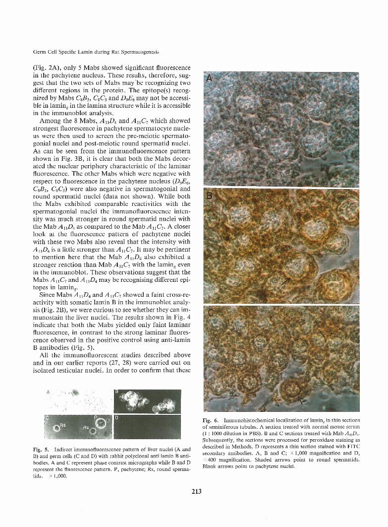
(Fig. 2A), only 5 Mabs showed significant fluorescencein the pachytene nucleus. These results, therefore, sug-gest that the two sets of Mabsmay be recognizing twodifferent regions in the protein. The epitope(s) recog-nized by Mabs C853, CSC5 and DgE6 may not be accessi-ble in laming in the lamina structure while it is accessiblein the immunoblot analysis.Amongthe 8 Mabs, AnD4 and AnC7 which showedstrongest fluorescence in pachytene spermatocyte nucle-us were then used to screen the pre-meiotic spermato-gonial nuclei and post-meiotic round spermatid nuclei.As can be seen from the immunofluorescence patternshown in Fig. 3B, it is clear that both the Mabs decor-ated the nuclear periphery characteristic of the laminarfluorescence. The other Mabs which were negative withrespect to fluorescence in the pachytene nucleus (DgE^,Cg53, CsCs) were also negative in spermatogonial andround spermatid nuclei (data not shown). While boththe Mabs exhibited comparable reactivities with thespermatogonial nuclei the immunofluorescence inten-sity was much stronger in round spermatid nuclei withthe Mab AnD4as compared to the Mab AnC7. A closerlook at the fluorescence pattern of pachytene nucleiwith these two Mabsalso reveal that the intensity withAnD4is a little stronger than AuC-j. It may be pertinentto mention here that the MabAnD4also exhibited astronger reaction than Mab AnC-, with the laming evenin the immunoblot. These observations suggest that theMabs A\\C-i and AUDAmay be recognising different epi-topes in laming.Since Mabs AUD4and AnC-, showed a faint cross-re-activity with somatic lamin B in the immunoblot analy-sis (Fig. 2B), we were curious to see whether they can im-munostain the liver nuclei. The results shown in Fig. 4indicate that both the Mabs yielded only faint laminarfluorescence, in contrast to the strong laminar fluores-cence observed in the positive control using anti-laminB antibodies (Fig. 5).
All the immunofluorescent studies described aboveand in our earlier reports (27, 28) were carried out onisolated testicular nuclei. In order to confirm that these
Fig. 5. Indirect immunofluorescence pattern of liver nuclei (A andB) and germ cells (C and D) with rabbit polyclonal anti lamin B anti-bodies. A and C represent phase contrast micrographs while B and Drepresent the fluorescence pattern. P, pachytene; Rs, round sperma-tids. x1,000.
Fig. 6. Immunohistochemical localization of laming in thin sectionsof seminiferous tubules. A section treated with normal mouse serum(1 : 1000 dilution in PBS). B and C sections treated with Mab AnD4.Subsequently, the sections were processed for peroxidase staining asdescribed in Methods. D represents a thin section stained with FITCsecondary antibodies. A, B and C; x 1,000 magnification and D,x 400 magnification. Shaded arrows point to round spermatids.
Blank arrows point to pachytene nuclei.
213
K. Manjula, A. Karande and M.R.S. Rao
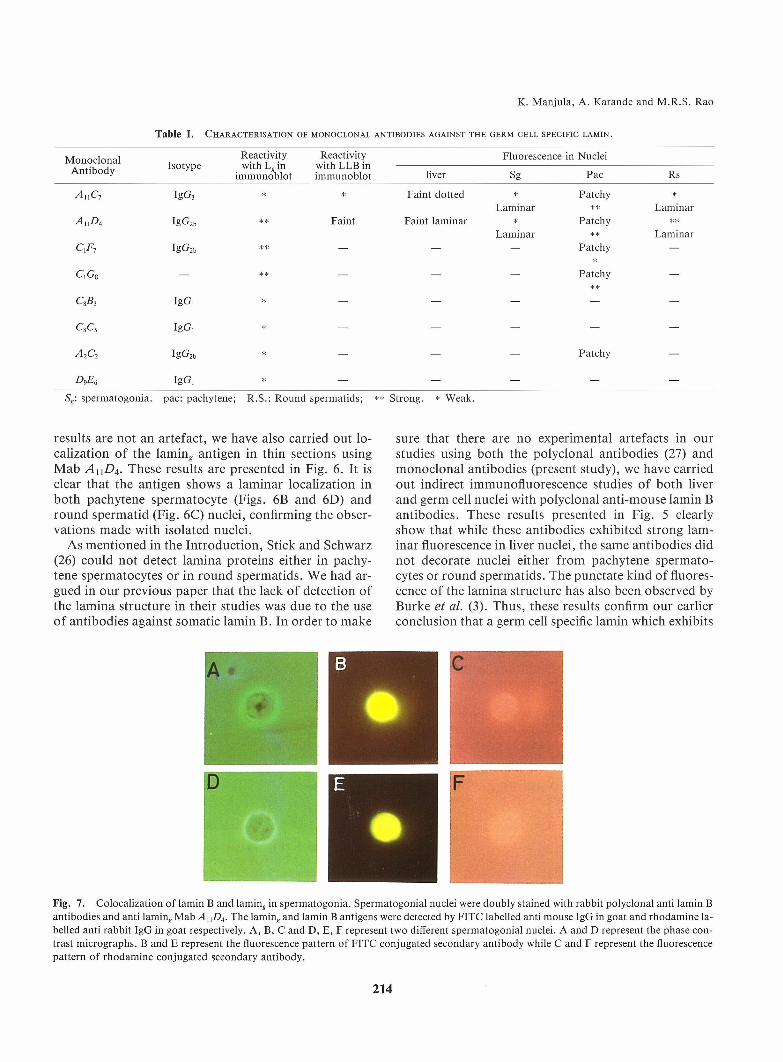
Table I. Characterisation of monoclonal antibodies against the germ cell specific lamin.
Mr.n r.rl r.na l Reactivity Reactivity Fluorescence in Nucleia t°h T Isotype with L.in with LLBinAntlDoay immunoblot immunoblot liver Sg Pac Rs
A U C7 IgG3 * * Faint dotted * Patchy *Laminar ** Laminar
A nD4 IgG2b ** Faint Faint laminar * Patchy **Laminar ** Laminar
C,F7 Ig G2b * * - - - P at chy -*
Ci Gs - ** - - - Patchy -
CSB, Ig G , * - - - - -
CSC5 IgG, * - - - - -
A2C2 IgG2b * - - - Patchy -
DgE6 Ig G , * - - - - -
Sg: spermatogonia. pac: pachytene; R.S.: Round spermatids; ** Strong. * Weak.
results are not an artefact, we have also carried out lo-calization of the laming antigen in thin sections usingMab AnD4. These results are presented in Fig. 6. It isclear that the antigen shows a laminar localization inboth pachytene spermatocyte (Figs. 6B and 6D) andround spermatid (Fig. 6C) nuclei, confirming the obser-vations madewith isolated nuclei.As mentioned in the Introduction, Stick and Schwarz(26) could not detect lamina proteins either in pachy-tene spermatocytes or in round spermatids. Wehad ar-gued in our previous paper that the lack of detection ofthe lamina structure in their studies was due to the useof antibodies against somatic lamin B. In order to make
sure that there are no experimental artefacts in ourstudies using both the polyclonal antibodies (27) andmonoclonal antibodies (present study), we have carriedout indirect immunofluorescence studies of both liverand germ cell nuclei with polyclonal anti-mouse lamin Bantibodies. These results presented in Fig. 5 clearlyshow that while these antibodies exhibited strong lam-inar fluorescence in liver nuclei, the same antibodies didnot decorate nuclei either from pachytene spermato-cytes or round spermatids. The punctate kind of fluores-cence of the lamina structure has also been observed byBurke et al. (3). Thus, these results confirm our earlierconclusion that a germ cell specific lamin which exhibits
Fig. 7. Colocalization of lamin B and lamine in spermatogonia. Spermatogonial nuclei were doubly stained with rabbit polyclonal anti lamin Bantibodies and anti laminf Mab AnD4. The laming and lamin B antigens were detected by FITC labelled anti mouse IgG in goat and rhodamine la-belled anti rabbit IgG in goat respectively. A, B, C and D, E, F represent two different spermatogonial nuclei. A and D represent the phase con-trast micrographs. B and E represent the fluorescence pattern of FITC conjugated secondary antibody while C and F represent the fluorescencepattern of rhodamineconjugated secondary antibody.
214

Germ Cell Specific Lamin during Rat Spermatogenesis
Fig. 8. Localization of lamin^ antigen in surface spread spermato-cytes. Surface spread spermatocytes were prepared as described inMethods. The microscope slide was first treated with MabAnD4andFITC-conjugated secondary antibody and visualized for the fluores-cence pattern (B). Subsequently the spread was processed for AgNO3staining to visualize the S.Cs (C) A, phase contrast micrograph. Thearrow in the phase contrast micrograph indicates two phase dense re-gions corresponding to the two telomeric ends of the chromosomesasis evident from the Ag-stained pattern in (C). The arrow in C pointsto the dense body which is also immunostained with MabAnD4.
very little cross-reactivity with somatic lamin B is pre-sent in meiotic and post-meiotic germ cells.A summaryof the observations made with all the 8Mabswith respect to the fluorescence pattern and theimmunoblot analysis of laming antigen is presented inTable I. The main conclusions from these studies agreevery well with those drawn from studies done with poly-clonal antibodies madein our previous study. Namely,(a) lamin^ antigen is a part of the lamina structure in thepre-meiotic germ cells, (b) As the germ cells enter meio-tic prophase, the laming antigen is retained in the pachy-tene nucleus even though the lamina structure has disas-sembled and (c) following meiotic division, the laminastructure is reformed using lamin^ as the only laminacomponent.Co-localization of lamin B and laming in the sper-matogonial cells. In the model proposed by us in ourprevious paper (27), we had speculated that both the so-matic lamins and lamin,, coexist in the nuclei of pre-mei-
otic spermatogonial cells. Uponentry into the meioticprophase, only lamin^ is retained and the same is relocal-ized into the lamina structure in round spermatids.However, we had not provided evidence for the coexist-ence of the lamin B and laming within the same nucleus.Nowsince wehave shownin this paper that anti-laminB antibodies do not react with laming (Fig. 5) and anti-lamin^ antibodies have negligible cross- reactivity withlamin B (Fig. 2B and Fig. 4), we have carried out doubleantibody labelling of spermatogonial nuclei. For thispurpose, nuclei were isolated from the tests of 12 dayold rats (containing predominantly spermatogonia anda few preleptotene spermatocytes) and then subjected todouble antibody labelling as mentioned in Methods.The lamin B was followed by Rhodamine labelled antirabbit IgG in goat and lamin^ by FITC labelled anti-mouse IgG in goat as secondary antibodies. The immu-nofluorescence pattern shown in Fig. 7 shows that bothlamin B and lamin^ coexist within the same spermato-gonial nucleus. A more confirmatory evidence for thiscolocalization needs to be obtained by immunoelectron-microscopic studies.
Localization of laming in surface spread pachytenespermatocytes. An event concomittant to the disas-sembly process of the nuclear lamina structure duringmeiotic prophase is the appearance of synaptonemalcomplexes (SC). The laming antigen was coisolated withsynaptonemal complexes, from rat pachytene spermato-cytes (27). However, the fluorescence patternin the pach-ytene nucleus did not indicate that it is localized toeither lateral elements or central elements of SCs. Boththe immunofluorescence and preliminary immuno-elec-tron microscopic studies showed it to be localized at thephase dense regions within the nucleus. These regionswere speculated to correspond to the attachment plaqueregions of the telomeric ends of the chromosomesthrough which they are attached to the nuclear mem-brane. To gain a further insight into the localization oflaming antigen, we have carried out immunolocalizationstudies in surface spread pachytene spermatocytes. Forthis purpose, testicular germ cells were given a mild hy-potonic treatment (0.5% NaCl) and then fixed with 4%paraformaldehyde. The spread was first reacted withAnD4 Mab and subsequently with FITC labelled goatanti-mouse IgG. The fluorescence pattern was recordedand then the sameslide was processed for silver stainingto visualize the SCs. A similar technique has been usedby Dresser and Moses (4). Fig. 8 shows the results ofsuch an experiment. Again, the fluorescence is observedat phase dense regions (Fig. 8B) in addition to a discon-tinuous fluorescence along the periphery of the nucleus.Froma comparison of the phase contrast microgragh(Fig. 8A) with that of the silver stained micrograph(Fig. 8C), it is very clear that the phase dense regionscorrespond to the telomeric ends of the paired homolo-
215

K. Manjula, A. Karande and M.R.S. Rao
gous chromosomes. The fluorescence is also observed atthe "dense body" region. The significance of these re-sults is discussed below.
DISCUSSION
The present investigation is a continuation of ourstudies on the immunological characterization of thegerm cell specific lamin^. In our previous study usingrabbit polyclonal antibodies, we had made several in-teresting observations (28). They were (a) lamin^ is re-tained in the pachytene spermatocytes even though thelamina structure is believed to be absent in these meioticprophase cells (26), (b) the lamina structure of haploidgametes is made up solely of this lamin^ and (c) lamin^is related but not identical to somatic lamin B. To gainfurther insight into the nature of laming protein, wehave produced and characterized 8 monoclonal anti-bodies. Although all these monoclonal antibodies re-acted strongly with laming in an immunoblot analysis,based on their reactivity with the antigen in immunoflu-orescence studies, they can be classified into 3 groups:(a) those showing a strong fluorescence (AnD4, AnCl9QG8); (b) those showing a medium intensity of fluores-cence (CiF7, A2C2); and (c) those showing no fluores-cence at all in the germ cells (C8£3, CSC5, D^E^. It is in-teresting to note that the monoclonal antibodies show-ing a strong fluorescence with germ cells, showed a veryfaint reaction with the laminar structure of liver nu-clei (Fig. 4) and also with lamin B in an immunoblotanalysis (Fig. 2B). These observations, in addition tostrengthening our above conclusion (c), also suggestthat there is only a small degree of homology in the epi-tope being recognized by these monoclonal antibodiesin laming and lamin B. The epitopes recognised by theother two sets of monoclonal antibodies (b and c) maybe unique to lamin^ which are absent in lamin B. Thedifferent levels of fluorescence generated by the threesets of monoclonal antibodies may reflect the differen-tial accessibility of the respectivey epitopes in germcells.
The lack of cross-reactivity, or, in some cases a lowlevel of cross-reactivity of the anti-lamin^ monoclonalantibodies with lamin B is rather intriguing, particular-ly considering the fact that somatic lamins A, B and Care immunologically related as reported by Raymondand Gagnon (23). These workers have obtained 2 mono-clonal antibodies (34B6 and 55B3) which reacted withall the three lamins A, B and C while another (36C2) re-acted only with lamins A and C. One of the 2 monoclon-al antibodies 31B5 reacted specifically with lamin Bonly. Using an IgM monoclonal antibody, Burke et al.(3) had earlier shown that it reacts with all the three so-matic lamins and this epitope is localized to a 46 kDachymotryptic fragment generated from each of the lam-
ins. In addition to their immunological relatedness, thelamins also share antigenic determinants with intermedi-ate filament proteins (23). It may be pertinent to pointout here, that even the rabbit anti-lamin^ polyclonal an-tibodies reacted very faintly with lamin B (27). Conver-sely, the experiments described here with anti somaticlamin B antibodies revealed that they do not light upgerm cells in immunofluorescence studies (Fig. 5).
Therefore, lamin^ stands out distinctly among the lam-ina proteins and it becomes very crucial to identify andcharacterize the epitopes recognized by the anti-lamingmonoclonal antibodies that we have generated. Thesestudies are nowin progress in our laboratory.An important observation made in this study is thecolocalization of lamin B and laming in spermatogonialcells, which supports the model proposed by us (27). Al-though both the somatic lamin B and lamin^ are presentin the premeiotic germ cells, only lamin^ is selectively re-tained in the pachytene nucleus. Therefore, we believethat laming must have someunique properties that areabsent in lamin B in order to facilitate the organizationof the meiotic chromosomes. Interestingly, lamin B isconstitutively expressed in all the tissues while laminsA/C are acquired only upon tissue maturation (25). Re-cently, we have shown that lamin^ is evolutionarily con-served in the germ cells of many eukaryotic cells (28).This antigen, however was not observed in somatic cellsof these organisms, stressing the functional importanceof laming in the germ cells. It is very essential, there-fore, to comparethe sequenceorganization and molecu-lar properties of laming and lamin B. For this purposewe are presently characterizing the CDNAclone for lam-ing which should provide valuable information to an-swer manyof the questions raised above.
Another series of experiments described in the pre-sent study are concerned with the localization of lamin^in pachytene spermatocytes which included both iso-lated nuclei as well as thin sections of seminiferous tu-bules period. The results obtained with monoclonal anti-bodies confirm our earlier conclusions, namely it is lo-calized in the phase dense regions of the pachytene sper-matocyte nucleus. Our results on the double staining ofsurface spread pachytene spermatocytes with FITC andsilver have clearly shown that the phase dense regionscorrespond to the telomeric ends of the meiotic chromo-somes. A question that arises at this juncture is whylam-in^ is retained in the pachytene nucleus when there isno lamina structure. However, there is one importantdifference in the organization of nuclear structure be-tween a mitotic cell and a meiotic cell particularly inmammals. Although the lamina structure is disassem-bled in both the cases, the nuclear membraneis retainedin the meiotic cell nucleus but not in the mitotic cell.Both ends of the synaptonemal complex appear to be incontact with the inner nuclear membranein manyorgan-
216

Germ Cell Specific Lamin during Rat Spermatogenesis
isms (31). Therefore, we believe that the retention oflamin^ is necessary to facilitate the attachment of the tel-pmeric ends of the chromosomes to the inner nuclearmembrane. In fact, lamin B is believed to provide theanchorage sites for the surrounding lipid bilayer mem-brane (7). The regions of attachment of the telomericends of the chromosomes to the nuclear membranehave been termed cytologically as "attachmentplaques"or "chromosomal bouquets". Recently, Klein et al. (14)have shown that RAP1(repressor activator protein 1) islocalized similarly to the telomeric ends of meiotic chro-mosomesin Saccharomyces cerevesiae using similartechnique. The functional significance of the localiza-tion of RAP1 at telomeric ends, however, is not known.In addition to the staining of the telomeric ends of thechromosomes, we have also observed a faint discontinu-ous fluorescence pattern around the nuclear periphery(Fig. 8) which was also evident in the immunohistochem-ical staining of thin sections of seminiferous tubules(Fig. 6B and 6D). Therefore we believe that there areremnants of a lamina structure in the meiotic prophasenucleus attached to the inner nuclear membrane. Re-cently Vester et al. (1993) have also observed using abroad specificity antibody, lamina structure in rat sper-matocytes involving a lamin protein that is closely re-lated to lamin B which is in conformity with our find-ings.
Recently we have also come across a paper byFurukawa and Hotta (6) describing the isolation andcharacterization of a CDNAclone for a germ cell spe-cific lamin in mouse. They have termed it as lamin B3which arises out of alternate splicing from lamin B2. Itremains to be seen whether the laming that we have de-scribed in the rat is the same protein as lamin B3 ofmouse.
Acknowledgement. This work was financially supported bygrants from the Department of Biotechnology, NewDelhi. The gener-ous gift of rabbit polyclonal anti mouse lamin B antibodies from Dr.Veena Parnaik, CCMB,Hyderabad, is gratefully acknowledged.
REFEREN CES
Behal, A., Kulkarni, P., and Rao, M.R.S. 1987. Identifica-tion of a meiotic prophase-specific nuclear matrix protein in therat. /. Biol Chem., 262: 10898-10902.
Bhown, A.S., Mole, J.E., Hunter, F., and Bennett, J.C.
1980. High sensitivity sequence determination of proteinsquantitatively recovered from sodium dodecyl sulphate gelsusing an improved electrodialysis procedure. Anal. Biochem.,103: 184-190.
Burke, B., Tooze, J., and Warren, G. 1983. A monoclonalantibody which recognises each of the nuclear lamin polypep-tides in mammalian cells. EMBOJ., 2: 361-367.Dresser, M.E. and Moses, M.J. 1980. Synaptonemal com-
plex karyotyping in spermatocytes of the Chinese hamster (Crice-tulus griseus). IV. Light and electron microscopy of synapsis
and nucleolar development by silver staining. Chromosoma,(Berl) 76: 1-22.
Dwyer, N. and Blobel, G. 1976. A modified procedure forthe isolation of a pore complex-lamina fraction from rat liver nu-
clei. /. CellBioL, 70: 581-591.Furukawa, K. and Hotta, Y. 1993. CDNAcloning of a germcell specific lamin B3 from mouse spermatocytes and analysis ofits function by ectopic expression in somatic cells. EMBOJ., 12:
97-106.
Gerace, L., Blum, A., and Blobel, G. 1978. Immunocyto-chemical localization of the major polypeptides of the nuclearpore complex lamina fraction. Interphase and mitotic distribu-
tion. /. CellBioL, 79: 546-566.Gerace, L. and Blobel, G. 1980. The nuclear envelope lami-na is reversibly depolymerized during mitosis. Cell, 19: 277-
287.
Goodpasture, C. and Bloom, S.E. 1975. Visualization of nu-cleolar organizer regions in mammalianchromosomesusing sil-
ver staining. Chromosoma, 53: 37-50.Hager, D.A. and Burgess, R.R. 1980. Elution of proteinsfrom sodium dodecyl sulphate-polyacrylamide gels, removal ofsodium dodecyl sulphate, and renaturation of enzymatic activi-ty: results with sigma subunit of escherichia coli RNApolymer-ase, wheat germ DNAtopoisomerase, and other enzymes. Anal.
Biochem., 109: 76-86.
Harlow, E. and Lane, D. 1988. Antibodies. A LaboratoryManual. Cold Spring Harbour laboratory.Hawkes, R., Niday, E., and Gordon, J. 1982. A dot-immu-nobinding assay for monoclonal and other antibodies. Anal.
Biochem., 119: 142-147.Kaufmann, S.H. 1989. Additional membersof the rat liver la-min polypeptide family. Structural and immunological charac-terization. /. Biol. Chem., 264: 13946-13955.Klein, F., Laroche, T., Cardenas, M.E., Hofmann, J.F.-X.,and Schweizer, D. 1992. Localization of RAP1 and topoiso-merase II in nuclei and meiotic chromosomes of yeast. /. Cell.
Biol, 117: 935-948.
Krohne, G. and Benavente, R. 1986. The nuclear lamins. Amultigene family of proteins in evolution and differentiation.Exp. Cell. Res., 162: 1-10.
Laemmli, U.K. 1970. Cleavage of structural proteins duringthe assembly of the head of bacteriophage T4. Nature, 227:680-685.
Lehner, C.F., Kurer, V., Eppenberger, H.M., and Nigg,E.A. 1986. The nuclear lamin protein family in higher ver-tebrates: Identification of quantitatively minor lamin proteinsby monoclonal antibodies. /. Biol. Chem., 261: 13293-13301.Lowry, O.H., Rosebrough, N.J., Farr, A.L., and Randall,R.J. 1951. Protein measurement with the folin phenol re-
agent. /. Biol. Chem., 193: 265-275.
Merril, C.R., Goldman, D., Sedman, S.A., and Ebert, M.H.1981. Ultrasensitive stain for proteins in polyacrylamide gels
shows regional variation in cerebrospinal fluid proteins. Sci-ence, 211: 1437-1438.
Mura, C.V. and Stollar, B.D. 1981. Serological detection ofhomologies of HI with H5 and HI histones. /. Biol. Chem.,
256: 9767-9769.
Platz, R.D., Meistrich, M.L., and Grimes, S.R. 1977. Lowmolecular weight basic proteins in spermatids. Methods in Cell
Biol, XVI (297).
Rao, M.R.S., Rao, B.J., and Ganguly, J. 1982. Localizationof testis variant histones on rat tesis chromatin. Biochem. J.,205: 15-21.
217
K. Manjula, A. Karande and M.R.S. Rao
Raymond, Y. and Gagnon, G. 1988. Lamin B shares a num-ber of distinct epitopes with lamins A and C with intermediate
filament proteins. Biochem., 27: 2597-2603.
Singh, J. and Rao, M.R.S. 1987. Chromatin organization ofsonication-resistant spermatid nuclei of rat testes. Ind. J. Bio-chem. Biophys., 24: 181-188.
Stewart, C. and Burke, B. 1987. Teratocarcinoma stem cellsand early mouse embryos contain only a single major lamin poly-peptide closely resembling lamin B. Cell, 51: 383-392.Stick, R. and Schwarz, H. 1982. The disappearance of thenuclear lamina during spermatogenesis: Anelectron microscop-ic and immunofluorescence study. Cell Differ., ll: 235-243.Sudhakar, L. and Rao, M.R.S. 1990. Stage specific changesin the localization of a germ cell specific lamin during mam-
malian spermatogenesis. /. Biol Chem., 265: 22526-22532.
Sudhakar, L., Sivakumar, N., Behal, A., and Rao, M.R.S.1992. Evolutionary conservation of a germ cell specific laminpersisting through mammalian spermiogenesis. Exp. Cell. Res.,
198: 78-84.
29. Towbin, H., Staehelin, T., and Gordon, J. 1979. Electro-phoretic transfer of proteins from polyacrylamide gels to nitro-cellulose sheets: Procedure and some applications. Proc. Natl.Acad. Sci. USA, 76: 4350-4354.
30. Vester, B., Smith, A., Krohne, G., and Benavente, R.1993. Presence of a nuclear lamina in pachytene spermato-cytes of the rat. /. Cell. ScL, 104: 557-563.
31. von Wettstein, D., Rasmussen, S.W., and Holm, P.B.
1984. The synaptonemal complex in genetic segregation. Ann.Rev. Genet., 18: 331-431.
32. Westerwoudt, R.J., Naipal, A.M., and Harrison, C.M.H.1984. Improved fusion technique. II stability and purity of hy-brid clones. /. Immunol. Meth., 68: 89-101.
{Received for publication, March ll, 1994and in revised form, May 12, 1994)
218