BD Stem Cell Enumeration Kit · The BD Stem Cell Enumeration kit, suff icient for 50 tests when...
31
03/2015 23-7867-04 BD, BD Logo and all other trademarks are property of Becton, Dickinson and Company. © 2015 BD Becton, Dickinson and Company BD Biosciences 2350 Qume Drive San Jose, CA 95131 USA Benex Limited Pottery Road, Dun Laoghaire, Co. Dublin, Ireland Tel +353.1.202.5222 Fax +353.1.202.5388 BD Biosciences European Customer Support Tel +32.2.400.98.95 Fax +32.2.401.70.94 [email protected] Becton Dickinson Pty Ltd, 4 Research Park Drive, Macquarie University Research Park, North Ryde NSW 2113, Australia Becton Dickinson Limited, 8 Pacific Rise, Mt. Wellington, Auckland, New Zealand bdbiosciences.com [email protected] IVD BD™ Stem Cell Enumeration Kit Catalog No. 344563
Transcript of BD Stem Cell Enumeration Kit · The BD Stem Cell Enumeration kit, suff icient for 50 tests when...

03/2015 23-7867-04
BD, BD Logo and all other trademarks are property of Becton, Dickinson and Company. © 2015 BD
Becton, Dickinson and CompanyBD Biosciences2350 Qume DriveSan Jose, CA 95131 USA
Benex LimitedPottery Road, Dun Laoghaire,Co. Dublin, IrelandTel +353.1.202.5222Fax +353.1.202.5388
BD BiosciencesEuropean Customer SupportTel +32.2.400.98.95Fax [email protected]
Becton Dickinson Pty Ltd,4 Research Park Drive,Macquarie University Research Park,North Ryde NSW 2113, Australia
Becton Dickinson Limited,8 Pacific Rise, Mt. Wellington,Auckland, New Zealand
IVD
BD™ Stem Cell Enumeration Kit
Catalog No. 344563

iii
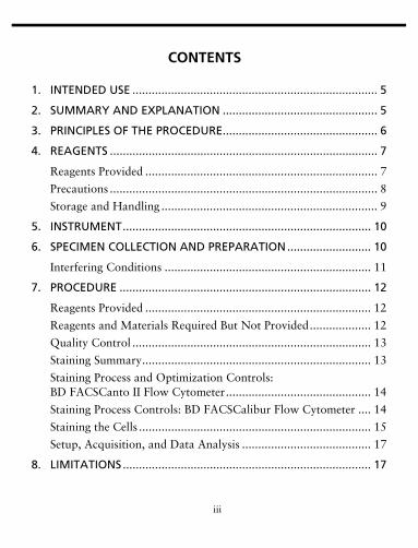
CONTENTS
1. INTENDED USE ............................................................................ 5
2. SUMMARY AND EXPLANATION ................................................ 5
3. PRINCIPLES OF THE PROCEDURE................................................ 6
4. REAGENTS ................................................................................... 7
Reagents Provided ........................................................................ 7
Precautions ................................................................................... 8
Storage and Handling ................................................................... 9
5. INSTRUMENT............................................................................. 10
6. SPECIMEN COLLECTION AND PREPARATION.......................... 10
Interfering Conditions ................................................................ 11
7. PROCEDURE .............................................................................. 12
Reagents Provided ...................................................................... 12
Reagents and Materials Required But Not Provided................... 12
Quality Control .......................................................................... 13
Staining Summary....................................................................... 13
Staining Process and Optimization Controls: BD FACSCanto II Flow Cytometer............................................. 14
Staining Process Controls: BD FACSCalibur Flow Cytometer .... 14
Staining the Cells ........................................................................ 15
Setup, Acquisition, and Data Analysis ........................................ 17
8. LIMITATIONS............................................................................. 17

iv
9. EXPECTED RESULTS................................................................... 18
Reference Intervals ..................................................................... 18
10. PERFORMANCE DATA .............................................................. 19
Agreement .................................................................................. 19
Precision ..................................................................................... 27
Linearity ..................................................................................... 28
Stability ...................................................................................... 29
WARRANTY..................................................................................... 29
REFERENCES .................................................................................... 30

5
1. INTENDED USE
The BD™ Stem Cell Enumeration (SCE) kit provides simultaneous enumeration of viable dual-positive CD45+/CD34+ hematopoietic stem cell populations in CD34+ absolute counts (cells/µL) as well as the percentage of the total viable leucocyte count that is CD34+ (%CD34). The following specimens can be analyzed with this kit: normal and mobilized peripheral blood, fresh and thawed leucopheresis products, fresh and thawed bone marrow, and fresh and thawed cord blood. The kit is intended for in vitro diagnostic (IVD) use on either a BD FACSCalibur™ flow cytometer using BD CellQuest™ or BD CellQuest™ Pro software or a BD FACSCanto™ II flow cytometer using BD FACSCanto™ clinical software.
2. SUMMARY AND EXPLANATION
Transplantation of hematopoietic progenitor cells is used increasingly in the treatment of blood disorders, malignancies, and genetic abnormalities.1-3 Progenitor cells are rare and are found primarily in the bone marrow, with extremely low frequencies in peripheral blood. However, with the arrival of mobilization regimens (G-CSF, GM-CSF, and chemotherapy), peripheral blood has become the preferred source of stem cells.1-3
The CD34 antigen is present on immature hematopoietic precursor cells and hematopoietic colony-forming cells in bone marrow and blood, including unipotent and pluripotent progenitor cells.4
An accurate measure of the CD34 cell count is necessary for dose requirement protocols in stem cell transplantation.2 An incorrectly high result could lead to an infusate with less than the recommended threshold dose of CD34+ cells. Quantitating the CD34+ cell population can also be useful during mobilization.

6
Fluorochrome-conjugated monoclonal antibodies directed against the CD34 molecule can be used to identify CD34+ cells by flow cytometry. Flow cytometric applications for CD34+ cell identification and enumeration provide a rapid, quantitative, and reproducible method to evaluate the progenitor cell population.
Significant site-to-site variation has been reported with flow cytometric methods for determining the percentages and absolute numbers of CD34+ cells.5 Single-platform flow cytometric absolute cell counting protocols have been shown to provide increased robustness of CD34 enumeration by limiting potential sources of imprecision.6 The BD Stem Cell Enumeration assay incorporates BD Trucount™ tubes to determine the absolute cell count, thereby eliminating variability associated with hematology-derived absolute counts.5 Enumeration of the cell populations in this assay is obtained using either an automated or a manual method for gating and analysis. The automated method is described in the BD Stem Cell Enumeration Application Guide for BD FACSCanto II Flow Cytometers. The manual method is described in the BD Stem Cell Enumeration Application Guide for BD FACSCalibur Flow Cytometers. The assay includes a no-wash fixative-free ammonium chloride solution for red blood cell lysis and the viability dye 7-aminoactinomycin D (7-AAD) to exclude dead cells.
3. PRINCIPLES OF THE PROCEDURE
The single-tube assay is performed by staining the sample with the reagent in individual BD Trucount tubes for absolute counts.6 When a sample is added to the reagent, the fluorochrome-labeled antibodies in the reagent bind specifically to the cell surface. Additionally, the lyophilized pellet in the BD Trucount tube dissolves, releasing a known number of fluorescent beads.
The dye 7-AAD is added to assess viability of the cells. Cells that are 7-AAD+ are not viable. Ammonium chloride is added to lyse erythrocytes before the sample is acquired on a flow cytometer.

7
During analysis of the sample, the concentration of viable CD34+ cells and viable CD45+ cells, and the percentage of viable CD34+ cells in the viable CD45+ cell population, are calculated.
4. REAGENTS
Reagents Provided
The BD Stem Cell Enumeration kit, sufficient for 50 tests when used as directed, is provided with the following reagents:
• BD Stem Cell reagent (CD45/CD34) provided in phosphate-buffered saline (PBS) containing bovine serum albumin (BSA) and 0.1% sodium azide
The reagent contains CD45 FITC, clone 2D1, and CD34 PE, clone 8G12.
• 7-AAD reagent
7-AAD is a nucleic-acid dye used to identify dead cells.
• 10X ammonium chloride lysing solution
The ammonium chloride solution is a fixative-free solution for red blood cell lysis.
• 50 BD Trucount tubes
Each single-use tube contains a freeze-dried pellet of fluorescent beads.
CD45 recognizes a 180- to 220-kilodalton (kDa) human leucocyte antigen that is a member of the leucocyte common antigen (LCA) family.7,8 The CD45 antigen is present on all human leucocytes and is weakly expressed on hematopoietic progenitor cells.

8
CD34 recognizes the class III human progenitor cell antigen (HPCA). The CD34 antigen is present on immature hematopoietic precursor cells and all hematopoietic colony-forming cells in bone marrow and blood, including unipotent and pluripotent progenitor cells.7,9-12
CD45 and CD34 antibodies are each composed of mouse IgG1 heavy chains and kappa light chains.
BD Trucount tubes are used with this reagent. By adding the reagent and the sample directly to the BD Trucount tube, the absolute count of the cell population of interest can be directly determined. See the appropriate BD Stem Cell Enumeration Application Guide for either the BD FACSCanto II or BD FACSCalibur™ flow cytometer.
Concentration values are listed in the following table:
Precautions
• For In Vitro Diagnostic use.• Do not use the reagent if you observe any change in appearance.
Precipitation or discoloration indicates instability or deterioration.• The antibody reagent contains sodium azide as a preservative;
however, take care to avoid microbial contamination, which can cause erroneous results.
• Do not decontaminate ammonium chloride lysed samples with bleach.
• To achieve an accurate result, it is critical to add a precise volume of specimen to BD Trucount tubes. Calibrate pipettes to deliver 100 µL. Use reverse pipetting or a positive displacement pipette to
Reagent Concentration (µg/mL)
CD45 12.5
CD34 10.0
7-AAD 45 – 65

9
aliquot samples. See the pipette manufacturer’s instructions for more information.
• The assigned bead count varies by the lot of BD Trucount tubes. It is critical to use the bead count shown on the current lot of BD Trucount tubes when entering this value in the software or when manually calculating absolute counts. Do not mix multiple lots of tubes in the same assay.
• BD Trucount tubes are designed for use with a specific lyse/no-wash procedure. Do not threshold on forward scatter (FSC) for data collection.
WARNING All biological specimens and materials coming in contact with them are considered biohazards. Handle as if capable of transmitting infection13,14 and dispose of with proper precautions in accordance with federal, state, and local regulations. Never pipette by mouth. Wear suitable protective clothing, eyewear, and gloves.
Storage and Handling
• Store the BD Stem Cell (CD45/CD34) reagent at 2°C–8°C. Do not use after the expiration date shown on the label. Do not freeze the reagent or expose it to direct light during storage or incubation with cells. Keep the reagent vial dry.
• Store BD Trucount tubes in their original foil pouch at 2°C–25°C. To avoid potential condensation, open the pouch only after it has reached room temperature and carefully reseal the pouch immediately after removing a tube. Examine the desiccant each time you open the pouch. If the desiccant has turned from blue to lavender, discard the remaining tubes. An unopened pouch is stable until the expiration date shown on the packaging. Do not open and use tubes after this expiration date. Use tubes within 1 hour after removal from the foil pouch. Use remaining tubes within 1 month after opening the pouch.
• Store 7-AAD at 2°C–8°C. Protect from light.

10
• Store 10X ammonium chloride at 2°C–8°C.
5. INSTRUMENT
The BD Stem Cell Enumeration kit is designed for use on either the BD FACSCanto II or the BD FACSCalibur flow cytometer.
To set the photomultiplier tube (PMT) voltages and fluorescence compensation, and to check instrument sensitivity before acquisition, use one of the following:
• For the BD FACSCanto II flow cytometer, use BD FACS™ 7-color setup beads with BD FACSCanto clinical software v2.4 for analysis. This assay requires an additional optimization step for PE–%7- AAD. See the BD Stem Cell Enumeration Application Guide for BD FACSCanto II Flow Cytometers for assay-specific setup information and for acquisition and analysis instructions.
• For the BD FACSCalibur flow cytometer, use BD Calibrite™ 3 beads and the 3-color or 4-color lyse/no-wash (LNW) assay with BD FACSComp™ software, v4.2 through v6.0. Use BD CellQuest (v3.3) or BD CellQuest Pro (v4.0.2, v5.2.1, or v6.0) software for analysis. See the BD Stem Cell Enumeration Application Guide for BD FACSCalibur Flow Cytometers for acquisition and analysis instructions.
Vortex the cells thoroughly, at low speed, to resuspend cells and beads and reduce aggregation before running them on the flow cytometer.15 Acquire data using the appropriate software.
6. SPECIMEN COLLECTION AND PREPARATION
• Follow the Clinical and Laboratory Standards Institute (CLSI) (H42-A2) guidelines for specimen storage and handling.16
• Optimally, keep undiluted specimens stored at 2°C–8°C.16,17 Labs should validate their pre-analytical sample storage conditions.
• Stain specimens within 24 hours of collection.18

11
• Store stained samples on wet ice* and acquire within 1 hour of lysing.
• The following anticoagulants have been verified for use with this assay:– EDTA, ACD-A, heparin, and CPD.– For leucopheresis, a mixture of ACD-A, heparin, and EDTA can
also be used with this assay.
A minimum of 100 µL of neat/diluted specimen is required per test.
Perform a white blood cell (WBC) count on all specimens to be evaluated. If the WBC count is greater than 40 x 103 cells/µL, dilute the specimen according to standard laboratory procedures, using PBS with 0.5% BSA.5 Use reverse pipetting to make dilutions. Record and enter the dilution factor for the calculation of the final CD34 result into either the Dilution Factor column of the worklist for BD FACSCanto clinical software with BD Stem Cell Enumeration module, v1.0, or the template for BD CellQuest or BD CellQuest Pro software. See the appropriate BD Stem Cell Enumeration Application Guide for either the BD FACSCanto II or BD FACSCalibur flow cytometer for acquisition and analysis instructions.
Interfering Conditions
Do not use previously fixed and stored specimens. Reject hemolyzed, clotted, or clumped specimens.
* Wet ice: Ice in a small amount of water. Allows better contact with the tube so that the contents chill quickly.

12
7. PROCEDURE
Reagents Provided
See Section 4, Reagents.
Reagents and Materials Required But Not Provided
• BD FACS 7-Color Setup Bead Kit (Catalog No. 335775, including BD Bead Dilution Buffer) for use on a BD FACSCanto II flow cytometer
• BD Calibrite 3 beads (Catalog No. 340486) for use on a BD FACSCalibur flow cytometer
• reagent-grade (deionized) water• BD Vacutainer® EDTA blood collection tubes or equivalent• vortex mixer• timer• ice-water bath• Falcon®* disposable 12 x 75-mm polystyrene test tubes or
equivalent• calibrated micropipettor with tips capable of delivering 20 µL• calibrated micropipettor with tips capable of delivering 100 µL• bulk dispenser or pipettor for dispensing 2 mL of ammonium
chloride lysing solution• 1X PBS (Dulbecco’s modified, pH 7.2 ±0.2) with 0.5% BSA5, if
sample dilution is necessary• BD™ Stem Cell Control kit (Catalog No. 340991)
* Falcon is a registered trademark of Corning Incorporated.
13
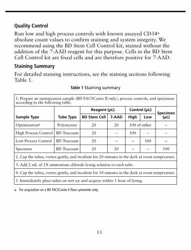
Quality Control
Run low and high process controls with known assayed CD34+ absolute count values to confirm staining and system integrity. We recommend using the BD Stem Cell Control kit, stained without the addition of the 7-AAD reagent for this purpose. Cells in the BD Stem Cell Control kit are fixed cells and are therefore positive for 7-AAD.
Staining Summary
For detailed staining instructions, see the staining sections following Table 1.
Table 1 Staining summary
1. Prepare an optimization sample (BD FACSCanto II only), process controls, and specimens according to the following table.
Sample Type Tube Type
Reagent (µL) Control (µL)Specimen
(µL)BD Stem Cell 7-AAD High Low
Optimizationa
a. For acquisition on a BD FACSCanto II flow cytometer only.
Polystyrene 20 20 100 of either –
High Process Control BD Trucount 20 – 100 – –
Low Process Control BD Trucount 20 – – 100 –
Specimen BD Trucount 20 20 – – 100
2. Cap the tubes, vortex gently, and incubate for 20 minutes in the dark at room temperature.
3. Add 2 mL of 1X ammonium chloride lysing solution to each tube.
4. Cap the tubes, vortex gently, and incubate for 10 minutes in the dark at room temperature.
5. Immediately place tubes on wet ice and acquire within 1 hour of lysing.
14
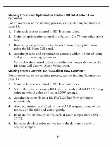
Staining Process and Optimization Controls: BD FACSCanto II Flow Cytometer
For an overview of the staining process, see the Staining Summary on page 13.
1. Stain each process control in BD Trucount tubes.
2. Stain the optimization control in a Falcon 12 x 75-mm polystyrene tube.
3. Run Setup using 7-color setup beads followed by optimization using the BD Stem Cell panel.
4. Acquire process and optimization controls within 1 hour of lysing and prior to staining specimens.
Verify that the control values are within the ranges shown on the BD Stem Cell Control Assay Values sheet.
Staining Process Controls: BD FACSCalibur Flow Cytometer
For an overview of the staining process, see the Staining Summary on page 13.
1. Stain each process control in BD Trucount tubes.
2. Set up the cytometer using BD Calibrite beads and BD FACSComp software with 3-color or 4-color LNW settings.
3. Acquire the controls on a BD FACSCalibur flow cytometer immediately.
4. After acquisition, add 20 µL of the 7-AAD reagent to one of the tubes. Cap the tube and vortex gently.
5. Incubate for 10 minutes in the dark at room temperature (20°C–25°C).
6. Immediately place tubes on wet ice in the dark until ready to acquire samples.

15
7. Run the control stained with 7-AAD reagent on a BD FACSCalibur flow cytometer within 1 hour of lysing.
Staining the Cells
1. For each patient specimen, label a BD Trucount tube with the sample identification.
NOTE Before use, verify that the BD Trucount bead pellet is under the metal retainer at the bottom of the tube. If this is not the case, discard the BD Trucount tube and replace it with another. Do not transfer beads to another tube.
2. Pipette 20 µL of the BD Stem Cell reagent into the bottom of the tube.
Pipette just above the stainless steel retainer of the BD Trucount tube. Do not touch the bead pellet.
NOTE Always change tips between tubes.
3. Pipette 20 µL of 7-AAD reagent into the tube.
4. Pipette 100 µL of a well-mixed specimen onto the side of the tube just above the retainer.
NOTE Avoid smearing the specimen down the side of the tube. If any specimen remains on the side of the tube, it will not be stained with the reagent but can be resuspended by the lysing solution and therefore can affect results.
Accurate pipetting is critical when using a BD Trucount tube. Use the reverse pipetting technique, or a positive displacement pipettor, to pipette specimen onto the side of the tube just above the retainer.
16
For reverse pipetting, depress the button to the second stop. Insert the pipettor into the specimen and release the button. When you release the button, excess specimen is drawn up into the tip. When dispensing, press the button to the first stop to expel a precise volume of specimen. This leaves excess specimen in the tip.
Always change to a new tip between tubes. Discard tips in an appropriate biohazard container.
5. Cap each tube and vortex gently to mix.
6. Incubate for 20 minutes in the dark at room temperature (20°C–25°C).
7. Add 2 mL of 1X ammonium chloride lysing solution to each tube to lyse red blood cells.
NOTE Each day, prepare enough 1X ammonium chloride lysing solution for use. To make, dilute 1 part of 10X ammonium chloride solution with 9 parts of distilled water. Store and use at room temperature (20°C–25°C).
8. Cap each tube and vortex gently to mix.
9. Incubate for 10 minutes in the dark at room temperature (20°C–25°C).
10. Immediately, place tubes on wet ice in the dark until ready to acquire samples.
Acquire samples on either a BD FACSCanto II or BD FACSCalibur flow cytometer within 1 hour of lysing. Highly manipulated or processed samples can be more susceptible to increased cell death after preparation.17
For information on setup, acquisition, and data analysis, see the BD Stem Cell Enumeration Application Guide for either the BD FACSCanto II or BD FACSCalibur flow cytometer.
17
Setup, Acquisition, and Data Analysis
For setup and acquisition on a BD FACSCanto II instrument and data analysis using BD FACSCanto clinical software with the BD Stem Cell Enumeration module, v1.0, see the BD Stem Cell Enumeration Application Guide for BD FACSCanto II Flow Cytometers.
For setup and acquisition on a BD FACSCalibur instrument and data analysis using BD CellQuest or BD CellQuest Pro software, see the BD Stem Cell Enumeration Application Guide for BD FACSCalibur Flow Cytometers.
8. LIMITATIONS
• For accuracy in determining absolute counts, it is critical that you use the reverse pipetting technique or a positive displacement pipettor to add the sample to each BD Trucount tube.
• Using an incorrect bead count from the BD Trucount tubes pouch label will result in inaccurate absolute counts.
• Erroneous results will occur if tubes are not placed immediately on wet ice in the dark until acquired or if samples are not run on a flow cytometer within 1 hour of lysing.
• Insufficient mixing of samples will result in inaccurate results.• Due to the temperature requirements of this assay, the BD FACS™
Loader cannot be used to acquire samples.• Laboratories must establish their own CD34+ viability
requirements for each specimen type.• A platelet streak might appear in the CD34-PE vs SSC dot plot
when running samples collected in heparin anticoagulant. If this occurs, see the BD Stem Cell Enumeration Application Guide for either the BD FACSCanto II or BD FACSCalibur flow cytometer for troubleshooting information.

18
9. EXPECTED RESULTS
Validation of the reference interval using the BD Stem Cell Enumeration kit was performed at BD Biosciences laboratories in San Jose, CA.
Reference Intervals
Reference intervals for peripheral blood were validated with whole blood specimens obtained from individuals from reference sample groups at BD Biosciences laboratories in San Jose, CA. See Limitations (previous section) for more information about reference intervals. Validated reference intervals are shown in Table 2.
We recommend that laboratories and other users establish their own reference intervals for their patient populations using the BD Stem Cell Enumeration kit to reflect potential sources of variability, such as patient gender, race, and age, and preparation techniques.
Table 2 Representative adult reference intervals
Measures Reported n Median SD
95% Reference Interval
Lower (90% conf. bounds)
Upper (90% conf. bounds)
CD34 absolute counts (cells/µL)
169 2.2 1.7 0.7(0.6, 0.8)
6.9(6.1, 7.9)
%CD34 cells 169 0.037 0.025 0.013(0.011, 0.014)
0.11(0.096, 0.12)

19
10. PERFORMANCE DATA
Agreement
CD34+ absolute counts were enumerated and percentages of CD34+ cells were determined with the BD Stem Cell Enumeration kit on both BD FACSCanto II and BD FACSCalibur flow cytometers and compared with results from the predicate reagent* on a BD FACSCanto II flow cytometer.
Normal and mobilized peripheral blood, leucopheresis, cord blood, and bone marrow specimens were collected from donors from four clinical laboratories.
Table 3 and Table 4 summarize the results of the BD Stem Cell Enumeration kit accuracy study on the BD FACSCanto II and the BD FACSCalibur flow cytometers, respectively. For each evaluable specimen, the differences (bias) between the values of the investigational and predicate systems for absolute viable CD34 and % viable CD34 in CD45 were calculated. The results were pooled to produce mean biases of CD34 and %CD34 for each bin, along with the 95% confidence interval (CI).
* Beckman Coulter Stem-Kit™ Reagents

20
Regression plots and statistics for viable CD34 counts and %CD34 in CD45 on the BD FACSCanto II flow cytometer are shown in Figure 1 and Figure 3. Regression plots and statistics for viable CD34 counts and %CD34 in CD45 on the BD FACSCalibur flow cytometer are
Table 3 Accuracy study results of the BD Stem Cell Enumeration kit on the BD FACSCanto II flow cytometer compared with the predicate method
Variables Bin N
Absolute DifferenceRelative Difference to
Predicate
Mean Absolute
Bias 95% CI
Mean Relative
Bias 95% CI
Viable CD34 Low 167 –0.1 (–0.5, 0.3) NA NA
Mid 496 –0.7 (–1.4, –0.04) –1.6 (–3.1, –0.2)
High 255 –1.4 (–4.3, 1.6) –1.0 (–2.4, 0.4)
%CD34 in CD45 Low 512 –0.004 (–0.01, 0.0002) –2.2 (–4.4, –0.1)
High 406 0.08 (0.04, 0.1) 2.1 (–0.1, 4.3)
Table 4 Accuracy study results of the BD Stem Cell Enumeration kit on the BD FACSCalibur flow cytometer compared with the predicate method
Variables Bin N
Absolute DifferenceRelative Difference to
Predicate
Mean Absolute
Bias 95% CI
Mean Relative
Bias 95% CI
Viable CD34 Low 156 –0.2 (–0.4, –0.05) NA NA
Mid 492 –0.5 (–1.3, 0.2) –0.9 (–2.3, 0.5)
High 257 –3.6 (–6.8, –0.3) –1.7 (–3.1, –0.2)
%CD34 in CD45 Low 487 –0.002 (–0.01, 0.001) –0.8 (–3.0, 1.3)
High 418 0.2 (0.09, 0.2) 3.2 (0.9, 5.5)

21
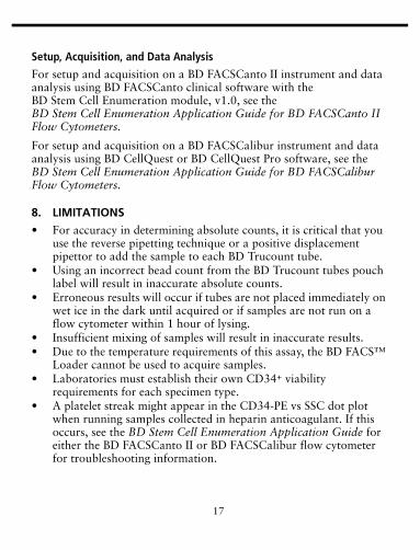
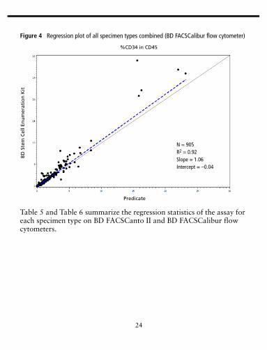
shown in Figure 2 and Figure 4. The solid line in each plot is the fitted line. The dotted line in each plot is the identity line where the predicate results are equal to the BD Stem Cell Enumeration kit results.
Figure 1 Regression plot of all specimen types combined (BD FACSCanto II flow cytometer)
N = 918R2 = 0.94Slope = 0.96Intercept = –0.15
Viable CD34 cells/µL
BD
Ste
m C
ell E
nu
mer
atio
n K
it
Predicate
22
Figure 2 Regression plot of all specimen types combined (BD FACSCalibur flow cytometer)
BD
Ste
m C
ell E
nu
mer
atio
n K
it
Viable CD34 cells/µL
Predicate
N = 905R2 = 0.95Slope = 0.97Intercept = –0.11

23
Figure 3 Regression plot of all specimen types combined (BD FACSCanto II flow cytometer)
BD
Ste
m C
ell E
nu
mer
atio
n K
it
Predicate
%CD34 in CD45
N = 918R2 = 0.92Slope = 1.05Intercept = –0.03
24
Figure 4 Regression plot of all specimen types combined (BD FACSCalibur flow cytometer)
Table 5 and Table 6 summarize the regression statistics of the assay for each specimen type on BD FACSCanto II and BD FACSCalibur flow cytometers.
BD
Ste
m C
ell E
nu
mer
atio
n K
it
Predicate
%CD34 in CD45
N = 905 R2 = 0.92Slope = 1.06Intercept = –0.04

25
Table 5 Regression statistics on BD FACSCanto II by specimen type
Specimen Type Variable N R2 Slope Intercept
Peripheral blood Viable CD34 188 (normal: 57,
mobilized: 131)
0.94 0.96 (0.93, 0.99)
–0.07 (–0.21, 0.07)
%CD34 in CD45
188(normal: 57,
mobilized: 131)
0.98 0.99 (0.93, 1.06)
0.00 (–0.01, 0.00)
Leucopheresis Viable CD34 341 (fresh: 232, frozen: 109)
0.97 0.96 (0.94, 0.98)
–0.02 (–0.2, 0.17)
%CD34 in CD45
341 (fresh: 232, frozen: 109)
0.95 0.96 (0.94, 0.99)
–0.02 (–0.03, 0.00)
Cord blood Viable CD34 241 (fresh: 124, frozen: 117)
0.88 0.97 (0.93, 1.02)
–0.52 (–0.92, -0.11)
%CD34 in CD45
241 (fresh: 124, frozen: 117)
0.87 1.02 (0.94, 1.09)
–0.02 (–0.04, 0.01)
Bone marrow Viable CD34 148 (fresh: 75, frozen: 73)
0.95 1.00 (0.96, 1.03)
–0.02 (–0.13, 0.10)
%CD34 in CD45
148 (fresh: 75, frozen: 73)
0.89 1.21 (1.13, 1.28)
–0.05 (–0.10, 0.00)

26
Table 6 Regression statistics on BD FACSCalibur by specimen type
Specimen Type Variable N R2 Slope Intercept
Peripheral blood Viable CD34 167 (normal: 52, mobilized: 115)
0.94 1.00 (0.96, 1.03)
–0.13 (–0.32, 0.05)
%CD34 in CD45
167 (normal: 52, mobilized: 115)
0.97 0.97 (0.9, 1.05)
0.00 (–0.01, 0.01)
Leucopheresis Viable CD34 342 (fresh: 232, frozen: 110)
0.97 0.98 (0.96, 0.99)
0.04 (–0.06, 0.15)
%CD34 in CD45
342 (fresh: 232, frozen: 110)
0.96 0.98 (0.96, 1.00)
–0.02 (–0.03, 0.00)
Cord blood Viable CD34 245 (fresh: 122, frozen:123)
0.94 0.96 (0.93, 0.99)
–0.28 (–0.52, -0.04)
%CD34 in CD45
245 (fresh: 122, frozen:123)
0.87 1.02 (0.92, 1.11)
–0.02 (–0.05, 0.02)
Bone marrow Viable CD34 151 (fresh: 73, frozen: 78)
0.94 0.96 (0.92, 1.00)
–0.03 (–0.09, 0.03)
%CD34 in CD45
151 (fresh:73, frozen: 78)
0.88 1.23 (1.15, 1.31)
–0.08 (–0.12, –0.03)

27
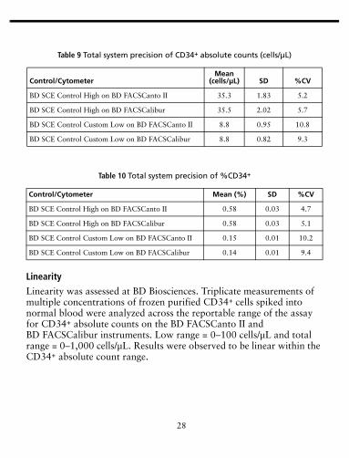
Precision
Estimates of assay precision were determined at BD Biosciences using BD SCE Control High (with a range of >10 to ≤100 cells/µL) and BD SCE Control Custom Low (with a range of ≤10 cells/µL) process controls stained in duplicate with the BD Stem Cell Enumeration kit and run on the BD FACSCanto II and BD FACSCalibur flow cytometers. Two separate runs were analyzed during each of the 21 days. Means, standard deviations (SDs), and coefficients of variation (CVs) are provided for within-run and total system precision in Table 7 through Table 10.
Table 7 Within-run precision of CD34+ absolute counts (cells/µL)
Control/CytometerMean
(cells/µL) SD %CV
BD SCE Control High on BD FACSCanto II 35.3 1.83 5.2
BD SCE Control High on BD FACSCalibur 35.5 1.69 4.8
BD SCE Control Custom Low on BD FACSCanto II 8.8 0.90 10.3
BD SCE Control Custom Low on BD FACSCalibur 8.8 0.75 8.6
Table 8 Within-run precision of %CD34+
Control/Cytometer Mean (%) SD %CV
BD SCE Control High on BD FACSCanto II 0.58 0.03 4.7
BD SCE Control High on BD FACSCalibur 0.58 0.03 4.7
BD SCE Control Custom Low on BD FACSCanto II 0.15 0.01 9.6
BD SCE Control Custom Low on BD FACSCalibur 0.14 0.01 8.2
28
Linearity
Linearity was assessed at BD Biosciences. Triplicate measurements of multiple concentrations of frozen purified CD34+ cells spiked into normal blood were analyzed across the reportable range of the assay for CD34+ absolute counts on the BD FACSCanto II and BD FACSCalibur instruments. Low range = 0–100 cells/µL and total range = 0–1,000 cells/µL. Results were observed to be linear within the CD34+ absolute count range.
Table 9 Total system precision of CD34+ absolute counts (cells/µL)
Control/CytometerMean
(cells/µL) SD %CV
BD SCE Control High on BD FACSCanto II 35.3 1.83 5.2
BD SCE Control High on BD FACSCalibur 35.5 2.02 5.7
BD SCE Control Custom Low on BD FACSCanto II 8.8 0.95 10.8
BD SCE Control Custom Low on BD FACSCalibur 8.8 0.82 9.3
Table 10 Total system precision of %CD34+
Control/Cytometer Mean (%) SD %CV
BD SCE Control High on BD FACSCanto II 0.58 0.03 4.7
BD SCE Control High on BD FACSCalibur 0.58 0.03 5.1
BD SCE Control Custom Low on BD FACSCanto II 0.15 0.01 10.2
BD SCE Control Custom Low on BD FACSCalibur 0.14 0.01 9.4

29
Stability
A study was conducted at one clinical laboratory to assess the stability of the BD™ Stem Cell Enumeration kit using leucopheresis specimens, and the following was measured:
• changes associated with the storage of whole blood before staining• changes as a result of time between staining and data acquisition• the combined effect of the two
All specimens were maintained at 2°C–8°C before staining. Based on the results of this study using leucopheresis specimens, we recommend staining specimens within 24 hours of collection, keeping stained samples on wet ice, and analyzing stained samples within 1 hour of lysing.
WARRANTY
Unless otherwise indicated in any applicable BD general conditions of sale for non-US customers, the following warranty applies to the purchase of these products.
THE PRODUCTS SOLD HEREUNDER ARE WARRANTED ONLY TO CONFORM TO THE QUANTITY AND CONTENTS STATED ON THE LABEL OR IN THE PRODUCT LABELING AT THE TIME OF DELIVERY TO THE CUSTOMER. BD DISCLAIMS HEREBY ALL OTHER WARRANTIES, EXPRESSED OR IMPLIED, INCLUDING WARRANTIES OF MERCHANTABILITY AND FITNESS FOR ANY PARTICULAR PURPOSE AND NONINFRINGEMENT. BD’S SOLE LIABILITY IS LIMITED TO EITHER REPLACEMENT OF THE PRODUCTS OR REFUND OF THE PURCHASE PRICE. BD IS NOT LIABLE FOR PROPERTY DAMAGE OR ANY INCIDENTAL OR CONSEQUENTIAL DAMAGES, INCLUDING PERSONAL INJURY, OR ECONOMIC LOSS, CAUSED BY THE PRODUCT.

30
REFERENCES1 Shpall EJ, Jones RB, Bearman SI, et al. Transplantation of enriched CD34-positive
autologous marrow into breast cancer patients following high-dose chemotherapy: influence of CD34-positive peripheral-blood progenitors and growth factors on engraftment. J Clin Oncol. 1994;12:28-36.
2 Langenmayer I, Weaver C, Buckner CD, et al. Engraftment of patients with lymphoid malignancies transplanted with autologous bone marrow, peripheral blood stem cells or both. Bone Marrow Transplant. 1995;15:241-246.
3 Zander AR, Lyding J, Bielack S. Transplantation with blood stem cells. Blood Cells. 1991;17:301-309.
4 Greaves MF, Titley I, Colman SM, et al. CD34 cluster workshop report. In: Schlossman SF, Boumsell L, Gilks W, et al, eds. Leucocyte Typing V: White Cell Differentiation Antigens. New York, NY: Oxford University Press; 1995;1:840-846.
5 Sutherland DR, Anderson L, Keeney M, Nayar R, Chin-Yee I. The ISHAGE guidelines for CD34+ cell determination by flow cytometry. J Hematotherapy. 1996;5:213-226.
6 Brocklebank AM, Sparrow RL. Enumeration of CD34+ cells in cord blood: a variation on a single-platform flow cytometric method based on the ISHAGE gating strategy. Cytometry. 2001;46:254-261.
7 Schwinzer R. Cluster report: CD45/CD45R. In: Knapp W, Dörken B, Gilks WR, et al, eds. Leucocyte Typing IV: White Cell Differentiation Antigens. New York, NY: Oxford University Press; 1989:628-634.
8 Cobbold SP, Hale G, Waldmann H. Non-lineage, LFA-1 family, and leucocyte common antigens: new and previously defined clusters. In: McMichael AJ, ed. Leucocyte Typing III: White Cell Differentiation Antigens. New York, NY: Oxford University Press; 1987:788-803.
9 Loken MR, Shah VO, Dattilio KL, Civin CI. Flow cytometric analysis of human bone marrow. II. Normal B-lymphocyte development. Blood. 1987;70:1316-1324.
10 Civin CI, Strauss LC, Brovall C, Fackler MJ, Schwartz JF, Shaper JH. Antigenic analysis of hematopoiesis. III. A hematopoietic progenitor cell surface antigen defined by a monoclonal antibody raised against KG-1a cells. J Immunol. 1984;133:157-165.
11 Siena S, Bregni M, Brando B, et al. Flow cytometry for clinical estimation of circulating hematopoietic progenitors for autologous transplantation in cancer patients. Blood. 1991;77:400-409.
12 Civin CI, Trischmann TM, Fackler MJ, et al. Report on the CD34 cluster workshop. In: Knapp W, Dörken B, Gilks WR, et al, eds. Leucocyte Typing IV: White Cell Differentiation Antigens. New York, NY: Oxford University Press; 1989:818-825.
13 Protection of Laboratory Workers from Occupationally Acquired Infections; Approved Guideline—Third Edition. Wayne, PA: Clinical and Laboratory Standards Institute; 2005. CLSI document M29-A3.

31
14 Centers for Disease Control. Perspectives in disease prevention and health promotion update: universal precautions for prevention of transmission of human immunodeficiency virus, hepatitis B virus, and other bloodborne pathogens in health-care settings. MMWR. 1988;37:377-388.
15 Jackson AL, Warner NL. Preparation, staining, and analysis by flow cytometry of peripheral blood leukocytes. In: Rose NR, Friedman H, Fahey JL, eds. Manual of Clinical Laboratory Immunology. 3rd ed. Washington, DC: American Society for Microbiology; 1986:226-235.
16 Enumeration of Immunologically Defined Cell Populations by Flow Cytometry; Approved Guideline—Second Edition. Wayne, PA: Clinical and Laboratory Standards Institute; 2007. CLSI document H42-A2.
17 Antonenas V, Garvin F, Webb M, Sartor M, Bradstock KF, Gottlieb D. Fresh PBSC harvests, but not BM, show temperature-related loss of CD34 viability during storage and transport. Cytotherapy. 2006;8:158-165.
18 Keeney M, Brown W, Gratama J, Papa S, Lanza F, Sutherland DR. Single platform enumeration of viable CD34pos cells. J Biol Regul Homeost Agents. 2003;17:247-253.


![l>lf·· E ·B; -I,:,C-·-1·1V · cat. no.i bd lj.657 bd lj.6]5 bd 4630 bd 4·627 bd 4628 bd 4886 bd 4546 bd 4·545 bd 4544 bd 4542 bd lj,588 bd lj.593 bd 0102 bd 4636 bd 4632 bd](https://static.fdocuments.in/doc/165x107/5f7c69bb7d840d18665ab1e6/llf-e-b-ic-11v-cat-noi-bd-lj657-bd-lj65-bd-4630-bd-4627-bd-4628-bd.jpg)