BCHS 6229 Protein Structure and Function - University of …nsmn1.uh.edu/yeo/doc/BCHS6229/Lecture...
46
1 BCHS 6229 Protein Structure and Function Protein Structure and Function Lecture 7 (November, 2011) From Sequence to Function (II): Sequences and Topology Structural Biology Knowledgebase
Transcript of BCHS 6229 Protein Structure and Function - University of …nsmn1.uh.edu/yeo/doc/BCHS6229/Lecture...
1
BCHS 6229
Protein Structure and FunctionProtein Structure and Function
Lecture 7 (November, 2011)
From Sequence to Function (II):
Sequences and Topology
Structural Biology Knowledgebase
2
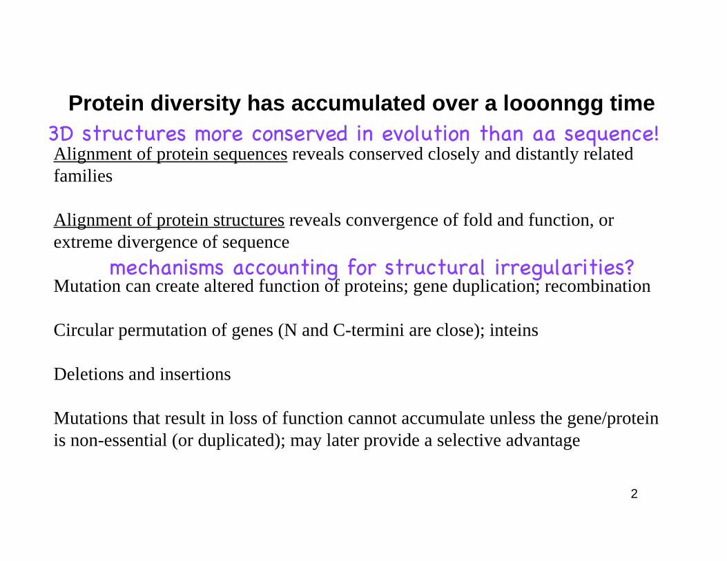
Protein diversity has accumulated over a looonngg time
Alignment of protein sequences reveals conserved closely and distantly related
families
Alignment of protein structures reveals convergence of fold and function, or
extreme divergence of sequence
Mutation can create altered function of proteins; gene duplication; recombination
Circular permutation of genes (N and C-termini are close); inteins
Deletions and insertions
Mutations that result in loss of function cannot accumulate unless the gene/protein
is non-essential (or duplicated); may later provide a selective advantage
mechanisms accounting for structural irregularities?
3D structures more conserved in evolution than aa sequence!
3
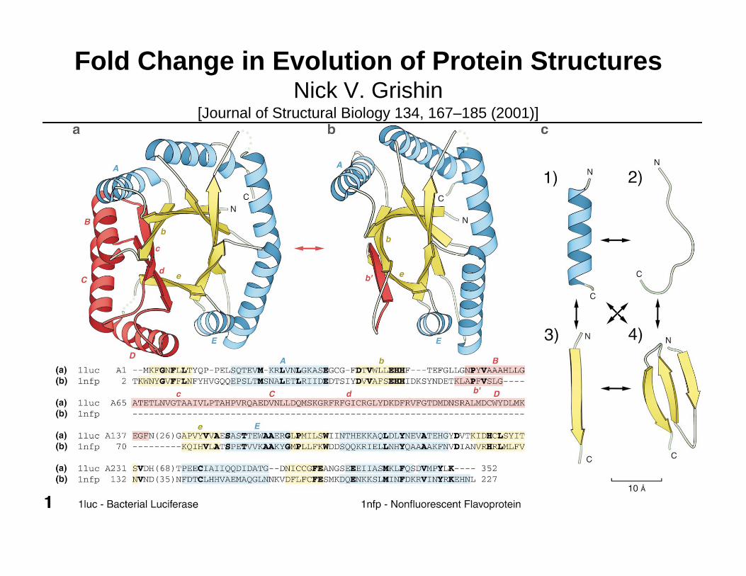
Fold Change in Evolution of Protein StructuresNick V. Grishin
[Journal of Structural Biology 134, 167–185 (2001)]
4
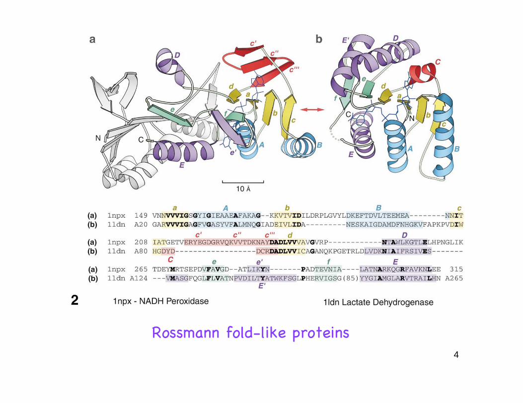
Rossmann fold-like proteins
5
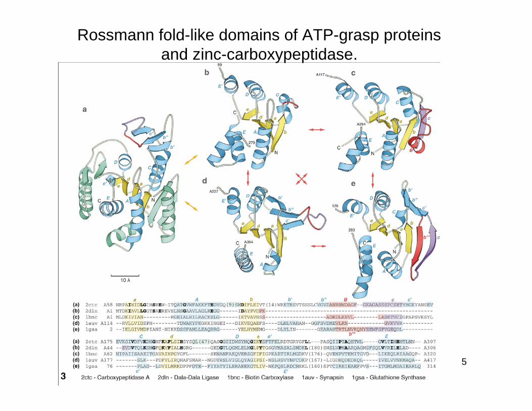
Rossmann fold-like domains of ATP-grasp proteins
and zinc-carboxypeptidase.
6
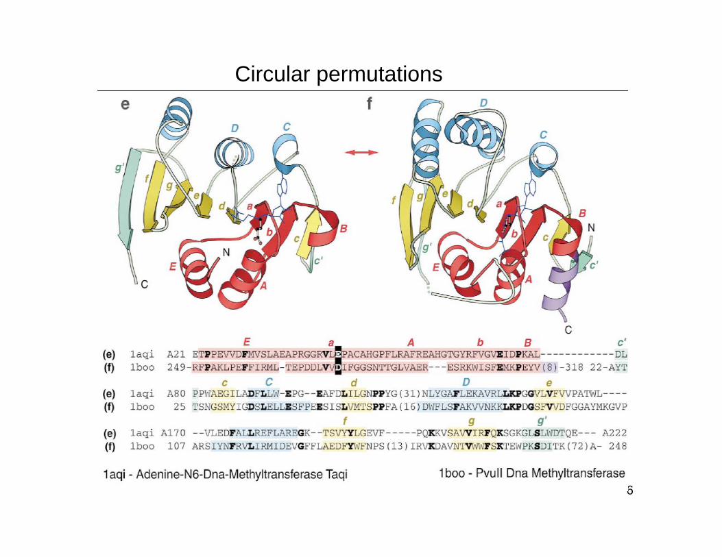
Circular permutations
7
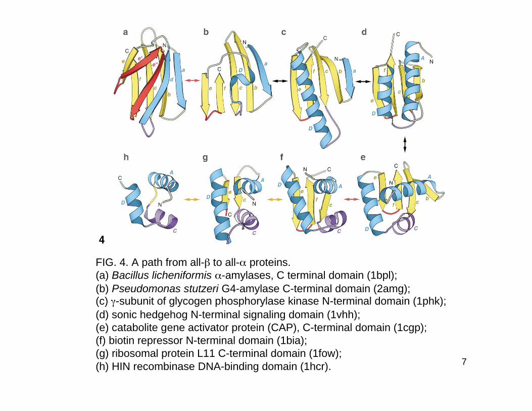
FIG. 4. A path from all- to all- proteins.
(a) Bacillus licheniformis -amylases, C terminal domain (1bpl);
(b) Pseudomonas stutzeri G4-amylase C-terminal domain (2amg);(c) -subunit of glycogen phosphorylase kinase N-terminal domain (1phk);
(d) sonic hedgehog N-terminal signaling domain (1vhh);
(e) catabolite gene activator protein (CAP), C-terminal domain (1cgp);
(f) biotin repressor N-terminal domain (1bia);
(g) ribosomal protein L11 C-terminal domain (1fow);
(h) HIN recombinase DNA-binding domain (1hcr).
8
Protein Design Principles
Need a stable hydrophobic core with constrained rotamer conformation for
individual side chains (high stability can be achieved without a well-ordered
core)
Core depends on three-dimensional arrangement of secondary structureelements and vice versa (breaking helix, over/underpacking a core)
Natural deviations in core and surface can be associated with disease states
resulted from misassociated to misfolded proteins
Amino acid diversity is ultimately required to reflect naturally occurring
proteins
Solution experiments on designed proteins can test the computational
methods
9
Topological variation in functionally diverse
enzyme superfamily
(Current Opinion in Structural Biology 2011, 21:391–397)
1. The haloalkanoic acid dehalogenase (HAD) SF: cap
domain variations enable divergent evolution of many
different reaction and substrate specificities.
2. The vicinal oxygen chelate (VOC) SF: mixing and
matching subdomains for functional versatility.
3. The thioredoxin (Trx)-fold like SFs: varied inserts and
domain additions extend the redox repertoire of the
canonical Trx-fold
10
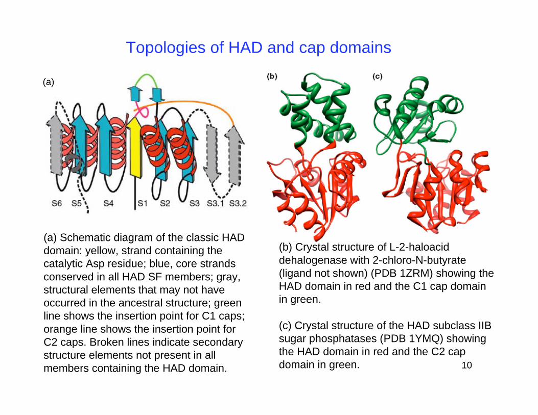
(b) Crystal structure of L-2-haloacid
dehalogenase with 2-chloro-N-butyrate
(ligand not shown) (PDB 1ZRM) showing the
HAD domain in red and the C1 cap domain
in green.
(c) Crystal structure of the HAD subclass IIB
sugar phosphatases (PDB 1YMQ) showing
the HAD domain in red and the C2 cap
domain in green.
Topologies of HAD and cap domains
(a)
(a) Schematic diagram of the classic HAD
domain: yellow, strand containing the
catalytic Asp residue; blue, core strands
conserved in all HAD SF members; gray,
structural elements that may not have
occurred in the ancestral structure; green
line shows the insertion point for C1 caps;
orange line shows the insertion point for
C2 caps. Broken lines indicate secondary
structure elements not present in all
members containing the HAD domain.
11
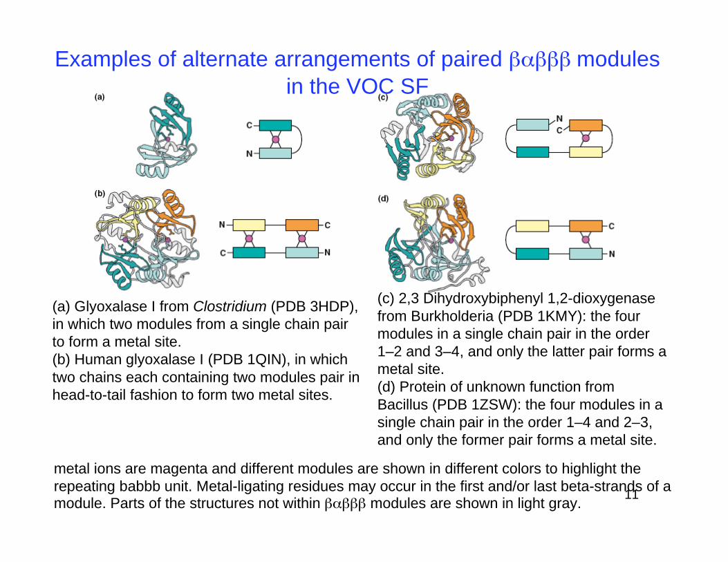
metal ions are magenta and different modules are shown in different colors to highlight the
repeating babbb unit. Metal-ligating residues may occur in the first and/or last beta-strands of amodule. Parts of the structures not within modules are shown in light gray.
Examples of alternate arrangements of paired modules
in the VOC SF
(a) Glyoxalase I from Clostridium (PDB 3HDP),
in which two modules from a single chain pair
to form a metal site.
(b) Human glyoxalase I (PDB 1QIN), in which
two chains each containing two modules pair in
head-to-tail fashion to form two metal sites.
(c) 2,3 Dihydroxybiphenyl 1,2-dioxygenase
from Burkholderia (PDB 1KMY): the four
modules in a single chain pair in the order
1–2 and 3–4, and only the latter pair forms a
metal site.
(d) Protein of unknown function from
Bacillus (PDB 1ZSW): the four modules in a
single chain pair in the order 1–4 and 2–3,
and only the former pair forms a metal site.
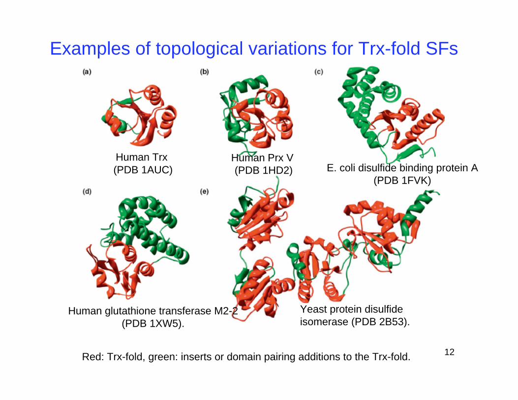
12Red: Trx-fold, green: inserts or domain pairing additions to the Trx-fold.
Human Trx
(PDB 1AUC)Human Prx V
(PDB 1HD2) E. coli disulfide binding protein A
(PDB 1FVK)
Human glutathione transferase M2-2
(PDB 1XW5).
Yeast protein disulfide
isomerase (PDB 2B53).
Examples of topological variations for Trx-fold SFs
13
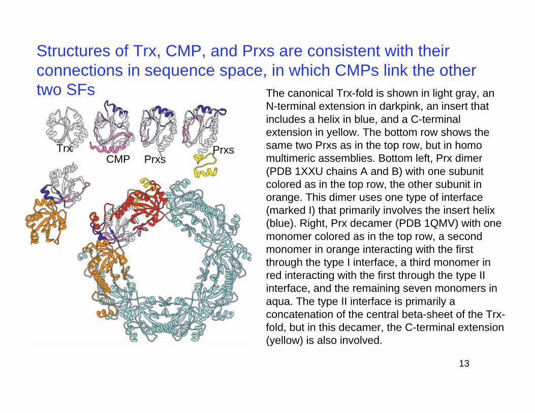
Structures of Trx, CMP, and Prxs are consistent with their
connections in sequence space, in which CMPs link the other
two SFs The canonical Trx-fold is shown in light gray, an
N-terminal extension in darkpink, an insert that
includes a helix in blue, and a C-terminal
extension in yellow. The bottom row shows the
same two Prxs as in the top row, but in homo
multimeric assemblies. Bottom left, Prx dimer
(PDB 1XXU chains A and B) with one subunit
colored as in the top row, the other subunit in
orange. This dimer uses one type of interface
(marked I) that primarily involves the insert helix
(blue). Right, Prx decamer (PDB 1QMV) with one
monomer colored as in the top row, a second
monomer in orange interacting with the first
through the type I interface, a third monomer in
red interacting with the first through the type II
interface, and the remaining seven monomers in
aqua. The type II interface is primarily a
concatenation of the central beta-sheet of the Trx-
fold, but in this decamer, the C-terminal extension
(yellow) is also involved.
TrxCMP Prxs
Prxs
14Version 2
PSI SBKB
The Structural Biology Knowledgebase
by Protein Structure Initiative (PSI)
and Nature Publishing Group (NPG)
Adopted from Materials prepared by Jennifer Williams, Ph.D.(Updated: Q1 2011)
www.openhelix.com
15
PSI SBKB
PSI SBKB: http://www.sbkb.org/
1. Introduction and Credits
2. Structural Biology Update
3. Sequence or Structure Search
4. Text Searches
5. Additional Features
6. Summary
7. Exercises
16
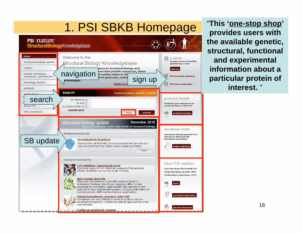
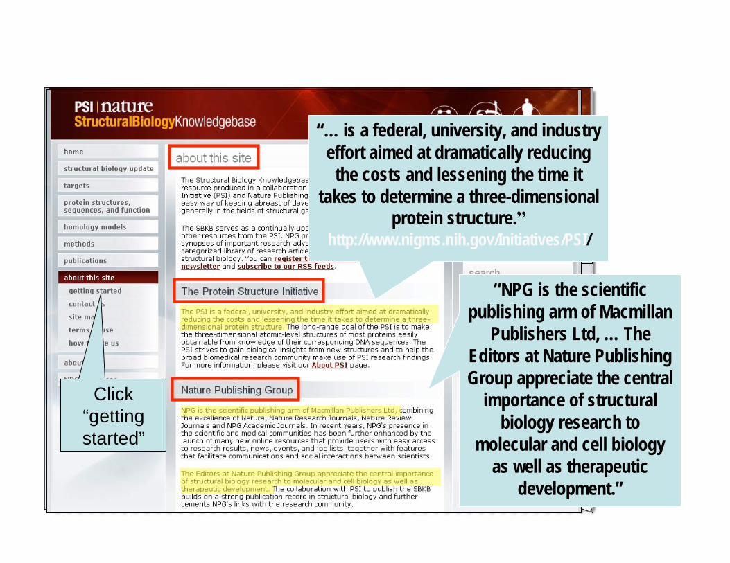
1. PSI SBKB Homepage “This ‘one-stop shop’
provides users with
the available genetic,
structural, functional
and experimental
information about a
particular protein of
interest. ”
SB update
search
sign upnavigation
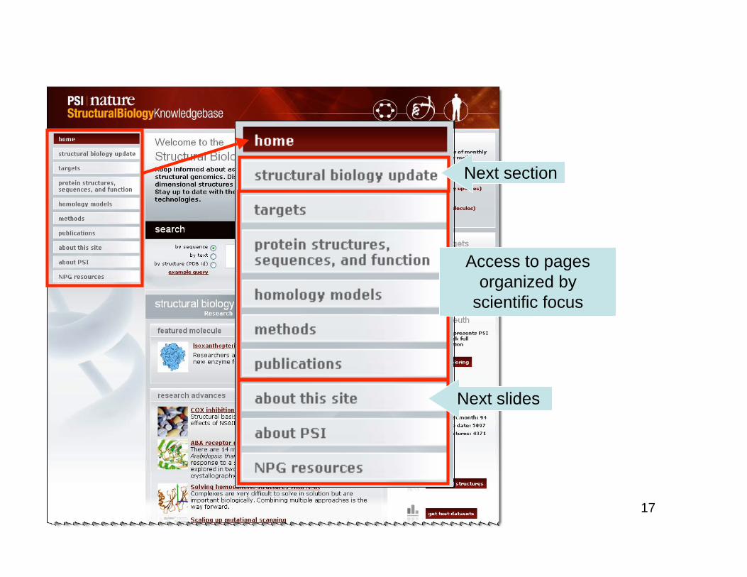
17
Next section
Next slides
Access to pages
organized by
scientific focus
18
FAQs Download
Tour
tutorial
by
OpenHelix
Interactive
tutorial
by SBKB
“NPG is the scientific
publishing arm of Macmillan
Publishers Ltd, … The
Editors at Nature Publishing
Group appreciate the central
importance of structural
biology research to
molecular and cell biology
as well as therapeutic
development.”
“… is a federal, university, and industry
effort aimed at dramatically reducing
the costs and lessening the time it
takes to determine a three-dimensionalprotein structure.”
http://www.nigms.nih.gov/Initiatives/PSI/
Click
“getting
started”
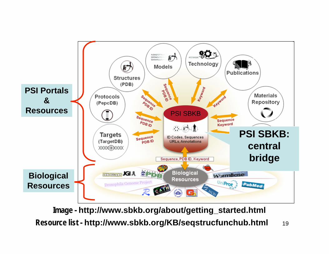
19
PSI SBKB Overview
Image - http://www.sbkb.org/about/getting_started.html
PSI Portals
&
Resources
Biological
Resources
Resource list - http://www.sbkb.org/KB/seqstrucfunchub.html
PSI SBKB:
central
bridge
PSI SBKB
20
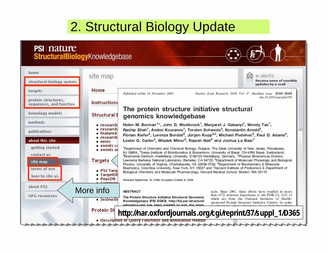
http://nar.oxfordjournals.org/cgi/reprint/37/suppl_1/D365
More info
2. Structural Biology Update
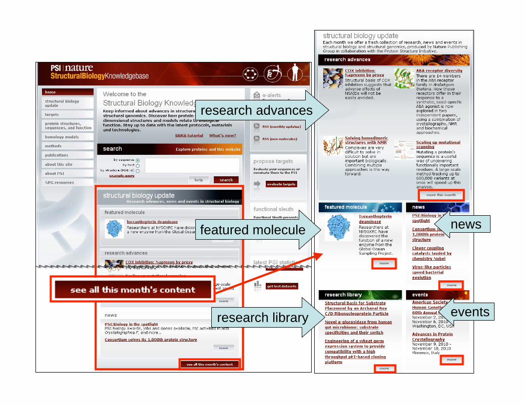
21
research advances
featured molecule news
eventsresearch library
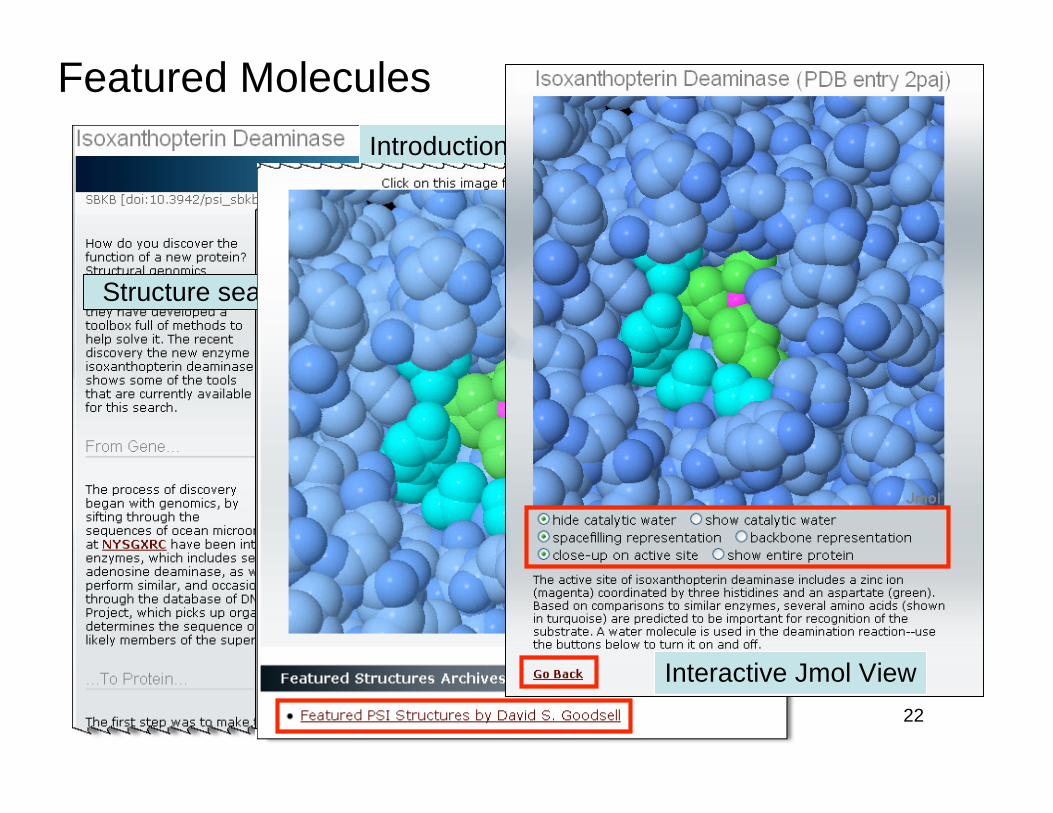
22
Featured Molecules
Introduction
Structure search
References
Click
Interactive Jmol View
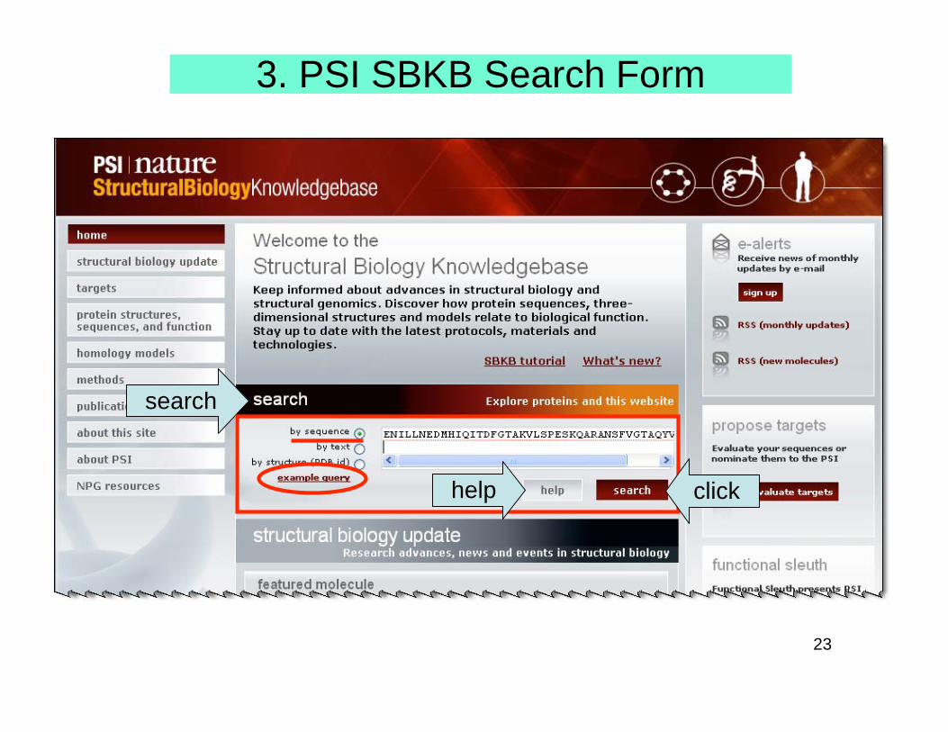
23
3. PSI SBKB Search Form
search
clickhelp
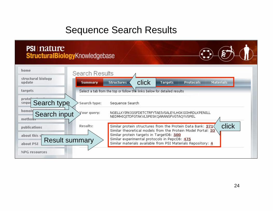
24
Sequence Search Results
Search type
Search input
Result summary
click
click
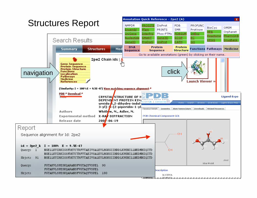
25
Glossary
navigation click
Structures Report
26
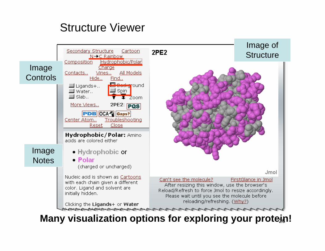
Structure Viewer
Many visualization options for exploring your protein!
Image
Controls
Image
Notes
Image of
Structure
27
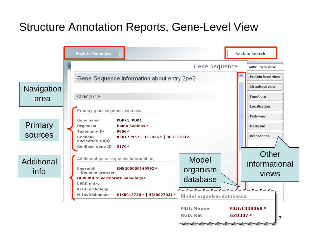
Structure Annotation Reports, Gene-Level View
Navigation
area
Primary
sources
Additional
info
Model
organism
database
Other
informational
views
28
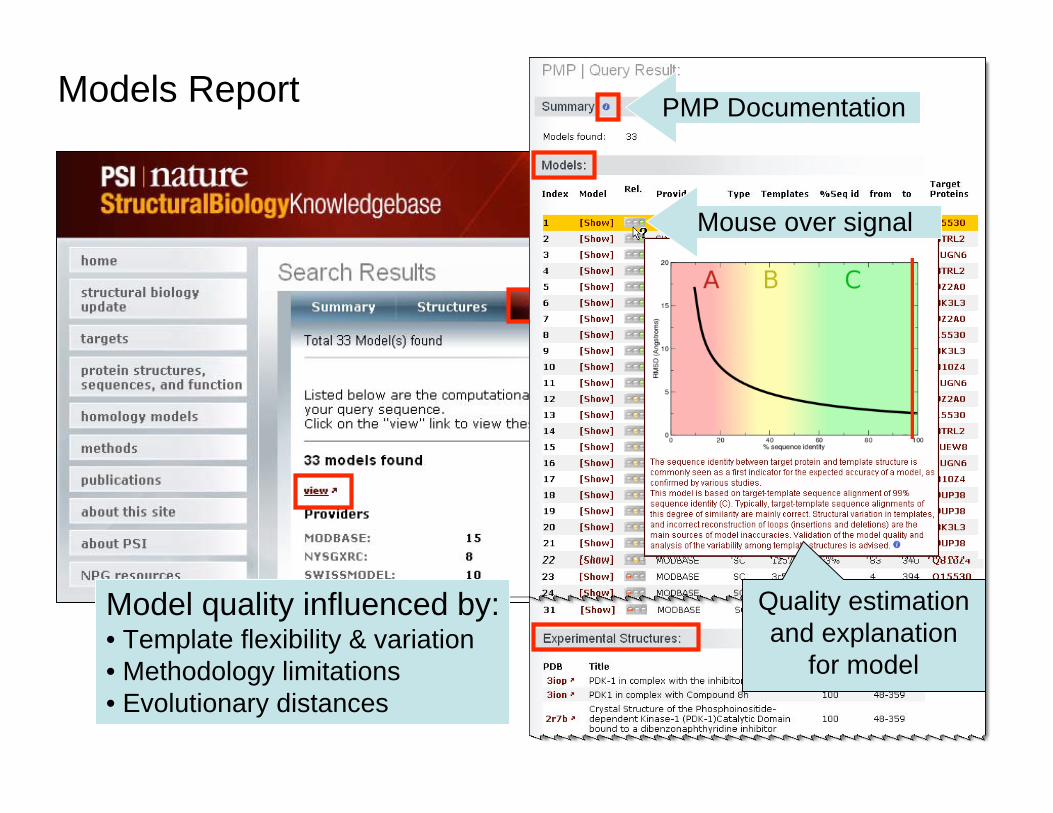
Models ReportPMP Documentation
Quality estimation
and explanation
for model
Mouse over signal
Model quality influenced by:• Template flexibility & variation
• Methodology limitations
• Evolutionary distances
29
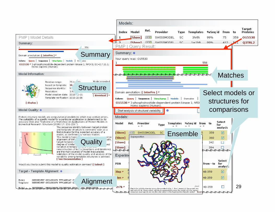
Query PMP ReportSummary
Structure
Quality
Matches
Select models or
structures for
comparisons
Alignment
Ensemble
30
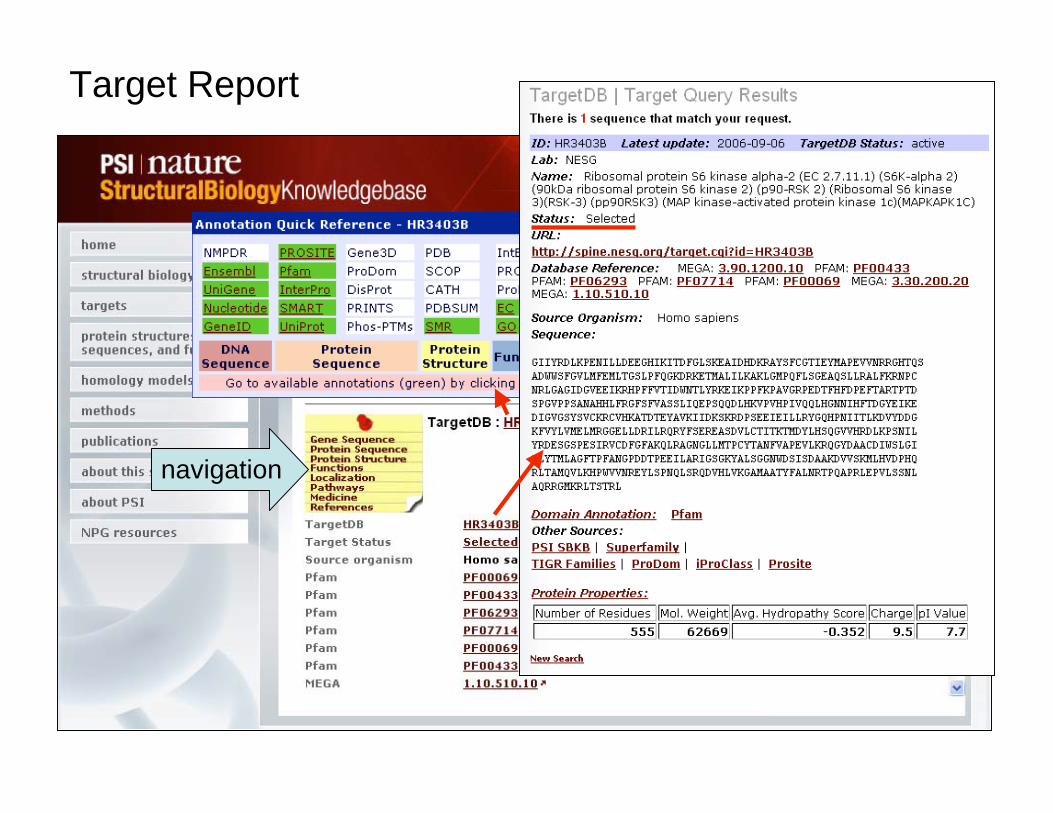
Target Report
navigation
31
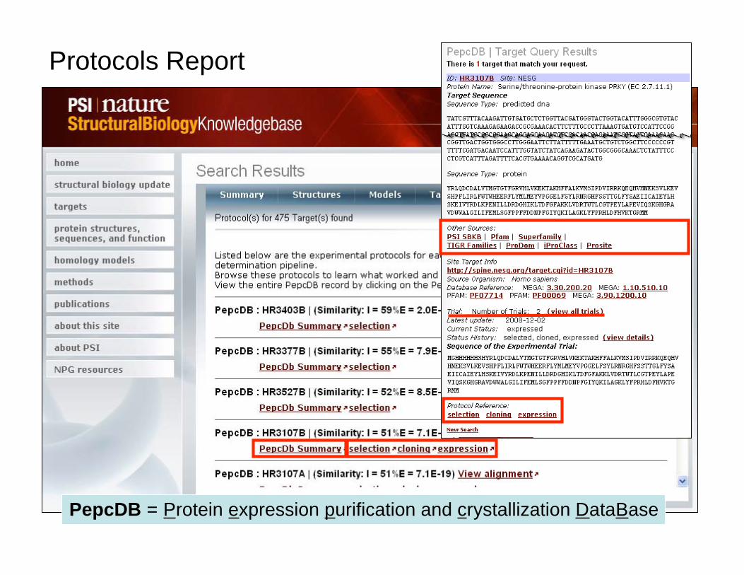
Protocols Report
PepcDB = Protein expression purification and crystallization DataBase
32
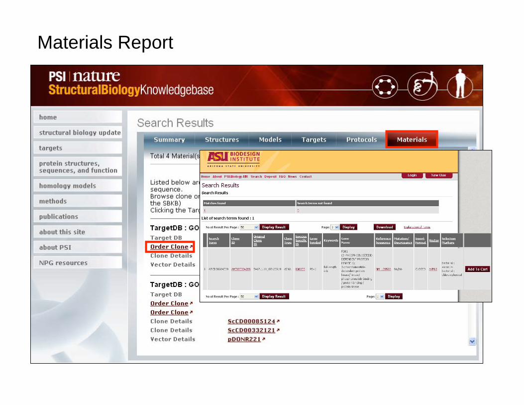
Materials Report
33
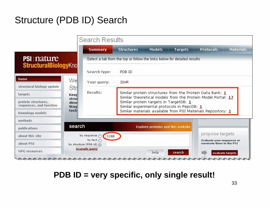
Structure (PDB ID) Search
PDB ID = very specific, only single result!
34
SBKB homepage: http://www.sbkb.org/
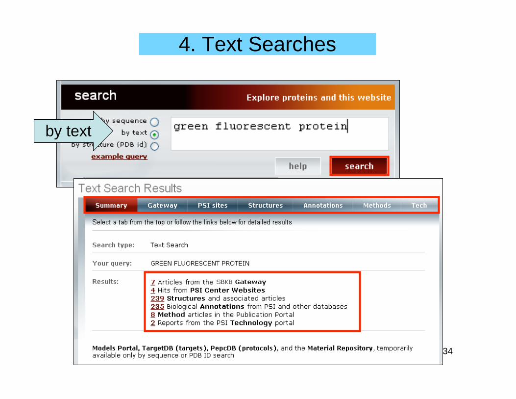
4. Text Searches
by text
35
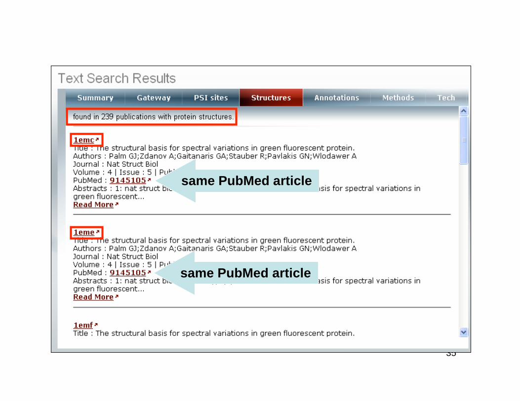
Text Search Structure Results
same PubMed article
same PubMed article
36
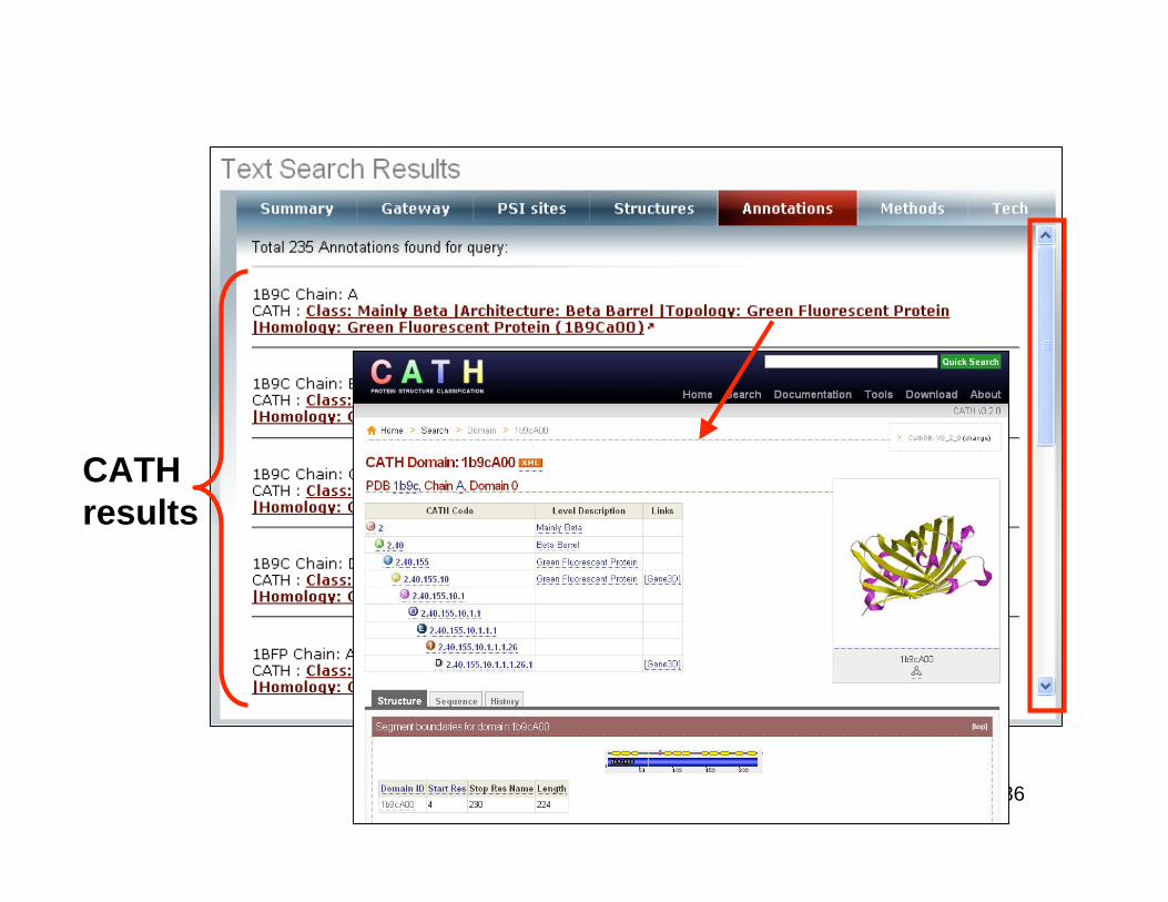
Annotation Results
CATH
results
37
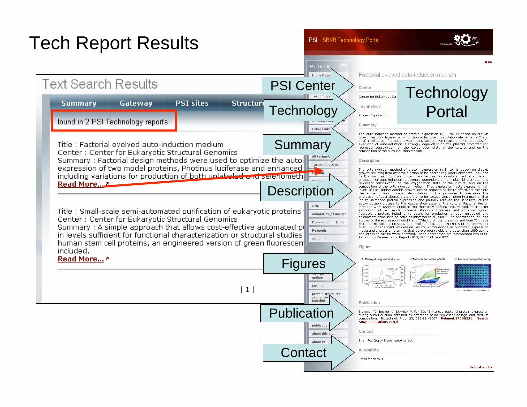
Tech Report Results
Technology
Portal
PSI Center
Technology
Summary
Description
Contact
Publication
Figures
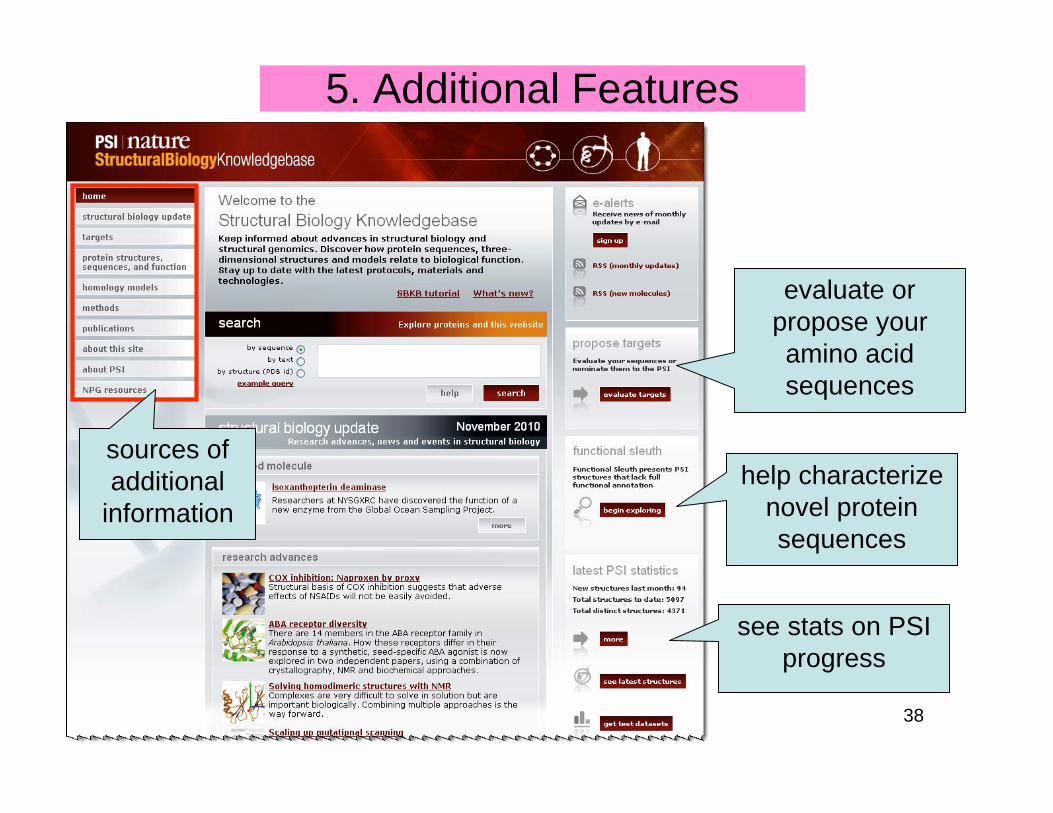
38
5. Additional Features
evaluate or
propose your
amino acid
sequences
help characterize
novel protein
sequences
see stats on PSI
progress
sources of
additional
information
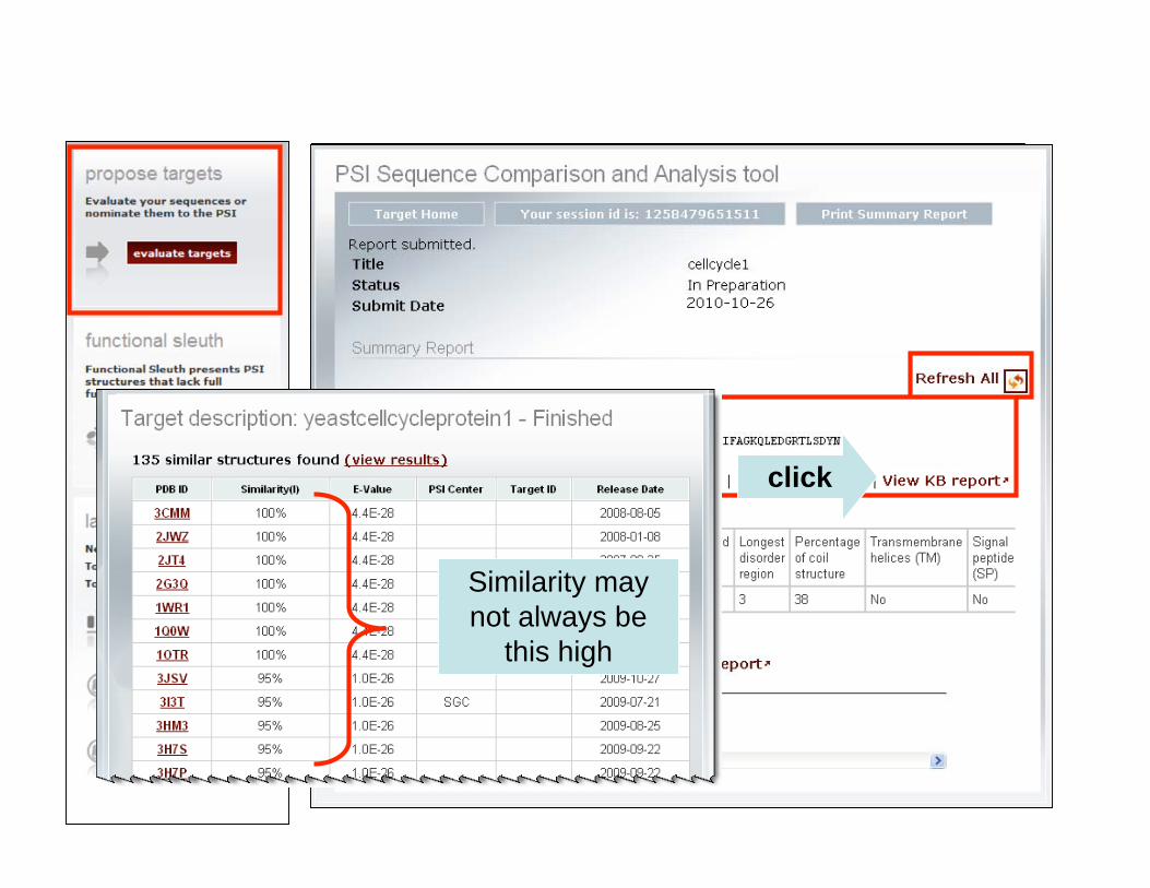
39
Proposed Targets
Sequence in
FASTA format
cellcycle1
title & email addressclick
Similarity may
not always be
this high
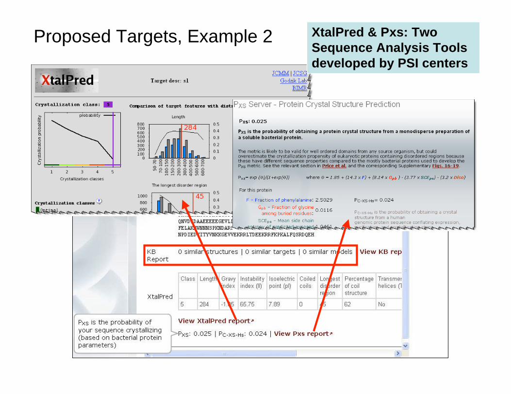
40
XtalPred & Pxs: Two
Sequence Analysis Tools
developed by PSI centers
Proposed Targets, Example 2
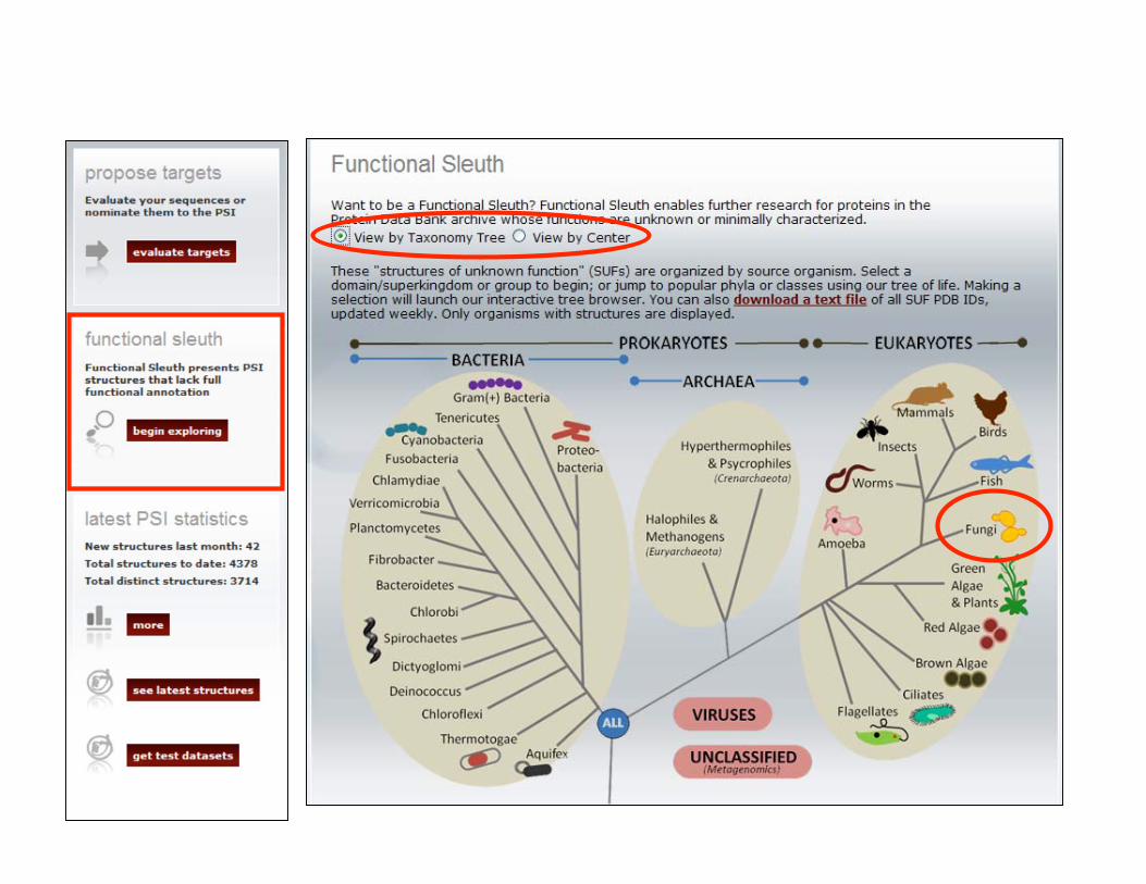
41
Functional Sleuth
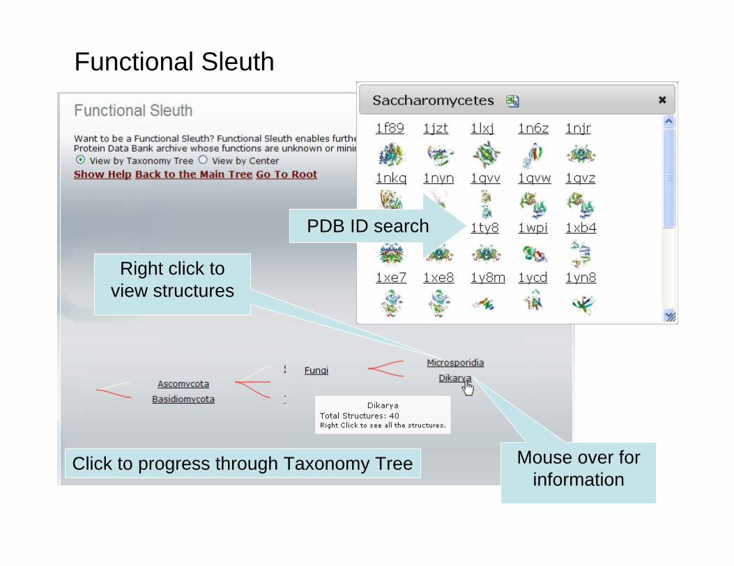
42
Functional Sleuth
Mouse over for
information
Right click to
view structures
Click to progress through Taxonomy Tree
PDB ID search
43
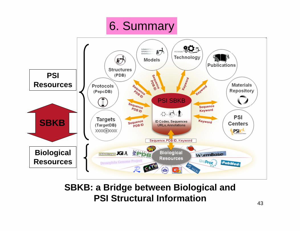
SBKB: a Bridge between Biological and
PSI Structural Information
PSI
Resources
Biological
Resources
SBKB
PSI SBKB
6. Summary
44
7. Exercises
1. You recently read an interesting paper that mentioned the
PDB structure xxx. Search the PSI SBKB to learn more
about this structure.
45
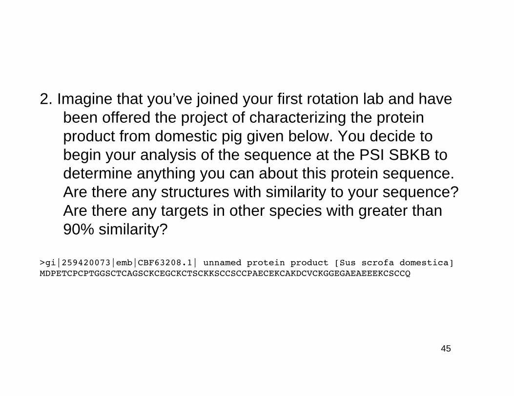
2. Imagine that you’ve joined your first rotation lab and have
been offered the project of characterizing the protein
product from domestic pig given below. You decide to
begin your analysis of the sequence at the PSI SBKB to
determine anything you can about this protein sequence.
Are there any structures with similarity to your sequence?
Are there any targets in other species with greater than
90% similarity?
>gi|259420073|emb|CBF63208.1| unnamed protein product [Sus scrofa domestica]MDPETCPCPTGGSCTCAGSCKCEGCKCTSCKKSCCSCCPAECEKCAKDCVCKGGEGAEAEEEKCSCCQ
46
3. You are working on a project trying to crystallize a
membrane protein, but you are having trouble with your
protocols. What helpful information can you find from the
SBKB Research Library as well as the rest of the PSI
SBKB?