BBasis of.indd 1asis of.indd 1 111/3/15 10:08:01 AM1/3/15 ... · his book “Exercitatio anatomica...
104
Basis of.indd 1 Basis of.indd 1 11/3/15 10:08:01 AM 11/3/15 10:08:01 AM
Transcript of BBasis of.indd 1asis of.indd 1 111/3/15 10:08:01 AM1/3/15 ... · his book “Exercitatio anatomica...

Basis of.indd 1Basis of.indd 1 11/3/15 10:08:01 AM11/3/15 10:08:01 AM

Basis of.indd 2Basis of.indd 2 11/3/15 10:08:02 AM11/3/15 10:08:02 AM
Jorge C. Trainini Benjamín Elencwajg - Néstor López-Cabanillas
Jesús Herreros - Noemí E. Lago Jorge A. Lowenstein - Alejandro Trainini
Basis of the New Cardiac Mechanics
The Suction Pump
Basis of.indd 3Basis of.indd 3 11/3/15 10:08:02 AM11/3/15 10:08:02 AM
Original title:
©2015 Editorial y Distribuidora Lumen SRL
Translator: Amalia Pujadas and Elena Lascano
©2015 Editorial y Distribuidora Lumen SRL
Grupo Editorial Lumen
Montevideo 604, 2.o piso (C1019ABN) Buenos Aires, Argentina
Tel.: (54-11) 4373-1414 • Fax: (54-11) 4375-0453
www.lumen.com.ar
All rights reserved
No part of this book may be reproduced, translated, stored in a retrieval system, or transmitted, in any form or by any means, electronic, mechanical, photoco-pying, microfi lming, recording, or otherwise, without written permission from the Publisher.
Printed in Argentina by Libris SRL.
Basis of the new cardiac mechanics : the suction pump /Jorge Carlos Trainini, ... [et al.]. - 1.a ed. . - Ciudad Autónoma de Buenos Aires : Lumen, 2015. 104 p. ; 22 x 15 cm.
Traducción de: Amalia Pujadas ; Elena Lascano. ISBN 978-987-00-1071-5
1. Salud. I. Trainini, Jorge Carlos, II. Pujadas, Amalia, trad. III. Lascano, Elena , trad. CDD 613
Basis of.indd 4Basis of.indd 4 11/3/15 10:08:02 AM11/3/15 10:08:02 AM
5
Jorge C. Trainini. Cardiovascular surgeon. Hospital Presidente Perón, Buenos Aires, Argentina. Fundación y Centro de Ingeniería Biomédica y Tec-nologías Sanitarias, Madrid, España.
Benjamín Elencwajg. Electrophysiologist. Hospital Presidente Perón, Buenos Aires, Argentina.
Néstor López-Cabanillas. Electrophysiologist. Hospital Presidente Perón, Buenos Aires, Argentina.
Jesús Herreros. Cardiovascular surgeon. Universidad Católica San An-tonio (UCAM), Murcia. Fundación y Centro de Ingeniería Biomédica y Tec-nologías Sanitarias, Madrid, España.
Noemí E. Lago. Cardiologist. Hospital Presidente Perón, Buenos Aires, Argentina.
Jorge A. Lowenstein. Chief of the Cardio Diagnosis Division. Investiga-ciones Médicas, Buenos Aires, Argentina
Alejandro Trainini. Cardiovascular surgeon. Hospital Presidente Perón, Buenos Aires, Argentina.
Basis of.indd 5Basis of.indd 5 11/3/15 10:08:03 AM11/3/15 10:08:03 AM

Basis of.indd 6Basis of.indd 6 11/3/15 10:08:03 AM11/3/15 10:08:03 AM
7
Dedication To Francisco Torrent Guasp (1931-2005)
There is need to grasp the past. To retrace in its steps towards the bifurcations that determine the mesh of history. Precision fades with time and circumstances. It becomes uncertain and misses the cross-roads. Progress is made over the incomprehension of oblivion, tread-ing over those brown and crispy leaves that return to the anonymous dust in spinning crosswinds. Which are destroyed shedding the pollen that had constituted their purpose of “existence”. The incomprehension of what occurred with the knowledge developed by Francisco Torrent Guasp is stirring. We arrive at the site of memory. Then, from the street that borders his house, we perceive the man who loved the stoicism of effort, the observation of nature and the skepticism in ephemeral, futile conquests. He did not desire success nor took refuge in inconsequential interests; he barely explored the imagined human existence. He became an artist able to paint realities in the dark, those that testify fi gures simi-lar to the torn sound of a string, slashing the air to produce the cry.
Francisco loved the sublime of the infi nite task. He launched in fren-zied days of research, in opposition to the established dogma and to the disbelief distilled by the fear of change. Today life seems to soothe him with the achieved explanation of his original theory. In the face of the rigidity of paradigms, this idea defi ed the inviolability determined by history, in an attempt to undermine an unbeaten foundation, never de-feated by the evolution of knowledge; only defi ed by time as the slave of his evolution, which in turn dominated him in an enduring monot-ony. Torrent Guasp realized that as long as there is movement there is time and that with the construction of his own historicity revision was possible, creating the coordinates to which man belongs.
And in this need for comprehension Torrent Guasp’s chronic be-comes similar to our daily experience until we fuse with his own promi-nence, that which sought the reality of things without being blinded by
Basis of.indd 7Basis of.indd 7 11/3/15 10:08:03 AM11/3/15 10:08:03 AM
Basis of the New Cardiac Mechanics
8
the uncertainty of success, equipped with a self-criticism that prevented him from the celebrity settled in the temporary and the trivial. We are guided by the refl ective glow emanating from his wisdom. We perceive him entering our consciousness from above, similar to a bird that ob-serves the knowledge he generated, diluting the darkness.
The authors, 2015
Basis of.indd 8Basis of.indd 8 11/3/15 10:08:03 AM11/3/15 10:08:03 AM
9
Auspices
Argentine Society of CardiologyAssociation of Cardiac SurgeonsCardiac Bioassisst AssociationCossio Foundation. Buenos Aires, Argentina.Foundation for Biomedical Engineering and Health Technology,
Madrid, Spain.Research Group of Biomedical Engineering and Health Technology
of the Catholic University San Antonio of Murcia (UCAM), Spain.Spanish Society of Surgical Research (SEIQ).
Basis of.indd 9Basis of.indd 9 11/3/15 10:08:03 AM11/3/15 10:08:03 AM

Basis of.indd 10Basis of.indd 10 11/3/15 10:08:04 AM11/3/15 10:08:04 AM
11
Acknowledgments
Juan Carlos ChachquesJuan Cosín-AguilarEduardo Otero CotoDivision of Cardiovascular Surgery, Hospital PresidentePerón, Ar-
gentina.To Salvador Mercé Vives and Salvador Mercé Cervelló because they
believed in this project and with their support made the book’s publica-tion possible.
To Jesús Valle Cabezas. His knowledge in engineering and computa-tional mechanics generated a new research project on Torrent Guasp’s model
To Jaime Herreros for his collaboration in the treatment of iconog-raphy.
To Antonio Teja. Without his collaboration this development would have been impossible.
Basis of.indd 11Basis of.indd 11 11/3/15 10:08:04 AM11/3/15 10:08:04 AM

Basis of.indd 12Basis of.indd 12 11/3/15 10:08:04 AM11/3/15 10:08:04 AM
13
Foreword
When the authors of this original and splendid book asked me to write some introductory lines, I was greatly honored by their request and also felt a great pleasure to perform the task at hand, as I consider its contents crucial for the progress of knowledge on cardiac contraction and subsequent relaxation, and thus of ventricular mechanics.
I have been a cardiologist for over fi ve decades and we have always con-sidered the heart as a pump which is active during systole and passive dur-ing diastole, but when my brilliant colleague Dr. Jorge Trainini put me in touch with the ideas and work of Dr. Francisco Torrent Guasp, and of the recent contributions of those who wrote this book, I was captivated refl ecting on them. I quickly realized that cardiac diastole plays an active phase in the heart’s pumping function since the effectiveness of manual or external cardiac massage would be impossible, if a previous mechanism of blood withdrawal or suction from the ventricles were not present, to then prompt an acceptable blood ejection by wringing them. An isolated fi rst pumping cannot possibly generate “vis a tergo”.
This work, with the historical contributions of Dr. Torrent Guasp and the subsequent work of the authors, discusses in detail the anatomical structures that make up Torrent Guasp’s ventricular myocardial band, its functional bas-es, electrical propagation, cardiac apex composition, electrophysiological and mechanical effects generated by muscle contraction and twisting, those that cause the ventricular suction effect during diastole (the authors’ “plunger” mechanism).
The originality of this physiological concept is that the fi rst phase of ven-tricular diastole (protodiastole), the isovolumic phase, has an active character due to the late myocardial contraction that produces left ventricular lengthen-ing by separating the base from the apex (cardiac suction). This phase of ven-tricular mechanics becomes an intermediate phase followed by the real diastole (mesodiastole) which produces ventricular expansion and fi lling by decom-pressing the elastic elements of the heart, completing the cycle with ventricular presystolic atrial contraction and the subsequent ventricular systole. All this establishes the three-stage heart mechanics described in the text.
Basis of.indd 13Basis of.indd 13 11/3/15 10:08:04 AM11/3/15 10:08:04 AM
Basis of the New Cardiac Mechanics
14
Faced with these facts there arises for the future a thorough study and anal-ysis that will bring a wealth of knowledge to the prognosis and solutions for the treatment of heart failure and ventricular dysfunction, perspectives well discussed in this book.
Dr. Torrent Guasp’s work is stimulating, and the deep study on the fur-therance of the research performed by the authors of this book allows me to say without doubt that it is a great contribution to the better understanding of cardiac mechanics and future management of heart failure.
Pedro Ramón Cossio
Basis of.indd 14Basis of.indd 14 11/3/15 10:08:04 AM11/3/15 10:08:04 AM
15
Preface of the authors
Any knowledge is a sequence of the previous, but it is not excerpted of being considered a turmoil. It happens when the bases that are in-tended to be reformed are very anchored in the time and the praxis.The history of the circulation of the blood evolved through three fun-damental jumps. The initial one, through Claudius Galeno (second cen-tury A.D.) that persisted until 1628, when William Harvey published his book “Exercitatio anatomica de motu cordis et sanguinis in animalibus”, establishing the modern physiology of circulation. This movement of the blood lacked the description of the pathway for returning of the blood to the heart. The appearance of the microscope allowed Marcelo Malpighi in 1661 to close he breach between arteries and veins with the discovery of the capillary vessels. His writing “De pulmonibus ob-servations anatomicae”, that appeared four years after Harvey´s death, shows excellent pictures, taking as an investigation model the lungs of the frog.
The fi rst visual demonstration of the circulation of the blood cor-responded to the cleric Lazarus Spallanzini in 1771, as he observed in a chicken embryo the blood circulating from the arteries to the veins. His relation describes the event with these words: “The room where I was didn´t have enough light, and willing anyway to satisfy my need, I decided to examine the egg under the direct light of the sun. Once placed the egg in Lyon-net device, I directed the lens, and, in spite of the great clarity that surrounded it, I was able, by sharpening my sight, to observe the blood running through the complete circuit of umbilical vessels, arterial and venous. Overwhelmed then by an unexpected happiness I exclaimed eureka! Eureka!”.
In spite of the righteous of this idea, it was not simple Harvey´s dis-covery to supplant the fourteen centuries of the hegemony imposed by Galeno´s system. The blood returned, it did not remained in the perifery as thouth by the Galeno. In relation to cardiac movement, through his observations Harvey explains the function of this organ in two phases: systole and diastole, considering the later one as a simple cardiac dilata-tion. With this position, he opposed to Galeno´s doctrine, who believed
Basis of.indd 15Basis of.indd 15 11/3/15 10:08:05 AM11/3/15 10:08:05 AM
Basis of the New Cardiac Mechanics
16
that the activity of the heart was manifested by the dilation, through the “vis pulsifi ca”. By 1980 Francisco Torrent Guasp originates fi ssures in the understanding of the dominant cardiac mechanics. He conceives an anatomic explanation adapted to the real facts of its function, which will advance in the theoretical understanding of its mechanics, in works together with Gerard Buckberg (2001).
The investigation in human beings in relation to the electrophysi-ological aspects of the heart that we expose in this text modifi es the conception of the cardiac function. It allows to understand the anatomi-cal-functional unity, the propagation of the electric impulse through the myocardium not visualized until now and to sustain the concept of a three phased heart.
Cardiac electrical activation is a consequence of the propagation of the stimuli through the muscular structure of the heart, both of Torrent Guasp´s myocardial band and the rest of the fi bers that constitute its structure. The cardiac mechanism of aspiration and expulsion requires an integration of the structure/function that takes into account several mechanisms of its dynamic due to the propagation of the excitation. This implies that the diastolic isovolumetric phase is an active process of contraction with a growing aspiration action that we called by its similarity as a plunger mechanism, which at a certain level of intraven-tricular pressure level produces the opening of atrioventricular valves, originating the rapid ventricular fi lling.
In our experience, we found a pathway of spreading of the electric impulse different from the one described by Torrent Guasp that ex-plains the phase of torsion of the heart, defi ned as a rotation movement opposed of the base and the apex.
This activation conceives, between the systole and the diastole, a third phase of active coupling of suction, with muscular contraction, energetic spending and remarkable fall in the intraventricular pressure, defi ning a three phase heart.
The basically anatomic model of Torrent Guasp, amplifi ed and mod-ifi ed by our investigation from an electromechanically point of view, offers countless possibilities of development both at a theoretical level as well as from tis clinical a therapeutic application. It is probable that a great part of the cardiology should be reexamined in light of this new paradigm, with potential unpredictable results.
Buenos Aires, 2015
Basis of.indd 16Basis of.indd 16 11/3/15 10:08:05 AM11/3/15 10:08:05 AM
17
Index
Intertext . . . . . . . . . . . . . . . . . . . . . . . . . . . . . . . . . . . . . . . . . 21Genesis of the idea. Torrent Guasp’s “key doubt” . . . . . 21
Chapter I. Torrent Guasp´s Ventricular Myocardial Band In Ventricular Anatomy . . . . . . . . . . . . . . . . . . . . . . 271. Ventricular myocardial band anatomy . . . . . . . . . . . . . 272. Phylogenetic development of the circulatory system . . . . . . . . . . . . . . . . . . . . . . . . . . . . . . . . . . . . . . . . . . 353. The cardiac apex . . . . . . . . . . . . . . . . . . . . . . . . . . . . . . . . 37
Chapter II. Electrophysiological Interpretation Of The Ventricular Myocardial Band . . . . . . . . . . . . . . . 391. Historical concepts on myocardial electrical activation . . . . . . . . . . . . . . . . . . . . . . . . . . . . . . . 392. Stimuli propagation, muscle torsion and cardiac suction effect through electrophysiological research . . . 403. Electrocardiographic correlation . . . . . . . . . . . . . . . . . . 564. Functional aspects of the ventricular myocardial band . . . . . . . . . . . . . . . . . . . . . . . . . . . . . . . . . . 58
Chapter III. The Three Stage Heart . . . . . . . . . . . . . . . . . 631. Chronology of the suction mechanism concept . . . . . 632. Cardiac mechanics . . . . . . . . . . . . . . . . . . . . . . . . . . . . . . 643. How is diastolic suction produced? . . . . . . . . . . . . . . . 704. Structural basis of diastolic left ventricular limitation in the suction mechanism . . . . . . . . . . . . . . . . . . . . . . . . . . 735. Echocardiographic concepts . . . . . . . . . . . . . . . . . . . . . . 77
Basis of.indd 17Basis of.indd 17 11/3/15 10:08:05 AM11/3/15 10:08:05 AM

Basis of the New Cardiac Mechanics
18
Chapter IV. Clinical, Surgical And Electrophysiological Perspectives Derived From This Research . . . . . . . . . . . . . . . . . . . . . . . . . . . . . . . . . . . . 811. Extent of heart failure . . . . . . . . . . . . . . . . . . . . . . . . . . . 812. Clinical perspectives . . . . . . . . . . . . . . . . . . . . . . . . . . . . 833. Surgical perspectives . . . . . . . . . . . . . . . . . . . . . . . . . . . . 844. Electrophysiological therapy perspectives . . . . . . . . . . 90
References . . . . . . . . . . . . . . . . . . . . . . . . . . . . . . . . . . . . . . . 95
Basis of.indd 18Basis of.indd 18 11/3/15 10:08:05 AM11/3/15 10:08:05 AM
19
The heart has the gift of oblivion. The grace that allows each dawn to restart the utopia of remaining in force. Despite memory and boredom. Of the personal history that refl ects in its own mir-ror the circular fl ow of blood and destinies.
Jorge Carlos Trainini
“Heart mechanics is homologous to that of the circular fi bers of blood vessels, which carry out their function without fi xed fulcrums”
Francisco Torrent Guasp (68)
Basis of.indd 19Basis of.indd 19 11/3/15 10:08:05 AM11/3/15 10:08:05 AM

Basis of.indd 20Basis of.indd 20 11/3/15 10:08:06 AM11/3/15 10:08:06 AM
21
Intertext
Genesis of the idea. Torrent Guasp’s “key doubt”
“Francisco [Torrent Guasp] never believed that blood could enter the left ventricle other than by a suction device”
Juan Cosín-Aguilar (personal interview, Valencia, June 2010)
The disciple slightly shrugged his shoulders recalling the question Francisco Torrent Guasp asked himself. -You know…- he said, facing us with his light-colored eyes while slowly stirring the coffee: -… Paco’s thesis began with a key doubt.
Juan Cosín-Aguilar, the speaker and faithful friend of the person we were trying to unravel held upon us a remarkable fascination in that warm night of Valencia. No one had been so close to the master’s “doubt” in those essential years when different hypotheses on heart functioning were being developed.
-What was the doubt- we asked in unison, impertinently driven by the force of our anxiety.
An external uproar fi ltered through all the cracks in the tavern, orna-mented with red and yellow headscarves on that festive day. Even the most frivolous had been caught by the merriment. At a moment when the excitement took a break he began outlining the answer stored in the memory.
- “Man, [said Paco] blood does not return to the heart vis a tergo or by a peripheral pressure difference with the heart. This is small. The gradient that returns it is ventricular suction”. To this statement I retorted with another: “So the heart is a suction pump?” At that point his shyness turned into a candle. He ignited. “Look Juan, all dissections made in animals and humans
Basis of.indd 21Basis of.indd 21 11/3/15 10:08:06 AM11/3/15 10:08:06 AM

Basis of the New Cardiac Mechanics
22
clearly explain this possibility. The heart is formed by a large muscle band that begins at the insertion of the pulmonary artery and ends at the level of the aorta, forming a double helical structure which limits the ventricles. The two ventricular chambers wrapped by the large muscle band are the left chamber with ellipsoidal shape and the right chamber of a semilunar structure. Well, the contraction of this band not only explains heart’s systole but also blood suction”.(72)
After a pause owing to the outside din and with his undisguised sensitive condition of a Spanish native he proceeded. -’I sometimes became the devil’s advocate. “Paco, to allow the bands surrounding the ventricles to contract, they should need a rigid support just as a tendon uses the bone insertion as lever Are there any in the heart?” In those moments he would make a quiet patient pause and from his working place in the attic would pensively gaze for a while at the long alley of his house in Denia, his adoptive village in Alicante, which appeared and disap-peared at every turn of its curves. “You do not need that support. When the heart fi lls with blood it behaves as a bone insertion. The large muscle band is a double helix suspended between the pulmonary artery and the aorta using the hemoskeleton i.e. cardiac fi lling as its fulcrum. At this point the left ventricle turns counter-clockwise and the right ventricle turns clockwise. Exactly like wringing out a towel”.(68, 87)
“Free, master of his time and his ideas”
Jesús Herreros, Eduardo Otero Coto, Salvador Mercé and his father Mercé Vives, the other participants of the meeting, must have looked astonished as Jorge Trainini lashed in the memory of the disciple trying to exhume the magic that the fi gure of Francisco Torrent Guasp stirred upon him.(82)
-Cosín … Torrent must have been a special man! I picture him working in anonymity, far from the media noise which often appears with the advances in our profession. What was hisinner-self?
-Paco was a free man, master of his time and his ideas.(19) To know him you had tolet go of prejudices. He lacked the need to enter a career of honors.
-If you are free you’ll die alone- managed to interrupt Trainini.-Something of that emerged in his recognition. He was cultivated, lucid,
curious, imaginative, spontaneous, nonconformist, rebellious, enthusiastic, committed.(19) Since 1954, while still a student, he devoted himself to the physio- structural study of cardiac mechanics. He was born to change these
Basis of.indd 22Basis of.indd 22 11/3/15 10:08:06 AM11/3/15 10:08:06 AM

Basis of the New Cardiac Mechanics
23
things of the heart. I think the rest of his life was an addition to that passion. He was an anonymous and colossal worker. He used more than a thousand bovine, horse, dog, pig, sheep, cat, hen, turtle, fi sh and obviously human hearts.
-He reminds me of Galen. The pergamum dissected all kinds of animals, even an elephant. However, since autopsies were forbidden in Rome [second century A.D.] he could only do so in a corpse found fl oating after a fl ood had swept his tomb.(79)
-He had an artistic mind. He liked to paint and even exhibited his paintings in Paris. Perhaps this explains his way of being. He had ideas and action. He lacked the dialectic.
-I picture him with the characteristics of a rebel. Dialectic, essential to be recognized is not usually well valued by men who are pioneers.
The interviewee agreed, nodding with a slight tilt of his head. Then he added, almost resignedly: -especially with today’s computing progress the space you do not occupy is quickly usurped.
Trainini reiterated the words of the disciple -Juan, I have always be-lieved that personality has a lot to do with success. Extreme self-criticism con-spires against it. I have the sensitivity to understand that Torrent Guasp’s almost silent work still needs a posthumous tribute.
-But in the last years he received some honors and acknowledgements. His idea of the ventricular myocardial band still requires certain explorations in other fi elds such as electrophysiology.
-Incidentally, concerning this multidisciplinary approach that you confer to it, did Torrent explore into microscopy?- Now everyone asked in unison resembling a circumstantial choir.
-At fi rst he worked on it but quitted without clear results.-However, the helical physio-structure he proposes would have correspon-
dence with the microscopic universe known today. Sarcomeres are bundled by collagen fi bers in the shape of double helical bands whose function would be limiting expansion and setting the mechanical recoil, in addition to storing the restoring force of energy for relaxation- explained Trainini with a par-simony not devoid of passion. The evening continued further on with other conversations, but Trainini would return to the subject… outside the joy persisted without shapes or boundaries.
Basis of.indd 23Basis of.indd 23 11/3/15 10:08:06 AM11/3/15 10:08:06 AM
Basis of the New Cardiac Mechanics
24
Denia
… –Cosín, perhaps this invitation for a conference in the fi eld of electro-physiology that Torrent desired so much has been yet another scorn of fate in its insult to men?
One could feel in the air that everyone remained stationed in their own impressions. Time-detained silence waited for the affectionate reminiscence of the disciple. They all directed their eyes towards Cosín. The disciple relaxed slightly allowing time for the word not to become an emotional fraud. The glasses and coffee cups seemed suspended in the air waiting for a decision to break the spell. He coughed softly to clear his knotted voice. Perhaps to divert some tears.
-Paco was happy with the invitation to the electrophysiology meeting. He had always longed to talk with cardiologists to explain that the contraction of the heart began in the right ventricular outfl ow tract and ended at the left apex. According to him the “cardiac piston” worked in that way. So he went to Madrid despite having been for weeks in a wheelchair. His lecture was bold, char-acteristic of a leader, exultant. He was ebullient with the reception he had been offered. It was the 25th of February 2005. He died just after his last conference.
We walked away from the meeting at midnight. The last celebra-tions were fading in the streets of a bright city. Golden-walled build-ings seemed mirrors in a maze we were trying to solve thinking of that village, of that attic, of those projects developed by Francisco Torrent Guasp, indifferent and detached from the clamor that usually blesses the medical community. We would arrive to his home in Denia, even if only for a silent exercise of admiration.
So we did the next day. Denia was born Roman, was then Arabic, but defi nitely remained Spanish. An Alicant village, at a very short dis-tance, it runs behind a rock against another gem, Javea, belonging to the Valencian community. Located in the last rugged steps of the appeased mountains in the jagged coastline of the Mediterranean, it stands be-tween uneven streets and white houses that seem to lean against each other. Its past shows the offering of each conqueror. Its fi gure outlined by an infi nite light is drawn in the rolling emerald waters that conferred it glory and destiny. We meandered the street where Torrent Guasp had lived. We imagined that from his working attic a watchful eye was still peering to where the future of silent men reached. One of us remem-bered François Jacob [Nobel Prize in Medicine, 1965] who always re-peated that “humility does not suit the wise or the ideas he has to defend”.(32) In our return the sun spilled fully without casting any shadow. It had stationed at the exact point where emotion meets absence.
Basis of.indd 24Basis of.indd 24 11/3/15 10:08:06 AM11/3/15 10:08:06 AM
Basis of the New Cardiac Mechanics
25
Buenos Aires
When Jorge Trainini returned to Buenos Aires he carried in his mem-ory the words of Juan Cosín in relation to Torrent Guasp’s hypothesis: “His idea of a myocardial band still needs certain explorations in other fi elds such as electrophysiology”. In Buenos Aires, he met with Benjamin Elenc-wajg, Nestor López Cabanillas and Noemí Lago and together with Jesús Herreros in Spain they formed a working group. They articulated a human research project that did not infringe the ethics. The literature was meticulously examined. Several proposals were considered for electrophysiological testing. The recent advent of navigation systems and three-dimensional endocardial and epicardial mapping (CARTO, Biosense Webster, California, USA) provided the ideal instrument. Then the steps were accomplished in a continuous and tenacious re-search. The fi rst steps led to others in the required paradigm of science. The point reached and presented in the text opens a perspective for the physiological understanding of the heart and its resulting clinical, sur-gical and electrophysiological implications. Future research will help to improve all the aspects developed so far.
The authors, Buenos Aires, 2015
Basis of.indd 25Basis of.indd 25 11/3/15 10:08:07 AM11/3/15 10:08:07 AM

Basis of.indd 26Basis of.indd 26 11/3/15 10:08:07 AM11/3/15 10:08:07 AM
27
Chapter I
Torrent Guasp´s Ventricular Myocardial Band In Heart Anatomy
1. Ventricular myocardial band anatomy
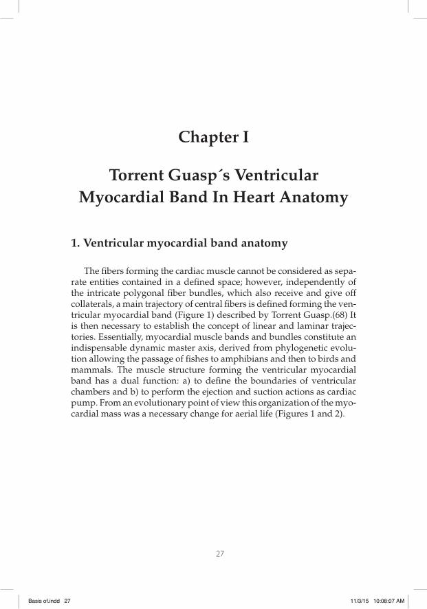
The fi bers forming the cardiac muscle cannot be considered as sepa-rate entities contained in a defi ned space; however, independently of the intricate polygonal fi ber bundles, which also receive and give off collaterals, a main trajectory of central fi bers is defi ned forming the ven-tricular myocardial band (Figure 1) described by Torrent Guasp.(68) It is then necessary to establish the concept of linear and laminar trajec-tories. Essentially, myocardial muscle bands and bundles constitute an indispensable dynamic master axis, derived from phylogenetic evolu-tion allowing the passage of fi shes to amphibians and then to birds and mammals. The muscle structure forming the ventricular myocardial band has a dual function: a) to defi ne the boundaries of ventricular chambers and b) to perform the ejection and suction actions as cardiac pump. From an evolutionary point of view this organization of the myo-cardial mass was a necessary change for aerial life (Figures 1 and 2).
Basis of.indd 27Basis of.indd 27 11/3/15 10:08:07 AM11/3/15 10:08:07 AM
Basis of the New Cardiac Mechanics
28
Figure 1. Torrent Guasp´s ventricular myocardial band. RV: Right ventricle; LV: Left ventricle; PIS: Posterior interventricular sulcus; AIS: Anterior inter-ventricular sulcus.
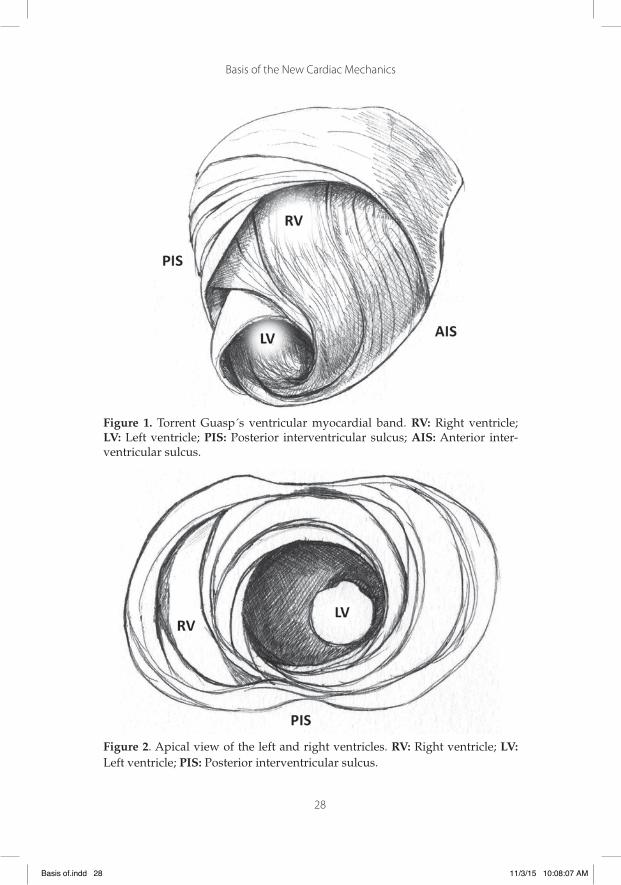
Figure 2. Apical view of the left and right ventricles. RV: Right ventricle; LV: Left ventricle; PIS: Posterior interventricular sulcus.
Basis of.indd 28Basis of.indd 28 11/3/15 10:08:07 AM11/3/15 10:08:07 AM

Basis of the New Cardiac Mechanics
29
Left ventricle. The entire apex belongs to the left ventricle. In its dis-tal part, called apical, a muscle layer with spiral trajectory extends from the surface to the center, undergoing a rotation that turns subepicardial into subendocardial fi bers, overlapped like the tiles of a roof. Conse-quently, the left ventricular distal end, the apex, surrounds a virtual tube with no muscular plane, lined at its two ends by the endocardium and epicardium. It is essential to consider that in the apex the fi bers un-dergo a helical spinning movement with sphincter-like arrangement as they transform from subepicardial to subendocardial fi bers, following a clockwise trajectory (apical view of the diaphragmatic surface of the heart in anatomical position) (Figure 3).(66)
Figure 3. Spiral arrangement of apical muscle layers. AIS: Anterior interven-tricular sulcus; LV: Left ventricle.
Basis of.indd 29Basis of.indd 29 11/3/15 10:08:08 AM11/3/15 10:08:08 AM

Basis of the New Cardiac Mechanics
30
In the left ventricular basal half, at the level of its free wall (Figure 4), the fi bers are ordered similarly to the apical half. A muscle layer with spiral trajectory extends from the surface to the center arranging par-aepicardial and paraendocardial regions from the outside to the inside. At this level their orientation is opposite to that of the apex, following a counter-clockwise trajectory (apical view of the diaphragmatic surface of the heart in anatomical position). This arrangement of the spiraling muscle layer limits a cavity which at the base of the heart is real and not virtual as in the apex.
Figure 4. Basal third of the left ventricle, showing the free wall muscle layers. PA: Pulmonary artery; A: Aorta; TV: Tricuspid valve; MT: Mitral valve.
The apex should be considered as a tunnel with a rim in its entire ring, while at the ventricular base this ring has two parts. One part cor-responds to the left ventricular free wall and the other to the interven-tricular septum. In addition, the superfi cial basal fi bers make contact with the fi brous mitral annulus, absent at the apical level. However, the essential functional difference between basal and apical segments is the opposite trajectory of their fi bers. This characteristic determines muscle torsion to achieve blood fl ow.
Right ventricle. According to their orientation, two types of fi bers can be identifi ed in its distal half: paraendocardial and paraepicardial
Basis of.indd 30Basis of.indd 30 11/3/15 10:08:08 AM11/3/15 10:08:08 AM
Basis of the New Cardiac Mechanics
31
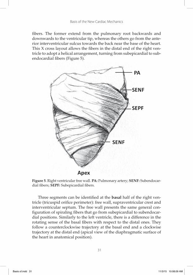
fi bers. The former extend from the pulmonary root backwards and downwards to the ventricular tip, whereas the others go from the ante-rior interventricular sulcus towards the back near the base of the heart. This X cross layout allows the fi bers in the distal end of the right ven-tricle to adopt a helical arrangement, turning from subepicardial to sub-endocardial fi bers (Figure 5).
Figure 5. Right ventricular free wall. PA: Pulmonary artery; SENF: Subendocar-dial fi bers; SEPF: Subepicardial fi bers.
Three segments can be identifi ed at the basal half of the right ven-tricle (tricuspid orifi ce perimeter): free wall, supraventricular crest and interventricular septum. The free wall presents the same general con-fi guration of spiraling fi bers that go from subepicardial to subendocar-dial positions. Similarly to the left ventricle, there is a difference in the rotating sense of the basal fi bers with respect to the distal ones. They follow a counterclockwise trajectory at the basal end and a clockwise trajectory at the distal end (apical view of the diaphragmatic surface of the heart in anatomical position).
Basis of.indd 31Basis of.indd 31 11/3/15 10:08:09 AM11/3/15 10:08:09 AM
Basis of the New Cardiac Mechanics
32
Interpretation. It can be seen that the spatial confi guration and ro-tating movement of the fi bers at the basal and distal levels of both ven-tricles correspond to Torrent Guasp’s ventricular myocardial band. The author considered that the ventricular myocardium is formed by the assembly of muscle fi bers twisted like a rope (rope theory) (Figure 6), fl attened laterally as a band, which presents two spiral turns defi ning a helical structure that limits the two ventricles and defi nes their func-tion.(70)
A classical interpretation of blood circulation through the differ-ent cardiac chambers has been made bearing no correlation with their muscle dynamics. However, this dynamic behavior is essentially the circulatory motor established by the muscle mass, which also defi nes the chamber boundaries through which blood fl ows. This arrangement of the ventricular myocardial band confers ventricular chambers a lead-ing role in cardiac function.
Cardiac muscle lacks fi xed points of attachment as those of the skel-etal system to develop force. In this sense, the ventricular myocardial band would act as the circular muscle of the arteries, supporting itself in its own cavity content (hemoskeleton). Moreover, attachment to the origin of the great vessels could also be considered an insertion point to drive the mechanics of the ventricular myocardial band.
Segmentation of the ventricular myocardial band. Ventricular chambers are defi ned by Torrent Guasps´s ventricular myocardial band, which describes two spiral turns with one end inserted in the pulmo-nary artery and the other in the aortic root. In its trajectory it adopts a helical confi guration forming the two ventricular chambers. The fi gure in 8 defi ned by its course outlines two loops: a basal and an apical loop. The basal loop extends from the root of the pulmonary artery to the central twist of the band. On the other hand, the apical loop courses from this twist to the aortic root. Moreover, each loop is formed by two segments. The basal loop consists of the right and left basal segments and the apical loop by the descending and ascending apical segments (Figure 6). In the general confi guration, the basal loop embraces the apical loop, so that the right ventricular chamber is more like an open slit in the muscle mass thickness forming both ventricles (Figure 1). The segments are defi ned by anatomical structures.
Basis of.indd 32Basis of.indd 32 11/3/15 10:08:09 AM11/3/15 10:08:09 AM
Basis of the New Cardiac Mechanics
33
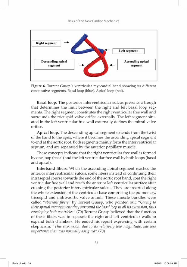
Figure 6. Torrent Guasp´s ventricular myocardial band showing its different constitutive segments. Basal loop (blue). Apical loop (red).
Basal loop. The posterior interventricular sulcus presents a trough that determines the limit between the right and left basal loop seg-ments. The right segment constitutes the right ventricular free wall and surrounds the tricuspid valve orifi ce externally. The left segment situ-ated in the left ventricular free wall externally defi nes the mitral valve orifi ce.
Apical loop. The descending apical segment extends from the twist of the band to the apex, where it becomes the ascending apical segment to end at the aortic root. Both segments mainly form the interventricular septum, and are separated by the anterior papillary muscle.
These concepts indicate that the right ventricular free wall is formed by one loop (basal) and the left ventricular free wall by both loops (basal and apical).
Interband fi bers. When the ascending apical segment reaches the anterior interventricular sulcus, some fi bers instead of continuing their intraseptal course towards the end of the aortic root band, coat the right ventricular free wall and reach the anterior left ventricular surface after crossing the posterior interventricular sulcus. They are inserted along the whole extension of the ventricular base comprising the pulmonary, tricuspid and mitro-aortic valve annuli. These muscle bundles were called “aberrant fi bers” by Torrest Guasp, who pointed out: “Owing to their spatial arrangement they surround the basal loop in all its extension, thus enveloping both ventricles”.(70) Torrent Guasp believed that the function of these fi bers was to separate the right and left ventricular walls to expand both chambers. He ended his report expressing with certain skepticism: “This expansion, due to its relatively low magnitude, has less importance than was normally assigned”.(70)
Basis of.indd 33Basis of.indd 33 11/3/15 10:08:09 AM11/3/15 10:08:09 AM
Basis of the New Cardiac Mechanics
34
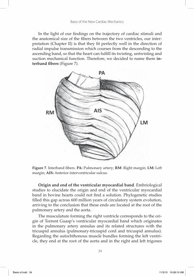
In the light of our fi ndings on the trajectory of cardiac stimuli and the anatomical size of the fi bers between the two ventricles, our inter-pretation (Chapter II) is that they fi t perfectly well in the direction of radial impulse transmission which courses from the descending to the ascending band, so that the heart can fulfi ll its twisting, untwisting and suction mechanical function. Therefore, we decided to name them in-terband fi bers (Figure 7).
Figure 7. Interband fi bers. PA: Pulmonary artery; RM: Right margin; LM: Left margin; AIS: Anterior interventricular sulcus.
Origin and end of the ventricular myocardial band. Embriological studies to elucidate the origin and end of the ventricular myocardial band in bovine hearts could not fi nd a solution. Phylogenetic studies fi lled this gap across 600 million years of circulatory system evolution, arriving to the conclusion that these ends are located at the root of the pulmonary artery and the aorta.
The musculature forming the right ventricle corresponds to the ori-gin of Torrent Guasp’s ventricular myocardial band which originates in the pulmonary artery annulus and its related structures with the tricuspid annulus (pulmonary-tricuspid cord and tricuspid annulus). Regarding the autochthonous muscle bundles forming the left ventri-cle, they end at the root of the aorta and in the right and left trigones
Basis of.indd 34Basis of.indd 34 11/3/15 10:08:10 AM11/3/15 10:08:10 AM

Basis of the New Cardiac Mechanics
35
(constituting the aortic annulus) (Figure 8). This arrangement allowed Torrent Guasp to deduce that the aorta, at its root, represents the end of the heart’s ventricular myocardial band.(69)
Figure 8. Septal portion of the aortic annulus between the right and left trigone. PA: Pulmonary artery; LT: Left trigone; RT: Right trigone; A: Aorta; MT: Mitral valve; TV: Tricuspid valve.
2. Phylogenetic development of the circulatory system
The circulatory apparatus of worms (Annelida, Nemerthea) consists of a closed system with two capillary beds (respiratory and systemic) with semicircular arterial and venous segments. In this unique circula-tory system blood is pumped by peristalsis (expression and suction) as it lacks a heart impulse (Figure 9 A).
In the evolution to fi shes three linear dilatations appear in the ve-nous semicircle (venous sinus, atrium and ventricle) forming a primi-tive heart. Although it is still a single circuit a pumping organ was in-serted increasing the possibility of intravascular pressure transmission (Figure 9 B).
Basis of.indd 35Basis of.indd 35 11/3/15 10:08:10 AM11/3/15 10:08:10 AM

Basis of the New Cardiac Mechanics
36
In the following evolutionary stage of amphibians and reptiles more prominent modifi cations are produced. At this stage of evolutionary development two circuits are identifi ed: the systemic and respiratory circuits, with the heart presenting two atria and one ventricle, this last generated by the incipient twist of the arterial circuit.(74) This self-rota-tion in a segment of the arterial semicircle (systemic circuit) is a crucial step in the evolutionary development of species (Figure 9 C). This rota-tion forms the future ventricular chambers in their defi nitive confi gu-ration, a fact well understood when the ventricular myocardial band forming the ventricles is unfolded.
Figure 9. Phylogenetic development of the circulatory system. VS: venous segment; AS: arterial segment. Arrows indicate the direction of circulation.
The development of the circulatory system of birds and mammals shows two atria and two ventricles. The twist of the tube which started in amphibians and reptiles is completed in the arterial semicircle form-ing a helical system. In this segment, the longitudinal incision, as a knife slit in the primitive arterial tube, gives rise to canals that make up the band separating the two ventricles (Figure 9 D). The pulmonary artery and aortic roots present today evidence of this evolutionary incision in the arterial circuit. This process succeeds in pumping blood from the loop exit into the systemic bed with low energy cost and high speed (in humans its reaches 300 cm/s) via the left ventricle and into the pulmo-nary artery via the right ventricle at pressures which are 20% those in the systemic circuit. This stratagem allowed the development of intra-vascular pressure in the arterial circuit segment that propels blood at a speed that manages to irrigate the whole organism.
If we step back to the primitive stage in the evolution of the circula-tory system we can appreciate the phylogenetic hallmarks of different species. The atria belong to the venous segment and the ventricles to the arterial segment. A subsequent more pronounced twist of the arterial segment puts into contact the atria with their corresponding ventricles. In summary, the comparison of fi sh and mammals shows that the single
Basis of.indd 36Basis of.indd 36 11/3/15 10:08:11 AM11/3/15 10:08:11 AM
Basis of the New Cardiac Mechanics
37
atrium and ventricle of the former become the right and left atria in the latter. In turn, the mammalian ventricles originate from the budding arterial segment twist corresponding to the arterial circuit of amphib-ians and reptiles.(68,74)
The concept of circulatory trajectory prevented a clear understand-ing of the muscle anatomical unity. This muscular mass forms the cham-bers where blood circulates, since its dynamic movement is determined by the ventricular myocardial band. The arrangement of both atria in a horizontal plane (chambers originated in the venous semicircle) attach-es to the ventricular component plane (arterial semicircle) where the chambers shaped by the myocardial band receive the motor impulse for ventricular contraction and suction.
In conclusion, according to Torrent Guasp’s concept, the unifi cation of the arterial and venous semicircles originating the circulatory sys-tem of birds and mammals determines the emergence of the ventricular myocardial band from an arterial loop and the differentiation of the right and left ventricular chambers when that tube segment is split into two.(68)
3. The cardiac apex
The apex -formed exclusively by the left ventricle- is a region situat-ed in a twist of the descending band in its ascending continuation. This helical rotation of its fi bers that from a subepicardial position become subendocardial, forms a coil of circularly interdigitating muscle layers that create a virtual rather than a real tunnel, as systolic contraction nar-rows it similarly to the mitral orifi ce. The apical “cul de sac” is lined in the inside by the endocardium and outside is covered by the epicardium.
The spatial confi guration of the double consecutive passage of the descending band situated posteriorly to the ascending band (Figure 6) would allow the apex to turn fi rst to the left during systole (seen from the apex) and then to the right, at the onset of the isovolumic diastolic phase with persisting contraction of the ascending segment. The con-tinuation of descending and ascending bands is a continuum that in this vertex allows the apical loop to act as a bellows that shortens dur-ing systole and lengthens during the isovolumic diastolic phase.
As a result of this anatomo-functional process the apex enables the approximation of the base to the tip of the heart during systole (shorten-ing) and its separation during diastole achieving ventricular elongation.
Basis of.indd 37Basis of.indd 37 11/3/15 10:08:11 AM11/3/15 10:08:11 AM
Basis of the New Cardiac Mechanics
38
This mainly longitudinal functional interplay favors the residual sys-tolic volume (30% of total diastolic volume) in the apical cul-de-sac.(81) At this point, we consider that the spatial arrangement of the double consecutive passage of the descending band situated posteriorly to the ascending band allows the non-ejection of part of the cardiac volume at end systole, remaining as residual volume. This remaining fl uid acts as a limiting layer for correct suction during the isovolumic diastolic phase (Chapter II).
The apex does not make any measurable movement. It remains practically immobile during the whole cardiac cycle producing only a certain pressure on the chest wall (apex beat). It is the base of the heart which shifts as it descends (reducing ventricular volume) and ascends (increasing ventricular volume).
During systole, the heart undergoes a jet propulsion motion (prin-ciple of action and reaction). The apex is the main subordinate region of the retrograde force affecting the ventricular chamber when blood is ejected during systole. Similarly to other body regions with stress overload (J.L. Petit triangle)1 it lacks muscle. In addition, it presents pre-carious irrigation and is submitted to a fi nal pressure in its cul-de-sac when the aortic valve closes. This relatively immobile avascular apical area, without interposing muscle, submitted to the maximum effect of left ventricular residual pressure, becomes the place where ventricular wall aneurysms originate in 90% of cases.
1. Weak area of the abdominal antero-lateral wall. It has a triangular shape whose sides are composed of the medial margin of the external abdominal oblique muscle, the lateral margin of the latissimus dorsi muscle and the iliac crest as inferior boundary. (A.N.)
Basis of.indd 38Basis of.indd 38 11/3/15 10:08:11 AM11/3/15 10:08:11 AM
39
Chapter II
Electrophysiological Interpretation Of The Ventricular Myocardial Band
1. Historical concepts of myocardial electrical activation
In 1998 Torrent Guasp wrote: “It would therefore be convenient, in order to validate the statement of a new concept of cardiac mechanics, to fi rst perform an experimental study exclusively directed towards demonstrating the reality of the new interpretation on the propagation of stimuli”.(70)
The propagation of stimuli throughout the ventricles has to follow a pattern that matches the topography of muscle bundles. This interpre-tation undoubtedly concerns the heart function in its contraction, suc-tion and dilatation phases. Structure and function are thus intimately linked and this concept has been the fundamental basis of our electro-physiological research to understand muscle twisting and untwisting and its cardiac suction effect.
In 1915, Thomas Lewis (39) had established that the stimuli arriving along the bundle of His are transmitted through the ventricular walls in an endocardial-epicardial direction, making the papillary muscles the fi rst electrically activated structures. This position was confi rmed by Parker in 1930.(52)
However, J. Robb and R. Robb posed in 1942 a fundamental ques-tion: “How is it possible that the transmission of electrical impulses occurs, as all electrical data indicate, from the endocardial to the epicardial surface, given that the ventricular wall is composed of well differentiated bundles separated by sheaths of collagen tissue?”(55) These same authors in dissection stud-ies performed in 1936 had already supported that propagation runs lon-
Basis of.indd 39Basis of.indd 39 11/3/15 10:08:11 AM11/3/15 10:08:11 AM

Basis of the New Cardiac Mechanics
40
gitudinally (axially) and not transversally, as defi ned by the classical view of Lewis with these statement: “These data indicate that the excitatory process is conducted axially in the muscles studied along a pathway parallel to fi ber direction”.(54)
Later, J. Armour and W. Randall (1) in 1970 demonstrated experi-mentally that subepicardial contraction precedes subendocardial con-traction. “Subepicardial muscle contraction rather forms a rigid shell within which the subsequent contraction of the remaining myocardial mass takes place, generating intraventricular pressure”. They also declared “the electri-cal activity propagates from the endocardium to the epicardium in the anterior surface of the left ventricular wall”. These concepts are in accordance with the anatomical arrangement of both ventricles within the myocardial mass, as in the integral loop confi guration, the basal loop embraces the apical loop, determining a right ventricular chamber that resembles an open slit in the muscle thickness forming both ventricles (Chapter I). C. Roy and J. Adami (57) confi rmed these conclusions in 1980 when they established the chronology between subepicardial contraction and mitral valve motion.
In 1987, García Civera, Cavadés and J. Cosin posed an open ques-tion abandoning the subtle discussion that “the sequence of epicardial ac-tivation does not bear exact correspondence with endocardial activation, the former being earlier and variable, even from individual to individual”.(25) Francisco Torrent Guasp confi rmed in 1998: “the succession of functional diffi culties of cardiac mechanics represents unequivocal proof of the longitu-dinal diffusion of stimuli along the ventricular myocardial band”.(70) This concept remained unchanged until the studies performed in the present investigation, based on the hypothesis that the axial transmission of the stimulus along the ventricular myocardial band supported by Torrent Guasp did not explain satisfactorily the twisting and untwisting move-ment indispensable to understand cardiac mechanics.
2. Stimuli propagation, muscle torsion and cardiac suction effect through electrophysiological research
Introduction
The hypothesis proposed by Torrent Guasp considers that the myo-cardium consists of a continuous single muscle band originating in the pulmonary valve and extending to the aortic root, thus limiting the two
Basis of.indd 40Basis of.indd 40 11/3/15 10:08:11 AM11/3/15 10:08:11 AM
Basis of the New Cardiac Mechanics
41
ventricular chambers. In its spatial trajectory two muscle bands can be identifi ed: the descending endocardial band and the ascending epicar-dial band, twisted in a double helical coil forming a basal loop (left and right basal segments) and an apical loop (descending and ascending apical segments). In this spatial arrangement, the descending and as-cending bands cross each other at a point we will call “band intersec-tion”.(9, 68) This anatomical peculiarity forming a fi gure of 8 and its stimulation sequence explains fundamental aspects of left ventricular dynamics: 1) the mechanism of ventricular torsion; 2) the physiology of rapid diastolic fi lling by the suction effect and 3) the residual systolic volume. Despite anatomical and then functional studies performed so far, it was necessary to establish a correlation with the endo-epicardial stimulation circuit, since there were no studies in humans.
Ventricular fi lling is generally assumed as a passive ventricular function, resulting from ventricular relaxation, elastic fi ber action and circulatory vis-a-tergo. Some authors (8,20,72) questioned that these mechanisms were suffi cient to explain rapid left ventricular fi lling. A mechanism was postulated by which at the onset of diastole (isovolu-mic diastolic phase) the ventricle actively aspirates blood by persistent muscle contraction of the “ascending band”. This action would produce apex-base lengthening with concomitant sudden decrease of intraven-tricular pressure until atrioventricular valve opening and subsequent rapid ventricular fi lling. (3-5,87) Various aspects of this theory have been challenged, mainly due to lack of an electrophysiological basis to sup-port it through the study of cardiac activation.(18) We investigated the sequence of endo and epicardial band electrical activation to explain ventricular torsion, the active suction effects in the isovolumic diastolic phase and the meaning of residual systolic volume.
Methods
The left ventricular endo and epicardial electrical activation se-quence has been studied using three-dimensional electroanatomic mapping (EAM) with a navigation system and Carto (Biosense Webster, California, USA) mapping, enabling three-dimensional anatomical rep-resentation, with activation maps and electrical propagation. Isochronic and activation sequence maps were performed, correlating them with surface ECG. An average of 50±8 endocardial and epicardial points were acquired for ventricular activation maps achieving detailed high density recordings. Apical, lateral and basal views were analyzed.
Basis of.indd 41Basis of.indd 41 11/3/15 10:08:12 AM11/3/15 10:08:12 AM
Basis of the New Cardiac Mechanics
42
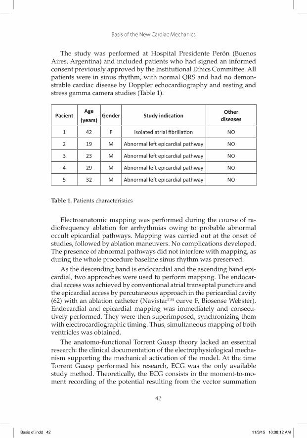
The study was performed at Hospital Presidente Perón (Buenos Aires, Argentina) and included patients who had signed an informed consent previously approved by the Institutional Ethics Committee. All patients were in sinus rhythm, with normal QRS and had no demon-strable cardiac disease by Doppler echocardiography and resting and stress gamma camera studies (Table 1).
Table 1. Patients characteristics
Electroanatomic mapping was performed during the course of ra-diofrequency ablation for arrhythmias owing to probable abnormal occult epicardial pathways. Mapping was carried out at the onset of studies, followed by ablation maneuvers. No complications developed. The presence of abnormal pathways did not interfere with mapping, as during the whole procedure baseline sinus rhythm was preserved.
As the descending band is endocardial and the ascending band epi-cardial, two approaches were used to perform mapping. The endocar-dial access was achieved by conventional atrial transeptal puncture and the epicardial access by percutaneous approach in the pericardial cavity (62) with an ablation catheter (Navistar™ curve F, Biosense Webster). Endocardial and epicardial mapping was immediately and consecu-tively performed. They were then superimposed, synchronizing them with electrocardiographic timing. Thus, simultaneous mapping of both ventricles was obtained.
The anatomo-functional Torrent Guasp theory lacked an essential research: the clinical documentation of the electrophysiological mecha-nism supporting the mechanical activation of the model. At the time Torrent Guasp performed his research, ECG was the only available study method. Theoretically, the ECG consists in the moment-to-mo-ment recording of the potential resulting from the vector summation
PacientAge
(years)Gender Study indicati on
Otherdiseases
1 42 F Isolated atrial fi brillati on NO
2 19 M Abnormal left epicardial pathway NO
3 23 M Abnormal left epicardial pathway NO
4 29 M Abnormal left epicardial pathway NO
5 32 M Abnormal left epicardial pathway NO
Basis of.indd 42Basis of.indd 42 11/3/15 10:08:12 AM11/3/15 10:08:12 AM
Basis of the New Cardiac Mechanics
43
of multiple simultaneous action potentials generated from the local ac-tivation of each area of the heart. Thus, the information provided by this method -invaluable in multiple aspects of cardiology- is limited to assess the discriminated spatio-temporal activation sequence for each ventricular region. The advent of clinical EAM overcame that limitation, as it not only allows the independent recording of different ventricular areas but also exclusive or integrated endocardial and epicardial areas.
We performed a high density and very detailed mapping of left ven-tricular endocardial and epicardial activation, according to the method-ology described above. Mapping was carried out simultaneously with surface ECG, providing a unifi ed temporal framework that enabled the correlation of both recordings and the synchronized view of the simul-taneous activation observed in different electroanatomic conditions.
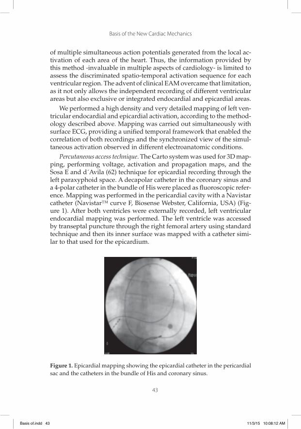
Percutaneous access technique. The Carto system was used for 3D map-ping, performing voltage, activation and propagation maps, and the Sosa E and d´Avila (62) technique for epicardial recording through the left paraxyphoid space. A decapolar catheter in the coronary sinus and a 4-polar catheter in the bundle of His were placed as fl uoroscopic refer-ence. Mapping was performed in the pericardial cavity with a Navistar catheter (Navistar™ curve F, Biosense Webster, California, USA) (Fig-ure 1). After both ventricles were externally recorded, left ventricular endocardial mapping was performed. The left ventricle was accessed by transeptal puncture through the right femoral artery using standard technique and then its inner surface was mapped with a catheter simi-lar to that used for the epicardium.
Figure 1. Epicardial mapping showing the epicardial catheter in the pericardial sac and the catheters in the bundle of His and coronary sinus.
Basis of.indd 43Basis of.indd 43 11/3/15 10:08:12 AM11/3/15 10:08:12 AM
Basis of the New Cardiac Mechanics
44
Results
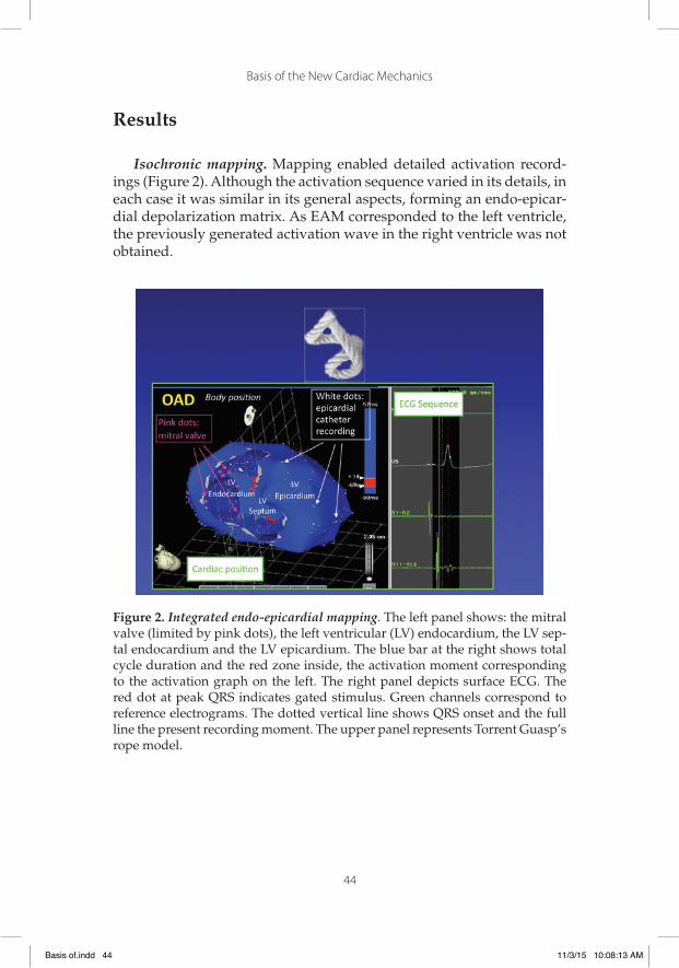
Isochronic mapping. Mapping enabled detailed activation record-ings (Figure 2). Although the activation sequence varied in its details, in each case it was similar in its general aspects, forming an endo-epicar-dial depolarization matrix. As EAM corresponded to the left ventricle, the previously generated activation wave in the right ventricle was not obtained.
Figure 2. Integrated endo-epicardial mapping. The left panel shows: the mitral valve (limited by pink dots), the left ventricular (LV) endocardium, the LV sep-tal endocardium and the LV epicardium. The blue bar at the right shows total cycle duration and the red zone inside, the activation moment corresponding to the activation graph on the left. The right panel depicts surface ECG. The red dot at peak QRS indicates gated stimulus. Green channels correspond to reference electrograms. The dotted vertical line shows QRS onset and the full line the present recording moment. The upper panel represents Torrent Guasp’s rope model.
Basis of.indd 44Basis of.indd 44 11/3/15 10:08:13 AM11/3/15 10:08:13 AM
Basis of the New Cardiac Mechanics
45
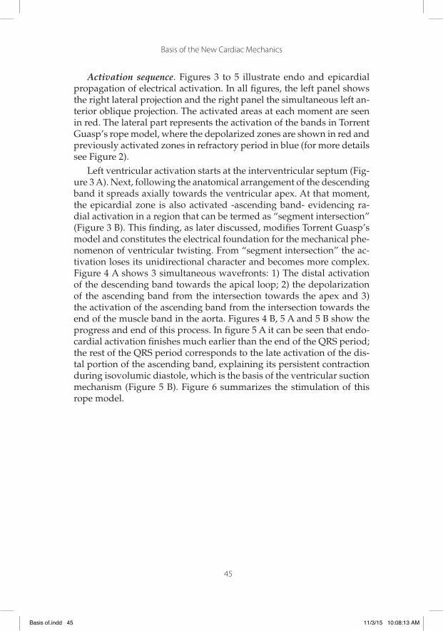
Activation sequence. Figures 3 to 5 illustrate endo and epicardial propagation of electrical activation. In all fi gures, the left panel shows the right lateral projection and the right panel the simultaneous left an-terior oblique projection. The activated areas at each moment are seen in red. The lateral part represents the activation of the bands in Torrent Guasp’s rope model, where the depolarized zones are shown in red and previously activated zones in refractory period in blue (for more details see Figure 2).
Left ventricular activation starts at the interventricular septum (Fig-ure 3 A). Next, following the anatomical arrangement of the descending band it spreads axially towards the ventricular apex. At that moment, the epicardial zone is also activated -ascending band- evidencing ra-dial activation in a region that can be termed as “segment intersection” (Figure 3 B). This fi nding, as later discussed, modifi es Torrent Guasp’s model and constitutes the electrical foundation for the mechanical phe-nomenon of ventricular twisting. From “segment intersection” the ac-tivation loses its unidirectional character and becomes more complex. Figure 4 A shows 3 simultaneous wavefronts: 1) The distal activation of the descending band towards the apical loop; 2) the depolarization of the ascending band from the intersection towards the apex and 3) the activation of the ascending band from the intersection towards the end of the muscle band in the aorta. Figures 4 B, 5 A and 5 B show the progress and end of this process. In fi gure 5 A it can be seen that endo-cardial activation fi nishes much earlier than the end of the QRS period; the rest of the QRS period corresponds to the late activation of the dis-tal portion of the ascending band, explaining its persistent contraction during isovolumic diastole, which is the basis of the ventricular suction mechanism (Figure 5 B). Figure 6 summarizes the stimulation of this rope model.
Basis of.indd 45Basis of.indd 45 11/3/15 10:08:13 AM11/3/15 10:08:13 AM
Basis of the New Cardiac Mechanics
46
Figure 3. A: Onset of left ventricular activation. The left panel illustrates the depo-larization of the ventricular septum, corresponding to the descending band. In the right panel, the ventricular epicardium (ascending band) has not been activated yet. B. Simultaneous band activation. The activation progresses in the left ventricular septum along the descending band (axial activation) and at the same time propa-gates to the epicardium (radial activation), activating the ascending band.
Figure 4. A: Bidirectional activation of the apex and the ascending band. The fi -nal septal activation is seen propagating towards the apex, synchronously with the epicardial activation in the same direction. At the same time, the epicardial activation propagates towards the base of the left ventricle. B: Propagation progress. The activation progresses in the directions of the previous fi gure.
Basis of.indd 46Basis of.indd 46 11/3/15 10:08:14 AM11/3/15 10:08:14 AM

Basis of the New Cardiac Mechanics
47
Figure 5. A: Late activation of the ascending band. At this moment, correspond-ing to approximately 60% of QRS duration, endocardial activation (descending band) has already been completed. The distal portion of the ascending band (epicardial) depolarizes lately. This phenomenon correlates with its persistent contraction at the initial phase of diastole. B: Final activation. In the right pan-el, the projection was modifi ed from left anterior oblique to left postero-lateral, showing the very late activation of the distal portion of the ascending band.
Figure 6. Rope model. Activation sequence of Torrent Guasp’s ventricular myo-cardial band (A-F) according to our fi ndings. Depolarization (red); repolariza-tion (blue).
Basis of.indd 47Basis of.indd 47 11/3/15 10:08:14 AM11/3/15 10:08:14 AM
Basis of the New Cardiac Mechanics
48
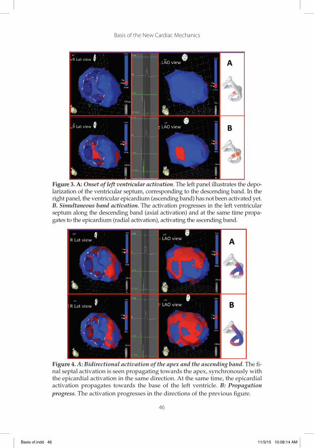
Figure 7. Rope model. Unidirectional propagation of excitation (red) in the ven-tricular myocardial band according to Torrent Guasp (A-D). Notice the differ-ence with Figure 6.
Discussion
Torrent Guasp’s continuous ventricular myocardial band hypoth-esis in cardiac mechanics implies a series of associated muscular move-ments. These occur in the band eliciting left ventricular narrowing, shortening, twisting, lengthening-untwisting and expansion phases during the cardiac cycle.
According to Torrent Guasp, longitudinal diffusion of stimuli along the ventricular myocardial band explained heart performance (Figure 7).(68) However, this sequential “peristaltic” activation did not cor-relate with some currently well-known fundamental phenomena, as clockwise and counter-clockwise twisting at the left ventricular apex and base, which are mainly responsible for its mechanical effi ciency (Figure 6). In an attempt to explain the mechanism of muscle twisting, we studied the sequence of ventricular electrical activation by means of simultaneous three-dimensional endo-epicardial segment mapping.
Electrical activation is the consequence of the propagation of stimuli through the muscular structure of the heart, both of Torrent Guasp’s ventricular myocardial band as the rest of the fi bers involved in its structural framework. The cardiac mechanism of suction and ejection requires structural-functional integration capable of unraveling the different dynamic aspects arising from the propagation of excitation. This indicates that the diastolic phase is an active process of contrac-tion producing increasing suction (due to its similarity we have called it “plunger mechanism”), (83) which at a certain level of intraventricular pressure opens the atrioventricular valves producing rapid fi lling.
Basis of.indd 48Basis of.indd 48 11/3/15 10:08:15 AM11/3/15 10:08:15 AM
Basis of the New Cardiac Mechanics
49
Stimulus propagation and left ventricular torsion
The integrated endo-epicardial three-dimensional mapping per-formed in this research supports the activation model that propagates along the descending and ascending bands. Figures 3 to 5 elucidate the activation sequence of the contractile areas and their entry into cardiac dynamics associated to the course of the excitation wave with a coordi-nated pattern according to muscle structure.
In this experience we found a stimulus trajectory different from that described by Torrent Guasp, but which explains the twisting phase of the heart, defi ned as the opposing rotational movement of the base and apex. At the point of band intersection the activation propagates from the endocardium to the epicardium (radial propagation), that is, from the descending to the ascending band. From the anatomical point of view, this passage could be mediated by “interband fi bers” (Torrent Guasp’s “aberrant fi bers”) (71) (Chapter I).
From this point onwards, the ascending band depolarizes in two senses: towards the apex and towards the base, at the same time that the descending band completes its activation towards the apex (Figure 5). Thus, two essential phenomena occur:
1. As the apical loop depolarizes from band intersection in two si-multaneous wavefronts (from the descending and from the as-cending bands) it generates their synchronized contraction.
2. The activation of the ascending band propagates from band inter-section in two opposing directions: towards the apex and towards the base (Figure 5). The resulting mechanical contraction will also have a divergent direction, giving origin to the apical and basal clockwise and counter-clockwise rotations, respectively.
According to Lewis (39), stimuli diffused from the endocardium to the epicardium through the muscle walls. Contrary to this concept, Robb (54) published in 1936 that stimuli propagation occurred longitu-dinally, and in 1942 inquired: “How is it possible that impulse transmission occurs from the endocardial to the epicardial surface… given that the ven-tricular wall is composed of well differentiated bundles, separated by sheaths of connective tissue?”.(55) Surprisingly, according to their experimental studies, Armour and Randall (1970) concluded that stimuli diffusion in the left ventricular anterior wall was generated from the endocardium to the epicardium.(1) This local event in the left ventricular anterior
Basis of.indd 49Basis of.indd 49 11/3/15 10:08:16 AM11/3/15 10:08:16 AM
Basis of the New Cardiac Mechanics
50
wall contrasts with previous concepts and with the remaining muscle mass where the electrical activity of subepicardial muscle bundles takes place before those in the subendocardium. However, this discrepancy of the impulse transmission theory through the ventricular myocardial band was not resolved until our research shed light on its understand-ing, with patent relevant considerations for cardiac mechanics. In 1960, Torrent Guasp expressed: “The subendocardial layers contracted by the de-scending segment come into activity before the subepicardial ones, which are components of the ascending segment” (66) and in 1988 he reaffi rmed “the descending segment and then the ascending segment successively enter into activity”.(70) Towards 2001, Buckberg and Torrent Guasp ratifi ed the hypothesis that excitation spreads unidirectionally along the ventricu-lar myocardial band (Figure 7).(8)
Our research modifi es these concepts since stimulus propagation is simultaneously axial and radial. This activation sequence has an im-portant anatomical component in cardiac architecture not evidenced by Torrent Guasp’s description (66), to the point of calling them “aber-rant fi bers” and that we will name “interband fi bers” (Chapter I), since they constitute the anatomical possibility of radial impulse propagation from the descending to the ascending band.
The ventricular narrowing phase (isovolumic systole) at the begin-ning of systole is produced by the contraction of the basal loop right and left segments. The overlapping shortening phase is due to the de-scent of the base, at the same time as twisting occurs, which is produced longitudinally, as the ring contracts before the apex. The fact that the apex remains fi xed, is due to the movement of the base, descending in systole and ascending in diastole. This is explained better because the ascending band, rigid in systole and at the beginning of diastole, acts as a tight tutor keeping the apex immobile. The pressure generated to eject the highest amount of blood at the onset of ejection during an interval lasting 20% of the systolic phase is feasible due to the twisting movement. This action is achieved because the electrical stimulation propagates towards the descending band (axial propagation) and si-multaneously to the ascending band (radial propagation). Although the electrical conduction progresses along the ventricular myocardial band, radial propagation towards the ascending band plays an essential role in ventricular twisting by allowing opposing forces on its longitudi-nal axis, generating the necessary intraventricular pressure to achieve abrupt blood ejection. The interband fi bers that cross from the descend-ing band to the ascending band would be responsible of impulse trans-mission between the bands (Chapter I). Thus, a twisting mechanism similar to “wringing a towel” would be produced.(80)
Basis of.indd 50Basis of.indd 50 11/3/15 10:08:16 AM11/3/15 10:08:16 AM

Basis of the New Cardiac Mechanics
51
The historical term of systole and diastole (79) did not take into ac-count the meaning of electrical activation and contraction, but only the hemodynamic concept of ventricular ejection and fi lling. It is therefore necessary to fi nd a relationship between activation and the mechani-cal outcome. The explanation is provided by the simultaneous axial and radial electrical conduction when it reaches band intersection, also confi rmed by the spatial arrangement of fi bers, with subendocardial fi bers on the right side and subepicardial fi bers on the left.(58) This layout also agrees with the evolutionary loop of the circulatory system forming the two developed ventricles in birds and mammals. Torsion -different rotation between the apex and base of the heart- generates: a) high pressures, b) reduces ventricular stress, and c) homogenizes its distribution in the ventricular wall thickness.
Torrent Guasp declared that “the sequence of ventricular muscle entry into activity in the different ventricular regions takes place along the band”, (70) similarly to a peristaltic movement (Figure 7). Then, how could the ventricle achieve its twisting movement, since this action requires two opposing forces at the same time? Unidirectional activation does not explain twisting or the evolutionary-structural development designed to apply a force capable of ejecting the ventricular content at a speed of 300 cm/s at low energetic cost. This is understood by the simultane-ous axial and radial activation we have found, whose sequence can be illustrated as (Diagram 1):
Diagram 1. References: r, right segment; l, left segment; sa, simultaneous ac-tivation of the ascending and descending segments (interband fi bers); A, as-cending segment; D, descending segment; T, torsion; IDP, isovolumic diastolic phase (active).
Basis of.indd 51Basis of.indd 51 11/3/15 10:08:16 AM11/3/15 10:08:16 AM
Basis of the New Cardiac Mechanics
52
Active suction in the isovolumic diastolic phase
The investigation of another previously unexplained point was to consider ventricular fi lling as an active phenomenon generated by myo-cardial contraction tending to increase left ventricular apex-base dis-tance after ejection, thus producing a suction effect through a “plunger” mechanism. This mechanism is explained by the persistence of the as-cending band contraction during isovolumic diastole.
We have found that the endocardium depolarizes completely dur-ing the fi rst part of the QRS. In turn, Buckberg found that the mechani-cal contraction triggered by this electrical mechanism is initiated 50 ms after its occurrence and persists for approximately 350 ms. If the depo-larization of the ascending band starts 50 ms after that of the descend-ing band and its contraction persists for the same length of time, the contractile state will last approximately 400 ms. If ventricular systole lasts about 300 ms, the remaining 100 ms correspond to the isovolumic diastolic phase (usually called isovolumic relaxation, though it can be seen that there is ventricular contraction). To summarize, during the initial part of this phase, the ascending band remains contracted as a consequence of the depolarization that took place during the QRS. The explanation of this late contraction does not require depolarizations af-ter the QRS, as postulated by Pedro Zarco.(88)
In our investigation, the fi nal part of the QRS corresponds to the activation of the ascending band (Figure 5), resulting in the necessary contraction to generate suction (“plunger effect”) during the isovolu-mic diastolic phase.
With the initiation of untwisting during isovolumic diastole, the as-cending band progressively lengthens, generating negative intraven-tricular pressure while it is still contracted (active process), as an ener-getic residue of the twisting process. On this point, Zarco expressed in 1998: “there is a point about which we cannot agree: that the straightening of the ascending segment is due to an active contraction of the cardiac muscle in full diastole”.(88) This is precisely what we have found in our research, establishing the mechanism that achieves rapid ventricular fi lling dur-ing a short interval in diastole.(61)
Basis of.indd 52Basis of.indd 52 11/3/15 10:08:16 AM11/3/15 10:08:16 AM
Basis of the New Cardiac Mechanics
53
Interpretation of the active suction phase
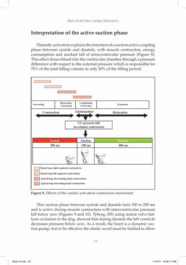
Diastolic activation explains the insertion of a suction active coupling phase between systole and diastole, with muscle contraction, energy consumption and marked fall of intraventricular pressure (Figure 8). This effect draws blood into the ventricular chamber through a pressure difference with respect to the external pressure which is responsible for 70% of the total fi lling volume in only 20% of the fi lling period.
Figure 8. Effects of the cardiac activation-contraction mechanism
This suction phase between systole and diastole lasts 100 to 200 ms and is active during muscle contraction with intraventricular pressure fall below zero (Figures 9 and 10). Tyberg, (85) using mitral valve bal-loon occlusion in the dog, showed that during diastole the left ventricle decreases pressure below zero. As a result, the heart is a dynamic suc-tion pump, but to be effective the elastic recoil must be limited to allow
Basis of.indd 53Basis of.indd 53 11/3/15 10:08:17 AM11/3/15 10:08:17 AM

Basis of the New Cardiac Mechanics
54
an effective subsequent systole. The effi cacy in surgeries that bypass the right ventricle (Fontan-Kreutzer) have shown this left ventricular suc-tion effect, (73) similarly to left mechanical support with univentricular devices, where blood is delivered from the left ventricle into the aorta.
Figure 9. Diagram of the electromechanical activity
Figure 10. Electrical activation and mechanical contraction of the ascending and descending bands (enlarged Figure 9 box).
In the traditional model, cardiac fi lling is only determined by venous pressure. Actually, atrial pressure is too low to explain this situation. Tor-rent Guasp developed the concept of active suction to explain this “key doubt”, supported by the physio-muscular structure he described.(82)
Basis of.indd 54Basis of.indd 54 11/3/15 10:08:17 AM11/3/15 10:08:17 AM
Basis of the New Cardiac Mechanics
55
The last contracting areas of the ascending band produce a suction ef-fect to draw blood towards the left ventricle. The high fi lling velocity at low pressures establishes that this is an active phenomenon.
Systolic residual volume
Systolic residual volume represents 30% of total diastolic volume. From the point of view of fl uid mechanics, between systole and diastole we fi nd a closed chamber that only contains blood, which is an incom-pressible fl uid, and hence, at any level of muscle contraction, this cham-ber volume cannot change. Therefore, this phase is isovolumic, and as it cannot change the volume, muscle contraction produces a depression (pressure fall) in the chamber favoring subsequent diastolic fi lling. In these conditions, the depression generated will depend on chamber ca-pacity of muscle contraction and geometry affecting the internal pres-sure distribution. It is physically necessary to have an incompressible fl u-id to produce this pressure fall, and that the chamber geometry and blood volume content allow an adequate depression (“plunger” mechanism).
There is a range of optimal residual systolic volume for suction. If it is higher it will require an important muscle contraction to develop the necessary depression, whereas if is lower, the interaction between walls will impair suction and hence the diastolic boundary layer phenomena will alter fi lling. From a physical standpoint, an excessive end-systolic volume will affect the suction capacity, which will thus be related with the volume pumped during systole.(22)
Conclusions
Our study shows a “three-stage heart”: systole, suction and diastole. The data obtained are especially important, as they were recorded in humans, with structurally normal hearts and in physiological condi-tions. According to these fi ndings, we may conclude:
1. Endo-epicardial 3-dimensional mapping shows a sequence of electrical activation in the region of the apical loop in agreement with the synchronous contraction of the descending and ascend-ing bands.
Basis of.indd 55Basis of.indd 55 11/3/15 10:08:18 AM11/3/15 10:08:18 AM
Basis of the New Cardiac Mechanics
56
2. The simultaneous and opposing activation of the ascending band to the starting point of its radial activation from the descending band, agrees with synchronous and opposite rotation of the apical and basal areas (mechanism of ventricular twisting).
3. The late activation of the ascending band, compatible with its per-sistent contraction during the initial phase of isovolumic diastole (untwisting and suction period), occurs without need of postulat-ing electrical activation after the QRS.
3. Electrocardiographic correlation
The onset of the QRS correlates with early endocardial depolariza-tion of the left ventricular anteroseptal wall. The depolarization of the epicardial segment starts about 50-60 ms after endocardial depolariza-tion (corresponding approximately to the fi nal 40% of QRS duration, coincident with peak positive dP/dt), generating the contraction of the apical loop ascending segment as shown in electrophysiological endo-epicardial mapping recordings. Certainly, small differences between QRS onset and end must be accounted for, as well as those of endocar-dial and epicardial potentials, similar to the ones recorded in electro-physiological studies.
Since Harvey and then Einthoven, contraction and electrical activa-tion of the heart have been considered as almost linear and homoge-neous processes. According to the classical concept, normal ventricular activation would have to be initiated in the septum, to continue in the apex and end in the basal region. In this way, contraction would occur “en bloc” during systole and relaxation would ensue almost homoge-neously during diastole. Systole is synonym of cardiac contraction and diastole of relaxation. These have been the classical concepts, and at this stage of knowledge they must be considered more complex.
Although several aspects of ventricular electrical stimulus propaga-tion have long been known, the advent of three-dimensional navigators and electroanatomic mapping have enabled a more detailed study of electrical propagation in the human heart in completely physiological clinical situations, revealing various interesting characteristics:
1. Normal electrical activation, though basically ordered and homo-geneous, is very complex especially in lately depolarized areas.
Basis of.indd 56Basis of.indd 56 11/3/15 10:08:19 AM11/3/15 10:08:19 AM
Basis of the New Cardiac Mechanics
57
2. Intraventricular activation “occupies” approximately the initial 60% of surface QRS duration. The “rest” of the QRS corresponds to myocardial and epicardial activation. The onset of ventricular activation evidenced by the QRS complex is “exclusively” en-docardial; during the intermediate phase both activations coex-ist and at the end, during suction, the activation is “exclusively” myo-epicardial.
3. The end of the apical loop ascending segment contraction is dia-stolic, indicating that the classically termed isovolumic relaxation phase is an active process.
In conclusion, ventricular electrical activation is a very complex process generating intricate patterns in the ventricular contraction it elicits. This complexity is further complicated by the addition of electro-mechanical coupling factors, intrinsic contractility of different cardiac regions and mechanical coupling, among others, which substantially modify basic concepts of cardiac mechanics evidenced so far.
It is possible to hypothesize about some aspects of this research:
a. Although there is no problem in defi ning the onset of systole as the moment in which the mitral valve closes and that of diastole as closure of the aortic valve, this does not imply that the process of contraction and relaxation start at those precise moments. On the contrary, it is obvious that the ventricular contraction gener-ating the increase in pressure required for mitral valve closure occurred before this point (that is, during diastole) and the re-laxation which reduced intraventricular pressure allowing aortic valve closure also started before this took place, that is, during systole. Likewise, it is reasonable to infer that not all myocardial fi bers contract or relax simultaneously, but that at certain periods (impossible to quantify for each individual fi ber) the fi bers coex-ist at different stages of contraction and relaxation, both during systole and diastole.
b. The fi nal state of contraction achieved (and hence the general ven-tricular geometrical confi guration and specially that of its cham-ber) must depend to a certain degree, and perhaps very signifi -cantly, on the state of contraction or relaxation of adjacent fi bers or structures. It seems logical to assume that the fi bers that con-tract in the fi rst place, surrounded by relaxed fi bers, may reach their maximum degree of potential contraction; conversely, if the
Basis of.indd 57Basis of.indd 57 11/3/15 10:08:19 AM11/3/15 10:08:19 AM
Basis of the New Cardiac Mechanics
58
adjacent fi bers are already contracted and “pulling” in an oppo-site direction, the degree of effective contraction will be lower.
c. Something similar might happen if we consider a structure such as the ventricle in its entirety and in the opposite situation of fi nd-ing a region of the heart relaxed while another is contracted. In this case, muscle shortening is completed producing chamber dilatation.
d. Electromechanical coupling. Even though electrical depolariza-tion triggers mechanical contraction, from this moment two dif-ferent and autonomous processes take place. Membrane activa-tion may occur without mechanical contraction, as evidenced by neuronal electrical activation eliciting the release of neurotrans-mitters with no contractile activity. This would be similar to hav-ing a selective inhibitor of actin-myosin activation producing a normal depolarization without muscle contraction.
On the other hand, the temporal relationship between electrical ac-tivity and mechanical contraction is probably not linear. It is known that above all, there is latency between electrical and mechanical activ-ity. Moreover, the time between electrical activation and contraction (at least, maximal contraction) probably varies depending on numerous factors: the type of fi ber, its metabolic condition, its place in myocardial wall thickness, the level of intracellular calcium, the contraction or re-laxation of neighboring fi bers, etc.
Let us consider typical situations: the QRS lasts approximately 100 ms, and systole 300 ms. Therefore, the QRS effects on contraction “per-sist” at least for those 300 ms after its end. Furthermore, fi bers activated at the end of the QRS contract or remain contracted at the beginning of diastole. Therefore, depolarizing potentials in the T wave would not be necessary to explain muscle contraction during diastole (which, as already mentioned, is not synonym for relaxation of all cardiac fi bers).
4. Functional aspects of the ventricular myocardial band
The analysis of the anatomical description of Torrent Guasp’s ven-tricular myocardial band and the ensuing electrophysiological research in patients performed by us indicates the need of providing a clear interpretation of some fundamental aspects. Of the three descending
Basis of.indd 58Basis of.indd 58 11/3/15 10:08:19 AM11/3/15 10:08:19 AM
Basis of the New Cardiac Mechanics
59
band turns relative to the ascending band, the fi rst two successively pass in front and behind the ascending band forming the basal loop. The last step, after the twist of the large band that turns it in the de-scending segment, it becomes again posterior to the ascending band, with a failed helicoidal arrangement in this space of the apical loop. This spatial anatomical situation of the muscle bands establishes a sig-nifi cant correlation with cardiac function.
The ventricular narrowing movement takes place at the base of the heart, conditioned by the consecutive basal loop right and left segment activation that produces the narrowing phase (systolic contraction). The progression of this phase, in the stimulation process of the descend-ing band (axial), together with the concomitant propagation (radial) to-wards the ascending band found in our investigations, determines a he-lical rotating motion with the ensuing shortening of the left ventricular vertical axis (ejection). The cardiac apex, composed of the subepicardial arrangement of fi bers that in their twist become subendocardial, consti-tute a free apex with a “cul de sac” to support the intraventricular pres-sure produced by the heart impulse during ventricular shortening. The subsequent movement as a result of the ascending band contraction, its straightening and ventricular lengthening therefore determine an active mechanism during isovolumic diastole that maintains the cham-ber in an isovolumic condition but with intraventricular pressure fall (untwisting and active suction phase) to give place to ventricular fi lling with atrioventricular opening (expansion phase). These left ventricular shortening (base descent) and lengthening (base ascent) movements correlate with the principle of Newton´s action and reaction principle.
As a result of this anatomo-functional process, the apex (space be-tween the two bands) has in normal conditions the power of annular narrowing (sphincter-like mechanism) to bear the retrograde intraven-tricular pressure produced by blood ejection.
At the beginning of the isovolumic (active) phase, erroneously con-sidered as belonging to diastole, which instead should be named suc-tion phase, the persistent contraction of the ascending band with left ventricular lengthening generates the fall of intraventricular pressure, suffi cient to achieve ventricular suction. This pressure fall produced by the persistent contraction of the ascending band during the isovolumic phase with every left ventricular orifi ce closed, works as a “plunger” mechanism (suction phase) at the moment it starts lengthening. When this pressure is suffi ciently low (less than 10 mmHg) and with the ventricle expanded and “unscrewed”, the mitral valve opens and the abrupt fl ow of blood from the atrium takes place (fi lling phase).
Basis of.indd 59Basis of.indd 59 11/3/15 10:08:19 AM11/3/15 10:08:19 AM
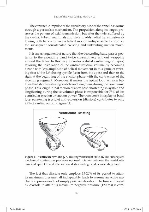
Basis of the New Cardiac Mechanics
60
The contractile impulse of the circulatory tube of the annelids worms through a peristalsis mechanism. The propulsion along its length pre-serves the pattern of axial transmission, but after the twist suffered by the cardiac tube in mammals and birds it adds radial transmission al-lowing both bands to have a helical motion indispensable to produce the subsequent concatenated twisting and untwisting-suction move-ments.
It is an arrangement of nature that the descending band passes pos-terior to the ascending band twice consecutively without wrapping around the latter. In this way it creates a distal cardiac region (apex) favoring the installation of the cardiac residual volume by becoming a zone with less amplitude of helical movement in this game of twist-ing fi rst to the left during systole (seen from the apex) and then to the right at the beginning of the suction phase with the contraction of the ascending segment. Moreover, it makes the apical loop act as a bel-lows that shortens during systole and lengthens during the isovolumic phase. This longitudinal motion of apex-base shortening in systole and lengthening during the isovolumic phase is responsible for 75% of left ventricular ejection or suction power. The transverse interplay of basal loop narrowing (systole) and expansion (diastole) contributes to only 25% of cardiac output (Figure 11).
Figure 11. Ventricular twisting. A. Resting ventricular state. B. The subsequent mechanical contraction produces opposed rotation between the ventricular base and apex. C: band intersection; d: descending band; a: ascending band.
The fact that diastole only employs 15-20% of its period to attain its maximum pressure fall indisputably leads to assume an active me-chanical process and not simply passive relaxation. The time employed by diastole to attain its maximum negative pressure (120 ms) is com-
Basis of.indd 60Basis of.indd 60 11/3/15 10:08:20 AM11/3/15 10:08:20 AM
Basis of the New Cardiac Mechanics
61
parable to that used by systole to achieve its maximum ejective pres-sure (140 ms). Sarcomere architecture in the spatial integration and its contractile framework, as well as its biochemical structure (elastin, titin) implicates elastic properties that add to the active phase. The force gen-erating negative intraventricular pressure through the ascending band activation acts on myocardial elastic properties to achieve the optimal effective sarcomere recoil intime and limit for adequate relaxation. The violation of this limit imposed by the architecture of cardiac muscle will have great impact on heart failure.
Neither is systole synonym for contraction nor diastole for relax-ation. This denomination is a dialectic that keeps no relation with the real movements that originate ventricular systole, suction and diastole, the three heart stages.
Basis of.indd 61Basis of.indd 61 11/3/15 10:08:20 AM11/3/15 10:08:20 AM

Basis of.indd 62Basis of.indd 62 11/3/15 10:08:20 AM11/3/15 10:08:20 AM

63
Chapter III
The Three Stage Heart
1. Chronology of the suction mechanism concept
Erasistratus of Keos, a Greek anatomist and physician in Alexandria (300-250 B.C.), detailed the anatomy of the heart, described its valves, giving its current name to the tricuspid valve, and referred to the inter-mediate system between arteries and veins, which he called “synanas-tomoseis”, as a fi rst approach to the capillaries. The latter would have led Erasistratus, had he not been infl uenced by the pneumatic theory -according to which the pneuma had to be transported by the arteries along the body to the veins by means of “synanastomoseis”- to a more precise knowledge of circulation.(79) Erasistratus considered that ar-teries did not carry blood, but pneuma, and assumed that the funda-mental movement of the heart was dilatation and was produced by a contracting muscle.(70) This concept persisted in history by the work of Galen of Pergamus (130-200 A.D.). In 1542, Andrea Vesalius in his work “De Humani Corporis Fabrica” stated “when the left ventricle expands again, breath and blood are attracted once more towards the ventricle”. (79)
In 1628, William Harvey in the fundamental work of human physi-ology “De Motu Cordis” regarded systole as the main fact of cardiac function. With this perspective he opposes Galen, who believed that the activity of the heart was manifested in its expansion, by means of the vis pulsifi ca.(79) It was necessary to wait until 1954 when Francisco Torrent Guasp (67) using a theoretical approach describes diastolic ventricular suction as an active contractile process, leading G. Brecher (4) (1956) to assume that “a demonstrative experimental evidence of diastolic ventricular suction is very diffi cult”. However, this same author elaborated on this concept and in 1958 established the certain possibility of ventricular suction.(5) The efforts, mainly of anatomical research, together with an intense refl ective analysis on the subject, led to the acknowledge-
Basis of.indd 63Basis of.indd 63 11/3/15 10:08:21 AM11/3/15 10:08:21 AM
Basis of the New Cardiac Mechanics
64
ment of the ventricular myocardial band postulated by Torrent Guasp (67-70) though without a clear physiological interpretation. This situa-tion would progress in the physiological understanding of ventricular pressure in the different phases of the cardiac cycle through the work performed by Francisco Torrent Guasp (68), D. Streeter (63), P. Lunken-heimer (40) and Gerard Buckberg.(7,8)
The experimental study in pigs by Juan Cosín Aguilar et al. in 2009, using piezoelectric crystals on the ventricular wall, considered the possi-bility of myocardial contraction in the isovolumic diastolic phase.(20)
2. Cardiac mechanics
Background. The mechanical activity of the heart is complex because it results from integrating its ejection, suction and fi lling properties, in different short successive phases, correlated in short periods. Anatomy texts describe the heart but pay little attention to the position adopted by myocardial fi bers. Physiologists do not even mention it, as if the mechanical heart had no bearing on the scaffolding design developed by muscle fi bers in the wall of the ventricles. It is impossible to inter-pret cardiac function measurements with diagnostic purposes without taking into account its morphological structure. Cardiologists and clini-cal researchers focus on the molecular aspects to explain heart failure following neurohormonal models. However, drugs that act upon neu-rohormonal activation alleviate but do not cure heart failure. Size and geometrical changes are responsible for the abnormal myocytes and myocardial structure that worsen cardiac function, enhance neurohor-monal activity and reduce cardiovascular system response. The mor-bidity and mortality prognosis of patients with heart failure is directly related to ventricular dilatation.(65)
Myocardial capacity is conditioned by cardiac fi ber orientation. In that regard, the conical heart has helical and circumferential myocardial fi bers. Fiber orientation determines heart function. Thus, the ejection fraction is 60% when the normal helical fi bers contract and drops to 30% if the transverse fi bers shorten.(7) The development of a spherical con-fi guration modifi es the orientation of the helical fi bers until a transverse dilatation is achieved with consequent decrease in contractile force.
Regarding the mechanism of ventricular fi lling two hypotheses have been generated:
Basis of.indd 64Basis of.indd 64 11/3/15 10:08:21 AM11/3/15 10:08:21 AM

Basis of the New Cardiac Mechanics
65
a) Classical model. According to Harvey (79), ventricular fi lling is achieved by atrial contraction or by pressure gradient, the latter being the hypothesis proposed by Wiggers in the 1920s.(86)
The atria have two main functions: one as transportation or pump-ing and another as reservoir for ventricular fi lling. Like the ventricles, the atria respond to an increase in fi ber length with an increase in con-tractile force and at rest atrial contraction contributes 20% to ventricular fi lling. Increase in atrial contractility and deviation to the left of atrial function curves or deviation to the right of velocity-force curves may re-sult from sympathetic stimulation, vagal inhibition or inotropic agents. In atrial fi brillation, circulatory reserve mechanisms may sustain car-diac output at rest, or otherwise, loss of atrial contraction may result in serious consequences for ventricular fi lling and heart function.
At the onset of the isovolumic phase, left ventricular pressure falls below left atrial pressure shortly after peak left atrial V wave. Moreover, the ventricular rapid fi lling phase at the end of the isovolumic phase coincides with the continuous atrial pressure decline that began during mitral opening, while the end of rapid ventricular fi lling and the begin-ning of slow ventricular fi lling is characterized by a change in the rise of the ventricular volume curve. During slow ventricular fi lling, left atrial and ventricular pressures increase slowly until the next atrial systole. Atrial contraction and the concomitant increase of ventricular fi lling are manifested by increased pressure and ventricular volume.
b) Suction mechanism model. The ventricle is an active suction pump during diastole. This hypothesis was proposed by Erasistratus in the third century B.C. and later by Galen (second century A.D.) and Vesalius (1542).(70,79) Diastolic fi lling by an active suction mechanism is supported by Torrent Guasp’s hypothesis (71) and by experimental and clinical studies.(4,20) The three components of the heart’s three-dimensional reduction interact synergistically through the ventricu-lar myocardial band rotating around itself to achieve the ventricular twisting and then untwisting-suction mechanism. In systole there is a three-dimensional reorientation of cardiac architecture with helical shortening in a longitudinal and transverse sense, producing a twisting movement. The atrioventricular annulus contracts, the aortic annulus slightly expands, the mitral and aortic planes descend and the left ven-tricular outfl ow tract remains open.
Suction occurs in early diastole. During this period (isovolumic phase) presence of negative intraventricular pressure has been docu-mented both in human clinical studies as in animals with balloon mitral valve occlusion.(23,56,61,85)
Basis of.indd 65Basis of.indd 65 11/3/15 10:08:21 AM11/3/15 10:08:21 AM
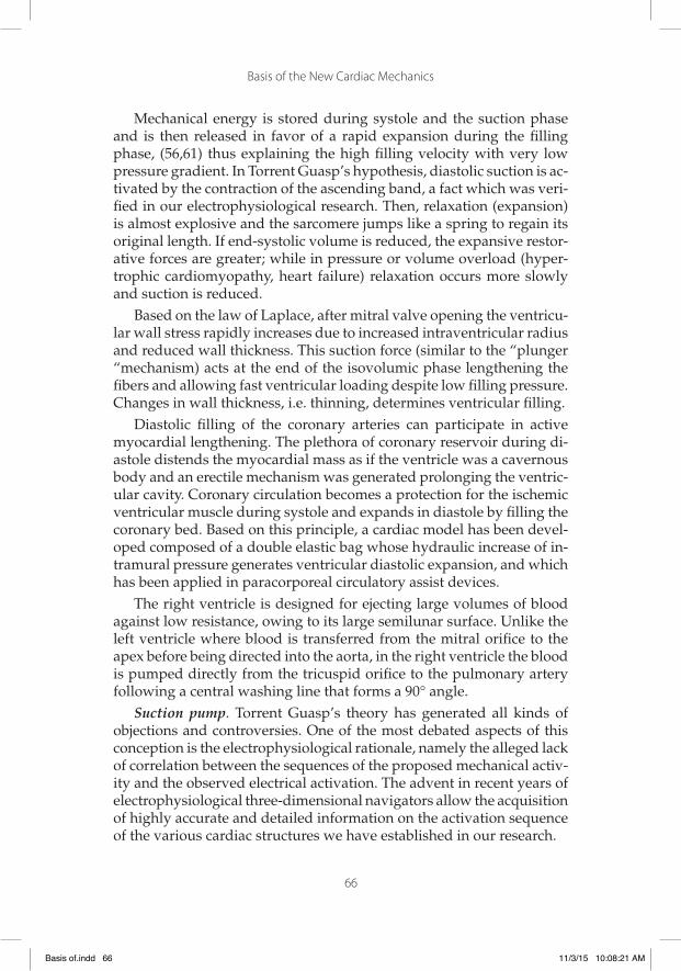
Basis of the New Cardiac Mechanics
66
Mechanical energy is stored during systole and the suction phase and is then released in favor of a rapid expansion during the fi lling phase, (56,61) thus explaining the high fi lling velocity with very low pressure gradient. In Torrent Guasp’s hypothesis, diastolic suction is ac-tivated by the contraction of the ascending band, a fact which was veri-fi ed in our electrophysiological research. Then, relaxation (expansion) is almost explosive and the sarcomere jumps like a spring to regain its original length. If end-systolic volume is reduced, the expansive restor-ative forces are greater; while in pressure or volume overload (hyper-trophic cardiomyopathy, heart failure) relaxation occurs more slowly and suction is reduced.
Based on the law of Laplace, after mitral valve opening the ventricu-lar wall stress rapidly increases due to increased intraventricular radius and reduced wall thickness. This suction force (similar to the “plunger “mechanism) acts at the end of the isovolumic phase lengthening the fi bers and allowing fast ventricular loading despite low fi lling pressure. Changes in wall thickness, i.e. thinning, determines ventricular fi lling.
Diastolic fi lling of the coronary arteries can participate in active myocardial lengthening. The plethora of coronary reservoir during di-astole distends the myocardial mass as if the ventricle was a cavernous body and an erectile mechanism was generated prolonging the ventric-ular cavity. Coronary circulation becomes a protection for the ischemic ventricular muscle during systole and expands in diastole by fi lling the coronary bed. Based on this principle, a cardiac model has been devel-oped composed of a double elastic bag whose hydraulic increase of in-tramural pressure generates ventricular diastolic expansion, and which has been applied in paracorporeal circulatory assist devices.
The right ventricle is designed for ejecting large volumes of blood against low resistance, owing to its large semilunar surface. Unlike the left ventricle where blood is transferred from the mitral orifi ce to the apex before being directed into the aorta, in the right ventricle the blood is pumped directly from the tricuspid orifi ce to the pulmonary artery following a central washing line that forms a 90° angle.
Suction pump. Torrent Guasp’s theory has generated all kinds of objections and controversies. One of the most debated aspects of this conception is the electrophysiological rationale, namely the alleged lack of correlation between the sequences of the proposed mechanical activ-ity and the observed electrical activation. The advent in recent years of electrophysiological three-dimensional navigators allow the acquisition of highly accurate and detailed information on the activation sequence of the various cardiac structures we have established in our research.
Basis of.indd 66Basis of.indd 66 11/3/15 10:08:21 AM11/3/15 10:08:21 AM
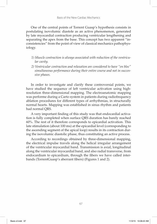
Basis of the New Cardiac Mechanics
67
One of the central points of Torrent Guasp’s hypothesis consists in postulating isovolumic diastole as an active phenomenon, generated by late myocardial contraction producing ventricular lengthening and separating the apex from the base. This concept has two apparent “in-consistencies” from the point of view of classical mechanics pathophys-iology.
1) Muscle contraction is always associated with reduction of the ventricu-lar cavity.
2) Ventricular contraction and relaxation are considered to have “en bloc” simultaneous performance during their entire course and not in succes-sive phases.
In order to investigate and clarify these controversial points, we have studied the sequence of left ventricular activation using high-resolution three-dimensional mapping. The electroanatomic mapping was performe during a Carto system in patients during radiofrequency ablation procedures for different types of arrhythmias, in structurally normal hearts. Mapping was established in sinus rhythm and patients had normal QRS.
A very important fi nding of this study was that endocardial activa-tion is fully completed when surface QRS duration has barely reached 60%. The rest of it therefore corresponds to epicardial activation. This late stimulation (about 100 ms) at the epicardial level (corresponding to the ascending segment of the apical loop) results in its contraction dur-ing the isovolumic diastolic phase, thus constituting an active process.
According to recordings obtained by three-dimensional mapping, the electrical impulse travels along the helical irregular arrangement of the ventricular myocardial band. Transmission is axial, longitudinal along the ventricular myocardial band, and also radial transverse, from endocardium to epicardium, through the fi bers we have called inter-bands (TorrentGuasp’s aberrant fi bers) (Figures 1 and 2).
Basis of.indd 67Basis of.indd 67 11/3/15 10:08:22 AM11/3/15 10:08:22 AM
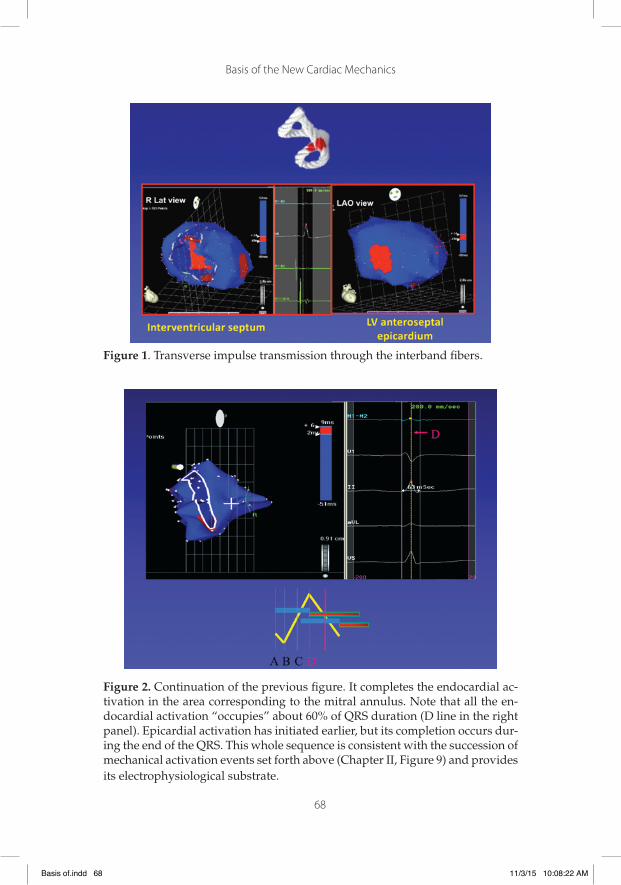
Basis of the New Cardiac Mechanics
68
Figure 1. Transverse impulse transmission through the interband fi bers.
Figure 2. Continuation of the previous fi gure. It completes the endocardial ac-tivation in the area corresponding to the mitral annulus. Note that all the en-docardial activation “occupies” about 60% of QRS duration (D line in the right panel). Epicardial activation has initiated earlier, but its completion occurs dur-ing the end of the QRS. This whole sequence is consistent with the succession of mechanical activation events set forth above (Chapter II, Figure 9) and provides its electrophysiological substrate.
Basis of.indd 68Basis of.indd 68 11/3/15 10:08:22 AM11/3/15 10:08:22 AM
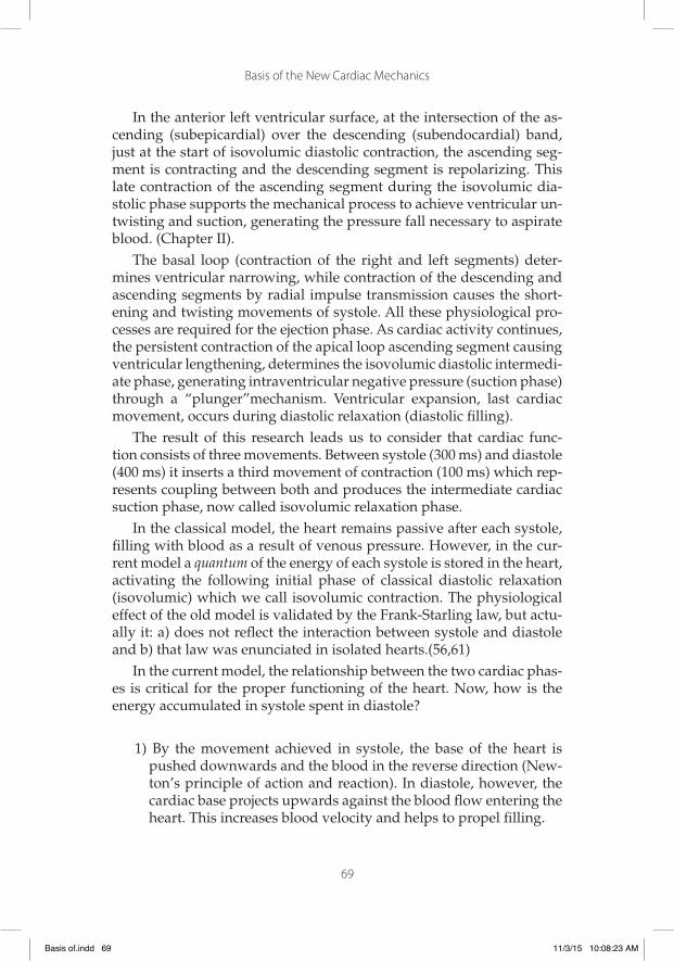
Basis of the New Cardiac Mechanics
69
In the anterior left ventricular surface, at the intersection of the as-cending (subepicardial) over the descending (subendocardial) band, just at the start of isovolumic diastolic contraction, the ascending seg-ment is contracting and the descending segment is repolarizing. This late contraction of the ascending segment during the isovolumic dia-stolic phase supports the mechanical process to achieve ventricular un-twisting and suction, generating the pressure fall necessary to aspirate blood. (Chapter II).
The basal loop (contraction of the right and left segments) deter-mines ventricular narrowing, while contraction of the descending and ascending segments by radial impulse transmission causes the short-ening and twisting movements of systole. All these physiological pro-cesses are required for the ejection phase. As cardiac activity continues, the persistent contraction of the apical loop ascending segment causing ventricular lengthening, determines the isovolumic diastolic intermedi-ate phase, generating intraventricular negative pressure (suction phase) through a “plunger”mechanism. Ventricular expansion, last cardiac movement, occurs during diastolic relaxation (diastolic fi lling).
The result of this research leads us to consider that cardiac func-tion consists of three movements. Between systole (300 ms) and diastole (400 ms) it inserts a third movement of contraction (100 ms) which rep-resents coupling between both and produces the intermediate cardiac suction phase, now called isovolumic relaxation phase.
In the classical model, the heart remains passive after each systole, fi lling with blood as a result of venous pressure. However, in the cur-rent model a quantum of the energy of each systole is stored in the heart, activating the following initial phase of classical diastolic relaxation (isovolumic) which we call isovolumic contraction. The physiological effect of the old model is validated by the Frank-Starling law, but actu-ally it: a) does not refl ect the interaction between systole and diastole and b) that law was enunciated in isolated hearts.(56,61)
In the current model, the relationship between the two cardiac phas-es is critical for the proper functioning of the heart. Now, how is the energy accumulated in systole spent in diastole?
1) By the movement achieved in systole, the base of the heart is pushed downwards and the blood in the reverse direction (New-ton’s principle of action and reaction). In diastole, however, the cardiac base projects upwards against the blood fl ow entering the heart. This increases blood velocity and helps to propel fi lling.
Basis of.indd 69Basis of.indd 69 11/3/15 10:08:23 AM11/3/15 10:08:23 AM
Basis of the New Cardiac Mechanics
70
2) Systole contracts both the elastic elements of the heart and its muscle fi bers to the point of inducing the natural tendency of the ventricles to expand even without any external fi lling. Thus, an intraventricular negative pressure or suction is generated.
Let us remember that in the traditional model the output is solely de-termined by the venous fi lling pressure of the right heart. Actually, the difference between peripheral and atrial pressure is too low to explain heart fi lling. Upon this “key doubt”, related to the classical explana-tion, Francisco Torrent Guasp developed the concept of active suction pump supported by the physiologic-muscular structure he described. The last contracted areas of the apical loop ascending segment produce blood suction from the atrium into the left ventricle. With variations to Torrent Guasp’s interpretation, the research we conducted allows to set solid bases on cardiac mechanics produced by the ventricular myocar-dial band.
Cardiac movements during systole and diastole correspond to phe-nomena of inertia and elastic recoil and not to static fi lling pressures. Mammalian hearts persist in their emptying and fi lling after being ex-cised and placed in buffered solution. The length reached by cardiac muscle fi bers is determined not only by the position of cardiac fi lling but also by the intervening suction mechanism.
Conversely, if there is failure of systolic contraction, as occurs in sys-tolic heart failure, it is reasonable to think that energy release will not be suffi cient in the suction phase. The role of venous fi lling pressure in these hearts is here more relevant according to the Frank-Starling law. (56,61, 81)
3. How is diastolic suction produced?
There is evidence from current functional anatomy to microscopy. The irregular helical anatomical description of Torrent Guasp’s ventric-ular myocardial band mimics in its torsion the supporting microscopic connective structure of the heart, a condition that reinforces his inves-tigation. Lengthening resides both in the sarcomere structure as in the muscle cell components that make up the cytoskeleton. The sarcomere expands laterally and increases its diameter. This lateral enlargement extends the Z discs. Thus it constitutes the storage mechanism of part of the energy of contraction that could be used as expansion energy.
Basis of.indd 70Basis of.indd 70 11/3/15 10:08:23 AM11/3/15 10:08:23 AM
Basis of the New Cardiac Mechanics
71
Connective tissue is here involved. The outer surface of the muscle cells is covered by collagen and elastin connective fi bers which have stress-strain properties arranged in a network. This network structure returns muscle cells to their original confi guration by preventing exces-sive stretching of the sarcomeres in order to achieve suitable suction during the relaxation phase.
The fi brous cytoskeleton should be understood as the sum of the fi brous interweaved constituents essential for the preservation of ven-tricular geometry. The collagenous scaffolding coordinates the muscle fi bers by collecting them in bundles of increasing structures, to keep an optimal stretching with the aim of achieving an effective subsequent contraction. This cytoskeleton is formed by a grid-shaped mesh that individually enfolds the sarcomeres, which are gathered in bundles of connective structures called straps or ropes, twisting helically around their own axis.(2)
In normal conditions, myocardial sarcomeres undergo length chang-es ranging from 1.85 micrometers during contraction to 2.05 microm-eters in the resting period. During systole, sarcomere shortening is only of 12%, but with this small percentage the left ventricle empties by 70%. Due to this effi cacy ejection is achieved at a speed of 300 cm/s at a pres-sure of 120 mm Hg, allowing a left ventricular three-dimensional reduc-tion of 15% in its longitudinal, transverse and anteroposterior axes. The helical disposition of muscle fi bers (macro and microscopic) enables this effi ciency resembling, in the words of Torrent Guasp a “heart pis-ton”. In this three-dimensional reduction there are three components: a) a transverse component of the basal loop resulting in myocardial constriction in the mentioned plane; b) a longitudinal movement of ventricular shortening due to the contraction of the descending apical loop segment that acts as a bellows approximating the base to the apex; and c) a twisting motion of the helical component, counter-clockwise in the left ventricle and clockwise in the right ventricle.
These straps of connective tissue, with have a similar disposition to that of helically twisted guy cables of suspension bridges, have sug-gested the idea of systolic energy storage, which when released in the last stages of the process allow the effect of a diastolic suction pump.
Mammalian hearts placed in buffered solution are self-propelled due to the strap structure. In contrast, there is no such impulse in the frog heart because it lacks these moorings that act as inter fi ber fi xing elements. Paradoxically, an invertebrate such as the squid, draws water through a hollow chamber wrapped in a muscular structure achieving with its ejection the effect of jet propulsion. This can be done due to the straps present in the muscles.(56)
Basis of.indd 71Basis of.indd 71 11/3/15 10:08:23 AM11/3/15 10:08:23 AM
Basis of the New Cardiac Mechanics
72
Left ventricular recoil lifts the right ventricle favoring its rapid and accelerated fi lling. The whole heart motion contributes to its fi lling. As a result of this mechanism the increased contractility of the left side enhances the effi ciency of the right side.(37)
In short, as a result of the helical ventricular elastic recoil, suction is an active ventricular process. During the misnamed left ventricular isovolumic relaxation the ascending apical loop segment contracts. Suction in this phase is explained by a “plunger-like” mechanism. As the ventricular walls that exert suction give in and dilate, the “plunger mechanism” becomes unsteady, thus establishing, through this concept, a different assessment of heart failure and its clinical severity.
Likewise, mitral valve opening which increases wall stress and de-creases its thickening, lengthens the fi bers allowing the ventricle to fi ll up quickly. The high fi lling velocity with low pressures would be ex-plained by the suction phenomenon. This active mechanism of Torrent Guasp’s ventricular myocardial band on the diastolic effect opens a wide perspective for surgical repair techniques both in shape and vol-ume and on the consequent left ventricular function.
Francisco Torrent Guasp never believed that blood could enter the left ventricle other than by a suction device. He considered that it can-not be achieved “vis a tergo” since the difference between peripheral blood pressure and heart pressure is small. The gradient that returns it is ventricular aspiration by a suction mechanism. The heart is a struc-ture formed by a large muscular band that begins at the insertion of the pulmonary artery and ends at the level of the aorta, forming a double helix which limits the ventricles. The two chambers wrapped by the ventricular myocardial band are the left ventricle with ellipsoidal shape and the right ventricle of semilunar structure. Hence, this band’s con-traction explains not only heart’s systole but also blood suction.
The inevitable question that arises is whether the bands surrounding the ventricles should do so on a rigid fulcrum, as does a tendon using a bone insertion as lever. Are there any in the heart? The heart does not need that support. When it fi lls with blood it behaves as a bone inser-tion. The ventricular myocardial band is a large double helix suspended from the aorta and pulmonary artery that uses the hemoskeleton, i.e. cardiac fi lling, as fulcrum. On this point, by rotating the base and the apex of the left ventricle in opposite directions, muscular twisting is generated. This counter-clockwise movement of the apex can generate high pressures reducing deformation. Exactly as if a towel was wrung out.(80,81,83,87) This mechanical activity is now clearly explained by the simultaneous axial and radial stimulation trajectory found in this investigation.
Basis of.indd 72Basis of.indd 72 11/3/15 10:08:23 AM11/3/15 10:08:23 AM
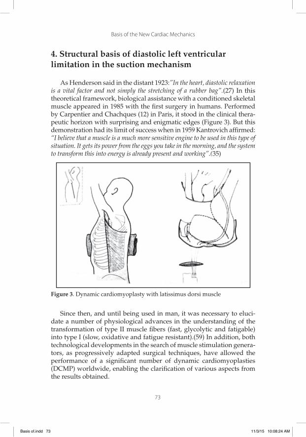
Basis of the New Cardiac Mechanics
73
4. Structural basis of diastolic left ventricular limitation in the suction mechanism
As Henderson said in the distant 1923:”In the heart, diastolic relaxation is a vital factor and not simply the stretching of a rubber bag”.(27) In this theoretical framework, biological assistance with a conditioned skeletal muscle appeared in 1985 with the fi rst surgery in humans. Performed by Carpentier and Chachques (12) in Paris, it stood in the clinical thera-peutic horizon with surprising and enigmatic edges (Figure 3). But this demonstration had its limit of success when in 1959 Kantrovich affi rmed: “I believe that a muscle is a much more sensitive engine to be used in this type of situation. It gets its power from the eggs you take in the morning, and the system to transform this into energy is already present and working”.(35)
Figure 3. Dynamic cardiomyoplasty with latissimus dorsi muscle
Since then, and until being used in man, it was necessary to eluci-date a number of physiological advances in the understanding of the transformation of type II muscle fi bers (fast, glycolytic and fatigable) into type I (slow, oxidative and fatigue resistant).(59) In addition, both technological developments in the search of muscle stimulation genera-tors, as progressively adapted surgical techniques, have allowed the performance of a signifi cant number of dynamic cardiomyoplasties (DCMP) worldwide, enabling the clarifi cation of various aspects from the results obtained.
Basis of.indd 73Basis of.indd 73 11/3/15 10:08:24 AM11/3/15 10:08:24 AM
Basis of the New Cardiac Mechanics
74
Since heart failure poses different problems derived from its own complexity, and has led to various analyses based on the segmentation of the problem, we believe that the same cartesian reasoning should be followed in interpreting the results of DCMP. Mechanical assistance during systole would be the most logical effect to accept, but this event has no explanation in the small increase observed in left ventricular ejection fraction. Similarly, the lack of difference between assisted and unassisted beats at postoperative mid-term, casts doubt on this func-tion. Neovascularization, i.e. the possibility of establishing irrigation channels between the latissimus dorsi muscle and the myocardium has not been demonstrated in humans so far.
If something has been demonstrated by DCMP, is its effectiveness in stopping the process of expansion of the ventricular chambers which occurs in dilated cardiomyopathy pathophysiology. This process of continuous dilatation carries in itself the greatest risk of death in these patients, as confi rmed by the SOLVD group of researchers.(65)
Capouya, (11) as well as Mott (48) have suggested that the proce-dure serves as an interruption of progressive dilatation. Carpentier (13) reports stability in cardiothoracic index values at three-year follow-up. Our experience has been similar in this respect.(76) Moreira, (46) on the other hand, reports clinical evidence of reduced wall stress, decreased left ventricular chamber diameters and increased maximum left ven-tricular elastance. Kass (36) also found benefi cial effects associated with “reverse remodeling”in cases studied analyzing pressure-volume rela-tionships, reporting left ventricular decreased end-diastolic and end-systolic volume and improved ventricular function. Wall stress reduc-tion, a central determining factor of oxygen consumption, would be ex-plained by DCMP prevention of progressive ventricular dilatation.(34)
We have seen in our own statistics these favorable cardiomyoplasty effects regarding the progression of ventricular dilatation.(76,78) Among the 15 patients who underwent this technique, 12 patients exceeded the absolute threshold of two years (follow-up average 31.3 months per patient). In these patients the value of left ventricular diastolic diameter (echocardiography) was: 72.7 mm ± 3 mm preoperatively, 73.6 mm ± 7 mm at one-year follow-up and 72.3 mm ± 8 mm at two years.(83) The difference between these values was not signifi cant, i.e., the left ven-tricle preserved its initial values, revealing no progression of dilatation. In the heart structure proper and in the pathological consequence im-plied by progressive and permanent dilatation we must acknowledge the benefi cial effects of dynamic cardiomyoplasty in dilated cardiomy-opathy.
Basis of.indd 74Basis of.indd 74 11/3/15 10:08:24 AM11/3/15 10:08:24 AM

Basis of the New Cardiac Mechanics
75
The force generated by systolic contraction exerts a compression on the elastic-heart muscle elements of such a magnitude that even with-out internal diastolic fi lling the ventricular tendency is to expand. This negative pressure determines a suction pump mechanism. The negative intraventricular pressures determining this effect were fi rst described in 1930. For this dynamic suction pump to be effective, the elastic recoil process must have a limit that allows a subsequent effective systole. Therefore, the possibility of sarcomere lengthening is engraved both in the muscle structure proper as in the fi brous cytoskeleton. The arrange-ment of the muscle straps, similar to guy cables on a suspension bridge, have suggested the idea of systolic energy storage, which once released in the diastolic process would allow the effect of a ventricular suction pump (Figure 4).(55,61,75)
Figure 4. Collagen cytoskeleton consisting of a grid-shaped mesh surrounding the sarcomere.
The muscle girth obtained with DCMP provides an elastic constric-tion limiting cardiac dilatation. This brings a benefi t to diastolic func-tion, producing a decrease in end-diastolic pressure, which is enhanced when the cardiomyostimulator is switched off. Similarly, once the cardiomyostimulator has been discontinued, there tends to be a more delayed ventricular fi lling, indicating that the contraction of the latis-simus dorsi muscle improves fi lling.(14,78) This situation of improved diastolic function performance would determine a decrease of mitral annulus and left atrial diameters, as was the tendency in patients of the different series published.(13.76)
Basis of.indd 75Basis of.indd 75 11/3/15 10:08:25 AM11/3/15 10:08:25 AM

Basis of the New Cardiac Mechanics
76
Figure 5. Collagen cytoskeleton surrounding the sarcomere; a, b and c: different expansion phases; d and e: collagen fi bers that constitute the ropes that bind sarcomere bundles and achieve an integral unit.
Dynamic cardiomyoplasty is an operative situation that contributes to restore the dilated heart muscle strength in order to obtain the re-quired internal gradient for fi lling through the suction pump mecha-nism, and has irrefutably demonstrated a signifi cant improvement in these patients’ functional class. In our experience, at two-year follow-up, it had passed from 3.06 ± 0.2 to 1.7 ± 0.6, with a survival rate of 75% during this period, similar to other reports.(77) To date, functional class III should be considered as the only one capable of being subjected to this technique. In some of this functional class series, long-term follow-up has shown (13.48) a survival rate of 66% at 7 years. Other publications have reported less optimistic results: about 56% at two years.(24) In or-der to achieve the diastolic effects of DCMP, tissue network shave been developed which are inserted around the ventricles through a minis-ternotomy. It has been considered that they control the deterioration of cardiac function by stabilizing the dimensions of the heart.(53)
It is understood that the straps between sarcomeres coordinate the action of a large number of muscle cells (Figure 5). The fact of providing solidary architecture for heart deformation allows better suction and a faster recovery of its initial length. Given the heart’s anatomo-physi-ological composition and the application of Laplace’s law to the pro-gressive dilatation mechanism, the benefi cial diastolic effect observed
Basis of.indd 76Basis of.indd 76 11/3/15 10:08:25 AM11/3/15 10:08:25 AM

Basis of the New Cardiac Mechanics
77
in DCMP monitoring is explained through the basis of left ventricular structural control in the active suction mechanism. The simplest inter-pretation of these fi ndings is that the grid structure of the collagen fi -bers prevents excessive sarcomere stretching. Cardiac muscle damage determines poor suction. With no limit to recoil, the mechanical activity of suction affects ventricular fi lling with the consequent failure of the suction pump, as there is no available intraventricular pressure drop in an adequate degree and suitable time.
5. Echocardiographic concepts
Echocardiography is currently able to provide noninvasive infor-mation on the complex mechanism of myocardial contraction. By ana-lyzing 2D deformation with speckle tracking technique the opposite rotation between the apex and the base of the heart (Figure 6) is evi-denced, thus achieving ventricular twisting (systolic contraction) and subsequent untwisting (suction mechanism, “plunger” effect) (Figure 7). Apical rotation (counter-clockwise, viewed from the apex) (Figure 8) is considered positive while basal rotation (clockwise, viewed from the apex) is considered negative (Figure 9). To calculate the degree of twist-ing, the software performs an algebraic subtraction (it adds the value of the apical positive twisting to the negative value of the base). In normal subjects it is about +11 degrees, always with prevalent tip rotation.
Figure 6. Twisting (T) considers the distance between the rotating points.
Basis of.indd 77Basis of.indd 77 11/3/15 10:08:25 AM11/3/15 10:08:25 AM

Basis of the New Cardiac Mechanics
78
Figure 7. Peak twisting and untwisting velocity.
Figure 8. Apical rotation (counter-clockwise, viewed from the apex).
Basis of.indd 78Basis of.indd 78 11/3/15 10:08:26 AM11/3/15 10:08:26 AM

Basis of the New Cardiac Mechanics
79
Figure 9. Basal rotation (clockwise, viewed from the apex).
The deviation from this value is generally a marker of heart disease, although it is necessary to consider that the normal values of left ven-tricular rotation and twisting are variable and depend on the technique used, the location of the area of interest (subendocardium, subepicar-dium), subject age and loading conditions.
Ventricular twisting and untwisting can be explained by the angular distribution of the ascending and descending fi bers.(15) Rotation is pro-duced by contraction of the helical fi bers and the strength and direction are given by the balance of subendocardial and subepicardial fi bers. If subendocardial fi bers are responsible for longitudinal deformation, we must consider that the mid-fi bers and especially the subepicardial fi bers contribute fundamentally to rotation and twisting.(26)
The radius of subepicardial rotation is greater than that of subendo-cardial rotation; therefore, the subepicardium provides greater rotation-al force than the subendocardium, and as result subepicardial rotation expresses more signifi cantly at the apical level.(49)
The mathematical difference between twisting and torsion is that the latter also considers the distance between the two points that rotate in opposite directions, although in practice and in the literature they are confused and are often used as synonyms.
Basis of.indd 79Basis of.indd 79 11/3/15 10:08:27 AM11/3/15 10:08:27 AM

Basis of the New Cardiac Mechanics
80
At the papillary muscle level rotation is neutral, constituting a tran-sition point between counter-clockwise apical rotation and clockwise basal rotation, so in general it is not considered. There is a clear rela-tionship between twisting and age, showing that it increases with age possibly by progressive deterioration of the subendocardial fi bers.(51) In coronary disease with subendocardial perfusion defi cit, longitudinal strain is attenuated. Myocardial ischemia does not affect twisting un-less infarctions are very extensive and transmural (affecting circumfer-ential and subepicardial fi bers).
In dilated cardiomyopathy, twisting is reduced proportionally to systolic function, and is responsible for the attenuation of apical rota-tion, whereas basal rotation may be normal or reduced. In some of these patients the apex and base rotate in the same clockwise direction.(60) In patients who respond to Cardiac Resynchronization Therapy a rapid normalization can predict inverse remodeling at 6 months.
In each of the different pathologies, rotation, twisting and untwist-ing are variably affected depending on the extent and distribution of the fi bers affected, adopting characteristic patterns in ventricular hypertro-phy, diabetes, amyloidosis, valvulopathies and pericardial diseases.
In conclusion rotation/twisting is fundamental for the heart con-traction and suction mechanism. Its echocardiographic assessment is an accurate and feasible method to detect both systolic and diastolic myocardial dysfunction in daily practice, even in the early stages of cardiovascular disease.
Basis of.indd 80Basis of.indd 80 11/3/15 10:08:28 AM11/3/15 10:08:28 AM
81
Chapter IV
Clinical, Surgical And Eletrophysiological Perspectives Derived From This Research
1. Extent of heart failure
Heart failure is one of most important public health problems due to its social, economic and especially human impact with a prevalence of 5 million patients, 300,000 deaths per-year and 500,000 new cases per year (16) in the United States. The implications for health systems are 15 million visits per year; 6.5 million days of hospitalization and 38,000 million dollars of health costs. In Europe, the incidence is 1.3 cases per 1000 inhabitants per year in subjects over 25 years, reaching 11.6 cases per 1000 inhabitants per year in patients over 85 years (21), while 5% of the European population has problems associated to heart failure.(17,50)
The main cause of heart failure is coronary artery disease also re-sponsible for 50% of cases in the United States. In ischemic heart dis-ease, acute myocardial infarction is the fi rst single factor, with a tenfold increased risk of heart failure than in the normal population during the fi rst year post infarction and up to twenty times higher in the following years. After acute myocardial infarction, there is loss of cardiomyocytes which added to the process of ventricular remodeling triggers heart failure. This remodeling is a complex phenomenon involving molec-ular, neurohormonal and genetic processes leading to left ventricular dilatation, abnormal morphology and dysfunction.
Early revascularization of acute myocardial infarction with angio-plasty and stenting has not reduced the incidence of dysfunction and left ventricular remodeling. Thus, if in old series with conventional
Basis of.indd 81Basis of.indd 81 11/3/15 10:08:28 AM11/3/15 10:08:28 AM

Basis of the New Cardiac Mechanics
82
treatment, 20% of patients with transmural infarction developed ven-tricular dilatation and dysfunction; in recent series of myocardial in-farction treated with angioplasty and stenting during the acute phase, 30% develop alterations in the shape or function at six months. The evolution of these patients regarding mortality and complications, is directly related to ventricular dilatation.(28,44,45)
In 5% of cases, patients present with Grade IV heart failure, are very symptomatic, require frequent hospitalizations and survival is less than 30% at one year. (33) The increase in ventricular volume and the ac-quisition of a spherical shape is responsible for the progression of the disease. The classic surgical treatment of these patients is heart trans-plantation, with a survival rate of over 70% at fi ve years and 25% at 20 years.(64) However, the disproportion between the number of recipi-ents and donors, only allows less than 20% of patients with Grade IV heart failure to benefi t from heart transplantation.
Hence it is necessary to preserve cardiac transplantation for patients who have no other treatment options and concomitantly develop other alternatives. Recent therapeutic strategies are designed to integrate bi-ology and new medical technologies, generating alternatives that may improve the prognosis and the functional status of these patients. Treat-ments include surgical ventricular repair, passive ventricular restraint, permanent ventricular assistance or as bridge to recovery, immunoad-sorption and tissue engineering.(30)
Neurohormonal models do not explain heart failure progression, and drug treatments that act on neurohormonal activation delay but do not stop disease progression, or are ineffective.(41,42) Size and geo-metrical changes are responsible for myocyte and extracellular matrix structural abnormalities that aggravate cardiac function, increase neu-rohormonal activity and reduce cardiovascular response. The study of the anatomical basis is necessary for the application of new techniques aimed at restoring ventricular geometry to the native volume and ven-tricular conical confi guration. The prognosis of patients with heart fail-ure is directly related with ventricular dilatation.
Myocardial capacity is conditioned by the orientation of cardiac fi -bers. In that respect, the conical heart has helical and circumferential myocardial fi bers. Fiber orientation determines function; thus, ejection fraction is 60% when the normal helical fi bers contract and falls to 30% if only the transverse fi bers shorten. The development of a spherical confi guration modifi es the helical fi ber orientation towards a transverse dilatation and decreases contractile force. Thus, the development of surgical ventricular repair techniques has reenacted the work of Tor-
Basis of.indd 82Basis of.indd 82 11/3/15 10:08:28 AM11/3/15 10:08:28 AM
Basis of the New Cardiac Mechanics
83
rent Guasp and his hypothesis of the ventricular myocardial band (7) as well as cardiac mechanics based on the research of cardiac electrical activation developed by us.
2. Clinical perspectives
The conclusions of the research presented in this manuscript can be summarized as follows:
1. Endo-epicardial three-dimensional mapping shows a sequence of apical loop electrical activation consistent with the synchronous contraction of the descending and ascending bands.
2. The simultaneous and opposed activation of the ascending band, to the starting point of its radial activation from the descending band, is consistent with the simultaneous clockwise and coun-ter-clockwise rotation of the apical and basal regions (ventricular twisting mechanism).
3. The late activation of the ascending band, compatible with its persistent contraction during the initial phase of the diastolic iso-volumic period, occurs without postulating electrical activations after the QRS.
The novel activation sequence of Torrent Guasp’s ventricular myo-cardial band found in this work, explains the previous process that trig-gers the ventricular twisting and suction mechanism. Furthermore, it confi rms that the activation of the ascending band completes the QRS. This fi nding demonstrates the contractile persistence of this muscle seg-ment during the fi rst part of diastole, cancelling the traditional concept of passive relaxation.
Our work suggests the existence of a “three-stage heart “: systole, suction and diastole. We believe that the data obtained are of special importance since they were recorded in humans, with structurally nor-mal hearts and in physiological (non-experimental) conditions. Further research is needed to elucidate what happens in different diseases.
In recent years, the importance of pathological ventricular fi lling and diastolic failure has been put into evidence.(38) In this respect, most studies have been based on the alterations of the passive myocardial properties.
Basis of.indd 83Basis of.indd 83 11/3/15 10:08:28 AM11/3/15 10:08:28 AM

Basis of the New Cardiac Mechanics
84
It is possible however, that at least some of these diseases are due to ventricular contractile dysfunction during diastole. Similarly, systolic alterations may be due in some cases to modifi cations in apical loop ac-tivation. These phenomena have very signifi cant clinical and therapeu-tic implications, and probably heart failure classifi cations based on the suction phase can be developed, as well as pharmacological, surgical and device-implemented therapies that take into account the regulation of diastolic contractile persistence of the ascending band, the timing and/or synchronization of the two bands’ contraction, etc. In fact, the pathophysiological basis of cardiac resynchronization therapy could correspond to this phenomenon. We have also rescued the value of the apex in the solution of ventricular reduction techniques, as well as the anatomical speculation of the myocardial band in relation to the dif-ferent functioning of the basal and apical loops, the former committed to the ejective phase and the second one to suction and fi lling phases. The approach is open, mainly because we agree that an enlarged heart has no adequate suction phase and consequently a subsequent ineffi -cient contraction. This active mechanism of the ventricular myocardial band on the diastolic effect opens a wide scenario for surgical repair techniques both of the heart shape and volume and the resultant left ventricular function.(6)
3. Surgical perspectives
Ventricular dilatation is an adaptive condition to different cardio-vascular diseases. As a result, there is cardiac remodeling presenting increased wall tension and progressive dilatation. In the myocyte this process leads to irreversible stretching with subsequent deviation of the pressure/volume curve to the right, increased ventricular volumes and mitral regurgitation. This continuous dilatation determines increased risk of mortality in these patients.(10) Therefore, the study of altered ventricular geometry in heart failure is of major interest for the practice of new surgical therapies that try to restore this situation in order to improve prognosis.
Leonardo da Vinci (1452-1519) had already drawn geometrical de-signs for his analyses of the left ventricle and aortic root.(79) On the other hand, William Harvey, in his anatomical “Exercitatio anatomica de motu cordis et sanguinis in animalibus” work (1628), made a description of the left ventricle with a long narrow shape during the ejective phase, tending towards a sphere during diastole.(79) The interest in these
Basis of.indd 84Basis of.indd 84 11/3/15 10:08:29 AM11/3/15 10:08:29 AM
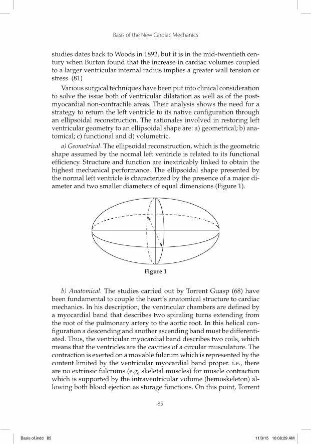
Basis of the New Cardiac Mechanics
85
studies dates back to Woods in 1892, but it is in the mid-twentieth cen-tury when Burton found that the increase in cardiac volumes coupled to a larger ventricular internal radius implies a greater wall tension or stress. (81)
Various surgical techniques have been put into clinical consideration to solve the issue both of ventricular dilatation as well as of the post-myocardial non-contractile areas. Their analysis shows the need for a strategy to return the left ventricle to its native confi guration through an ellipsoidal reconstruction. The rationales involved in restoring left ventricular geometry to an ellipsoidal shape are: a) geometrical; b) ana-tomical; c) functional and d) volumetric.
a) Geometrical. The ellipsoidal reconstruction, which is the geometric shape assumed by the normal left ventricle is related to its functional effi ciency. Structure and function are inextricably linked to obtain the highest mechanical performance. The ellipsoidal shape presented by the normal left ventricle is characterized by the presence of a major di-ameter and two smaller diameters of equal dimensions (Figure 1).
Figure 1
b) Anatomical. The studies carried out by Torrent Guasp (68) have been fundamental to couple the heart’s anatomical structure to cardiac mechanics. In his description, the ventricular chambers are defi ned by a myocardial band that describes two spiraling turns extending from the root of the pulmonary artery to the aortic root. In this helical con-fi guration a descending and another ascending band must be differenti-ated. Thus, the ventricular myocardial band describes two coils, which means that the ventricles are the cavities of a circular musculature. The contraction is exerted on a movable fulcrum which is represented by the content limited by the ventricular myocardial band proper. i.e., there are no extrinsic fulcrums (e.g. skeletal muscles) for muscle contraction which is supported by the intraventricular volume (hemoskeleton) al-lowing both blood ejection as storage functions. On this point, Torrent
Basis of.indd 85Basis of.indd 85 11/3/15 10:08:29 AM11/3/15 10:08:29 AM
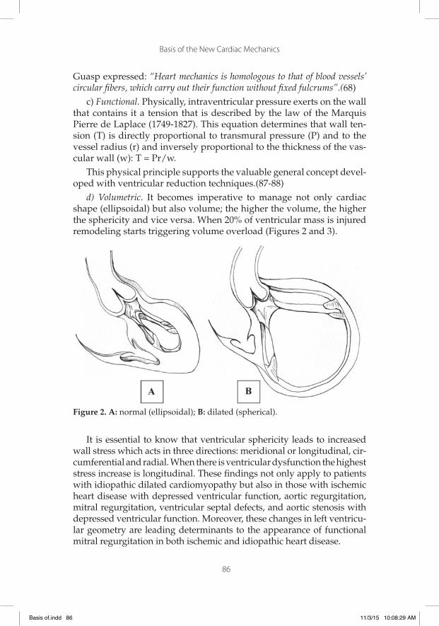
Basis of the New Cardiac Mechanics
86
Guasp expressed: “Heart mechanics is homologous to that of blood vessels’ circular fi bers, which carry out their function without fi xed fulcrums”.(68)
c) Functional. Physically, intraventricular pressure exerts on the wall that contains it a tension that is described by the law of the Marquis Pierre de Laplace (1749-1827). This equation determines that wall ten-sion (T) is directly proportional to transmural pressure (P) and to the vessel radius (r) and inversely proportional to the thickness of the vas-cular wall (w): T = Pr/w.
This physical principle supports the valuable general concept devel-oped with ventricular reduction techniques.(87-88)
d) Volumetric. It becomes imperative to manage not only cardiac shape (ellipsoidal) but also volume; the higher the volume, the higher the sphericity and vice versa. When 20% of ventricular mass is injured remodeling starts triggering volume overload (Figures 2 and 3).
Figure 2. A: normal (ellipsoidal); B: dilated (spherical).
It is essential to know that ventricular sphericity leads to increased wall stress which acts in three directions: meridional or longitudinal, cir-cumferential and radial. When there is ventricular dysfunction the highest stress increase is longitudinal. These fi ndings not only apply to patients with idiopathic dilated cardiomyopathy but also in those with ischemic heart disease with depressed ventricular function, aortic regurgitation, mitral regurgitation, ventricular septal defects, and aortic stenosis with depressed ventricular function. Moreover, these changes in left ventricu-lar geometry are leading determinants to the appearance of functional mitral regurgitation in both ischemic and idiopathic heart disease.
Basis of.indd 86Basis of.indd 86 11/3/15 10:08:29 AM11/3/15 10:08:29 AM

Basis of the New Cardiac Mechanics
87
Figure 3. A: normal (ellipsoidal); B: dilated (spherical). The dilated heart shows separation between the descending and ascending bands.
In view of this situation, we have implemented ellipsoidal recon-struction according to a technique that links the anatomical and func-tional geometrical bases presented here.(80,81) The left ventricular el-lipsoidal repair technique we have researched and performed (80,81) assumes the possibility of restoring ventricular geometry within the shape needed for its mechanical function. It consists of the following steps (Figure 4):
1) A longitudinal incision along the anterior descending artery in the avascular left ventricular wall (Figure 5).
2) The left incision margin is brought by means of a continuous suture to the level of the preserved interventricular septum (Figure 6).
3) The remaining right margin is sutured to the left ventricular free wall (Figure 7).
4) Both the original incision as well as the size of the fl aps must observe the cavity to be preserved in order to reduce ventricular volume.
Figure 4. Diagram of the successive steps in ellipsoidal reconstruction.
Basis of.indd 87Basis of.indd 87 11/3/15 10:08:30 AM11/3/15 10:08:30 AM
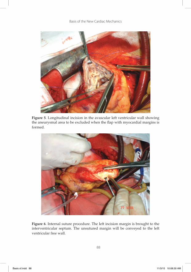
Basis of the New Cardiac Mechanics
88
Figure 5. Longitudinal incision in the avascular left ventricular wall showing the aneurysmal area to be excluded when the fl ap with myocardial margins is formed.
Figure 6. Internal suture procedure. The left incision margin is brought to the interventricular septum. The unsutured margin will be conveyed to the left ventricular free wall.
Basis of.indd 88Basis of.indd 88 11/3/15 10:08:30 AM11/3/15 10:08:30 AM

Basis of the New Cardiac Mechanics
89
Figure 7. External suture procedure. The right margin with the part of the ex-cluded septum is sutured to the left ventricular free wall.
This technique adds a number of benefi ts when compared to tradi-tional techniques (Jatene, Dor, Batista) (10,29,31) and follows the strat-egy presented by Matsui (43) and Menicanti (44,45):
a) Preservation of the heart muscle by acting on the area bounded by the ascending and descending segments of the ventricular myo-cardial band according to the work of Torrent Guasp. (68) In this geographical tip of the heart, called the apex, a virtual conduct is defi ned in which the endocardium attaches to the epicardium constituting a weak zone favorable to dyskinesias.(66, 78)
b) This topographical heart area is avascular, which prevents pull-ing the arterial system during resection.
c) Preservation of the circumfl ex artery by choosing the left ven-tricular wall along the anterior descending artery as avascular incision site. In other techniques, with lateral approach of the left ventricle, circumfl ex branches are sacrifi ced.
d) The geometrical fl ap effect made with this technique has the ef-fect of a ventricular girdle.
e) The fl ap built with the overlapping incision margins excludes the remodeled area (anterior and septal tip) leaving the cardiac
Basis of.indd 89Basis of.indd 89 11/3/15 10:08:31 AM11/3/15 10:08:31 AM

Basis of the New Cardiac Mechanics
90
apex with new vitality to bear heart pressure as it is rebuilt with healthy tissue.
f) In the thin apical septum with distortion produced by remodeling, this technique eliminates dyskinesia, providing septal rigidity.
g) This technique does not involve placing synthetic patches, thus avoiding noncontractile areas on the left ventricular contraction surface.
h) This surgical approach approximates the ascending and descend-ing apical loop segments, which are separated during heart fail-ure, contributing to improve ventricular function.
i) We undertook treating both septal distortion and diastolic func-tion with this technique.
j) The recovery of the heart’s ellipsoidal shape might act upon the structurally altered mitral apparatus. We have experienced that once the ventricle is reduced, the insuffi cient valves did not re-quire surgical repair.(78)
Conclusion. The restitution of ventricular geometry has stimulated both the use of techniques to assess it as well as having solved spatial distortion in heart failure.(29) Certainly the sphericity to which the left ventricle turns during heart failure has an ominous prognostic value. Ventricular geometry is a sensitive marker of function and prognosis. Be it cause or consequence, the presence of spherical geometry in the patient determines increased oxygen consumption through increased wall stress. It is certainly the consequence, but it also carries the blame of perpetuating the alteration.
4. Electrophysiological therapy perspectives
Throughout time, there were always situations in daily practice where classical pathophysiology was not enough to interpret them correctly. Often, only after the appearance -and acceptance- of diametrically op-posed and resisted alternatives to those currently existing, therapeutic interpretations and appropriate and effective behaviors were achieved. From Harvey’s seminal description to the implantable cardioverterde-fi brillator, passing through heart surgery and the use of beta blockers in heart failure, there appeared new concepts which marked milestones in cardiology. Sometimes these ideas were implemented immediately
Basis of.indd 90Basis of.indd 90 11/3/15 10:08:31 AM11/3/15 10:08:31 AM
Basis of the New Cardiac Mechanics
91
but in other cases they were described years earlier and had to wait for further studies to prove their validity. This is probably the case of Torrent Guasp’s model. In the research presented in this text his initial ideas have been summarized, completed and reinterpreted in the light of new evidence.
The clinical implications of this model remain to be discussed. It will probably take years to verify them in the various syndromes and heart diseases. However, a situation in which its application may result immediate is in the loss of ventricular synchrony and its counterpart, biventricular resynchronization therapy.
In patients with end-stage heart failure of different etiologies, this is signifi cantly aggravated by biventricular dissynchrony. When left bundle branch block occurs in an already severely damaged heart, left ventricular contraction is delayed. Therefore, cardiac contraction ceases to be simultaneous or synchronous and becomes dissynchronous, fur-ther worsening cardiac mechanics.
Resynchronization Therapy (RCT) is a procedure that restores bi-ventricular synchrony. This resynchronization is achieved via a heart stimulator similar to a sequential pacemaker, which stimulates not only the right ventricle but the left ventricle as well. However, to achieve proper resynchronization the stimulation of the left ventricular cham-ber is not enough; it must also be achieved from specifi c sites or “effec-tive areas”, typically the medial lateral or posterolateral area of the left ventricle.
The usual catheter implantation procedure is coronary sinus cannu-lation and then a tributary vein that reaches the “effective” area. How-ever, this method has numerous problems and disadvantages failing in 20% to 30% of patients in whom RCT is performed. These patients have been referred to as “non-responders”.
Before proceeding, it is important to note the signifi cance of this cir-cumstance. Resynchronization Therapy is a crucial procedure. It is per-formed in patients with end-stage heart failure in whom all other avail-able pharmacological and surgical treatment options were ineffective. If it is successful, a signifi cant change is achieved in the quality of life and survival of the patient; its failure means remaining in Functional Class III-IV. Although cardiac transplantation could be raised as an option, in practice only very few patients have the opportunity of accessing to it.
In this pathology and its therapy there are extraordinarily remark-able elements:
Basis of.indd 91Basis of.indd 91 11/3/15 10:08:32 AM11/3/15 10:08:32 AM
Basis of the New Cardiac Mechanics
92
1. Despite the tens of thousands of patients treated with this meth-od, current cardiac mechanics has failed to clearly elucidate the concept of biventricular dissynchrony. Even in cases of very se-vere mechanical cardiac dysfunction, the most advanced diagnos-tic methods, including the most sophisticated echocardiography, gamma camera or MRI techniques cannot diagnose this disease, at least with a useful and reliable sensitivity and specifi city. The diagnosis is still based on the presence of left bundle branch block, logically in the context of dilated cardiomyopathy in Functional Class III or IV. Even this apparently permanent and classical crite-rion is under discussion, debating whether its morphology (“typ-ical” or “atypical”), its critical duration, or some other as yet not elucidated criterion is important.
2. The same applies for the evaluation of RCT effectiveness. None of the mentioned techniques has been able to document a parameter with a suffi ciently signifi cant and early variation to consistently correlate with the almost surprising improvements in the clinical outcome of patients in whom RCT is successful.
In our opinion these “failures” are due to the fact that the assessed cardiac parameters of classical mechanics are neither responsible for the pathology nor for the therapeutic response. On the contrary, it is very attractive to reconsider these phenomena in the light of the recent fi ndings on stimulus propagation and subsequent cardiac mechanics resulting from this investigation.
The princeps “dyssynchrony” parameter is, as we have pointed out, left bundle branch block. This phenomenon, which we again mention, hardly explains the severity of mechanical disorders, on the other hand would seriously alter the mechanics of the ascending band. In fact, it would seem very logical to say that a fault in the activation at the level of “band intersection” could generate the image of left bundle branch block (and its “atypical” forms) and of course seriously alter the activa-tion sequence of the ascending band, but not in the way postulated by the “classical” conception. There would be an alteration in the “radial” activation of the descending endocardial band to the ascending epi-cardial band and of the “bidirectional longitudinal” activation of the epicardial band. The phenomena that these activations cause in cardiac twisting at the level of the apical loop and in the active protodiastolic ventricular suction mechanism have already been explained in detail.
We insist that the usual methods of ventricular mechanical assess-ment, especially of the left ventricle, usually do not take into account
Basis of.indd 92Basis of.indd 92 11/3/15 10:08:32 AM11/3/15 10:08:32 AM
Basis of the New Cardiac Mechanics
93
these essential factors. Therefore, it would not be surprising that these factors lack the accuracy to evaluate ventricular function alteration as well as its improvement with RCT.
The left ventricular stimulation catheter positioning has critical im-portance in the remarkable RCT effectiveness and in the failure of the current methods used. It has been suggested that the best position of the catheter is the one corresponding to the late ventricular activation area; although in many cases this is true, in others it is not. There is agreement, however, that the anatomical location in the medial area of the left ventricular posterolateral or lateral surface is effective whether or not it coincides with late potentials. This fact could be interpreted consistently with Torrent Guasp’s model and our fi ndings, as this loca-tion corresponds to the point of band intersection. The catheter would activate the left ventricle at the precise point required to “restore” the normal activation of the ascending segment in its double progression towards the apical loop and the ventricular base.
Finally, there is another fact that strongly reinforces this interpreta-tion; we have indicated that the normal pathway for left ventricular catheter implantation is through the coronary sinus, which gives ac-cess to the left ventricular epicardial veins, so that activation propa-gates from the endocardium to the epicardium. Although the catheter is properly located in the mentioned “effective area”, there are some “non-responder” patients. In recent studies (Alsync trial) (47) 20% of patients are non-responders although the catheter implanted through the coronary sinus is perfectly positioned. When implanted through an endocavitary pathway, 50% of these patients become responders. The explanation using the classical conception, is a fairly vague consider-ation in the sense that endo-epicardial activation is “more physiologi-cal” than the reverse.
From the perspective of our research, the explanation is much more consistent: the activation of the ascending band at the point of band intersection restores its longitudinal activation, with the benefi cial con-sequences mentioned above. However, the radial activation from the descending to the ascending band and the persistent normal distal ac-tivation of the descending segment is lost. Endocavitary stimulation (descending segment) would almost completely restore the normal electrical activation and therefore its mechanical result. Based on this principle, at Hospital PresidentePerón de Avellaneda we have been re-searching endocavitary RCT for several years. This has already been employed in 34 patients, with excellent surgical and clinical outcomes.
Basis of.indd 93Basis of.indd 93 11/3/15 10:08:32 AM11/3/15 10:08:32 AM

Basis of the New Cardiac Mechanics
94
In conclusion, Torrent Guasp’s hypotheses and fundamental ana-tomical model, extended and modifi ed in our electromechanical re-search offers countless opportunities for the development of both theo-retical and clinical and therapeutic applications. It is possible that much of cardiology should be reviewed in the light of this new paradigm, with perhaps unpredictable results.
Basis of.indd 94Basis of.indd 94 11/3/15 10:08:32 AM11/3/15 10:08:32 AM
95
References
1. Armour JA, Randall WC, Structural basis for cardiac function. Am J Physiol 1970;218:1517-23.
2. Becú L, Brusca G. El colágeno en el miocardio. Rev Argent Cardiol 1996; 64:235-43.
3. Brecher GA. Cardiac variations in venous return Studies with new Bristle fl owmeter. Am J Physiol 1954;176:423-30.
4. Brecher GA. Experimental evidence of ventricular diastolic function. Circ Res 1956;4:513-8.
5. Brecher GA. Critical review of recent works on ventricular diastolic suction. Circ Res 1958;6:554-6.
6. Brutsaert DL, Stanislas U, Gillibert TC. Diastolic failure: patho-physiology and therapeutics implications. J Am Coll Cardiol 1993;22:318-25.
7. Buckberg GD, Coghlan HC, Torrent Guasp F. The structure and func-tion of the helical heart its buttress wrapping. VI. Geometrics conceps of heart failure and use for structural correction. Sem Thorac Cardiovasc Surg 2001; 13: 386-401.
8. Buckberg GD, Coghlan HC, Torrent Guasp F. The structure and func-tion of the helical heart and its buttress wrapping.V. Anatomic and physiologic considerations in the healthy and failing heart. Semin Thorac Cardiovasc Surg 2001;132:358-85.
9. Buckberg GD. Ventricular structure and surgical history. Heart Fail-ure Reviews 2004;9:255-68.
10. Buckberg GD. La era post-STICH y su impacto. Cir Cardiovasc 2010;17:45-56.
11. Capouya ER, Gerber RS, Drinkwater DC, Pearl JM, Sack JB, Aharon AS et al. Girdling effect of nonstimulated cardiomyoplasty on left ventricular function. Ann Thorac Surg 12993; 56:867-71.
Basis of.indd 95Basis of.indd 95 11/3/15 10:08:32 AM11/3/15 10:08:32 AM

Basis of the New Cardiac Mechanics
96
12. Carpentier A, Chachques JC. Myocardial substitution with a stimu-lated skeletal muscle: fi rst succesful clinical case (letter). Lancet 1985; 1: 1267.
13. Carpentier A, Chachques JC, Acar C, Relland J, Mihaileanu S, Ben-sasson D et al. Dynamic cardiomyoplasty at seven years.J Thorac Cardiovasc Surg 1993; 106:42-54.
14. Chachques JC, Berrebi A, Hernigou A, Relland J, Mihaileanu S, Ben-sasson D y col. Study of muscular and ventricular function in dynamic cardiomyoplasty: A ten year follow-up. J Heart Lung Transplant 1997;16:854-68.
15. Cheng A, Nguyen TC, Malinowski M, Daughters GT, Miller DC, Ingels NB Jr. Heterogeneity of left ventricular wall thickening mechanisms. Circulation.2008, 118:713-21.
16. Chockalingan A, Chalmers J, Lisheng L, Labarthe D, MacMahon S, Martin I, Whitworth J. Prevention of cardiovascular diseases in developing countries: agenda for action (statement from a WHO-ISH Meeting in Beijing, October 1999). J Hipertensión 2000, 18: 1705-8.
17. Cleland J, Khand A, Clark A. The heart failure epidemia: exactly how big is it?.Eur Heart J 2001; 22: 623-6.
18. Coghlan C, Hoffman J. Leonardo da Vinci´s fl ights of the mind must continue: cardiac architecture and the fundamental relation of form and function revisited. Eur J Cardio-Thorac Surg 2006: 295; S4-S17.
19. Cosín Aguilar J. “Francisco Torrent Guasp (1931-2005)”. Rev Esp Cardiol 2005; 58:759-60.
20. Cosín Aguilar J, Hernándiz Martínez A, Tuzón Segarra MT, Agüero Ramón-Llin J, Torrent Guasp F. Estudio experimental de la lla-mada fase de relajación isovolumétrica del ventrículo izquierdo. Rev Esp Cardiol 2009;62:392-9.
21. Cowie MR, Word DA, Cotas AJ, Thompson SG, Poole-Wilson PA, Zurres V, et al. Incidence and aetiology of heart failure: a popula-tion based study. Eur Heart J 1999; 20: 421-8.
22. Cuenca Castillo J. El volumen telesistólico del ventrículo izquierdo es la clave para la indicación y el éxito de la restauración ven-tricular quirúrgica. Cir Cardiovasc 2010;17:37-9.
23. Donato M, Gelpi R. Nuevos conceptos (y otros antiguos reconsid-erados) en la fi siopatología de la diástole.`Rev Argent Cardiol 2000;68:121-7.
Basis of.indd 96Basis of.indd 96 11/3/15 10:08:33 AM11/3/15 10:08:33 AM

Basis of the New Cardiac Mechanics
97
24. Frazier OH, Myers TJ. Surgical therapy for severe heart failure.Cur-rent Problems in Cardiology 1998;23:723-64.
25. García Civera R, Cavadés A, Cosín J. Automatismo y Conducción Cardíacos. MCR 1987; p 700.
26. Geyer H, Caracciolo G, Abe H, Wilansky S, Carerj S, Gentile F, Nesser HJ, Khandheria B, Narula J, Sengupta PP. Assessment of myocardial mechanics using speckle tracking echocardiography: fundamentals and clinical applications.Am Soc Echocardiogr 2010;23:351-69.
27. Henderson Y. Volume changes of the heart. Physiol Rev 1923;3:165-70.
28. Herreros J. Cirugía coronaria. Evolución última década. Indicacio-nes y resultados. Rev Esp Cardiol 2005; 58: 1107-16.
29. Herreros J, Trainini JC, Menicanti L, Stolf N, Cabo J, Buffolo E. Cirugía de restauración ventricular después del estudio STICH. Cir Cardiovasc 2010;17:25-35.
30. Herreros J, Trainini JC, Chachques JC. Alternatives to heart trans-plantation: integration of biology with surgery. Frontiers Biosc 2011; E·3: 635-47.
31. Herreros J, Trainini JC, Bernal JM, Gutierrez F, Cabo J, Chachques JC. Tratamiento de la insufi ciencia cardíaca: nuevas estrategias terapéuticas. Cir Cardiovasc 2011;18:113-20.
32. Jacob F. “El juego de lo posible”. Grijalbo Ed., Barcelona, 1982.33. Jessup M, Brozena MS. Heart failure. N Engl J Med 2003; 348: 2007-
18. 34. Jondeau G, Dorent R, Bors V, Dib JC, Dubourg O, Benzidia R, Gand-
jbakhch Y, Bourdarias JP. Dynamic Cardiomyoplasty: Effect of discontinuing latissimus dorsi muscle stimulation on left ven-tricular systolic and diastolic performance and exercise capacity. J Am Coll Cardiol 1995;26:129-34.
35. Kantrowitz A, McKinnon WMP. The experimental use of the dia-phragm as an auxiliary myocardium.Surg Forum 1959; 9:266-8.
36. Kass DA, Baughman KL, Pak PH, Cho PW, Levin HR, Gardner TJ et al. Reverse remodeling from cardiomyoplasty in humans heart failure; external constraint versus active assist. Circulation 1995;91:2314-8.
37. Katz LN. The role played by the ventricular relaxation process in fi lling the ventricle. Am J Physiol 1930; 95:542-53.
Basis of.indd 97Basis of.indd 97 11/3/15 10:08:33 AM11/3/15 10:08:33 AM
Basis of the New Cardiac Mechanics
98
38. Lazar Mandinov L, Eberli F, Seiler C, Hess OM. Diastolic heart fail-ure. Cardiovasc Res 2000;45:813-25.
39. Lewis T, Rothschild MA. The excitatory process in dog´s heart.II- The ventricles. Philos Trans R Soc 1915;206:1981.
40. Lunkenheimer PP, Lunkenheimer A. Implicancias cardiodinámicas. Organización dualista de la estructura ventricular. En Torrent Guasp F, editor. Estructura y mecánica del corazón. Barcelona: Grass Ed., 1987.
41. Mann DL, Deswal A, Bozcurt B, Torre-Amione G.New therapeutics for chronic heart failure.Annu Rev Med 2002; 53: 59-74.
42. Mann DL, Bristow MR. Mechanisms and models in heart failure. The biomechanical model and beyond.Circulation 2005; 11: 2837-49.
43. Matsui Y, Fukuda Y, Suto Y. Overlapping cardiac volume reduction operation. J Thorac Cardiovasc Surg 2002;124:395-7.
44. Menicanti L. Surgical left ventricle reconstruction, pathophysiologic insights, results and experience from the STICH trial. Eur J Car-dio-thorac Surg 2004; 26 (Suppl.): S42-7.
45. Menicanti L. The Dor procedure: What has changed after fi fteen years of clinical practice. J Thorac Cardiovasc Surg 2002;124:886-90.
46. Moreira LF, Bocchi EA, Bacal F, Stolf N, Belotti G, Jatene AD. Pres-ent trends in clinical experience with dynamic cardiomyoplasty. Artif Organs 1995; 19:211-6.
47. Morgan J, Biffi M, Geller L, Ruffa F, Leclercq C, Tung S et al. Alsync Investigators. Novel superior-access, atrial transseptal approach to left ventricular endocardial lead implantation, feasibility and safety results of the alternate site cardiac resynchronización (Al-sync ) study. Heart Rhythm Society, Annual Scientifi c Sessions; May 7-10, 2014; San Francisco.https://clinicaltrials.gov/ct2/show/NCT01277783
48. Mott BD, Misawa Y, Helou J. Effects of dynamic cardiomyoplasty on ventricular function in a rapid pacing heart failure model (ab-stract). J Mol Cell Cardiol 1995; 27 (Suppl A):12.
49. Nakatani S. Left ventricular rotation and twist: why should we learn? J Cardiovasc Ultrasound 2011;19:1-6.
50. Navarro López F, de Teresa E, López- Sendón JL, Castro Beiras A. “Guidelines for the diagnosis and management of heart failure and cardiogenic shock”. Informe del Grupo de Trabajo de Insu-fi ciencia Cardiaca de la Sociedad Española de Cardiología. Rev Esp Cardiol 1999; 52 (Suppl. 2): S1-54.
Basis of.indd 98Basis of.indd 98 11/3/15 10:08:33 AM11/3/15 10:08:33 AM
Basis of the New Cardiac Mechanics
99
51. Notomi Y, Srinath G, Shiota T, Martin-Miklovic MG, Beachler L, Howell K, Oryszak SJ, Deserranno DG, Freed AD, Greenberg NL, Younoszai A, Thomas JD. Maturational and adaptive mod-ulation of left ventricular torsional biomechanics: Doppler tis-sue imaging observation from infancy to adulthood. Circulation 2006;113:2534-41.
52. Parker PS, Mac Leod AG, Alexander J. The excitatory process ob-served in the exposed human heart. Am Heart J 1930;15:720-8.
53. Raman JS, Power JM, Byrne M, Alferness C. Ventricular containment in advanced heart failure halts decline in cardiovascular function associated with experimental dilated cardiomyopathy. Presented at the 37th Annual Meeting of the Society of Thoracic Surgeons, January 2001, New Orleans, LA.
54. Robb JS, Robb RC. The excitatory process in the mamalian ventricle. Am J Physiol 1936;115:43-52.
55. Robb JS, Robb RC. The normal heart: Anatomy and physiology of the structural units. Am Heart J 1942;23:455-67.
56. Robinson TF, Factor SWM, Sonnenblick EH.The heart as a suction pump.Sci Am 1986; 254:84-91.
57. Roy CS, Adami JG. Heart-beat and pulse-wave. Practitioner 1980;44:81-94.
58. Sallin EA. Fiber orientation and ejection fraction in the human ven-tricle. Biophys J 1969, 9: 954-64.
59. Salmons S, Streter FA. Signifi cance of impulse activity in the trans-formation of skeletal muscle type. Nature 1976;263:30-4.
60. Sengupta PP, Krishnamoorthy VK, Korinek J, Narula J, Vannan MA, Lester SJ, Tajik JA, Seward JB, Khandheria BK, Belohlavek M. Left ventricular form and function revisited: applied transla-tional science to cardiovascular ultrasound imaging. J Am Soc Echocardiogr 2007;20:539-51.
61. Sonnenblick EH. The structural basis and importance of restoring forces and elastic recoil for the fi lling of the heart. Eur Heart J 1980;1:107-10.
62. Sosa E, Scanavacca M, d’Avila A, Pilleggi F. A new technique to per-form epicardial mapping in the electrophysiology laboratory.J Cardiovasc Electrophysiol 1996;7: 531–6.
63. Streeter DD, Vaishnav RN, Patel DJ, Spotnitz HM, Ross J, Sonnen-blick EH. Stress distribution in the canine left ventricle during diastole and systole. Biophysical J. 1970;10: 345-63.
Basis of.indd 99Basis of.indd 99 11/3/15 10:08:33 AM11/3/15 10:08:33 AM
Basis of the New Cardiac Mechanics
100
64. Taylor DO, Stehlik J, Edwards LB, Aurora P, Christie JD, Dobbels F, et al. Registry of the International Society for Heart and Lung Transplantation: Twenty-sixth offi cial adult heart transplant re-port-2009. J Heart Lung Transplant 2009; 28: 1007-22.
65. The SOLVD Investigators. Effect of enalapril on survival in patients with reduced left ventricular ejection fractions and congestive heart failure. N Engl Med 1991;325:293-302.
66. Torrent Guasp F. La estructuración macroscópica del miocardio ven-tricular. Rev Esp Cardiol 1980, 33: 265-87.
67. Torrent Guasp F. Comentarios sobre la forma y la función del corazón. Clin Cardiovasc 1982;1:85-8.
68. Torrent Guasp F. “Estructura y mecánica del corazón”. Grass Ed. Bar-celona, 1987.
69. Torrent Guasp F. Nuevos conceptos sobre la estructura miocárdica ventricular. En Torrent Guasp F, editor. Estructura y mecánica del corazón. Barcelona: Grass Ed., 1987, p. 35-97.
70. Torrent Guasp F. Estructura y función del corazón. Rev Esp Cardiol 1998;51:91-102.
71. Torrent Guasp F, Buckberg G, Carmine C, Cox J, Coghlan H, Gharib M. The structure and function of the helical heart and its but-tress wrapping. I. The normal macroscopic structure of the heart. Seminars in Thorac and Cardiovasc Surg 2001;13:301-19.
72. Torrent Guasp F. La mecánica agonista–antagonista de los segmen-tos descendente y ascendente de la banda miocárdica ventricular. Rev Esp Cardiol 2001; 54:1091-102.
73. Trainini JC, Flores JC, Cacheda J, Bogado R, Troyano S, Cusumano H y col. Exclusión experimental del ventrículo derecho. Rev Argent Card 1988; 56: 134-42.
74. Trainini JC, Aventín GE, Auricchio R. Evolución y ventrículo dere-cho. Rev Argent Card 1991; 59:86-9.
75. Trainini JC, Barisani JL, Varini S. Cardiomioplastia, su perspectiva en el tratamiento de la insufi ciencia cardíaca avanzada. Rev Ar-gent Card 1994;62:399-405.
76. Trainini JC, Barisani JL, Mouras JH, Cabrera Fischer E, Elencwajg E. Dynamic Cardiomyoplasty. Clinical Follow-Up in Argentina.Basic Appl Myol 1998; 8:191-5.
77. Trainini JC, Barisani JL, Mouras J, Cabrera Fischer EI, Christen A.I. Chronic aortic counterpulsation with latissimus dorsi: clini-cal follow-up. Cardiomyoplasty comparison. Basic Appl Myol 2000;10:119-25.
Basis of.indd 100Basis of.indd 100 11/3/15 10:08:33 AM11/3/15 10:08:33 AM
Basis of the New Cardiac Mechanics
101
78. Trainini JC, Cabrera Fischer EI, Juffé Stein A. Tratamiento de la Insu-fi ciencia Cardíaca. Librería Akadia, Buenos Aires, 2000, pp 179-99.
79. Trainini JC. “La circulación de la sangre”.Biblioteca Médica Aventis. Buenos Aires, 2003.
80. Trainini JC, Andreu E. ¿Tiene signifi cado clínico la remodelación reversa quirúrgica del ventrículo izquierdo? Rev Argent Cardiol 2005:73:44-51.
81. Trainini JC, Herreros J, Cabo J, Otero Coto E, Cosín Aguilar J. La bomba de succión cardíaca. Aplicación de la banda miocárdica de Torrent Guasp al tratamiento quirúrgico de la insufi ciencia cardíaca. Cir Cardiovasc 2011;18:103-12.
82. Trainini JC, Herreros J, Otero Coto E, Cosin Aguilar J. La “duda clave” de Torrent Guasp. Cir Cardiovasc 2011;18:77-81.
83. Trainini JC, Herreros J. ¿El corazón es una bomba de succión? Rev Argent Cardiol 2011:79:39-46.
84. Trainini JC, Chachques JC, Herreros J, Pulitani I, García I, Nistal JF, Cabo J. La contención ventricular ¿es una opción válida de la cardiomioplastia? Cir Cardiovasc 2011;18:199-206.
85. Tyberg JV, Keon WJ, Sonnenblick EH, Urschel J. Mechanics of ven-tricular disease. Cardiovasc Res 1970;4:423-8.
86. Wiggers CJ, Katz LN. The contour of the ventricular volume curves under different conditions. Am J Physiol 1922, 58: 439-47.
87. Zarco P. Mecánica de la contracción cardiaca. En Torrent Guasp F, editor. Estructura y mecánica del corazón. Barcelona: Grass Ed., 1987.
88. Zarco P. The ventricular rapid fi lling phase: a muscle relaxation or contraction process? Rev Esp Cardiol 2001;54: 1031-2.
Basis of.indd 101Basis of.indd 101 11/3/15 10:08:34 AM11/3/15 10:08:34 AM

Basis of.indd 102Basis of.indd 102 11/3/15 10:08:34 AM11/3/15 10:08:34 AM

Basis of.indd 103Basis of.indd 103 11/3/15 10:08:34 AM11/3/15 10:08:34 AM

Se terminó de imprimir en el mes de noviembre de 2015
en el establecimiento gráfi co LIBRIS S. R. L.
MENDOZA 1523 • (B1824FJI) LANÚS OESTE
BUENOS AIRES • REPÚBLICA ARGENTINA
Basis of.indd 104Basis of.indd 104 11/3/15 10:08:34 AM11/3/15 10:08:34 AM


![C. SALLUSTIUS CRISPUS BELLUM CATILINAE...C. SALLUSTIUS CRISPUS BELLUM CATILINAE [1] Omnis homines, qui sese student praestare ceteris animalibus, summa ope niti decet, ne vitam silentio](https://static.fdocuments.in/doc/165x107/61207c9c50521d5768544459/c-sallustius-crispus-bellum-catilinae-c-sallustius-crispus-bellum-catilinae.jpg)