
BASIC RESEARCH Efficacy of vitamin E and selenium … · of intra-abdominal adhesions in rats:...
5
BASIC RESEARCH Efficacy of vitamin E and selenium for the prevention of intra-abdominal adhesions in rats: uterine horn models Ali Said Durmus, I Hamit Yildiz, II Ihsan Yaman, III Halil Simsek IV I Department of Surgery, Faculty of Veterinary Medicine, Firat University, Elazig, Turkey. II Department of Obstetrics and Gynecology, Faculty of Veterinary Medicine, Firat University, Elazig, Turkey. III Department of Animal Breeding, Sivrice Vocational College, Firat University, Elazig, Turkey. IV Sanitary Services Vocational School of Higher Education, Bingol University, Bingol, Turkey. OBJECTIVE: This study compares the efficacies of vitamin E and selenium, both individually and in combination, for the prevention of postoperative intra-abdominal adhesions in rats. METHODS: Forty-seven female rats were divided into five groups. The sham animals (S group, n = 7) were given only laparotomies and intraperitoneally received 0.9% NaCl (2 ml). In the 40 other rats, abrasions of the left uterine horn were performed, followed by intraperitoneal administration of either 2 ml 0.9% NaCl (C group), 10 mg vitamin E (vitamin E group), 0.2 mg/kg selenium (Se group) or 10 mg vitamin E with 0.2 mg/kg selenium (vitamin E + Se group), with 10 animals in each treatment group. RESULTS: Adhesion formation was significantly reduced in animals in the Se and vitamin E + Se groups (p,0.05). Tissue catalase and glutathione peroxidase activities did not significantly differ between the groups. However, catalase and glutathione peroxidase activities and reduced glutathione levels were slightly increased in the vitamin E, Se and vitamin E + Se groups. In the vitamin E group, malondialdehyde concentrations were significantly lower than in the C group (p,0.05), but no significant differences were present among the S, C, Se and vitamin E + Se groups. Levels of nitric oxide were significantly higher in the C group than in the other groups (p,0.01). CONCLUSION: Intraperitoneal administration of selenium or combined vitamin E and selenium appears to be effective in preventing intra-abdominal adhesion formation in rat models through the reduction of lipid peroxidation products. KEYWORDS: Vitamin E; Selenium; Intra-abdominal adhesion; Histopathology; Rat. Durmus AS, Yildiz H, Yaman I, Simsek H. Efficacy of vitamin E and selenium for the prevention of intra-abdominal adhesions in rats: uterine horn models. Clinics. 2011;66(7):1247-1251. Received for publication on February 8, 2011; First review completed on February 25, 2011; Accepted for publication on March 25, 2011 E-mail: [email protected] / [email protected] Tel.: 90 424 237 00 00/3869 INTRODUCTION The development of intraperitoneal adhesions is a serious postoperative complication of intra-abdominal surgery. 1 A wide variety of therapeutics have been used in attempts to decrease postoperative adhesion formation in animal models. 1-5 Vitamin E plays a crucial role in the protection of cellular membranes against lipid peroxidation. Vitamin E is a physio- logic component of cellular membranes that reacts with peroxyl radicals more quickly than unsaturated fats, protecting mem- branes from damage caused by reactive oxygen species (ROS). Vitamin E has interesting biological properties and activities that suggest possible applications in preventing intraperitoneal adhesions. In vitro studies have shown that vitamin E has antioxidant, anti-inflammatory, anticoagulant, and antifibro- blastic effects and decreases collagen production. 6,7 Selenium is an essential trace element that regulates a major component of the antioxidant defense mechanism in all living tissues; it is an essential constituent of glutathione peroxidase (GPx), an enzyme that destroys hydrogen peroxide and organic hydroperoxides using reducing equivalents from glutathione. Selenium is unique in that it acts both as a pro-oxidant and as an antioxidant. 8 In response to reductions of the intracellular peroxide tone, it also induces anti-inflammatory intracellular metabolic processes. 9 This study investigated the efficacies of vitamin E and selenium, both individually and in combination, for preven- tion of intra-abdominal adhesion formation in a rat model. MATERIAL AND METHODS Animals and protocol design The guidelines followed for the care and use of the animals were approved by the local institution. The study Copyright ß 2011 CLINICS – This is an Open Access article distributed under the terms of the Creative Commons Attribution Non-Commercial License (http:// creativecommons.org/licenses/by-nc/3.0/) which permits unrestricted non- commercial use, distribution, and reproduction in any medium, provided the original work is properly cited. CLINICS 2011;66(7):1247-1251 DOI:10.1590/S1807-59322011000700021 1247
Transcript of BASIC RESEARCH Efficacy of vitamin E and selenium … · of intra-abdominal adhesions in rats:...
BASIC RESEARCH
Efficacy of vitamin E and selenium for the preventionof intra-abdominal adhesions in rats: uterine hornmodelsAli Said Durmus,I Hamit Yildiz,II Ihsan Yaman,III Halil SimsekIV
I Department of Surgery, Faculty of Veterinary Medicine, Firat University, Elazig, Turkey. II Department of Obstetrics and Gynecology, Faculty of Veterinary
Medicine, Firat University, Elazig, Turkey. III Department of Animal Breeding, Sivrice Vocational College, Firat University, Elazig, Turkey. IV Sanitary Services
Vocational School of Higher Education, Bingol University, Bingol, Turkey.
OBJECTIVE: This study compares the efficacies of vitamin E and selenium, both individually and in combination, forthe prevention of postoperative intra-abdominal adhesions in rats.
METHODS: Forty-seven female rats were divided into five groups. The sham animals (S group, n = 7) were given onlylaparotomies and intraperitoneally received 0.9% NaCl (2 ml). In the 40 other rats, abrasions of the left uterine hornwere performed, followed by intraperitoneal administration of either 2 ml 0.9% NaCl (C group), 10 mg vitamin E(vitamin E group), 0.2 mg/kg selenium (Se group) or 10 mg vitamin E with 0.2 mg/kg selenium (vitamin E + Se group),with 10 animals in each treatment group.
RESULTS: Adhesion formation was significantly reduced in animals in the Se and vitamin E + Se groups (p,0.05).Tissue catalase and glutathione peroxidase activities did not significantly differ between the groups. However,catalase and glutathione peroxidase activities and reduced glutathione levels were slightly increased in the vitaminE, Se and vitamin E + Se groups. In the vitamin E group, malondialdehyde concentrations were significantly lowerthan in the C group (p,0.05), but no significant differences were present among the S, C, Se and vitamin E + Segroups. Levels of nitric oxide were significantly higher in the C group than in the other groups (p,0.01).
CONCLUSION: Intraperitoneal administration of selenium or combined vitamin E and selenium appears to beeffective in preventing intra-abdominal adhesion formation in rat models through the reduction of lipidperoxidation products.
KEYWORDS: Vitamin E; Selenium; Intra-abdominal adhesion; Histopathology; Rat.
Durmus AS, Yildiz H, Yaman I, Simsek H. Efficacy of vitamin E and selenium for the prevention of intra-abdominal adhesions in rats: uterine hornmodels. Clinics. 2011;66(7):1247-1251.
Received for publication on February 8, 2011; First review completed on February 25, 2011; Accepted for publication on March 25, 2011
E-mail: [email protected] / [email protected]
Tel.: 90 424 237 00 00/3869
INTRODUCTION
The development of intraperitoneal adhesions is a seriouspostoperative complication of intra-abdominal surgery.1 Awide variety of therapeutics have been used in attempts todecrease postoperative adhesion formation in animal models.1-5
Vitamin E plays a crucial role in the protection of cellularmembranes against lipid peroxidation. Vitamin E is a physio-logic component of cellular membranes that reacts with peroxylradicals more quickly than unsaturated fats, protecting mem-branes from damage caused by reactive oxygen species (ROS).Vitamin E has interesting biological properties and activitiesthat suggest possible applications in preventing intraperitonealadhesions. In vitro studies have shown that vitamin E has
antioxidant, anti-inflammatory, anticoagulant, and antifibro-blastic effects and decreases collagen production.6,7
Selenium is an essential trace element that regulates amajor component of the antioxidant defense mechanism in allliving tissues; it is an essential constituent of glutathioneperoxidase (GPx), an enzyme that destroys hydrogenperoxide and organic hydroperoxides using reducingequivalents from glutathione. Selenium is unique in that itacts both as a pro-oxidant and as an antioxidant.8 In responseto reductions of the intracellular peroxide tone, it alsoinduces anti-inflammatory intracellular metabolic processes.9
This study investigated the efficacies of vitamin E andselenium, both individually and in combination, for preven-tion of intra-abdominal adhesion formation in a rat model.
MATERIAL AND METHODS
Animals and protocol designThe guidelines followed for the care and use of the
animals were approved by the local institution. The study
Copyright � 2011 CLINICS – This is an Open Access article distributed underthe terms of the Creative Commons Attribution Non-Commercial License (http://creativecommons.org/licenses/by-nc/3.0/) which permits unrestricted non-commercial use, distribution, and reproduction in any medium, provided theoriginal work is properly cited.
CLINICS 2011;66(7):1247-1251 DOI:10.1590/S1807-59322011000700021
1247

was approved by the committee on Animal Research atUniversity of Firat, Elazig, Turkey. In total, 47 female, 4.5-month-old Sprague-Dawley rats weighing between 200 and220 g were housed in a climate-controlled (relative humidityof 40-60% and temperature of 21 to 24 C) animal care facilitywith a 12-hour light/dark cycle. They had free access towater and standard rodent feed.
The rats were randomly divided into four groups, andeach rat was anaesthetized with an intramuscular injectionof 85 mg/kg ketamine hydrochloride (Parke-Davis, Ketalar,ketamine hydrochloride, 50 mg/ml) and 6 mg/kg xylazinehydrochloride (Bayer, Rompun, xylazine hydrochloride,23.32 mg/ml). Their abdomens were shaved and preparedwith 1% antiseptic povidone-iodine solution (Kim-Pa,Poviiodeks, 10% povidone-iodine), and a 3-cm midlinelaparotomy was made. The sham group (S, n = 7) underwentonly a laparotomy and received 2 ml of 0.9% saline solutionintraperitoneally. In the remaining 40 rats, the small bowelwas retracted and the uterus exposed. In each of these rats,punctate serosal hemorrhages were generated by scrapingwith a No. 15 scalpel blade at the abdominal sidewall andthe antimesenteric surface of the left uterine horn to createadhesions. A vitamin E solution, prepared by dissolving2 ml of vitamin E (Evigen Ampul, Aksu Farma, Istanbul,Turkey, 300 mg/2 ml dl-alpha tocopherol acetate) in 58 mlof olive oil, was sterilized in an autoclave and culturedbefore use. The lesion areas created in the control (C, n = 10),vitamin E-treated (vitamin E group (VE), n = 10), selenium-treated (Se, n = 10) and vitamin E-and-selenium-treated(VE+Se, n = 10) groups were coated and instilled in theperitoneal cavity with either 2 ml of 0.9% saline solution,10 mg of vitamin E in 2 ml solution, 0.2 mg/kg body weightselenium (sodium selenate, Aldrich, USA) dissolved in 2 mlof distilled water, or 10 mg of vitamin E with 0.2 mg/kgbody weight selenium in a 2 ml solution, respectively.
The abdominal incision was then closed. The animalswere allowed to resume their diets until 14 days post-operation.
Histopathological examinationsFourteen days after creation of the lesions, all animals
were anaesthetized as previously described, and theirabdomens were exposed through U-shaped incisions,providing maximal exposure. All animals were theneuthanized by puncture of the vena porta.
The left uterine horn and any adherent material wereimmediately collected after euthanization for histopatholo-gical examination. These tissue samples were fixed in 10%neutral buffered formalin, embedded in paraffin wax, cutinto 5 mm sections and stained with hematoxylin and eosin.The severity of the histologic changes (presence of adhe-sions, edemas, cell infiltration and fibrosis) was evaluated at5 different locations with a light microscope at 10Xmagnification. Histological changes were classified as mild(1+), moderate (2+) or severe (3+).
Biochemical examinationsSamples from each tissue were washed twice with cold
saline solution, placed in glass bottles and stored at -30 C fora maximum of 10 hours until use. After weighing, the tissue(1 g) was placed on ice, cut into small pieces with scissorsand homogenized for 2 minutes at 3000 x g in ice-cold Tris-HCl buffer (50 mM, pH 7.4, 155, w/v) using a glass-Teflonhomogenizer (Caliskan Cam Teknik, Ankara, Turkey). All
procedures were performed at 4 C. Immediately after theaddition of butylhydroxytoluol (4 ml per ml), the tissuehomogenate levels for nitric oxide (NO), malondialdehyde(MDA), GPx, catalase (CAT) and reduced glutathione (GSH)were assayed.
Lipid peroxidation intensities in the tissue homogenateswere measured with a thiobarbituric acid reaction using themethod of Yildiz et al.10 The GSH content of the tissuehomogenate was measured at 412 nm using the method ofSedlak and Lindsay.11 The tissue GSH-Px activities werespectrophotometrically measured at 37 C and 412 nm, asdescribed by Lawrence and Burk.12 The results areexpressed as mmol/g of proteins. Tissue protein contentwas measured using the method of Lowry et al.13 withbovine serum albumin as a standard. CAT activity wasassayed in the tissue homogenate with the Aebi method,14
and the results are expressed as katal/g of tissue. NOcontent was measured using the method of Cortas andWakid,15 and the results are expressed as mmol/g of tissue.
Statistical analysisAdhesion score, fibrosis, edema and mononuclear cell
infiltration data were analyzed using the Kruskal-Wallistest, and biochemical data were analyzed using one-wayanalysis of variance (ANOVA). The Duncan test wasperformed for multiple comparisons using SPSS 11.0 forWindows. Data are expressed here as mean¡standard error(SEM). The results were considered statistically significantat p,0.05.
RESULTS
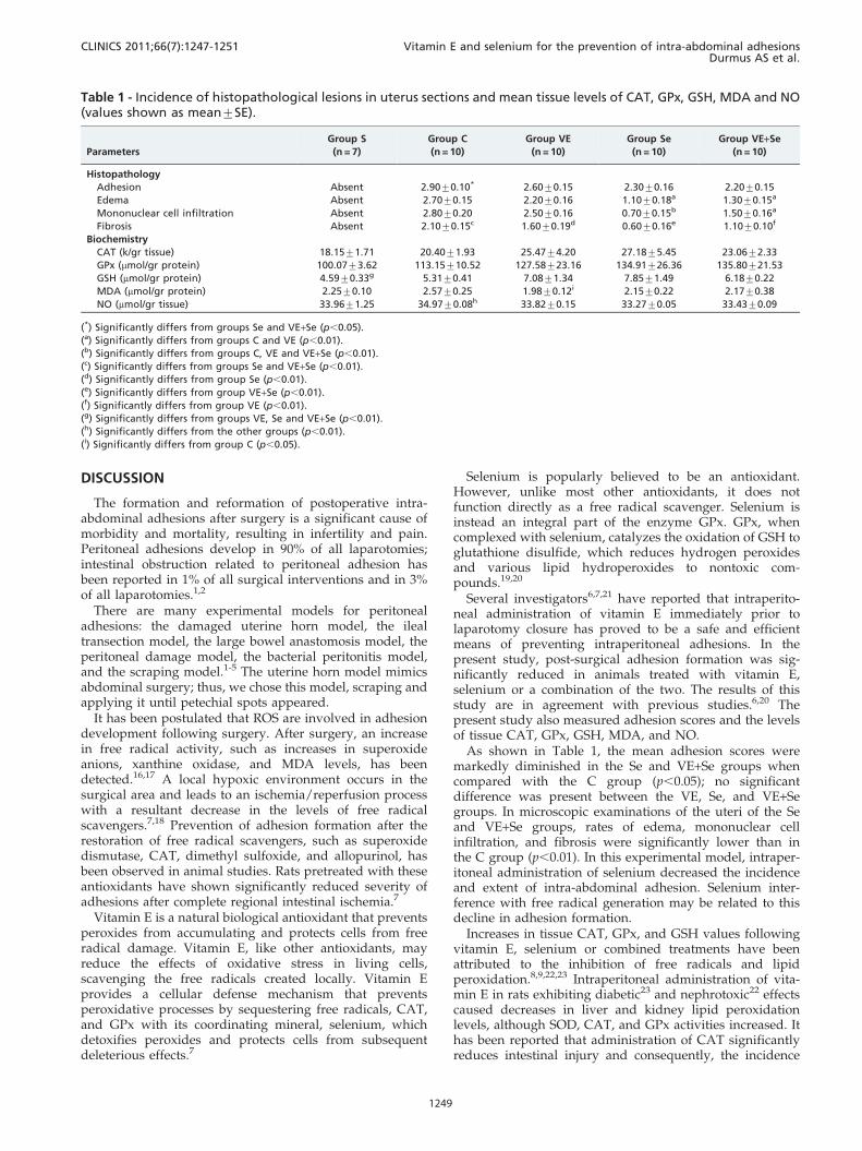
The average adhesion scores were 2.90¡0.10, 2.60¡0.15,2.30¡0.16, and 2.20¡0.15 in the C, VE, Se, and VE+Segroups, respectively. The adhesion scores for the Se andVE+Se groups were significantly lower than those of the Cgroup (p,0.05); scores in the VE group were also decreased(Table 1).
No pathological symptoms were observed in microscopicexaminations of the uteri of the S group. In the C group,adhesions between uteri and neighboring tissues werevisible. Edema, thickness, and capillary vascular prolifera-tions were noted in adhesion regions. Focal microabscesses,fibrosis, and intensive mononuclear cell infiltrations in themuscular layer of the uterus were also observed (Figure 1A).Adhesions, edema, thickness, capillary vascular prolifera-tion, and mononuclear cell infiltrations in the uterine serosawere less severe in the VE group than in the C group(Figure 1B). Similar lesions were found in the Se and VE+Segroups but were less severe than in the other groups(Figure 1C, Figure 1D). The distribution of pathologicallesions among the groups is shown in Table 1.
Tissue CAT, GPx, GSH, MDA, and NO levels weremeasured in all groups; these data are shown in Table 1.CAT and GPx activities did not differ significantly amongthe groups. However, CAT and GPx activities and GSHlevels were slightly increased in the VE, Se, and VE+Segroups. GSH levels were significantly lower in the S groupthan in the VE, Se, and VE+Se groups (p,0.01). In the VEgroup, MDA concentrations were significantly lower than inthe C group (p,0.05), but those of the S, C, Se, and VE+Segroups did not differ significantly. Levels of NO weresignificantly higher in the C group than in the other groups(p,0.01).
Vitamin E and selenium for the prevention of intra-abdominal adhesionsDurmus AS et al.
CLINICS 2011;66(7):1247-1251
1248
DISCUSSION
The formation and reformation of postoperative intra-abdominal adhesions after surgery is a significant cause ofmorbidity and mortality, resulting in infertility and pain.Peritoneal adhesions develop in 90% of all laparotomies;intestinal obstruction related to peritoneal adhesion hasbeen reported in 1% of all surgical interventions and in 3%of all laparotomies.1,2
There are many experimental models for peritonealadhesions: the damaged uterine horn model, the ilealtransection model, the large bowel anastomosis model, theperitoneal damage model, the bacterial peritonitis model,and the scraping model.1-5 The uterine horn model mimicsabdominal surgery; thus, we chose this model, scraping andapplying it until petechial spots appeared.
It has been postulated that ROS are involved in adhesiondevelopment following surgery. After surgery, an increasein free radical activity, such as increases in superoxideanions, xanthine oxidase, and MDA levels, has beendetected.16,17 A local hypoxic environment occurs in thesurgical area and leads to an ischemia/reperfusion processwith a resultant decrease in the levels of free radicalscavengers.7,18 Prevention of adhesion formation after therestoration of free radical scavengers, such as superoxidedismutase, CAT, dimethyl sulfoxide, and allopurinol, hasbeen observed in animal studies. Rats pretreated with theseantioxidants have shown significantly reduced severity ofadhesions after complete regional intestinal ischemia.7
Vitamin E is a natural biological antioxidant that preventsperoxides from accumulating and protects cells from freeradical damage. Vitamin E, like other antioxidants, mayreduce the effects of oxidative stress in living cells,scavenging the free radicals created locally. Vitamin Eprovides a cellular defense mechanism that preventsperoxidative processes by sequestering free radicals, CAT,and GPx with its coordinating mineral, selenium, whichdetoxifies peroxides and protects cells from subsequentdeleterious effects.7
Selenium is popularly believed to be an antioxidant.However, unlike most other antioxidants, it does notfunction directly as a free radical scavenger. Selenium isinstead an integral part of the enzyme GPx. GPx, whencomplexed with selenium, catalyzes the oxidation of GSH toglutathione disulfide, which reduces hydrogen peroxidesand various lipid hydroperoxides to nontoxic com-pounds.19,20
Several investigators6,7,21 have reported that intraperito-neal administration of vitamin E immediately prior tolaparotomy closure has proved to be a safe and efficientmeans of preventing intraperitoneal adhesions. In thepresent study, post-surgical adhesion formation was sig-nificantly reduced in animals treated with vitamin E,selenium or a combination of the two. The results of thisstudy are in agreement with previous studies.6,20 Thepresent study also measured adhesion scores and the levelsof tissue CAT, GPx, GSH, MDA, and NO.
As shown in Table 1, the mean adhesion scores weremarkedly diminished in the Se and VE+Se groups whencompared with the C group (p,0.05); no significantdifference was present between the VE, Se, and VE+Segroups. In microscopic examinations of the uteri of the Seand VE+Se groups, rates of edema, mononuclear cellinfiltration, and fibrosis were significantly lower than inthe C group (p,0.01). In this experimental model, intraper-itoneal administration of selenium decreased the incidenceand extent of intra-abdominal adhesion. Selenium inter-ference with free radical generation may be related to thisdecline in adhesion formation.
Increases in tissue CAT, GPx, and GSH values followingvitamin E, selenium or combined treatments have beenattributed to the inhibition of free radicals and lipidperoxidation.8,9,22,23 Intraperitoneal administration of vita-min E in rats exhibiting diabetic23 and nephrotoxic22 effectscaused decreases in liver and kidney lipid peroxidationlevels, although SOD, CAT, and GPx activities increased. Ithas been reported that administration of CAT significantlyreduces intestinal injury and consequently, the incidence
Table 1 - Incidence of histopathological lesions in uterus sections and mean tissue levels of CAT, GPx, GSH, MDA and NO(values shown as mean¡SE).
Parameters
Group S
(n = 7)
Group C
(n = 10)
Group VE
(n = 10)
Group Se
(n = 10)
Group VE+Se
(n = 10)
Histopathology
Adhesion Absent 2.90¡0.10* 2.60¡0.15 2.30¡0.16 2.20¡0.15
Edema Absent 2.70¡0.15 2.20¡0.16 1.10¡0.18a 1.30¡0.15a
Mononuclear cell infiltration Absent 2.80¡0.20 2.50¡0.16 0.70¡0.15b 1.50¡0.16a
Fibrosis Absent 2.10¡0.15c 1.60¡0.19d 0.60¡0.16e 1.10¡0.10f
Biochemistry
CAT (k/gr tissue) 18.15¡1.71 20.40¡1.93 25.47¡4.20 27.18¡5.45 23.06¡2.33
GPx (mmol/gr protein) 100.07¡3.62 113.15¡10.52 127.58¡23.16 134.91¡26.36 135.80¡21.53
GSH (mmol/gr protein) 4.59¡0.33g 5.31¡0.41 7.08¡1.34 7.85¡1.49 6.18¡0.22
MDA (mmol/gr protein) 2.25¡0.10 2.57¡0.25 1.98¡0.12i 2.15¡0.22 2.17¡0.38
NO (mmol/gr tissue) 33.96¡1.25 34.97¡0.08h 33.82¡0.15 33.27¡0.05 33.43¡0.09
(*) Significantly differs from groups Se and VE+Se (p,0.05).
(a) Significantly differs from groups C and VE (p,0.01).
(b) Significantly differs from groups C, VE and VE+Se (p,0.01).
(c) Significantly differs from groups Se and VE+Se (p,0.01).
(d) Significantly differs from group Se (p,0.01).
(e) Significantly differs from group VE+Se (p,0.01).
(f) Significantly differs from group VE (p,0.01).
(g) Significantly differs from groups VE, Se and VE+Se (p,0.01).
(h) Significantly differs from the other groups (p,0.01).
(i) Significantly differs from group C (p,0.05).
CLINICS 2011;66(7):1247-1251 Vitamin E and selenium for the prevention of intra-abdominal adhesionsDurmus AS et al.
1249

and severity of peritoneal adhesion formation.24 In thecurrent literature, there are no studies reporting the effectsof vitamin E and selenium treatments on the prevention ofadhesion formation in rats or on the tissue MDA or GSHlevels or GPx activities. In the VE, Se and VE+Se groups,tissue GSH content and CAT and GPx activities wereelevated. These increases may be due to their depletion orinhibition as a result of the increased production of freeradicals. The results of this study are consistent withprevious studies24 using CAT to prevent intraperitonealadhesion formation. These data suggest that ROS may playa role in intra-abdominal adhesion formation.
MDA, a product of lipid peroxidation, is generated as aresult of the toxic effects of active oxygen radicals thatdestroy unsaturated fatty acids in membranes.23,25 Locallygenerated free radicals, such as superoxides, peroxides andhydroxyl radicals, are potential oxidizers of polyunsatu-rated fatty acids and are therefore postulated to induceperitoneal adhesions through damage to cellular mem-branes.7 In this study, levels of MDA in the vitamin E groupwere significantly lower than in the control group. It hasbeen demonstrated that tissue MDA levels are decreased byvitamin E. This may be related to the antioxidative and freeradical-scavenging effects of vitamin E.
This study showed that selenium is a potent inhibitor ofadhesion formation and suggests that its action is due to itsanti-inflammatory and antioxidant effects, as shown by thelower observed tissue concentration of MDA, higheractivities of CAT and GPx and high tissue GSH content inthe VE, Se and VE+Se groups. These data indicate that theadministration of vitamin E and selenium can preventadhesion formation in rat uteri by modifying tissueantioxidant enzyme activities.
NO is a free radical gas molecule that is produced from L-arginine by the enzyme nitric oxide synthase (NOS).Depending on its redox state and cellular source, NO maybe toxic or protective.26 Increased NO, especially associatedwith oxidative stress, is harmful to tissue.16 Gurel et al.27
reported that acute administration of vitamin E decreasedNO concentrations in both ipsilateral and contralateral renaltissues compared to an ischemia-reperfusion group. Gorgunet al.28 showed that vitamin E was effective in reducing NOSactivity in streptozotocin-induced diabetic rats, suggestingthat a-tocopherol acts as a beneficial antioxidant. Vitamin Emay also increase NO efficiency by scavenging oxygenradicals in diabetic animals. Vitamin E supplementation isbeneficial in reducing nitric oxidation mechanisms. Studieson the role of NO in adhesion formation are limited.
Figure 1 - (A) Histopathological view of uterus section in the control group. a) Edema and thickness in serosa, b) capillary vascularproliferation, c) microabscess (H&E, 100 x). (B) Histopathological view of uterus section in the vitamin E-treated group. a) Edema andthickness on serosa, b) capillary vascular proliferation (H&E, 100 x). (C) Histopathological view of a uterus section in the selenium-treated group. a) Edema on serosa (H&E, 100 x). (D) Histopathological view of uterus section in the animals treated with both vitamin Eand selenium. a) Edema and thickness on serosa (H&E, 100 x).
Vitamin E and selenium for the prevention of intra-abdominal adhesionsDurmus AS et al.
CLINICS 2011;66(7):1247-1251
1250
Intraperitoneal administration of melatonin,29 aminoguani-dine,30 and resveratrol31 has been reported to decrease theincidence and extent of peritoneal adhesions and cause adecrease in NO levels. In this study, levels of NO weresignificantly higher in the C group than in other groups(p,0.01). Tissue NO levels were decreased by vitamin E andselenium. These results show that NO may play animportant role in adhesion formation, and the decrease inadhesion formation from vitamin E and selenium treatmentmay be related to their antioxidant and free radicalscavenging effects. The observed lower levels of tissue NOin the VE, Se, and VE+Se groups may be evidence of theseantioxidative effects.
Selenium was shown to be effective in reducing intra-abdominal adhesion formation. However, the efficacy ofvitamin E used together with selenium was not superior tothat of selenium alone.
CONCLUSION
The results of this study suggest that postoperative intra-abdominal adhesion formation in female rats resulted inincreased oxidative stress, but that vitamin E and seleniumsupplementation may reduce the imbalance between uncon-trolled free radical generation and scavenging enzymeactivity. Intraperitoneal administration of vitamin E, sele-nium, and a combination of the two may be effective inpreventing intra-abdominal adhesion formation in rat mod-els. The antioxidative effects of selenium may have helped toprevent intraperitoneal adhesions between serosal surfaces.
REFERENCES
1. Durmus AS, Han MC. Comparison of the effects of different concentra-tions of sodium carboxymethylcellulose on prevention of intraabdominaladhesions in rats. Rev Med Vet Toulouse. 2006;157:535-8.
2. Durmus AS, Han MC. Effect of bovine amniotic fluid on intraabdominaladhesions. Indian Vet J. 2006;83:621-3.
3. Emre A, Akin M, Isikgonul I, Yuksel O, Anadol AZ, Cifter C.Comparison of intraperitoneal honey and sodium hyaluronate -carboxymethylcellulose (SeprafilmTM) for the prevention of postopera-tive intra-abdominal adhesions. Clinics. 2009;64:363-8, doi: 10.1590/S1807-59322009000400016.
4. Irkorucu O, Ferahkose Z, Memis L, Ekinci O, Akın M. Reduction ofpostsurgical adhesions in a rat model: a comparative study. Clinics.2009;64:143-8.
5. Aksakal O, Yilmaz B, Gungor T, Sirvan L, Sut N, Inan I, et al. Arandomised controlled trial on melatonin and rosiglitazone for preven-tion of adhesion formation in a rat uterine horn model. Arch GynecolObstet. 2010;282:55-61, doi: 10.1007/s00404-009-1240-8.
6. De La Portilla F, Ynfante I, Bejarano D, Conde J, Fernandez A, Ortega JM,et al. Prevention of peritoneal adhesions by intraperitoneal administra-tion of vitamin E: an experimental study. Dis Colon Rectum.2005;47:2157-61, doi: 10.1007/s10350-004-0741-6.
7. Yildiz H, Durmus AS, Simsek H. Surgery-induced changes in red bloodcell and plasma lipid peroxidation, enzymatic and non-enzymaticantioxidants, and blood hematology of female rats: protective role ofmethylene blue and vitamin E. Eur J Obstet Gynecol Reprod Biol.2011;155:89-93, doi: 10.1016/j.ejogrb.2010.11.003.
8. Forceville X. Seleno-enzymes and seleno-compounds: the two faces ofselenium. Crit Care. 2006;10:180, doi: 10.1186/cc5109.
9. Forceville X. Effects of high doses of selenium, as sodium selenite, inseptic shock patients a placebo-controlled, randomized, double-blind,multi-center phase II study- Selenium and sepsis. J Trace Elem Med Bio.2007;21:62-5, doi: 10.1016/j.jtemb.2007.09.021.
10. Yıldız H, Durmus AS, Simsek H, Yaman M. Protective effect of sildenafilcitrate on contralateral testis injury after unilateral testicular torsion/detorsion. Clinics, 2011;66:137-42.
11. Sedlak J, Lindsay RHC. Estimation of total, protein bound and non-protein sulfhydryl groups in tissue with Ellmann’s reagent. AnalBiochem. 1968;25:192-205, doi: 10.1016/0003-2697(68)90092-4.
12. Lawrence RA, Burk RF. Glutathione peroxidase activity in selenium-deficient rat liver. Biochem Bioph Res Co. 1976;71:952-8, doi: 10.1016/0006-291X(76)90747-6.
13. Lowry OH, Rosebrough NJ, Farr AL, Randall RJ. Protein measurementwith the Folin-Phenol reagent. J Biol Chem. 1951;193:265-75.
14. Aebi H. Catalase in vitro. Method Enzymol. 1984;105:121-6, doi: 10.1016/S0076-6879(84)05016-3.
15. Cortas NK, Wakid NM. Determination of inorganic nitrate in serum andurine by a kinetic cadmium-reduction method. Clin Chem. 1990;36:1440-3.
16. Alpay Z, Saed GM, Diamond MP. Female infertility and free radicals:Potential role in adhesions and endometriosis. J Soc Gynecol Investig.2006;13:390-8, doi: 10.1016/j.jsgi.2006.05.002.
17. Bentes De Souza AM, Rogers MS, Wang CC, Yuen PM, Pui SN.Comparison of peritoneal oxidative stress during laparoscopy andlaparotomy. J Am Assoc Gynecol Laparosc. 2003;10:65-74, doi: 10.1016/S1074-3804(05)60237-X.
18. Andraus W, Souza GFP, Oliveira MG, Haddad LBP, Coelho AMM,Galvao FH, et al. S-Nitroso-n-acetylcysteine ameliorates ischemia-reperfusion injury in the steatotic liver. Clinics. 2010;65:715-21, doi: 10.1590/S1807-59322010000700011.
19. Bright MWR, Beate T, Peter ET, Martin AV, David HS. Effect ofpancreatic enzyme preparations on erythrocyte glutathione peroxidaseactivities and plasma selenium concentrations in cystic fibrosis. FreeRadical Bio Med. 1998;25:242-9, doi: 10.1016/S0891-5849(98)00061-6.
20. Sarada SKS, Sairam M, Dipti P, Anju B, Pauline T, Kain AK, et al. Role ofselenium in reducing hypoxia-induced oxidative stress: an in vivo study.Biomed Pharmacother. 2002;56:173-8, doi: 10.1016/S0753-3322(02)00173-7.
21. Corrales F, Corrales M, Schirmer CC. Preventing intraperitonealadhesions with vitamin E and sodium hyaluronate/carboxymethylcel-lulose. A comparative study in rats. Acta Cir Bras. 2008;23:36-41.
22. Armagan A, Kutluhan S, Yilmaz M, Yilmaz N, Bulbul M, Vural H, et al.Topiramate and vitamin E modulate antioxidant enzyme activities, nitricoxide and lipid peroxidation levels in pentylenetetrazol-inducednephrotoxicity in rats. Basic Clin Pharmacol Toxicol. 2008;103:166-70,doi: 10.1111/j.1742-7843.2008.00271.x.
23. Kinalski M, Sledziewski A, Telejko B, Zarzycki W, Kinalska I. Lipidperoxidation and scavenging enzyme activity in streptozotocin-induceddiabete. Acta Diabetol. 2000;37:179-83, doi: 10.1007/s005920070002.
24. Yuzbasioglu MF, Ezberci F, Imrek E, Bulbuloglu E. The effect ofintraperitoneal catalase on prevention of peritoneal adhesion formationin rats. J Invest Surg. 2008;21:65-9, doi: 10.1080/08941930701883616.
25. Budin SB, Othman F, Louis SR, Bakar MA, Das S, Mohamed J. The effectsof palm oil tocotrienol-rich fraction supplementation on biochemicalparameters, oxidative stress and the vascular wall of streptozotocin-induced diabetic rats. Clinics. 2009;64:235-44, doi: 10.1590/S1807-59322009000300015.
26. Lipton SA, Choi YB, Pan ZH, Lei SZ, Chen HSV, Sucher NJ, et al. Aredox-based mechanism for the neuroprotective and neurodestructiveeffects of nitric oxide and related nitroso-compounds. Nature.1993;364:626-32, doi: 10.1038/364626a0.
27. Gurel A, Armutcu F, Sahin S, Sogut S, Ozyurt H, Gulec M, et al.Protective role of alpha-tocopherol and caffeic acid phenethyl ester onischemia-reperfusion injury via nitric oxide and myeloperoxidase in ratkidneys. Clin Chim Acta. 2004;339:33-41, doi: 10.1016/j.cccn.2003.09.013.
28. Gorgun FM, Gumustas MK, Altug T, Kokoglu E. Vitamin E supple-mentation in streptozotocin-treated rats alters cerebellar and plasmanitric oxide metabolism. J Toxicol Env Heal A. 2002;65:631-7, doi: 10.1080/152873902317349772.
29. Ara CT, Kirimlioglu H, Bay Karabulut A, Coban S, Hascalik S, Celik O,et al. Effect of melatonin against oxidative stress on adhesion formationin the rat cecum and uterine horn model. Life Sci. 2005;77:1341-50, doi:10.1016/j.lfs.2005.01.024.
30. Ara C, Bay Karabulut A, Kirimlioglu H, Yilmaz M, Kirimliglu V, YilmazS. Protective effect of aminoguanidine against oxidative stress in anexperimental peritoneal adhesion model in rats. Cell Biochem Funct.2006;24:443-8, doi: 10.1002/cbf.1245.
31. Sogutlu G, Bay Karabulut A, Ara C, Cinpolat O, Isik B, Piskin T, et al. Theeffect of resveratrol on surgery-induced peritoneal adhesions in anexperimental model. Cell Biochem Funct. 2007;25:217-20, doi: 10.1002/cbf.1324.
CLINICS 2011;66(7):1247-1251 Vitamin E and selenium for the prevention of intra-abdominal adhesionsDurmus AS et al.
1251