Basement Membrane Heparan Sulfate in Atherogenesis and Intimal
69
Thesis for doctoral degree (Ph.D.) 2008 Basement Membrane Heparan Sulfate in Atherogenesis and Intimal Hyperplasia Karin Tran Lundmark Thesis for doctoral degree (Ph.D.) 2008 Karin Tran Lundmark Basement Membrane Heparan Sulfate in Atherogenesis and Intimal Hyperplasia
Transcript of Basement Membrane Heparan Sulfate in Atherogenesis and Intimal

Thesis for doctoral degree (Ph.D.)2008
Basement Membrane Heparan Sulfate in Atherogenesisand Intimal Hyperplasia
Karin Tran Lundmark
Thesis for doctoral degree (Ph.D.) 2008
Karin Tran Lundm
ark B
asemen
t Mem
brane H
eparan Su
lfate in A
therogen
esis and In
timal H
yperplasia

From the Department of Molecular Medicine and Surgery
Karolinska Institutet, Stockholm, Sweden
Basement Membrane Heparan Sulfate in Atherogenesis and Intimal Hyperplasia
Karin Tran Lundmark, MD
Stockholm 2008

All previously published papers were reproduced with permission from the publisher. Published by Karolinska Institutet. Printed by Larserics Digital Print AB, Sundbyberg, Sweden. Cover page: Perlecan staining of ApoE0 aortic root section. © Karin Tran Lundmark, 2008 ISBN 978-91-7357-501-0

To my family


ABSTRACT Cardiovascular disease due to atherosclerosis has become the leading cause of mortality in the world. Atherosclerosis is a progressive disease characterized by the accumulation of lipids, inflammatory cells, smooth muscle cells (SMCs) and extracellular matrix in the wall of large and medium-sized arteries. Surgical treatment of atherosclerosis cause mechanical injury to the vessel wall, which in many cases leads to restenosis and graft stenosis and recurrence of symptoms. Intimal SMC proliferation contributes to the stability of atherosclerotic plaques but it is also the main feature of intimal hyperplasia, which contributes to restenosis. It is therefore important to understand the mechanisms that control SMC growth in order to achieve a balanced healing response following interventions. This can be illustrated by the use of stents that elute anti-proliferative drugs, which has recently been associated with a higher risk of late stent thrombosis due to impaired intimal healing. Here, the role of basement membrane heparan sulfate (HS) in intimal hyperplasia and atherogenesis was investigated. Exogenously added heparin and HS are known inhibitors of SMC proliferation. However, the role of perlecan, which is the major arterial HS proteoglycan, in vascular disease was previously largely unknown. In vitro, the HS chains of perlecan lead to altered interactions between SMCs and fibronectin, possibly due to conformational changes in the fibronectin molecule. Such interactions may influence SMC function in atherogenesis and vascular repair processes. The use of transgenic mice expressing an HS-deficient perlecan showed increased SMC proliferation in vitro and increased intimal hyperplasia in vivo, confirming a growth inhibitory role for perlecan HS. A possible mechanism is decreased bioavailability of heparin-binding growth factors like FGF-2 at the cell surface due to sequestering in the basement membrane. In order to study the role of HS in atherogenesis the HS-deficient mice were cross-bred with apolipoprotein E null mice, which develop atherosclerosis. The results from that study indicate that the perlecan HS chains are pro-atherogenic in mice through increased lipoprotein retention, and the ability of HS to inhibit SMC growth may contribute to lesion instability. However, when binding and retention are not limiting factors, the perlecan HS chains may be anti-atherogenic by reducing endothelial permeability to lipoproteins. To investigate how perlecan can be pharmacologically regulated we explored the effect of all-trans-retinoic acid (AtRA) on perlecan expression in SMCs. AtRA was shown to up-regulate perlecan and the inhibition of SMC proliferation by AtRA is secondary to an increased expression of perlecan and dependent upon its HS chains. In summary, the role of basement membrane HS in vascular disease is complex. It may enhance lipoprotein retention, but also decrease endothelial permeability to lipoproteins. In addition, it can reduce restenosis, but maybe also cause plaque instability. This makes perlecan a difficult but very intriguing target for pharmacological interventions.

LIST OF PUBLICATIONS This thesis is based on the following original articles and manuscripts, which will be referred to in the text by their roman numerals:
I. Lundmark K, Tran PK, Kinsella MG, Clowes AW, Wight TN and Hedin U. Perlecan Inhibits Smooth Muscle Cell Adhesion to Fibronectin: Role of Heparan Sulfate. J Cell Physiol. 2001 Jul;188(1):67-74
II. Tran PK, Tran-Lundmark K, Soininen R, Tryggvason K, Thyberg J and Hedin U. Increased Intimal Hyperplasia and Smooth Muscle Cell Proliferation in Transgenic Mice with Heparan Sulfate-Deficient Perlecan. Circ Res. 2004 Mar 5;94(4):550-8
III. Tran-Lundmark K, Tran PK, Paulsson-Berne G, Fridén V, Soininen R, Tryggvason K, Wight TN, Kinsella MG, Borén J, Hedin U. Heparan Sulfate in Perlecan Promotes Mouse Atherosclerosis: Roles in Lipid Permeability, Lipid Retention and Smooth Muscle Cell Proliferation. Manuscript submitted to Circulation Research
IV. Tran-Lundmark K, Tran PK, Rauch B, Ekstrand J, Wight TN, Hedin U, Kinsella MG. Heparan Sulfate Chains on Perlecan are Required for the Inhibition of Arterial Smooth Muscle Cell Proliferation by all-trans-Retinoic Acid Manuscript submitted to ATVB

CONTENTS 1 AIMS OF THE THESIS ..............................................................................1 2 INTRODUCTION........................................................................................2
2.1 Clinical background............................................................................2 2.2 The normal artery wall .......................................................................3
2.2.1 The vascular cells ...................................................................4 2.2.2 The extracellular matrix .........................................................4 2.2.3 Interactions between cells and the extracellular matrix ........8 2.2.4 The proteoglycans of the artery wall .....................................8
2.3 Atherosclerosis..................................................................................15 2.3.1 Lipoprotein retention by proteoglycans...............................15 2.3.2 Inflammation ........................................................................16 2.3.3 Plaque progression ...............................................................17 2.3.4 Plaque rupture and plaque stability......................................19
2.4 Restenosis .........................................................................................19 2.4.1 Elastic recoil .........................................................................19 2.4.2 Negative remodeling ............................................................20 2.4.3 Intimal hyperplasia ...............................................................20 2.4.4 In-stent restenosis .................................................................21
2.5 AtRA .................................................................................................22 3 METHODS.................................................................................................23
3.1 Adhesion assay .................................................................................23 3.2 Intimal hyperplasia ...........................................................................23
3.2.1 Smooth muscle cell culture ..................................................24 3.2.2 In vivo model ........................................................................25
3.3 Atherosclerosis..................................................................................26 3.3.1 Aortic root sectioning...........................................................26 3.3.2 En face analysis ....................................................................27 3.3.3 Lipoprotein analysis .............................................................29
3.4 Animal models..................................................................................29 3.4.1 Heparan sulfate deficient mice.............................................29 3.4.2 Apolipoprotein E null mice..................................................30
3.5 Proteoglycan analysis .......................................................................31 3.6 Additional methods used ..................................................................33
4 RESULTS AND DISCUSSION................................................................34 4.1 Perlecan HS influence SMC function (paper I) ...............................34 4.2 Basement membrane HS in intimal hyperplasia (paper II) .............36 4.3 Basement membrane HS in atherogenesis (paper III).....................39 4.4 Pharmacological regulation of perlecan HS (paper IV) ..................43
5 CONCLUDING REMARKS.....................................................................45 6 FUTURE PERSPECTIVES.......................................................................46 7 ACKNOWLEDGEMENTS.......................................................................47 8 REFERENCES...........................................................................................50

LIST OF ABBREVIATIONS SMC smooth muscle cell HS heparan sulfate ECM extracellular matrix FN fibronectin CS chondroitin sulfate DS dermatan sulfate FGF fibroblast growth factor LDL low-density-lipoproteins apoB apolipoprotein B ox-LDL oxidized low-density-lipoproteins PDGF platelet derived growth factor Hb-EGF heparin-binding epidermal-growth-factor-like growth factor TNF-α tumor necrosis factor-α AtRA all-trans-retinoic acid ApoE0 apolipoprotein E null IGF-1 insulin-like growth factor-1 TGF-β transforming growth factor-β NO nitric oxide GAG glycosaminoglycan DIT diffuse intimal thickening

1
1 AIMS OF THE THESIS The general objective of this thesis work was to investigate the role of basement membrane components in vascular disease. Through the years I have continuously been working with both perlecan and laminin, but since the studies on perlecan were finished first we chose to focus on those for the dissertation. I, however, hope to be able to continue my work on laminin in the future. The specific aims for the included studies was to investigate:
1. The influence of perlecan/HS on SMC function in vitro (paper I). 2. The role of basement membrane HS in intimal hyperplasia (paper II). 3. The role of basement membrane HS in atherogenesis (paper III). 4. How perlecan/HS can be pharmacologically regulated (paper IV).
2
2 INTRODUCTION 2.1 CLINICAL BACKGROUND
Atherosclerosis is a progressive disease characterized by the accumulation of lipids, inflammatory cells, smooth muscle cells (SMCs) and extracellular matrix in the wall of large and medium-sized arteries. Manifestations of atherosclerosis (ischemic heart disease and stroke) were the leading causes of death in both high-income and low-and-middle-income countries when all deaths globally during 2001 were analyzed 1. The two conditions were together responsible for more than a fifth of all deaths worldwide. Epidemiological studies over the past 50 years have revealed numerous risk factors for atherosclerosis. Some of the important factors are high blood-cholesterol levels, smoking, hypertension, diabetes mellitus, physical inactivity and obesity 2. The signs and symptoms of atherosclerosis depend on its location within the vascular system. Some common locations are the coronary arteries, arteries in the lower extremities, the carotid arteries, the aorta and the renal arteries. Disease in the coronary arteries presents as angina pectoris or myocardial infarction, and in the lower extremities claudication and critical limb ischemia are the principal clinical manifestations. Atherosclerosis in the carotid arteries causes transient ischemic attacks and stroke, and disease in the aorta can lead to obstruction or aneurysm formation. Renal artery atherosclerosis may result in hypertension or ischemic nephropathy or both 3. Clinical symptoms of atherosclerosis can be either chronic or acute depending on the pathophysiological mechanism causing the symptoms. There can be a gradual decrease in vessel lumen diameter limiting the capacity to increase the blood-flow during exercise. This is the case in angina pectoris and claudication where symptoms appear during physical activity. There may also be a rupture or erosion of a lesion causing thrombosis at the site of the lesion and sudden occlusion of the vessel, leading to acute ischemia in the tissue distal from the occlusion. This is what happens in myocardial infarction. A third important mechanism is embolization of platelet-fibrin thrombi from atherosclerotic lesions. This is the most common mechanism in transient ischemic attacks and stroke since the cerebral circulation is interconnected via the circle of Willis, and therefore is less sensitive to hypoperfusion due to lesions located in carotid and vertebral arteries. Treatment for atherosclerosis can be either pharmacological or surgical and is often used in combination with lifestyle changes. Pharmacological treatment is today mostly used in patients with severe risk factor profiles to retard the progression of the disease, or to relieve symptoms, or as a secondary preventive measure. Surgical treatment is used when the clinical symptoms becomes too severe. Such symptoms include

3
debilitating chest pain, incapacitating pain in lower extremities with risk of gangrene, and recurring transient ischemic attacks (indicating an unstable carotid lesion). The surgical treatments used are either removal of the lesion, endarterectomy, or procedures that by-passes the diseased vessel segment with autologous veins or synthetic grafts. During the last two decades percutaneous catheter-based techniques, with and without use of metal stents, have also become an important complement to traditional surgical methods. The first percutaneous coronary intervention (PCI) was performed in 1977 4. It has since then become the most common revascularization procedure for coronary artery disease and endovascular approaches are nowadays also commonly used for peripheral artery disease 5. However, although PCI spares patients the need for costly and complex surgical revascularization, between 30% and 50% of patients who undergo conventional balloon angioplasty and 10-30% of patients who receive a bare-metal stent experience recurrence of symptoms due to so called restenosis 6. Restenosis is a healing response caused by the iatrogenic injury to the artery. Balloon inflation fractures the atherosclerotic plaque and this starts a cascade of molecular and cellular events, which results in narrowing of the vessel lumen and recurrence of symptoms. Restenotic lesions are generally stable and presents as a gradual recurrence of symptoms rather than as an acute event. The presence of restenosis therefore does not confer an increase in mortality, but the impact of restenosis on patient quality of life has been confirmed in prospective and retrospective clinical trials 7, 8. Risk factors for restenosis are for example diabetes mellitus, genetic predisposition, small vessel diameter, ostial lesions, lesion length, and runoff status 5, 6. These factors should be considered when choosing between percutaneous interventions and traditional surgical approaches. A pathological healing response can however also be a problem in vein grafts as well as in synthetic grafts. Graft stenosis is either a result of injury to the vein during surgery or due to altered hemodynamic conditions in the conduit 9. 2.2 THE NORMAL ARTERY WALL
Arteries are composed of three layers. The intima is the innermost layer and includes the endothelial lining. An elastic membrane called the internal elastic laminia separates the intima from the outer layers. The media is the muscular middle layer and the adventitia is the outermost connective tissue layer. The adventitia around large arteries also contains blood vessels (vasa vasorum) and nerves (nervi vascularis). Traditionally arteries are classified into three types on the basis of size and characteristics of the medial layer. The largest are the elastic arteries, which have sheets of elastic tissue in their wall. Functionally they serve primarily as conduction tubes, but they also maintain blood pressure and sustained flow during the relaxation phase of the cardiac cycle. Muscular arteries have more SMCs and less elastin in the media

4
compared to elastic arteries and most of the named arteries in the body are of this type. Arteries with less than eight layers of medial SMCs are called small arteries, and together with arterioles (one or two layers of SMCs) they serve as flow regulators to the capillary bed. 2.2.1 The vascular cells
The intima is dominated by endothelial cells, which line the entire vascular system from the heart to the smallest capillary. The endothelial cells control the passage of macromolecules and cells into and out of the bloodstream. The endothelium is also important for hemostasis, vasomotor tone (NO, endothelin etc), and recruitment of leukocytes in response to inflammatory stimuli 10. SMCs in the media are specialized muscle cells that participate in the regulation of blood pressure and distribution of blood flow. In the normal adult artery SMCs reside in a quiescent, differentiated state. They exhibit very low synthetic activity and express a unique repertoire of contractile proteins, ion channels, and signaling molecules required for the cells contractile functions 11. However, SMCs also have the ability to dramatically increase the rate of cell proliferation, migration, and protein synthesis in response to vascular injury. This phenotypic switch may be beneficial, but it also leads to exaggerated healing responses, as seen for example in restenosis. In the adventitia the fibroblasts predominate. They produce a matrix that anchors the vessel to the adjacent tissues and provides tensile strength to the vessel wall. 2.2.2 The extracellular matrix
The extracellular matrix (ECM) is the meshwork of macromolecules that surrounds the cells of multicellular organisms. The ECM of the vessel wall can be divided into basement membrane components and interstitial matrix. 2.2.2.1 The basement membrane
The basement membrane is a specialized ECM structure that remains in close proximity to the cell it was formed by and it is found underneath the endothelial cell layer and surrounds medial smooth muscle cells. The major components of basement membranes are laminins, collagen type IV, nidogens and the HS proteoglycan perlecan. The basic structure of the basement membrane is thought to be two distinct and independent networks of type IV collagen and laminin, linked together by nidogens and perlecan (Figure 1) 12. However, although the basic building blocks of basement membranes are the same variations in relative amounts of the components and site specific isoforms of the molecules result in unique functions of different basement membranes in different parts of the body.

5
Laminins are approximately cross-shaped heterotrimeric glycoproteins, composed of an α, β and γ chain. To date, 5 distinct α, 4 β, and 3 γ chains have been identified and they can combine to form 15 different isoforms. The laminin α chains are considered the functionally active portion of the heterotrimers since they carry the major domains that interact with cellular receptors. In the vasculature three different α chains are expressed. Endothelial cells express α4 and α5, and SMCs express α4, α5 and α2. The specific roles of different laminin isoforms in the vasculature are just starting to be investigated. For endothelial cells it has for example been shown that different laminin isoforms may contribute to the regulation of leukocyte recruitment 12. So far very little is known about the role of different laminin isoforms in regulation of SMC function. Type IV collagen is a nonfibrillar, network forming collagen, which is the most abundant protein in all basement membranes. Non-fibrillar collagens differ from fibrillar collagens by the presence of globular non-collagenous domains (NC-domains). In mammals, six distinct genes encode for six different chains of type IV collagen, known as α chains. All six isoforms of collagen IV appears to be highly conserved among vertebrates. The isoforms differ mostly in their NC1 domains. Type IV collagen is secreted from cells in the form of protomers composed of three α chains. Currently the existence of two different protomers has been confirmed in vivo and three additional ones are speculative. Protomers are the building units of the basement membrane collagen IV network 13.
Figure 1: The major basement membrane components and their proposed interactions. Modified from Hallmann et al, Physiol Rev 85:979-1000, 2005.
6
Nidogen-1 and –2 (Entactin-1 and –2) are glycoproteins that contain 10% carbohydrates. The core protein is composed of three G domains, which are separated by two rod-like segments. Two genes encode for the two kinds of nidogens. Both isoforms are ubiquitously expressed in all basement membranes throughout the body. Of the two nidogens nidogen-2 exhibits a more restricted pattern of expression and this is the predominant kind in most vascular basement membranes 13. The HS proteoglycan perlecan is also one of the major constituents of the vascular basement membrane as previously mentioned, but since the role of the HS chains of perlecan is the main focus of this thesis the structure and function of perlecan will be presented in more detail below. Basement membranes fulfill many biological functions. They can for example provide structural support for cells and separate different tissue compartments. In addition, they influence cell proliferation, migration, and differentiation. In the endothelium the basement membrane is also likely to contribute to the barrier function, to interactions with mural cells and thereby vessel stability, and to the transduction of mechanosensory signals from the lumen of the vessel to the vessel wall 12. The basement membrane around SMCs in the media is thought to regulate the differentiated and quiescent state of SMCs in the artery wall 14, this is however discussed in more detail below. 2.2.2.2 Interstitial matrix
In addition to basement membrane components the vascular ECM consists of a variety of molecules residing in the interstitial space between the cells. Some of these molecules that are relevant for this thesis will be presented briefly in the following sections. Fibronectin (FN) is a glycoprotein that is present in the ECM and in body fluids. FN plays critical roles in the maintenance of normal cell morphology, cell adhesion, migration, hemostasis, wound healing, differentiation and proliferation. Secreted FN forms dimers composed of two similar, but not necessarily identical, subunits of 250-280 kDa joined by a pair of disulfide bonds near their carboxyl terminal end 15. Fibronectin is encoded by a single gene that is alternatively spliced at three sites to generate the two major forms of FN, cellular FN and plasma FN. Plasma FN is secreted by hepatocytes and is enriched in blood, whereas cellular FN is secreted by fibroblasts, SMCs and many other cell types and is incorporated into a fibrillar matrix on the cell surface 16. Adhesion of SMCs to FN is mediated through an interaction between integrin α5β1 and an Arg-Gly-Asp (RGD) sequence, and can be inhibited by soluble RGD peptides 17. In addition to interactions with cells FN also has binding sites for molecules like collagen, fibrin and heparin 18. The strongest interaction between FN and heparin occurs in the carboxyterminal heparin-binding domain. For a schematic drawing of a FN dimer please see Figure 2 below.

7
Elastin is the major constituent of elastic fibers and is the principle component of the medial ECM in large elastic arteries. The soluble precursor of elastin, tropoelastin, exists as a single polypeptide chain and there are several different isoforms due to alternative splicing. The principle function of elastin is to provide resilience to the tissue 19. Elastin is however also an important regulator of cell function since deletion of the elastin gene leads to excessive proliferation of SMCs in the vessel wall intima 20. Collagens are a major component of all ECMs. Collagens consist of a triple helix of polypeptide chains (referred to as α chains) and there are many different isoforms. Collagen types I and III are the predominant collagens in the vascular interstitial matrix. In addition smaller quantities of type VI collagen exist in the normal vessel wall. Type I and III are organized into fibrils while type VI collagen forms a network 21. Collagens play a central role in maintaining the biophysical integrity and stability of the normal and the diseased vessel wall. The tensile strength and elastic resilience of the vessel wall are largely dependent on the composition and size of the collagen fibers22. Proteoglycans are molecules that consist of a core protein to which one or more glycosaminoglycan (GAG) chains are covalently attached. The major proteoglycans in the vascular interstitial matrix are versican, biglycan and decorin. In addition, hyaluronan is a GAG that exists as a free molecule in the interstitial ECM 21. All of
B A V
B A V
S S
FN Fibrin Collagen
FN Heparin Cells Fibrin
NH2 COOH
Type I Type II Type III
Figure 2: The structure of FN dimers. FN consists of 12 type I modules, 2 type II modules, and 15-17 type III modules. The alternatively spliced domains IIIA, IIIB and the V region are labeled A, B, and V. Binding domains for fibrin, collagen, cells and heparin are indicated. Modified from Mao and Schwarzbauer, Matrix Biology 24: 389-399, 2005.

8
these molecules will be presented in more detail in the section ‘Proteoglycans of the artery wall’. 2.2.3 Interactions between cells and the extracellular matrix
Cell-matrix interactions are primarily mediated by integrins. They comprise a large family of cell surface receptors and they are composed of two subunits, α and β. Each αβ combination has its own binding specificities and signaling properties. Most integrins recognize several ECM proteins, and individual matrix proteins (such as FN, laminins, and collagens) bind to several different integrins 23. For example integrins α6β1 and α3β1 are the major laminin α4 and α5 binding integrins and α5β1 integrin mediates RGD-dependent binding to FN as well as to laminin α5 12. This network of different interactions probably makes the system more stable, but it also makes it very complex. Integrins can signal through the cell membrane in either direction. The extracellular binding activity is regulated from inside the cell and binding of ECM molecules elicits signals that are transmitted into the cell 23. As integrins bind to ECM proteins they become clustered in the cell membrane and associate with the actin cytoskeleton and a signaling complex, forming so called focal adhesions (focal contacts) 24. Signaling via integrins play important roles in cell migration, proliferation, differentiation and survival. However, the ECM can affect cell behavior in more ways than by direct receptor interactions. Glycosaminoglycans like hyaluronan can for example absorb large amount of fluid, which provides space for cells to migrate and divide 25. The matrix can also bind and immobilize mitogens and chemotactic factors and thereby regulate their bioavailability through controlled release by protease activity 26. During development the ECM is also important for the formation of morphogen gradients and thereby for the correct migration of cells 27. 2.2.4 The proteoglycans of the artery wall
As previously mentioned proteoglycans are molecules composed of a core protein with a varying number of covalently linked GAG side chains. Proteoglycans have often been referred to as a group of molecules with similar structure and functions. They are however highly heterogeneous. In the vascular wall the majority of GAG chains linked to a proteoglycan core protein are either of the chondroitin-, dermatan-, or heparan sulfate subclass. GAG structures are based on disaccharide repeats and each GAG subclass is distinguished by a particular repeating disaccharide. The basic disaccharide for chondroitin sulfate (CS) is D-glucuronic-acid combined with N-acetyl-D-galactosamine, whereas heparan sulfate (HS) contains D-glucuronic-acid and N-acetyl-D-glucosamine (Figure 3). Dermatan sulfate (DS) is generated through epimerisation of D-glucuronic-acid in CS chains to L-iduronic acid.

9
GAG chains evolve by the stepwise addition of the disaccharide repeats to a linker tetrasaccharide on serine residues of proteoglycan core proteins, a process that requires transferases that belong to the EXT gene family. Polymerisation of the HS chain is then followed by modifications (Figure 4). In the first step, an N-deacetylase/N-sulfotransferase removes the N-acetyl group from N-acetyl-D-glucosamine, and usually substitutes it with a sulfate group. Next, the enzyme C-5 epimerase converts some glucoronic acids to iduronic acids, which can then be sulfated by a 2-O-sulfotransferase. The glucosamine residues can then be further sulfated at the C6 position by 6-O-sulphotranferases and the final and most rare modification is sulfation at the C3 position by the action of 3-O-sulphotransferases. Modification of the HS chain is incomplete and occurs in a hierarchial order. For example sulfation at the C6 position is more likely to occur if previous modifications have already occurred 28, 29. Heparin, which is produced by mast cells, is a highly sulfated variant of HS.
Figure 3: The basic disaccharides for chondroitin (left) and heparan sulfate (right) GAG chains. Glucoronic acid (GlcUA), N-acetyl-galactosamine (GalNAC), and N-acetyl-glucosamine (GlcN).
4. 6-O-sulphotransferase
3. 2-O-sulphotransferase
1. N-deacetylase/N-sulfotranferase
2. C-5 epimerase
Figure 4: Overview of different HS modifications. Modified from Mulloy and Rider, Biochem Soc Trans. 2006 Jun;34:414-17.

10
An important question has been the functional importance of the GAG modifications and to what extent protein-GAG interactions are specific, rather than non-specific ionic interactions between the negatively charged GAGs and positively charged amino acids. The first evidence of specificity was for the binding of antithrombin III to heparin, which is mediated by a specific pentasaccharide 30. Evidence has since then been sought for specificity of interactions with other proteins like FGF-2 and VEGF. The results have, however, so far suggested that the HS fine structure may be less critical than expected for many GAG-protein interactions. Mice with mutations of enzymes involved in HS modifications have been surprisingly normal in many ways (although kidney, lung, and skeletal abnormalities are seen) 31-33, whereas mice with a deletion of EXT1 (resulting in complete absence of HS) fail to undergo proper gastrulation in accordance with the recognized need for HS in early patterning events 34. This suggests that certain functionally important HS-protein interactions depend primarily on charge distribution, whereas others require specific saccharide components. What may be of importance is the distribution of domains containing a varying degree of sulfation, but very little is known so far 28. CS is less extensively modified compared to HS. As previously mentioned epimerisation of CS chains results in formation of DS chains and there is also varying degrees of sulfation by chondroitin 6-O-sulfotransferases and chondroitin 4-O-sulfotransferases 35. In the following sections some of the important proteoglycans present in the vascular wall will be presented. 2.2.4.1 Chondroitin/dermatan sulfate proteoglycans
Versican is the principal CS proteoglycan in the normal human vascular wall and immunohistochemistry reveals that it is prominent in the intima and adventitia of most human arteries. The amino-terminal domain of versican binds hyaluonan (hyaluronic acid; a molecule that exists as a free non-sulfated GAG without core protein). The mid portion contains two GAG attachment domains (versican can carry up to 30 CS-chains), and the carboxy-terminal terminal end is a selectin-like region that contains two epidermal growth factor (EGF) repeats, a C-type lectin domain and a complementary regulatory motif (Figure 5). Versican exists in at least four different isoforms created by alternative splicing of mRNA from a single gene (VO, V1, V2, and V3). These variant differ in length of the core protein and in number of attached GAGs.

11
Versican is present in advanced atherosclerotic lesions, at the borders of lipid-filled necrotic cores as well as at the plaque thrombus interface, suggesting roles in lipid accumulation, inflammation and thrombosis. Versican is also up-regulated after vascular injury and is a prominent component of restenotic lesions. It can also influence the assembly of ECM and controls elastic fiber fibrillogenesis, which is of importance in ECM remodeling in vascular disease processes 36. Biglycan is synthesized by endothelial cells and SMCs and consists of a core protein carrying two CS and/or DS side chains. Biglycan is a member of the small leucine-rich proteoglycan family, which is characterized by the presence of repeated sequences containing a high proportion of leucine residues (Figure 6).
αGAG βGAGHABR CE E L
Figure 5: Structure of versican. HABR: hyaluronan binding region αGAG and βGAG: GAG attachment regions E: epidermal growth factor repeats L: C-type lectin domain C: complementary regulatory motif
N-linked oligosaccharide
Leucine-rich repeat
GAGFigure 6: Biglycan.

12
Functionally, biglycan is important for collagen fibrillogenesis 37. It has also been shown to be increased in atherosclerotic lesions, and is thought to be important for lipid accumulation. It is the proteoglycan that shows the strongest co-localization with retained lipoproteins in human lesions. In addition, biglycan has also been shown to be up-regulated in restenotic lesions (in animal models and in humans) and biglycan overexpression in mouse vascular SMCs result in increased proliferation and migration 38. Biglycan null mice develop age-related osteopenia 37, and have also been shown to have a vascular phenotype with increased rate of aortic dissection and rupture in male mice 39. Decorin is structurally very similar to biglycan, but it carries only one GAG chain (Figure 7). Decorin null mice show skin fragility due to abnormal collagen fibril formation 40. It has also been shown that decorin binds lipoproteins in vitro, but it does not co-localize with lipoproteins in vivo and the core protein has properties that would be consistent with an anti-atherogenic role. It inhibits the effect of TGF-β, which would result in decreased amounts of biglycan and versican, and shorter and less sulfated chains attached to these molecules. The role of decorin in collagen fibril formation might also contribute to plaque stability, and it is able to inhibit activation of the classical pathway for complement activation 35. An anti-atherogenic role for decorin has recently been confirmed by overexpression of decorin in apolipoprotein E null mice 41. Decorin may also play an important role in restenosis since local overexpression causes a decreased SMC proliferation following arterial injury in rabbits and rats 42, 43. 2.2.4.2 Heparan sulfate proteoglycans
Perlecan is a large ECM heparan sulfate proteoglycan present in basement membranes. The size of the core protein is 396 kDa in mice 44, and 467 kDa in humans 45. The core
N-linked oligosaccharide
Leucine-rich repeat
GAGFigure 7: Decorin.

13
protein can be divided into five domains. The domains are numbered from I to V starting at the amino-terminal (Figure 8). Domain-I contains three ser-gly-asp (SGD) sequences that serve as attachment sites for three HS chains. Domain II has high homology with the LDL receptor and domain III shares homology with a laminin chain. In the mouse, but not in humans, domain III also contains the integrin-binding RGD-sequence 44. Domain IV contains immunoglobulin repeats similar to those found in the neural adhesion molecule (N-CAM). Domain V shows sequence homology to laminin and may also carry GAG chains 46, 47. The role of the N-terminal HS chains of perlecan in atherogenesis and restenosis is the main focus of this thesis so the functional role of perlecan will discussed in detail in the ‘Results and Discussion’ section. Agrin is an HS proteoglycan best known for its role as a crucial organizer of postsynaptic differentiation in neuromuscular junctions 48. It is also the most abundant HS proteoglycan in the glomerular basement membrane 49 and is present in small amounts also in other vascular basement membranes. Its role in vascular biology is largely unknown, but a recent study showed strong upregulation during blood vessel formation in liver cirrhosis and hepatocellular carcinoma, indicating a role in angiogenesis 50. Collagen XVIII is another HS proteoglycan present in basement membranes. It has been shown that both collagen XVIII and its proteolytically released fragment endostatin (which inhibits angiogenesis in cancer and atherosclerosis models) are differentially degraded in arteries affected by atherosclerosis. Loss of collagen XVIII/endostatin results in enhanced plaque neovascularization and vascular permeability to lipids in mice prone to develop atherosclerosis 51. Data from the same group also show that endostatin can interfere with LDL binding to biglycan 52. Syndecans and glypicans are cell-surface bound HS proteoglycans. The cell-surface proteoglycans are often discussed in the literature as one entity. There are however distinct differences. The syndecan family of cell-surface proteoglycans consists of transmembrane proteins with attached HS-chains and sometimes also CS chains. In contrast, the glypicans are linked to the plasma membrane by a
I II III IV V
Figure 8: Perlecan.

14
glycosylphospatidylinositol (GPI) anchor 29. The syndecans also have their HS chains distal from the plasma membrane, whereas the glypicans have their HS chains placed adjacent to the plasma membrane. Expression of both the syndecans and the glypicans is extensively regulated during embryogenesis and results in discrete adult expression patterns indicating specific functions 53. Cell-surface HS proteoglycans are important mediators for lipid transport. They bind lipoprotein lipase and lipoproteins and function either as co-receptors or act as independent receptors. The syndecans have been shown to be important for these processes. Their structure is suitable since the HS chains are located far from the cell-surface, which facilitates the approach of large bulky ligands like lipoproteins. In addition, the sulfate-rich heparin-like domains of the syndecan HS chains are located away from the attachment site to the core protein, which could also favor the binding of large ligands. The ability of syndecans to mediate binding, internalization and lysosomal delivery of lipoproteins was confirmed by transfection of cells with different syndecan expression vectors, and subsequent observations of increased lipoprotein transport 54. The glypicans are probably also involved in lipid transport and may mediate transport of lipoprotein lipase from adipocytes to the apical surface of endothelial cells 55. Heparin and HS are also important co-factors for growth factor signaling. They are for example involved in the organization of the FGF-receptor complexes and involvement of HS or heparin is an absolute requirement for full FGF signaling 56. It has been shown that both syndecans and glypicans can support bFGF-FGFR1 interactions and signaling 57. The syndecan-4 cytoplasmic domain may however also play a role in enhancing the FGF signaling process 58. In addition to their role in growth factor signaling syndecans have been shown to mediate cellular interactions with the ECM. They are important for formation of focal contacts and for signaling via focal contacts. An up-regulation of syndecans is seen following vascular injury. The exact roles of syndecans in vascular repair is unclear, but most likely they regulate migration and proliferation through interactions with growth factors and ECM components 29. Another important function for HS-containing molecules on the endothelium is the interaction with antithrombin III. All membrane associated HS proteoglycans were demonstrated to bind antithrombin III, but high affinity binding was more typical for glypican 59. The above described functions of cell-surface bound HS proteoglycans is just an introduction to this complex group of molecules to illustrate that they have very diverse and distinct functions although there seems to be some redundancy since many knock-outs have discrete phenotypes.

15
2.3 ATHEROSCLEROSIS
There are many reviews of atherogenesis in the literature, emphasizing different aspects of the process, but most authors agree on the fundamental steps of the process. 2.3.1 Lipoprotein retention by proteoglycans
Hyperlipidemia is a powerful risk factor for atherosclerosis and the increased risk of cardiovascular disease associated with hyperlipidemia is thought to reflect elevated plasma levels of cholesterol-rich lipoproteins, leading to subendothelial accumulation of lipids 60. Lipoprotein passage through the endothelial layer is poorly understood. There is evidence that LDL is transported across the endothelial cells by trancytosis via plasmalemmal vesicles 61, but passive diffusion through endothelial cell junctions is also possible. Alterations in endothelial permeability may probably influence atherogenesis, but it is not a requirement since the normal healthy endothelium leaks lipoproteins. What instead is crucial is the retention of lipoproteins in the vessel wall 62. Diffuse intimal thickening (DIT) is a thickened intima present in human arteries before atherosclerosis develops. The major components of DIT are SMCs, elastin and proteoglycans. DIT is seen from an early age in arteries that are considered to be prone to atherosclerosis, like the coronary and iliac arteries and the abdominal aorta 63, 64. Proteoglycans have been shown to accumulate in the outer layer of DIT and this is also where the extracellular lipids accumulate 65, 66. The direct interaction between negatively charged proteoglycans and cholesterol-rich lipoproteins is mediated through interactions with positively charged amino acids in apolipoprotein B (apoB). ApoB100 is a large protein that wraps around low-density-lipoprotein (LDL) particles. Studies of transgenic mice expressing human recombinant LDL with specific mutations in the positively charged proteoglycan binding site of
Figure 9: Atherosclerotic plaque. * indicates the lipid core and the arrowheads show the border between the lipid core and the fibrous cap. L= lumen.
*
L
16
apoB has confirmed the importance of this interaction in the initiation of atherosclerosis67. Triglyceride-rich chylomicron remnant lipoproteins are however also atherogenic despite the fact that they carry apoB48, a shorter form of apoB, instead of apoB100 and thereby lack the crucial binding site that was mutated in the above-mentioned study. It has however been shown that the binding of apoB48 is mediated through a proteoglycan binding site that is exposed only in carboxyl-truncated forms of apoB 68. Since binding of lipoproteins to proteoglycans is mediated through ionic interactions with the negatively charged GAG chains it is easy to assume that all proteoglycans are pro-atherogenic. This is however not true since proteoglycans are complex molecules with different core proteins with different functional properties and variable numbers of GAG side chains of different types and variable length. The CS/DS proteoglycans versican and biglycan, are considered pro-atherogenic due to their ability to bind lipoproteins in vitro and because of their locations in atherosclerotic lesions. Decorin on the other hand has been shown to be anti-atherogenic through mechanisms that probably involve the core protein more than the GAG chain 35. HS proteoglycans have been proposed as anti-atherogenic since decreased HS is associated with increased atherosclerosis in different species 69-73. In addition to the direct interaction of lipoproteins with proteoglycans, binding can be mediated via bridging molecules like lipoprotein lipase 74, and retention can be enhanced through enzymatic actions by secretory sphingomyelinase and secretory phospolipase A2
75. 2.3.2 Inflammation
Lipoproteins that are trapped in the intima undergo a mild oxidation, which gives rise to ‘minimally oxidized’ LDL species. When exposed to ‘minimally oxidized’ LDL endothelial cells express VCAM-1, which causes adherence of monocytes to the endothelium 76-78. Human endothelial cells and SMCs also produce monocyte chemotactic protein 1 (MCP-1) in response to ‘minimally oxidized’ LDL, which results in the migration of monocytes into the subendothelial space 79. In addition, ‘minimally oxidized’ LDL also induce the production of M-CSF and thereby the differentiation of monocytes into macrophages 80. Macrophages release reactive oxygen species (ROS) and enzymes like myeloperoxidase, which contribute to the conversion of the trapped, mildly oxidized LDL into heavily oxidized LDL (Ox-LDL). Ox-LDL is then recognized by macrophage scavenger receptors, like SR-A and CD36, and cellular uptake a leads to foam cell formation 2. Macrophages and vascular cells of the forming plaque also produce T cell attractants. Initially the recruitment of T cells is probably antigen-nonspecific. However, an efficient T cell response is generated first when the naïve T cells encounter antigen-

17
presenting cells (APC) that present antigenic peptides for which the T cells are specific 81. Probably dendritic cells scavenge atherosclerosis-related antigens, like parts of ox-LDL (apoB derived oligopeptides carrying adducts formed during oxidation), in the lesion and traffic to regional lymph nodes. In the lymph node the antigens are presented to T cells and this results in formation of memory T cells specific for plaque contents. When the memory T cells then enter a plaque macrophages expressing MHC class II molecules may present antigen to those T cells, leading to further rounds of activation and clonal expansion. Atherosclerosis is primarily a TH1 driven disease and a TH2 response may even be anti-atherogenic although the data are inconclusive. TH1 cells produce many cytokines that have been shown to promote atherosclerosis development. For example, IFNγ activates macrophages and increases their production of NO, pro-inflammatory cytokines, and pro-thrombotic mediators. Thus, T cells play an important role in the amplification of the inflammatory response during atherogenesis 82. Another mechanism for propagation of the inflammatory activity is CD40 ligation. Macrophages and T cells express CD40 and CD40L, as do SMCs, endothelial cells and platelets. CD40 ligation triggers secretion of cytokines and proteases, and expression of adhesion molecules. Importantly CD40 ligation also causes expression of the pro-coagulant tissue factor by macrophages 83. Infectious agents, like Chlamydia pneumoniae and CMV, have also been implicated in the atherosclerotic process, but evidence for a causal link between these agents and the chronic inflammatory response of atherosclerosis is scant. They may, however, contribute to amplification of the inflammatory response 84. Diabetes is also a factor that can aggravate the inflammatory response, possibly at least in part by the formation of advanced endproducts of glycation that interact with endothelial receptors 85. 2.3.3 Plaque progression
As the plaque grows and more inflammatory cells accumulate and more lipids are retained, the amount of SMCs in the lesion also increases. Probably the lipids and inflammatory cells trigger proliferation of pre-existing SMCs in the media, as well as migration of SMCs from the media to the intima. A migratory response is clearly visible in rodent and rabbit models of atherosclerosis where there is no pre-existing intimal population 86. Production of platelet-derived growth factor (PDGF) and heparin-binding epidermal-growth-factor-like growth factor (Hb-EGF) by macrophages is probably of importance for the recruitment of SMCs. The SMCs gather in the luminal part of the lesion, forming a fibrous cap, covering the lipid-laden foam cells. There are several possible mechanisms for cap formation. One is cell sorting through cadherins, which are expressed by SMCs and endothelial cells but not macrophages 87, 88. Another is migration of SMCs along an endothelial to medial gradient of chemoattractants since activated endothelium produces PDGF-BB 89 and insulin-like growth factor-1 (IGF-1) 90.

18
Another important event during atherogenesis is macrophage apoptosis. It occurs at all stages and is likely triggered by a combination of factors like ox-LDL, oxysterols, TNF-α, Fas ligand, NO and intracellular accumulation of unesterified cholesterol 91, 92. Initially macrophage apoptosis is an anti-atherogenic event since early atherosclerotic lesions are filled with cholesterol-laden, but yet otherwise functioning phagocytes, which efficiently clear the apoptotic macrophages and thus reduce the number of foam cells. This has been confirmed in animal studies where inhibition of early lesional macrophage apoptosis promotes the growth of early atherosclerotic lesions 93, 94. In contrast, in vivo observations and cell-culture studies show that clearance of apoptotic macrophages in advanced lesions is defective. The apoptotic cells that are not phagocytozed become secondarily necrotic. This results in formation of a necrotic core filled with extracellular lipids, inflammatory cytokines and tissue factor 95. As the lesion continues to grow it may narrow the arterial lumen and cause hypoperfusion, which leads to clinical symptoms like angina pectoris and claudication. However, in 1987 Glagov and colleagues reported the surprising finding that the lumen area of atherosclerotic human coronary arteries often remained constant through compensatory enlargement (outward or positive remodeling) of the artery until the percent stenosis exceeded 40%. At this point lumen diameter decreased, resulting in a restriction in flow 96. The study confirmed earlier findings in animal models 97, 98. The Glagov phenomenon is probably a result of a biological feedback system aimed at maintaining constant blood flow and an optimal hemodynamic shear stress at the endothelial surface.
1 2
3
4
5
67
Figure 10: Schematic illustration of atherogenesis. (1) The initiating step is retention of lipoproteins. (2) The lipoproteins are then ‘minimally oxidized’, (3) which attracts monocytes by induction of VCAM-1 and MCP-1. (4) ‘Minimally oxidized’ lipoproteins also induce the production of M-CSF and thereby the differentiation of monocytes into macrophages, that scavenge ox-LDL and become ‘foam cells’. (5) T cells are also recruited to the lesion and enhance the inflammatory response. (6) SMCs form a fibrous cap, (7) and cell death leads to formation of a necrotic lipid core filled with thrombogenic factors. Modified from Faxon et al. Circulation. 2004;109:2617-2625

19
2.3.4 Plaque rupture and plaque stability
The Glagov phenomenon with positive remodeling explains why very advanced plaques often cannot be detected with angiography. Angiography reveals only the plaques that narrow the lumen and thereby affect organ perfusion. However, most acute cardiovascular events occur due to plaque rupture or erosion and the risk is more dependent on plaque composition than size. Factors that indicate plaque vulnerability are a large lipid core, a thin fibrous cap depleted of SMCs and collagen, prominent infiltration of inflammatory cells in the shoulder region of the plaque and adventitia, and prominent positive remodeling. Production of MMPs by macrophages in the leukocyte rich shoulder region is assumed to be an important factor in plaque rupture 99. Following plaque rupture thrombosis is triggered when thrombogenic components of the core are exposed to the circulating blood. Thrombogenic components include collagen, oxidized lipoproteins and tissue factor 100. The outcome of a plaque rupture depends on whether the thrombosis proceeds to an occlusive thrombus or persists as a non-occlusive mural thrombus. A non-occlusive thrombosis may not cause symptoms at the time of rupture, but may trigger a healing response with SMC proliferation leading to an increase in plaque volume 101. However, although larger, the healed lesion may be more stable due to the increased SMC content. Even though fibrous cap rupture is the primary cause of coronary thrombosis, plaque erosion is responsible for 30-40% of acute coronary events. Plaque erosion does not lead to exposure of the necrotic core to the blood and instead the composition of the fibrous cap and the systemic thrombogenic state will determine the extent of thrombus formation. Exposure of a matrix rich in hyaluronan and versican at the site of erosion has been suggested as pro-thrombotic 102. 2.4 RESTENOSIS
Restenosis is a maladaptive response of the artery to the trauma induced during vascular intervention. Lumen loss after balloon angioplasty can be separated into three distinct stages: elastic recoil, negative remodeling, and intimal hyperplasia. 2.4.1 Elastic recoil
Elastic recoil occurs immediately following angioplasty and results in loss of 30-50% of the lumen area that is gained during angioplasty. Stent implantation reduces elastic recoil, but does not eliminate it 6.

20
2.4.2 Negative remodeling
The type of positive remodeling that Glagov described for atherosclerosis can occur after angioplasty, but negative remodeling can also ensue, contributing to restenosis. Current data suggest that negative remodeling is a result of adventitial thickening 103 and scar contraction following replacement of hyaluronan with collagen in the ECM 104. Like elastic recoil, negative remodeling can be avoided to a large extent by the use of stents 105. 2.4.3 Intimal hyperplasia
Intimal hyperplasia is the third component of restenosis and refers to the process by which SMCs are recruited to the site of vascular injury, proliferate and deposit extracellular matrix. Much of our knowledge of intimal hyperplasia is based on observations made in animal models, mainly the rat carotid balloon injury model 106, 107. In animal models the SMCs originate from the vessel media and to a variable degree from vascular progenitor cells 108. In the human situation it is probably a combination of recruitment of medial SMCs and progenitor cells and activation of pre-existing intimal clones 109. 2.4.3.1 SMC activation
As early as 24 hours after balloon injury to the rat carotid, DNA synthesis can be detected in SMCs in the arterial media 106. This is probably due to stimulation by fibroblast growth factor-2 (also known as basic FGF), which is released from HS proteoglycans in the ECM and from injured medial cells 110, 111. However, in order to become responsive to chemoattractants and mitogens medial SMCs must go through an activation process, which can be induced in response to mechanical injury, inflammation or flow changes. The cells undergo reversible modulation from a quiescent, contractile state to a synthetic phenotype that is able to proliferate 112, 113. Following mechanical injury the extent of activation correlates to the degree of injury. Endothelial denudation activates only the cells in the innermost layers, while medial tearing is associated with activation also in the deeper layers of the media 114. Activation of SMCs is associated with a breakdown of the basement membrane 115, which supports a contractile phenotype, and replacement by molecules like hyaluronan, versican and fibronectin. Hyaluronan and versican are thought to create space for the cells to migrate and proliferate 25 and fibronectin has been shown to facilitate cell-cycle progression 116. MMPs are probably important for the degradation of the basement membrane, and activation of MMPs may be a regulated step in relieving the inhibitory constraints on proliferation 86. 2.4.3.2 SMC migration
The activated SMCs in the media then need to be stimulated to migrate into the intima. This probably happens in response to a PDGF-BB gradient that is created by activated platelets, which adhere to the thrombogenic surface that is exposed to the flowing blood

21
following experimental injury or angioplasty 117. This is supported by the fact that platelet depletion decreases SMC migration without affecting SMC proliferation 118. 2.4.3.3 Intimal expansion
After balloon injury in the rat model SMCs in the intima are detected after about a week. These cells are exclusively in the synthetic state and respond to growth factor stimulation by proliferating and depositing ECM of the pro-proliferative type described above. The intima reaches its maximum size about 2 weeks after injury 106, 107 and 5-7 weeks after injury the SMCs have returned to a contractile state 115 and the ECM production has switched back to predominantly basement membrane components. 2.4.4 In-stent restenosis
Although bare-metal stents reduce early elastic recoil and lumen loss due to remodeling, they do not decrease intimal hyperplasia and may, in fact, amplify the proliferative component of restenosis due to increased inflammation 6. The problem with in-stent restenosis has been partially alleviated with the use of stents that elute anti-proliferative drugs, although the use of these devices recently has been associated with impaired intimal healing and risk of late stent thrombosis 119, 120.
Figure 11: Schematic drawing of the restenotic process. It should be noted that this illustrates what happens in an animal model of restenosis. In the human situation the process takes place within an atherosclerotic plaque. (1) Elastic recoil follows immediately after balloon dilatation. (2) SMCs in the media are activated and platelets adhere to the exposed thrombogenic surface. (3) Medial cells migrate into the intima and there is also recruitment of progenitor cells. (4) SMC proliferation continues in the intima. (5) The SMCs gradually re-differentiate and start to produce basement membrane components, and the endothelium recovers. (6) There is adventitial thickening and scar contraction following replacement of hyaluronan with collagen, which leads to negative remodeling. Please note that for the ligation model we used the triggering factor is not balloon dilatation and endothelial denudation, but flow changes and inflammation.
1
2 3 4
5
6

22
2.5 ATRA
Vitamin A (retinal) and its naturally occurring biologically active derivative all-trans-retinoic acid (AtRA) have been shown to influence several biological processes involved in restenosis, including SMC differentiation and proliferation 121. Retinoids exert their effects via regulation of gene expression through binding to their nuclear receptors RAR (RA receptors α, β, and γ) and RXR (retinoic X receptors α, β, and γ) 122. In paper IV we examine the effect of AtRA on perlecan gene expression.

23
3 METHODS 3.1 ADHESION ASSAY
Our primary aim for paper I was to study SMC function on a perlecan substrate. Highly purified perlecan isolated from mouse EHS sarcoma was a generous gift from Dr Gerry Castillo 123. In the initial experiments (not shown in the paper) pure perlecan diluted in PBS (from 2 to 20 µg/ml) was used for coating of cell culture plates. However passaged rat SMCs did not adhere to the pure perlecan substrate. Perlecan was then diluted in dH2O since we believed that the salt content in the PBS buffer could be affecting the binding of perlecan to the plastic surface. However, the change of diluent did not improve SMC adhesion. We therefore started to do systematic adhesion assays in 96-well plates and mixed perlecan with two well-characterized adhesion molecules, FN and laminin 124. A fixed concentration of either FN or laminin was mixed with increasing concentrations of perlecan. The molecules were pre-mixed in solution before they were used to coat the plastic wells. Wells were incubated with the coating solution overnight at 37ºC, remaining solution was removed, and the wells were blocked with 1% BSA in PBS for two hours. SMCs were detached by incubation in Versene (0.053 mM EDTA in PBS) for 10-20 min, followed by very brief trypsinization. The cells were then washed twice in soybean trypsin inhibitor (STI) in PBS, counted and resuspended in culture medium. Serum-free medium was used since serum contains large amounts of FN. 50 000 cells were seeded into each well and after 3 hours the medium was removed and non-adherent cells rinsed off with PBS. The cells were then fixed in 4% paraformaldehyde (PFA) for 5 min, and stained with 0.5% toluidine blue in 4% PFA for 5 min. After repeated washes in water the cells were solubilized in 1% SDS. Optical density, as an indirect measurement for the number of adhered cells, was determined at 595 nm in an ELISA plate reader. 3.2 INTIMAL HYPERPLASIA
In paper II we wanted to study the role of the perlecan HS chains in intimal hyperplasia. We used cultured aortic SMCs as an in vitro model for synthetic intimal SMCs and a flow-cessation model for in vivo studies. However, SMC in vitro cultures were used in all four papers.
24
3.2.1 Smooth muscle cell culture
Although in vitro culture of SMCs was described as early as 1913/1914 125, primary culture of SMCs was well characterized first in the 1970’s by Chamley-Campbell and co-workers 126. They described a change in structure and function of arterial SMCs isolated by enzymatic digestion when cultured in the presence of serum. This type of phenotypic modulation is similar to what is seen in vivo during intimal hyperplasia. The cells go from a “contractile” differentiated phenotype, which function is to contract and participate in the regulation of vascular tone, to a “synthetic” phenotype, which primary function is to generate new tissue by proliferation and synthesis of ECM components. Structurally the cytoplasm of contractile cells is filled with myofilaments and organelles involved in macromolecule synthesis, such as endoplasmic reticulum (ER), Golgi apparatus and free ribosomes, are small or few in number. In contrast, synthetic cells have few myofilaments, but are rich in synthetic organelles 127. Many of the biochemical markers for a differentiated phenotype have been shown to decline in culture. For example, SM α-actin decreases in cultured SMCs and instead the beta isoform increases 128. In a similar manner, decreased expression of desmin 129, SM myosin heavy chain 130, and smoothelin 131, 132 has been reported. It should however be noted that the concept of two phenotypes is an over-simplification. More likely, a series of phenotypes exists and a gradual transformation between the two extremes takes place. Once a vascular healing process has been accomplished the SMCs can, at least in animal models, revert to a contractile phenotype 115. An alternative to this hypothesis that all medial SMCs can go through phenotypic modulation is that a predisposed SMC subpopulation is responsible for the production of intimal hyperplasia. It has been shown that SMC heterogeneity exists within the vessel wall in both rats and humans 133, 134. Several groups have also shown that intimal and medial SMCs tend to maintain their distinct features in culture 135, 136. It thus seems like the phenotype of SMCs may depend on both intrinsic features and environment. As previously mentioned in the introduction other sources of intimal cells, like progenitor cells, have also been suggested. The importance of each source of intimal SMCs is much under debate and the contribution to intimal hyperplasia seems to depend on which injury model that is used 108. For the papers in this thesis we chose to use passaged cultures of medial SMC for studies of SMC function in vitro, and we believe that they are functionally closer to the ‘synthetic’ cells seen in intimal hyperplasia in vivo than to the contractile differentiated phenotype of the medial layer. SMCs from rat (paper I) and mouse (paper II, III and IV) aortas were isolated by collagenase digestion. The rat aortas were dissected and isolated, and separation of the adventitia from the media was done in a sterile petri dish. A different approach was required to isolate the media of mouse aortas. For mice, we found it more feasible to separate the adventitia from the media in situ, taking advantage of the intercostal branches that holds the vessel in place, providing traction and retraction. It is also good to keep the aorta attached proximally and distally as long as possible, since elastic
25
recoil otherwise makes dissection difficult. Only the thoracic part of the mouse aortas was used since it is easier to dissect. To establish a cell culture 4-5 animals are generally needed. We routinely used either Dulbecco’s Minimal Essential Medium (DMEM) or F-12 supplemented with Glutamax, 50 µg/ml streptomycin, 50 IU/ml penicillin, and 50 µg/ml L-ascorbic acid. The serum concentration varied between 10 and 20 % fetal bovine serum (FBS). Normally rat SMCs were cultured in 10% serum, while mouse isolates often required 15-20%. For synchronization of cultured SMCs, low serum or serum-free medium was used. Serum-free medium was supplemented with 0.1% BSA. Low serum was used whenever the cells did not survive under complete serum deprivation. Rat SMCs tolerated serum-free conditions very well, while mouse cultures often required 0.1-0.5% FBS for survival. 3.2.2 In vivo model
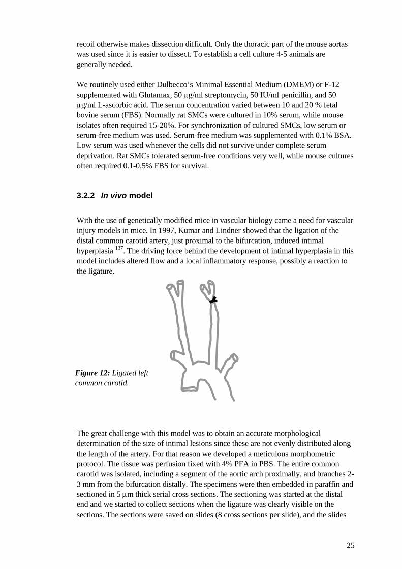
With the use of genetically modified mice in vascular biology came a need for vascular injury models in mice. In 1997, Kumar and Lindner showed that the ligation of the distal common carotid artery, just proximal to the bifurcation, induced intimal hyperplasia 137. The driving force behind the development of intimal hyperplasia in this model includes altered flow and a local inflammatory response, possibly a reaction to the ligature. The great challenge with this model was to obtain an accurate morphological determination of the size of intimal lesions since these are not evenly distributed along the length of the artery. For that reason we developed a meticulous morphometric protocol. The tissue was perfusion fixed with 4% PFA in PBS. The entire common carotid was isolated, including a segment of the aortic arch proximally, and branches 2-3 mm from the bifurcation distally. The specimens were then embedded in paraffin and sectioned in 5 µm thick serial cross sections. The sectioning was started at the distal end and we started to collect sections when the ligature was clearly visible on the sections. The sections were saved on slides (8 cross sections per slide), and the slides
Figure 12: Ligated left common carotid.

26
were marked with a serial number. In this way, we were able to keep track of the distance between any given cross section and the ligature. The next step was to get an overview of the size and distribution of the lesions along the vessel. Every third slide was stained with Masson’s Trichrome and digitalized images were taken of three representative sections from each slide. The amount of intimal lesion in each image was first subjectively rated on an 8-graded scale, where 0, 4, and 8 represented 0%, 50%, and 100% reduction of the lumen. For each rated vessel, a diagram was then drawn, showing the size and location of lesions in relation to the ligature. Typically the lumen was completely occluded by a massive intimal lesion right proximal to the ligature. The size of the lesion then tapered off to almost none within 1-2 mm from the ligature, but usually reappeared further proximally. For the definitive quantification, all diagrams were lined up, with the position of the ligature as the anchor point. A region encompassing 400 µm with equal distance from the ligature, across all diagrams, was then selected with the objective of excluding regions of complete occlusion or regions with no lesion. All the images within this 400 µm region were analyzed using an image analysis software. The luminal border, the internal elastic lamina (IEL), and the external elastic lamina (EEL) were manually traced. The software computed the areas within these tracings into pixels, which were then converted into mm2-units. 3.3 ATHEROSCLEROSIS
In paper III the role of the perlecan HS chains in atherogenesis was analysed. For quantitative assessment of atherosclerosis in mice two different methods were used. The reason for using two different methods is that atherosclerosis develops unevenly along the length of the aorta of apoE null mice. 3.3.1 Aortic root sectioning
For early lesion development analysis of plaque development in the aortic root is a well established and commonly used method. It was initially described in detail by Paigen et al 138, and entails sequential tissue sectioning from the origin of the aortic valves to a region in the ascending aorta. For our study the mice were euthanized at 15 and 33 weeks of age. The heart and ascending aorta were frozen in OCT following cryo-protective treatment with sucrose. The aortic roots were then sectioned according to a standardized protocol 139. Briefly, sections were collected starting 100 µm from the appearance of the aortic valve. Eight 10 µm sections at 100 µm intervals were used for lesion quantification. The sections were fixed with formaldehyde, rinsed with dH2O, immersed in 60% isopropanol for 1-2 minutes, and stained with Oil-Red-O for 20 minutes. The stained
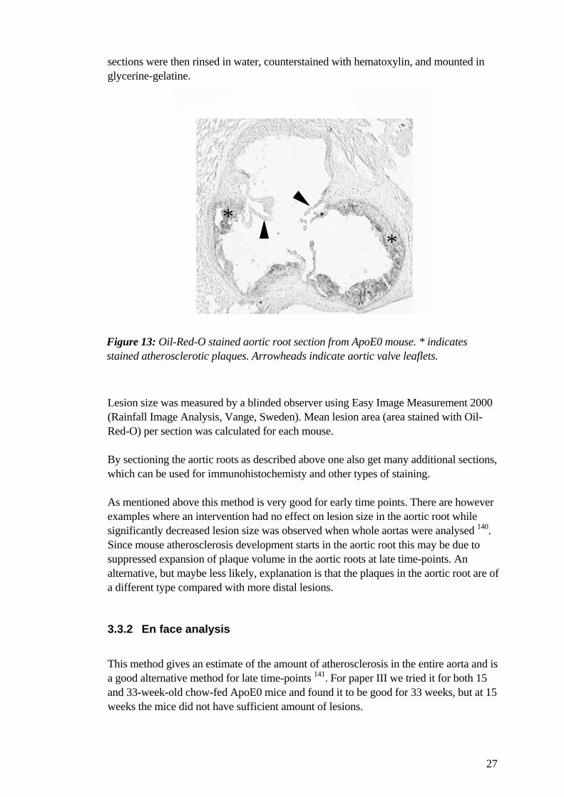
27
sections were then rinsed in water, counterstained with hematoxylin, and mounted in glycerine-gelatine. Lesion size was measured by a blinded observer using Easy Image Measurement 2000 (Rainfall Image Analysis, Vange, Sweden). Mean lesion area (area stained with Oil-Red-O) per section was calculated for each mouse. By sectioning the aortic roots as described above one also get many additional sections, which can be used for immunohistochemisty and other types of staining. As mentioned above this method is very good for early time points. There are however examples where an intervention had no effect on lesion size in the aortic root while significantly decreased lesion size was observed when whole aortas were analysed 140. Since mouse atherosclerosis development starts in the aortic root this may be due to suppressed expansion of plaque volume in the aortic roots at late time-points. An alternative, but maybe less likely, explanation is that the plaques in the aortic root are of a different type compared with more distal lesions. 3.3.2 En face analysis
This method gives an estimate of the amount of atherosclerosis in the entire aorta and is a good alternative method for late time-points 141. For paper III we tried it for both 15 and 33-week-old chow-fed ApoE0 mice and found it to be good for 33 weeks, but at 15 weeks the mice did not have sufficient amount of lesions.
**
Figure 13: Oil-Red-O stained aortic root section from ApoE0 mouse. * indicates stained atherosclerotic plaques. Arrowheads indicate aortic valve leaflets.

28
In brief, the aortas were dissected carefully while still in the animal. It was worth the effort to remove as much as possible of the adventitia already at this stage. Remaining adventitia may contain fat and cause confusion. The intimal surface was then exposed by a longitudinal cut through the inner curvature of the aortic arch and down the ventral aspect of the remaining aorta. To obtain a flat preparation a cut was also made through the greater curvature of the aortic arch to the subclavian branch. The vessels were then pinned to a dark surface using 0.2 mm thick stainless steel pins. For this purpose black wax plates are often prepared. After a recommendation from Dr Einar Eriksson, Karolinska Institutet, we however decided to used another technique where multiple layers of Parafilm® was covered with black plastic tape. The vessels were kept in paraformaldehyde after pinning, and when all the aortas had been pinned out they were stained with Sudan IV. Briefly, the aortas were rinsed in 70% ethanol for 5 min, stained with Sudan IV for 6 min. For the Sudan IV working solution 5g of Sudan IV was mixed with 500 ml 70% ethanol and 500 ml 100% acetone. The working solution can be used for several weeks. After staining, the plates were immersed twice in 80% ethanol for 3 min and then stored in PBS at 4ºC. To determine the extent of atherosclerosis lesion areas were traced manually by a blinded observer, measured using Easy Image Measurement 2000, and expressed as percentage of total aortic area. One disadvantage of this method is that it measures a two-dimensional lesion size without taking the thickness of the lesions into account. An alternative approach can be to measure the amount of cholesterol in the aortas, which can be done either by enzymatic analysis or gas chromatography 142.
Figure 14: Aorta stained with Sudan IV.

29
3.3.3 Lipoprotein analysis
In order to further explore the role of perlecan HS in atherogenesis we used both in vitro and in vivo methods for studies of lipoprotein-proteoglycan interactions. 3.3.3.1 In vitro lipoprotein binding
Initially we explored binding of human LDL to purified proteoglycans (the basic steps of proteoglycan purification are described in detail below) and compared HS-deficient proteoglycan preparations with control. This, however, raised two questions from reviewers. One was whether the mechanisms were relevant for the mouse model that was used since apoE null mice primarily suffer from elevated levels of remnant particles containing apoB48, and the other was whether we would get the same results if we used total ECM rather than purified proteoglycans. Thus, we ended up doing in vitro lipoprotein binding to both purified proteoglycans and total ECM, using both human LDL and mouse triglyceride-rich lipoproteins. This is discussed in more detail in the ‘Results and Discussion’ section. 3.3.3.2 In vivo lipoprotein uptake
Mouse triglyceride-rich lipoproteins and human LDL were 125I-labeled and injected retro-orbitally in 8 to 10-week-old mice. The age of the mice was chosen because at 8 to 10 weeks the mice are large enough and their atherosclerosis development is still at an early stage. After 20 min (for both types of lipoproteins) or 72 h (for human LDL) the mice were perfusion fixed and the aortas dissected and measured for radioactivity. We think that the early time-point mostly shows the endothelial permeability to lipoproteins, whereas the later time-point shows how the lipoproteins are retained in the matrix. Identical 20 min experiments were also performed with 125I-labeled albumin instead of lipoproteins in order to explore whether difference in endothelial permeability was selective or general. 3.4 ANIMAL MODELS
For this thesis two types of genetically modified mice were used, HS-deficient mice and apoE null mice. 3.4.1 Heparan sulfate deficient mice
In 1999, it was shown that a null mutation of the perlecan gene is embryonically lethal 143, 144. The mice showed defective cephalic development, severe cartilage defects, and hemopericardium. However, Dr Raija Soininen in Prof Karl Tryggvason’s laboratory at Karolinska Institutet, Stockholm, generated mice with a targeted deletion of exon-3 in domain-I of
30
the perlecan gene, which means that the major attachment sites for HS chains on the core protein are lost. The strain was called Hspg2∆3/∆3 145. The phenotype of these animals was however, at least initially, not as what had been expected since the primary aim was to explore the role of perlecan HS in kidney function. The presence of HS in basement membranes was first detected in the glomerular basement membrane 146, and it was suggested that HS establish a permeability barrier in the kidney. The Hspg2∆3/∆3 mice were however surprisingly normal. They survived embryonic development, appeared healthy, and did not develop proteinuria. Careful examination did however show that they had defects in lens development and later studies have shown that they do develop proteinuria following protein overload 147. In addition, they have been shown to have delayed wound healing, retarded FGF-2 induced tumor growth, and defective angiogenesis 148. The generation of the Hspg2∆3/∆3 mice gave us a unique opportunity to examine the role of endogenous basement membrane HS in vascular disease. 3.4.2 Apolipoprotein E null mice
Experimental atherosclerosis was reported as early as 1908 when Ignatowski reported intimal thickening with formation of large clear cells in the aorta of rabbits fed a diet rich in fat and animal proteins. For a long time the most useful animal models were relatively large animal like nonhuman primates, swine, and rabbits 149. In recent years there has, however, been an explosion in the number of in vivo studies of atherogenesis, largely attributable to the use of mouse models. The first mouse model of diet-induced atherosclerosis was characterized in the 1960’s. The diet used (30% fat, 5% cholesterol, and 2% cholic acid) gave aortic root atherosclerosis in C57BL/6 mice, but was however toxic and the mice lost weight and suffered from respiratory infections. Paigen et al modified the diet and introduced what is commonly called the ‘Paigen diet’ (15% fat, 1.25% cholesterol, and 0.5% cholic acid) 150. The diet-induced model was used quite widely, but there were many disadvantages. The lesions are small and largely confined to the aortic root, and usually do not develop beyond the early foam-cell, fatty-streak stage. The diet itself has also been shown to be inflammatory as it leads to the induction of acute phase reactants 151. In 1992, two groups independently reported the generation of apolipoprotein E null (ApoE0) mice 152, 153 and since then it has become the most widely used model for experimental atherosclerosis. Apolipoprotein E (apoE) is one of the apolipoproteins that coat the surface of lipoprotein particles and mediate their metabolism. It is synthesized primarily in the liver, although synthesis has been documented in virtually every tissue. ApoE resides on very-low, intermediate, and high-density-lipoproteins (VLDL, IDL, HDL), as well as on chylomicrons and their remnants. ApoE mediates the clearance of VLDL and IDL via low-density-lipoprotein (LDL) receptors and chylomicron remnants via both the LDL receptor and the LDL-receptor-related protein (LRP).
31
The ApoE0 mice develop spontaneously high cholesterol levels. Most of the elevation is in cholesterol-enriched remnants of chylomicrons and VLDL, but there is a significant elevation of LDL as well. In this context it should be noted that mice naturally have high levels of HDL and low levels of LDL, in contrast to humans that are high in LDL and low in HDL. The mice develop extensive atherosclerosis even without a high fat diet and the lesions progress to advanced lesions with necrotic cores surrounded by SMCs. Human apoE deficiency also exists. However, compared to what is seen in human subjects the hypercholesterolemic phenotype in mice is much more severe. This can probably be explained by species differences in hepatic apoB metabolism. Aside from apoE, apoB is the other physiological ligand for the LDL receptor. In rats and mice hepatic apoB mRNA is edited, and apoB is secreted as the truncated B48 (as it is in the gut in mice and humans), which does not contain the LDL receptor-binding region. In contrast, the human liver does not edit apoB mRNA and apoB is secreted as the full-length B100. In both humans and mice, the liver produces VLDL, which is processed in plasma to IDL and LDL. In humans, some of the VLDL and IDL are cleared by B100-mediated removal, whereas in mice these lipoproteins contain mainly B48 and clearance relies almost entirely on apoE. This is probably the explanation for the more severe hypercholesterolemia in ApoE0 mice compared with humans with homozygous null mutation 153. We used the ApoE0 mouse as a tool for studying the role of perlecan HS in atherogenesis, by cross-breeding the Hspg2∆3/∆3 mouse with apoE mutants. 3.5 PROTEOGLYCAN ANALYSIS
Because the GAG chains of proteoglycans are ubiquitously sulfated, radioactively labeled sulfate can be used to trace GAG chain production in cells. Here (paper II and III), subconfluent SMC cultures were synchronized in low serum medium, and labeled for 48 h with medium supplemented with 35S-sulfate. In order to analyze the total production of proteoglycans by SMCs in culture both molecules released into the culture medium and cell-layer proteoglycans (localized in intracellular compartments, incorporated into the plasma membrane, and accumulated in the ECM), must be retrieved. Thus, the culture medium was collected and the cell-layer was extracted with urea buffer and scraped off the dishes after 48 hours of labeling. Protease inhibitors were added to the samples immediately. The proteoglycans were then separated from other molecules by DEAE (Diethylaminoethyl-Sephacel) anion-exchange chromatography. DEAE binds glycosylated (negatively charged) molecules such as proteoglycans whereas the non-glycosylated proteins can be washed away. This procedure concentrates the sample and facilitates further analysis.

32
In order to analyze the specific subclasses of proteoglycans produced by SMCs deglycosylation with enzymes (heparinase and chondroitinase) was used. However, the enzymes work poorly in urea buffer, and chondroitinase is particularly sensitive to Triton X-100. The samples were therefore precipitated at -72ºC in 1% potassium acetate buffer in 70% ethanol to remove urea and detergents. To improve the yield, CS was added as a carrier in the first round of precipitation. Normally, two precipitations were enough to clean the samples. An alternative to ethanol precipitation is dialysis and this was used in paper III for the preparation of purified proteoglycans for lipoprotein binding. By using dialysis one can avoid the use of ‘cold’ CS as carrier, which would interfere with binding experiments. Following ethanol precipitation the proteoglycan pellets were allowed to air-dry for 30 min. Cocktails of the glycosidases heparinase and chondroitinase were always prepared fresh for immediate use. The enzymatic reactions were performed in volumes between 25 and 50 µl, for 2 hours at 37ºC and 1 hour at 42ºC. For further analysis of the purified proteoglycans gradient gel electrophoresis is particularly useful because it covers a wide span of molecular sizes. SDS-polyacrylamide gels were routinely made with a 3.5% stacking and 4-12% resolving gel. The height of the slab was 15 cm. This type of gel covers molecular sizes from 30 kDa up to over 1000 kDa. For complete analysis radiolabeled samples were enzyme treated in four different ways (see below). Sample buffer was then added and the samples were loaded onto the gel under reducing conditions. The bands of 35S-sulfate labeled samples on SDS-PAGE are usually very wide due to the broad range in molecular sizes of the GAG chains, and are therefore referred to as ‘smears’. In the gel, 35S-sulfate labeled samples without prior enzymatic deglycosylation (1) will appear as smears showing the size and amount of intact proteoglycans (with intact GAG chains). Deglycosylated proteoglycans will not appear in the gel since the fragmented GAG chains, containing the radioactive signals, are too small and will run off the gel. Samples treated with chondroitinase (2) will therefore be seen as smears in the gel representing the size and amount of heparan sulfate proteoglycans, whereas samples treated with heparinase (3) will be seen as smears representing chondroitin and dermatan sulfate proteoglycans. The forth type of sample treatment is double digestion with both chondroitinase and heparinase (4). No digestion (1) and double digestion (4) can be seen as controls, where (1) should contain all bands/smears present in a superimposed image of (2) and (3). Treatment (4) should be blank, with no intact GAGs remaining in the gel, showing complete digestion by both enzymes. For an illustration of the description above please see Figure 15 below.

33
3.6 ADDITIONAL METHODS USED
A number of additional methods, like different types of immunohistochemistry, Western blot, quantitative PCR etc, were used for this thesis but most of them were used in a very standardized way and are described better elsewhere.
Figure 15: Autoradiograph of 35S-sulfate labeled proteoglycans from wild-type (wt) and mutant (∆3) mouse aortic SMCs. Note the complete digestion when samples have been treated with both enzymes (4).

34
4 RESULTS AND DISCUSSION The overall aim of this thesis was to further explore the role of basement membrane structures in vascular disease, and since heparin and HS have been shown in many studies to be anti-proliferative for SMCs in vitro and in vivo 154-157 we chose to primarily focus on the basement membrane HS proteoglycan perlecan. Initially we studied the effect of perlecan HS on SMC function in vitro. We then explored the role perlecan HS in intimal hyperplasia and atherogenesis in vivo. Since perlecan null animals are lethal we used the Hspg2∆3/∆3 perlecan HS-deficient mice 145. This also gave us the opportunity to separate the role of the perlecan core protein from the role of the HS chains. In the forth paper we investigated the effect of AtRA on perlecan expression. 4.1 PERLECAN HS INFLUENCE SMC FUNCTION (PAPER I)
As explained in the Methods section it was difficult to study the effect of perlecan on SMC function since the perlecan preparation we used did not support SMC adhesion. This was in contrast to what had been reported previously 158, 159. It is possible that this difference can be explained by species differences in cell-surface receptors and also by different sources of perlecan. Instead, we combined perlecan with two well-characterized adhesive molecules, fibronectin (FN) and laminin, in an attempt to make functional studies of perlecan possible. The use of FN and laminin for the combined substrates was also interesting since activation of SMCs after arterial injury is coupled to the disappearance of basement membrane structures like laminin, whereas FN accumulates around migrating and proliferating cells in the arterial media and intima 115. In vitro, interaction of SMCs with FN has been shown to induce phenotypic modulation and to mediate cell-cycle entry 116, 124, whereas cells in contact with laminin retain differentiated properties 124. A combined substrate of FN and perlecan showed a reduced adhesion of rat aortic SMCs by 70-90% compared to FN alone (Figure 1, paper I). In contrast, perlecan did not affect adhesion to laminin. We then examined whether the anti-adhesive property of perlecan resided in the HS chains or in the core protein. The HS chains were removed by means of enzymatic digestion with heparinase I and II, and heparinase treated perlecan lost 60% of its anti-adhesive effect (Figure 3, paper I). Furthermore, pure HS as well as heparin reduced SMC adhesion to FN, whereas neither CS nor hyaluronan had any effect, indicating that the anti-adhesive effect of perlecan resided in the HS chains (Fig 4, 5, and 6, paper I). Morphologically, SMCs adhering to a substrate prepared by co-coating FN and perlecan or heparin were small, rounded, lacked focal contacts, and showed poorly developed stress fibers of actin (Figure 2, paper I). To explore whether heparin inhibited adhesion simply by sterically blocking the interaction between SMCs and FN, heparin was also added as a second coating to a
35
preformed FN matrix, but this did not affect cell adhesion (Figure 8, paper I). Other control experiments showed that co-coating with perlecan/heparin did not affect the amount of FN that bound to the plastic surface (Figure 7, paper I). Two different FN fragments that lack the main heparin binding domains were used in order to explore whether these sites were necessary for the anti-adhesive effect. Surprisingly, heparin inhibited adhesion to these fragments (Figure 9A, paper I). In addition, the use of 3H-heparin showed that it was not even incorporated in the substrate, or alternatively the amount that was incorporated was below the detection limit (Figure 9B, paper I). Our hypothesis is that perlecan/heparin induces a conformational change in the FN molecule in solution and that this has an effect on how the RGD sequence is presented when the molecules adhere to a solid surface. The ability of cells to regulate their attachment to the surrounding ECM is crucial for important processes like migration and proliferation. For example, cell migration requires a continuous cycle of assembly and disassembly of focal adhesions in order to allow movement 160. It is tempting to speculate that when basement membrane structures surrounding medial SMCs are degraded following arterial injury released fragments of perlecan HS chains may interact with FN that is produced and possibly modulate the adhesiveness of the newly formed matrix, thereby affecting the migration towards the intima. In addition perlecan HS in the basement membrane of the normal uninjured arteries may act as a shield to protect the SMCs from interactions with FN, which would otherwise result in activation. In retrospect, I find it necessary to discuss the experimental conditions used in this paper. The use of distilled water as a diluent instead of a buffering solution makes pH changes likely. It has previously been shown that pH changes or changes in ionic strength of a solution may induce conformational changes in the FN molecule 161, 162. On the other hand, tissue fluid is not always perfectly buffered under pathological conditions such as ischemia and tumor growth, and cellular processes in such situations may well occur in acidic pH. In addition, Gauer and co-workers used PBS in the coating solution and they still saw similar effects of perlecan on mesangial cell adhesion to FN 163, and heparin has been shown to induce changes in FN also at physiological pH, such as aggregation and extended shape 164, 165. The shape changes of FN were specific for heparin/HS and no effect was seen with CS and HA 164. In a subsequent study that is not part of this thesis we also showed that low molecular weight heparins, with diminished anticoagulant activity but similar sulfate contents, down to a size of 1 400 Da retained an anti-proliferative capacity for SMCs, whereas the effect on adhesion to FN was lost gradually with decreasing size 166. This indicates that it is not just the amount of negative charge introduced to the solution that causes the effect. There are additional concerns with the use of a two-dimensional substrate for studies of cell behavior. 3D gels are increasingly being used for studies of matrix organization and cell-matrix interactions 16. Assembly of a FN matrix in vivo is also regulated by intracellular signaling events. Binding to integrins, organization of the actin cytoskeleton, stimulation of cell contractility, and activation of kinase cascades all contribute to the assembly process 16, which complicates studies of FN in a physiological way in vitro.

36
In summary, it is very difficult to speculate if the observed effects on SMC adhesion are relevant in vivo. We can only conclude that the role of heparin/HS in the processes of intimal hyperplasia and atherogenesis may include effects on matrix organization and cell-matrix interactions. 4.2 BASEMENT MEMBRANE HS IN INTIMAL HYPERPLASIA (PAPER II)
The next step was to explore the role perlecan HS in intimal hyperplasia in vivo. When the work was initiated the vascular system of the Hspg2∆3/∆3 mice had not been thoroughly examined. Perlecan null embryos suffer from hemopericardium due to discontinuities in the myocardial wall, indicating a weakness in basement membranes in response to stress 143. A null mutation also results in an increased incidence of transposition of the great arteries 167. In order to study vascular development, Hspg2∆3/∆3 mice and wild-type controls were kept on chow diet for up to one year and the heart and aorta, including main branches, were analyzed by light and electron microscopy at different time-points. No differences were noted in vascular development, and SMCs as well as endothelial cells exhibited normal ultrastructural features with a basement membrane adjacent to the cells. It is possible that the core protein is more essential for vascular development. However, the HS chains of perlecan have been shown to interact with laminin and support the structural integrity of basement membranes 168. An alternative hypothesis is therefore that the remaining chain attachment site in domain V provides sufficient amounts of HS for proper basement membrane assembly. Due to the remaining HS chains attached to domain V, and the presence of other proteoglycans in the arterial wall that carry HS chains, it was necessary to explore how much the HS content of the vessel wall was affected by the perlecan exon-3 mutation. Biochemical analysis of proteoglycan production by aortic SMCs from Hspg2∆3/∆3 mice, using 35S-sulfate labeling, clearly showed that the Hspg2∆3/∆3 mutation results in a depletion of large HS proteoglycans (Figure 1A, paper II). Immunocytochemical analysis (‘modified ELISA’) of cultured SMCs and immunohistochemical analysis of arterial specimens from the animals also showed a dramatic reduction in HS accumulation, whereas the production and accumulation of the perlecan core protein remained unchanged, or possibly slightly increased (Figure 2 and 6, paper II). Our findings indicate that the majority of the HS chains produced by mouse aortic SMCs are attached to domain I on the perlecan protein core, and that perlecan is the predominant HS proteoglycan in the vessel wall ECM. As previously mentioned exogenously added heparin and HS have been shown to inhibit SMC proliferation in vitro and in vivo 154-157. Perlecan has also been shown to be low during the phase of maximal SMC proliferation up to 7 days after arterial injury in the rat, and to be dramatically increased after two weeks. The increase in accumulation of perlecan coincides with a decrease and arrest in SMC proliferation. The question whether proliferating cells simply produce less perlecan or whether it actually is important for growth regulation had already been answered to some extent since

37
heparinase treatment restored responsiveness of 5 weeks post-injury explants to PDGF-BB 169. However, as mentioned above the Hspg2∆3/∆3 mutation gave us the opportunity to address the role of the perlecan HS chains in proliferation and intimal hyperplasia without affecting other HS proteoglycans like syndecans, glypicans, agrin, and collagen XVIII. The importance of studying the effect of different HS proteoglycans separately can be illustrated by the fact that despite the convincing data showing that exogenous heparin/HS inhibits SMC proliferation and intimal hyperplasia, HS proteoglycans have been shown to act both as activators and suppressors of growth in vitro and in vivo 170. Removal of endogenous HS proteoglycans from the native artery wall largely prevents medial SMC proliferation within the first two days after balloon injury alone, and after addition of exogenous FGF-2 171, 172. This probably reflects the requirement for heparin/HS for dimerization of FGF high-affinity receptors and subsequent receptor phosphorylation and signaling 173. On the inhibitory side, overexpression of syndecan-1 can inhibit FGF2-mediated proliferation 174. This may be due to increased shedding of the soluble ectodomain, which, like exogenous HS proteoglycans secreted from endothelial cells 175, can inhibit FGF-2-mediated proliferation. Although perlecan has been shown to both promote and inhibit the proliferation of various tumor cells 176-178, it has so far always been shown to have an inhibitory effect on SMC growth in vitro 158,
179. Here, we now had the opportunity to study the specific role of endogenous perlecan HS chains in vitro and in vivo. The role of perlecan HS in intimal hyperplasia in vivo was analyzed using ligation of the common carotid as previously described 137. Analysis of the intimal lesions was done after 1 and 6 weeks. Vessels in Hspg2∆3/∆3 mice developed an increased intima/media ratio compared with wild-type controls already at 1 week after surgery. At 6 weeks these differences were even more pronounced (Figure 4D, paper II). Proliferative activity in the lesions was quantified at 1 week after ligation by staining for Ki-67 and the replication index was significantly higher in the Hspg2∆3/∆3 mice compared with wild-type controls (Figure 7, paper II). In vitro, Hspg2∆3/∆3 cells were shown to respond with increased DNA-synthesis compared with wild-type cells after stimulation with 10% FBS, PDGF-AA, PDGF-BB, FGF-2, and Hb-EGF, confirming the effect on SMC proliferation seen in vivo (Figure 3B, paper II). Our mechanistic hypothesis was that perlecan HS sequesters heparin-binding growth factors in the ECM and thereby limits the amount of growth factors that reach the cell surface HS proteoglycans and growth factor receptors. The explanation for the increased DNA synthesis seen also with non-heparin binding growth factors could be a defective capacity to sequester HS-binding autocrine co-factors. For example SMC growth stimulation with PDGF-BB, as well as with thrombin, leads to release of FGF-2 and an enhanced proliferative response 180, 181. Figure 16 below illustrates the mechanistic hypothesis.

38
P
Wild-type basement membrane
Hspg2∆3/∆3 basement membrane
ERK
Cell proliferation
Early signal
Early signal
Late signal
FGFRPDGF-BB Thrombin Release
Storage
Action
Syndecan ectodomain
Perlecan
ERK
Cell proliferation !!
Early signal
Early signal
Late signal
FGFRPDGF-BB Release
Storage
Action
Syndecan ectodomain
Thrombin
FGF-2
FGF-2
Perlecan
P
B
A
Figure 16: A shows the wild-type situation with storage of FGF-2 in the basement membrane and weak pro-proliferative signaling. B illustrates what happens in the Hspg2∆3/∆3 SMCs. Since the storage capacity is limited more growth factors are available at the cell surface for receptor interactions, which results in increased proliferation. Signaling through the PDGF-BB and thrombin receptors shows how FGF-2 can be an autocrine co-factor.

39
To test the hypothesis the binding capacity of ECM deposited by SMCs isolated from Hspg2∆3/∆3 mice was compared with control ECM (Figure 8, paper II). FGF-2 was used as a ligand due to its known importance for the proliferative response after arterial injury, in combination with its well-know heparin binding properties. The binding capacity of the HS-deficient ECM was only 25% of that of the control ECM. Pre-treatment with heparinase brought the binding capacity of all matrices down to 12.5% of wild-type control. This equals the binding capacity of the background collagen type IV matrix, which was used to coat the wells before the seeding of cells in order to get a well-attached ECM. In contrast, pre-digestion with chondroitinase did not alter the FGF-2 binding capacity. The results above makes a sequestering role for perlecan HS very likely. We can, however, not exclude the possibility that perlecan HS also have direct effects on cell signaling. Heparin has for example been shown to inhibit c-fos and c-myc expression 182, and to affect mitogen-activated protein kinase-dependent signal transduction 183. In addition, perlecan HS has recently been shown to regulate proliferation through activation of the tumor suppressor PTEN, which subsequently leads to inactivation of Akt 184. There is, however, also a possibility that these ‘direct’ effects on cell signaling may be an effect of interference with HS-binding autocrine co-factors. In summary, the HS chains of perlecan contribute to SMC growth control both in vitro and in vivo, possibly by sequestering heparin-binding mitogens such as FGF-2. 4.3 BASEMENT MEMBRANE HS IN ATHEROGENESIS (PAPER III)
Characterizing of the vascular system of the Hspg2∆3/∆3 mice demonstrated a dramatic decrease of the HS content of the vascular wall of mutant mice. We therefore decided to use this mouse strain to study the role of basement membrane HS in atherogenesis. Up to this point, HS was primarily regarded as an anti-atherogenic component of the vessel wall ECM, mainly because increased atherosclerosis had been associated with decreased arterial HS content in different species 69-73. We cross-bred the Hspg2∆3/∆3 mice with ApoE0 mice. We decided to use ApoE0 since they develop lesions spontaneously without a high-fat diet, which has been reported to have effects on the inflammatory response 151. In retrospect, LDL-receptor null mice could have been a good choice as well because of their different lipoprotein profile, with higher LDL levels more like human cardiovascular patients. Apart from an increase in SMC content in the lesions, we really did not know how the vascular phenotype of the double mutant mice would turn out. Enzymatic removal of HS had been shown to increase binding of LDL to endothelial matrix in vitro 185, but on the other hand HS had also been shown to bind LDL in vitro 186. HS had also been shown to reduce endothelial permeability for LDL 187, 188 and removal of HS from the matrix had been shown to increase monocyte binding in vitro 187, 189. Since apoE had been reported to influence perlecan expression 179 we started by comparing perlecan mRNA levels from ApoE0 aortas with C57BL/6 controls using

40
real time PCR. No difference was detected between the two groups at 35 weeks of age, indicating that the atherosclerosis model we had picked retained intact perlecan expression in the aorta. When the project was already initiated Vikramadithyan and co-workers published a study where heterozygous perlecan null mice had been cross-bred with ApoE0 and LDL receptor null mice 190. A decreased perlecan level was shown on the mRNA level. Their results showed a significant reduction of atherosclerosis with reduced perlecan expression, but only in ApoE0 mutants and only in young males. It was difficult to draw conclusions from that study since the perlecan mutation was only partial and it was not possible to separate the role of the core protein and the role of the HS chains. We analyzed atherosclerosis in the ApoE0/Hspg2∆3/∆3 mice at 15 and 33 weeks of age, in both female and male mice. As mentioned in the Methods section we chose to use both aortic root sectioning and en face analysis of whole aortas for quantification of lesions since the two methods sometimes have given different results at different time-points 142. Compared with ApoE0 control mice, aortic sinus lesion size was reduced in female ApoE0/Hspg2∆3/∆3 mice by 53% and 20% at 15 and 33 weeks, respectively (Figure 1B and C, paper III). En face preparations revealed a 59% reduction in lesion area in female ApoE0/Hspg2∆3/∆3 mice compared with controls at 33 weeks (Figure 1E, paper III). Based on the findings in female mice we analyzed aortic sinus atherosclerosis in males at 15 week and did en face analysis for the 33 weeks time-point. Identical relative reductions of atherosclerosis were seen in the males. The lesions were, however, smaller compared to females, as previously reported 191. In hindsight, it would have been nice to have data from aortic sinuses of 33-week-old male mice, since we then possibly could have answered the question whether the smaller difference in the females at 33 weeks was due to ‘crowding of lesions’ in the aortic root or due to different types of lesions in different parts of the vascular tree. We then explored the possible mechanisms behind the observed reduction in atherosclerosis. In order to analyze if the difference was due to decreased lipoprotein retention we started by doing lipoprotein binding in vitro. As mentioned in the Methods section we ended up doing in vitro lipoprotein binding to both purified proteoglycans and total ECM, using both human LDL and mouse triglyceride-rich lipoproteins. All of the assays showed the same result that the HS-deficient matrix bound less lipoprotein (Figure 4, paper III). This indicates that binding of lipoproteins to HS proteoglycans is relevant in mice and maybe also in the human situation. It also shows that both ApoB100 containing LDL and apoB48 containing remnant particles can bind to HS. This is of relevance since not only LDL but also remnant particles have been shown to be atherogenic 192. We then wanted explore whether increased retention could be seen in vivo. 125I-labeled mouse triglyceride-rich lipoproteins and human LDL were prepared and injected retro-orbitally in 8 to 10-week-old mice. After 20 min (for both types of lipoproteins) or 72 h (for human LDL), the mice were perfusion fixed, the aortas dissected and measured for radioactivity. Analysis 20 min after injection showed significantly more of both types of lipoproteins in the aortas of ApoE0/Hspg2∆3/∆3 mice compared with ApoE0 mice

41
(Table 2, paper III). However, when human LDL was analyzed after 72 h, no significant difference remained between the two groups (Table 2, paper III). Our interpretation of the results is that the endothelial permeability for lipoproteins is higher in the absence of perlecan HS, thereby allowing a more rapid influx into the vessel wall. However, the same amount of LDL retained at 72 h in ApoE0/Hspg2∆3/∆3 mice and controls suggests that the efflux of lipoproteins from the vessel wall of ApoE0/Hspg2∆3/∆3 mice is greater due to a reduced retention capacity of the HS-deficient matrix. To confirm decreased retention in the HS-deficient matrix we also did immunohistochemisty for apoB in aortic root lesions (15 weeks), which showed significantly less apoB in ApoE0/Hspg2∆3/∆3 mice compared with ApoE0 controls (Figure 5, paper III). Injections of 125I-labeled albumin showed no difference between the HS-deficient mice and controls when aortic radioactivity was measured 20 min after injection, suggesting a selective, rather than a general, increase in basement membrane permeability. Figure 17 shows a schematic illustration of the mechanistic hypothesis.
Figure 17: Illustration of how apoB-containing lipoproteins are transported into the subendothelial matrix (grey areas) and how some are retained and some are not. In the ApoE0/Hspg2∆3/∆3 mice to the left there is a greater influx of lipoproteins compared with ApoE0 controls to the right, but also a greater efflux due to decreased retention.
20 min
72 hours
15 weeks
-HS
-HS
-HS
+HS
+HS
+HS

42
In support of our observation that HS-deficiency cause increased permeability to lipoproteins HS has previously been reported to reduce endothelial permeability for LDL in vitro 187, 188. In addition, a recent study showed increased endothelial permeability to lipoproteins in collagen XVIII null mice cross-bred with mice prone to develop atherosclerosis 51. The overall effect of a collagen XVIII null mutation was however not reduced atherosclerosis as seen for perlecan, but increased atherosclerosis. In theory, this finding supports our conclusion since increased endothelial permeability for lipoproteins in the presence of an unaltered or increased retention capacity of the vascular ECM would rather promote than prevent the atherogenic process. The increased atherosclerosis in the collagen XVIII null mice is probably a combination of increased endothelial permeability to lipoproteins, possibly due to lack of HS, and increased plaque neovascularization and increased lipoprotein binding to biglycan, due to lack of endostatin (the proteolytically released fragment which inhibits angiogenesis and interferes with lipoprotein binding to biglycan) 52. Our findings suggest that retention of lipoproteins in the subendothelial matrix has a more pronounced effect on the amount of atherosclerosis compared with endothelial permeability. In support of this, the rate of LDL entry into the normal artery wall has previously been shown to vastly exceed LDL accumulation 193, indicating that changes in endothelial permeability are relevant for atherogenesis only if the lipoproteins are subsequently retained. In contrast to what was seen in when the proteoglycan binding site of LDL was mutated 67 we did not only see an effect on early atherogenesis. This can possibly be due to the binding of ox-LDL to proteoglycans via lipoprotein lipase in more advanced lesions. An interaction that would be affected by reduced proteoglycan levels, but not by the charge of the apolipoprotein since the binding to lipoprotein lipase is mediated by the lipid-binding domain of lipoprotein lipase 194. Since increased monocyte binding to ECM had been reported following heparinase treatment 187, 189 we quantified the amount of CD68 in the lesions, but did no see a difference between the ApoE0/Hspg2∆3/∆3 mice and controls (Figure 6G, H, and I, paper III). As we had expected the amount of α-actin staining was increased in ApoE0/Hspg2∆3/∆3 lesions compared with controls (Figure 6A, B, and C, paper III), which may indicate more stable plaques. However, we were not able to detect an increase in collagen content (Figure 6D, E, and F, paper III). In summary, the HS chains of perlecan promote atherogenesis in mice, most likely through increased retention of lipoproteins. In addition, the ability of perlecan HS to regulate SMC proliferation was found to influence SMC content in lesions, thus implicating a role for perlecan HS in plaque stability. It should however be noted that perlecan is one of the predominant proteoglycans in murine atherosclerotic plaques, whereas versican and biglycan are more abundant in human lesions. The role of perlecan in human atherogenesis is therefore difficult to determine and its role in endothelial permeability may be more crucial in the human situation.

43
4.4 PHARMACOLOGICAL REGULATION OF PERLECAN HS (PAPER IV)
As shown by the first three studies the role of basement membrane HS in vascular disease is very complex. It may enhance lipoprotein retention, but also decrease endothelial permeability to lipoproteins. In addition, it can reduce restenosis, but maybe also contribute to plaque instability. Much more knowledge is therefore needed about the role of perlecan in different situations, in different species, and in different cell types in order to predict if it is beneficial to up- or down-regulate perlecan. However, a situation where a transient up-regulation of perlecan most probably would be beneficial is in-stent restenosis, which involves intense SMC proliferation during a short period. Therefore, a drug that up-regulates perlecan might be suitable for drug eluting stents. Retinoic acid has been shown to inhibit proliferation of SMCs both in vitro and in vivo 136, 195-201 and to affect the expression of extracellular matrix molecules 202. We therefore hypothesized that retinoids may induce the expression of perlecan. Perlecan mRNA expression in murine SMCs after treatment with all-trans-retinoic acid (AtRA) was analyzed using quantitative PCR and Northern blot. mRNA levels were increased in a dose-dependent manner by AtRA and maximal perlecan mRNA levels were induced by 2.5 µg/ml AtRA (Figure 1A, paper IV). The perlecan up-regulation was confirmed on the protein level using immunocytochemistry and Western blot (Figure 2, paper IV). Since we had the unique possibility to use SMCs from the Hspg2∆3/∆3 mice it was possible to investigate whether the up-regulation of perlecan by AtRA was the mechanism behind the anti-proliferative effect of AtRA. In time-course experiments SMCs from wild-type and Hspg2∆3/∆3 mice were treated with AtRA and increased perlecan expression was evident in both wild-type and mutant cells by 12h with maximal levels of expression (~3 fold over control) reached by 24-48 h after AtRA addition (Figure 1B, paper IV). AtRA treatment substantially (>75%) inhibited serum-stimulated wild-type SMC growth, whereas the effect on Hspg2∆3/∆3 SMC growth was slight (<15%) (Figure 3A, paper IV). In support of these findings, AtRA also inhibited DNA synthesis in wild-type, but not Hspg2∆3/∆3 SMCs (Figure 3B, paper IV). Since AtRA has been shown to induce apoptosis of SMCs in some studies 201, 203 we wanted to explore whether the observed inhibitory effect was due to decreased cell proliferation or decreased cell survival. Using two different apoptosis assays we were not able to detect any signs of increased apoptosis rates with AtRA treatment of either of the cell types (Figure 4E + text, paper IV). This indicates that at least in our system the growth inhibitory effect seen with AtRA is due to decreased proliferation. Garl et al recently showed that perlecan might inhibit proliferation through activation of the tumor suppressor PTEN and subsequent inhibition of Akt 184. An effect that to a large extent seemed to be mediated by the HS chains. In order to strengthen the evidence that the AtRA effect was mediated by an up-regulation of perlecan we

44
explored whether the same effects on PTEN and Akt would be seen with AtRA. We therefore examined if AtRA treatment diffentially affected PTEN and Akt levels or phosphorylation in wild-type and Hspg2∆3/∆3 SMC. Although normalized levels of total Akt were not altered by AtRA in wild-type or mutant cells, relative Akt phosphorylation was decreased by AtRA in wild-type but not in Hspg2∆3/∆3 SMCs. In addition phosphorylation of PTEN, which probably reflects PTEN deactivation 184 was greatly decreased by AtRA treatment of wild-type SMC, but had little effect on mutant cells. The findings suggest that the HS-chains of perlecan are required for the reduction of PTEN phosphorylation and subsequent Akt inactivation by AtRA. As previously mentioned it is possible that these effects of perlecan HS on signaling are ‘direct’ effects. However, since the receptor that mediates these effects has not been identified it is also possible that the effects are due to sequestering of autocrine heparin-binding factors that cells release after growth stimulation. It should be mentioned that other factors that inhibit SMC proliferation such as heparin 204, apolipoprotein E 179, and transforming growth factor-β1 205 increase perlecan expression so we are not the first to show that anti-proliferative agents influence perlecan expression. Perlecan may be central mediator for growth inhibition and is probably affected by many more growth inhibitory agents that have not been analyzed yet. However, the use of Hspg2∆3/∆3 SMC gave us a unique opportunity to show that the AtRA mediated up-regulation of perlecan is functionally important and not just ‘a marker’ for growth inhibition. In summary, our results show that perlecan can be pharmacologically up-regulated with AtRA and that an up-regulation of perlecan HS is functionally important for the full inhibition of SMC proliferation by AtRA. The effect is due to a decreased proliferation and not decreased cell survival.

45
5 CONCLUDING REMARKS In conclusion, this thesis work shows that basement membrane HS can modify SMC function in several different ways. It may contribute to changes of the organization of the ECM and thereby regulate cell-matrix interactions. It is also an important inhibitory factor for SMC proliferation and can thereby influence processes such as restenosis and plaque stability. Basement membrane HS can also be pro-atherogenic through retention of ApoB containing lipoproteins, but may possibly be anti-atherogenic through decreased endothelial permeability to lipoproteins when retention is not a limiting factor. These findings show that perlecan is a very complex molecule with many different functions. This makes it an interesting but difficult target for pharmacological interventions. A potential drug needs to be given at the right time in the right place. Here, we show that perlecan can be pharmacologically regulated by compounds like AtRA. However, because of the diverse effects of AtRA, development of a more specific therapeutic alternative would be desirable.

46
6 FUTURE PERSPECTIVES A better understanding of the roles of different components of the arterial wall will hopefully help us understand why some individuals get severe atherosclerosis despite moderate cholesterol levels, and possibly make it easier for us to predict who would benefit from lipid lowering drugs from a quite young age. It may also be possible in the future to pharmacologically modulate the proteoglycan profile of the vascular wall. What is needed is probably to decrease the overall negative charge, and it will be necessary to do it without affecting important biological functions of the proteoglycans. One can for example imagine a drug that shortens the proteoglycan GAG chains just enough to decrease atherosclerosis, but without affecting important signaling events. It is possibly even more effective to target the proteoglycans compared with ApoB since both the early direct binding of ApoB to proteoglycans and the binding of ox-LDL to proteoglycans via lipoprotein lipase would be decreased. Due to the complex nature of proteoglycan biology pharmacological interventions are probably many years ahead. There are however some other things that I would like to explore in the near future. First of all, I suspect we have not really excluded an effect on inflammation in the ApoE0/Hspg2∆3/∆3 mice since we only looked at macrophage content in the lesions. A large number of immunomodulatory factors have been shown to bind HS or heparin. These factors include IL-2, -3, -4, -5, -6, -7, -8, -10, interferon-γ, RANTES, and MCP-1. Miller et al has also recently identified perlecan as a major splenic HS proteoglycan that binds IL-2, which may regulate the distribution and possibly the activity of IL-2 206. It would therefore be interesting study T cell function in the Hspg2∆3/∆3 mice. It would also be interesting to look at thrombus formation after arterial injury in the Hspg2∆3/∆3 mice. In a paper published 2000, Nugent et al showed that when endothelial cells were seeded onto three-dimensional polymeric matrices and implanted adjacent to porcine carotid arteries subjected to deep injury normal endothelial cells prevented occlusive thrombosis, while perlecan-deficient endothelial cells were completely ineffective 207. We did look for thrombosis in the Hspg2∆3/∆3 mice 24 hours after ligation of the common carotid, but since the endothelium was intact the HS proteoglycans on the endothelial surface probably prevented thrombosis effectively. It would therefore be interesting to use an endothelial denudation model. Another interesting field is pulmonary hypertension, a disease process where intimal hyperplasia develops in the pulmonary circulation. We are currently about to initiate a study where Hspg2∆3/∆3 mice and wild-type controls will be kept under hypoxic conditions in order to induce pulmonary hypertension. We have also already initiated studies on the effect of AtRA on intimal hyperplasia in vivo in the Hspg2∆3/∆3 mice. It will be interesting to see whether the in vitro data from paper IV will be confirmed in vivo.

47
7 ACKNOWLEDGEMENTS I would like to express my sincere gratitude to my family, friends, and colleagues for your support during these years. In particular, thanks to: Ulf Hedin, for being the perfect principle supervisor. You found the perfect balance between always being there when I needed you, in combination with giving me freedom to develop as a researcher. Thank you also for being a part of our family. Alex could not have a better Godfather, and thanks for the great diving trips with you and your daughters through the years. Mike Kinsella, for great scientific discussions, great methodological advice, and great orchids. I think you are the most generous person I know. Tom Wight, for always being so supportive and for letting me be part of you lab from time to time. Joy for being such a good friend. Thanks for being a great teacher during my first months in the lab, and thank you for just stopping by for a chat and some food or coffee now and then. Thank you Emma for taking care of him and for showing us Dalarna. Jesper Swedenborg, for being the ‘Grandfather’ in the lab and for dancing ‘Små grodorna’ with our son in fancy restaurants in Hanoi and everywhere else in Vietnam. Ann-Britt Wikström for great technical assistance and for not just doing the practical things, but also caring so much about my projects and me. Mariette Lengquist for great technical assistance and the great skiing trips. Siw Frebelius for knowing everything about everything in the lab and for help real time PCR. Eva Berlin for quick help when I desperately needed autoclaving or cell culture materials. All my friends that have passed through the lab during these years. Anton for being such a great friend, Johan for always having such a positive attitude, Maggie for being ’sprallig’ and a lot of fun to be around, Jun-Hang for being a great desk neighbour, Monsur for great indian food and great friendship, Piotr and Dorotha for great dinners and great postcards from you and your daughters, Bimma for your supportive attitude and for help with carrying construction material. Ulrika and Carl-Magnus for being bridges between the lab and the clinic. Einar for co-authorship and help with the ‘pinning technique’. Magnus for help with computer problems. Everyone at M3 for great coffee breaks. Everyone at the L1:00 administration. Special thanks to Helena Nässén for always making me feel welcome in your office.

48
Friends in the Wight lab in Seattle. You are a great gang and you really made me feel at home in Seattle. Special thanks to Kathy Braun for introducing me to molecular biology. The Hoopers for letting Kiet and me become part of your family. Thank you Bill for those wonderful late night scientific discussions, and thank you Judy for all the great dinners and birthday parties during our visits to Seattle. Everyone at the Vascular Surgery Clinic for great parties and great friendship. Special thanks to Björn Wiklund for the ’Examination of general and subject-specific knowledge’, and to Synnöve Nordström for help with practical details. All my fiends at CMM. Special thanks to Gabrielle for great methodological advice and to Lotta for trying to help me study T cell differentiation. Lydia Sorokin and Rupert Hallmann for your generosity and great advice. Alexander Clowes for great lab meetings and for your supportive attitude. Karl Tryggvason, Raija Soininen, Jill Thyboll, Jarkko Kortesmaa, and Masayuki Doi for great mice and great recombinant proteins. Special thanks to Jill for introducing me to the world of mouse farming. Nóra Kerekes and Tomas Hökfelt for introducing me to science. Scheele animal facility for taking such good care of our animals. My teachers and advisors through the years. You have meant a lot to me and I always felt your support. Pia, Maria, Annika, Malin, and Martina for great friendship. Medical school would not have been the same without you. I hope I will have time to call all of you more often now. Phung and Nguyet for being the world’s greatest parents in law. Uyen, Dean, Doan, and Robert for taking great care of Alex when his mum and dad had to feed cells on weekends. Anh and Linh for taking such great care of us every time we come to San Diego. All my relatives for being so supportive. Special thanks to Tore and Arne for asking now and then how things were going. Helena and Katarina for being like sisters to me. Mum and Dad for always being there for me. Kiet, Alex, and the baby to come, for being the most important thing in life.

49
This work was supported by generous stipends and grants from: Karolinska Institutet (MDPhD program and Forskar-AT) Erik and Edit Fernströms Stiftelse för Medicinsk Forskning The Swedish Research Council King Gustav V and Queen Victoria’s Fund Swedish Heart-Lung Foundation

50
8 REFERENCES 1. Lopez AD, Mathers CD, Ezzati M, Jamison DT, Murray CJ. Global and
regional burden of disease and risk factors, 2001: systematic analysis of population health data. Lancet. 2006;367:1747-1757.
2. Lusis AJ. Atherosclerosis. Nature. 2000;407:233-241. 3. Faxon DP, Fuster V, Libby P, Beckman JA, Hiatt WR, Thompson RW, Topper
JN, Annex BH, Rundback JH, Fabunmi RP, Robertson RM, Loscalzo J. Atherosclerotic Vascular Disease Conference: Writing Group III: pathophysiology. Circulation. 2004;109:2617-2625.
4. Gruntzig A. Transluminal dilatation of coronary-artery stenosis. Lancet. 1978;1:263.
5. Laird JR. Limitations of percutaneous transluminal angioplasty and stenting for the treatment of disease of the superficial femoral and popliteal arteries. J Endovasc Ther. 2006;13 Suppl 2:II30-40.
6. Weintraub WS. The pathophysiology and burden of restenosis. Am J Cardiol. 2007;100:3K-9K.
7. Weaver WD, Reisman MA, Griffin JJ, Buller CE, Leimgruber PP, Henry T, D'Haem C, Clark VL, Martin JS, Cohen DJ, Neil N, Every NR. Optimum percutaneous transluminal coronary angioplasty compared with routine stent strategy trial (OPUS-1): a randomised trial. Lancet. 2000;355:2199-2203.
8. Rinfret S, Grines CL, Cosgrove RS, Ho KK, Cox DA, Brodie BR, Morice MC, Stone GW, Cohen DJ. Quality of life after balloon angioplasty or stenting for acute myocardial infarction. One-year results from the Stent-PAMI trial. J Am Coll Cardiol. 2001;38:1614-1621.
9. Lemson MS, Tordoir JH, Daemen MJ, Kitslaar PJ. Intimal hyperplasia in vascular grafts. Eur J Vasc Endovasc Surg. 2000;19:336-350.
10. Sumpio BE, Riley JT, Dardik A. Cells in focus: endothelial cell. Int J Biochem Cell Biol. 2002;34:1508-1512.
11. Owens GK, Kumar MS, Wamhoff BR. Molecular regulation of vascular smooth muscle cell differentiation in development and disease. Physiol Rev. 2004;84:767-801.
12. Hallmann R, Horn N, Selg M, Wendler O, Pausch F, Sorokin LM. Expression and function of laminins in the embryonic and mature vasculature. Physiol Rev. 2005;85:979-1000.
13. LeBleu VS, Macdonald B, Kalluri R. Structure and function of basement membranes. Exp Biol Med (Maywood). 2007;232:1121-1129.
14. Hedin U, Roy J, Tran PK, Lundmark K, Rahman A. Control of smooth muscle cell proliferation--the role of the basement membrane. Thromb Haemost. 1999;82 Suppl 1:23-26.
15. Kaspar M, Zardi L, Neri D. Fibronectin as target for tumor therapy. Int J Cancer. 2006;118:1331-1339.
16. Mao Y, Schwarzbauer JE. Fibronectin fibrillogenesis, a cell-mediated matrix assembly process. Matrix Biol. 2005;24:389-399.
17. Hedin U, Bottger BA, Luthman J, Johansson S, Thyberg J. A substrate of the cell-attachment sequence of fibronectin (Arg-Gly-Asp-Ser) is sufficient to promote transition of arterial smooth muscle cells from a contractile to a synthetic phenotype. Dev Biol. 1989;133:489-501.
18. Magnusson MK, Mosher DF. Fibronectin: structure, assembly, and cardiovascular implications. Arterioscler Thromb Vasc Biol. 1998;18:1363-1370.
19. Vrhovski B, Weiss AS. Biochemistry of tropoelastin. Eur J Biochem. 1998;258:1-18.
20. Li DY, Toland AE, Boak BB, Atkinson DL, Ensing GJ, Morris CA, Keating MT. Elastin point mutations cause an obstructive vascular disease, supravalvular aortic stenosis. Hum Mol Genet. 1997;6:1021-1028.

51
21. Wight TN. The Vascular Extracellular Matrix. In: Fuster V, ed. Atherosclerosis and Coronary Artery Disease. Philadelphia: Lippincott-Raven Publishers; 1996.
22. Ottani V, Raspanti M, Ruggeri A. Collagen structure and functional implications. Micron. 2001;32:251-260.
23. Giancotti FG, Ruoslahti E. Integrin signaling. Science. 1999;285:1028-1032. 24. Burridge K, Chrzanowska-Wodnicka M. Focal adhesions, contractility, and
signaling. Annu Rev Cell Dev Biol. 1996;12:463-518. 25. Toole BP, Wight TN, Tammi MI. Hyaluronan-cell interactions in cancer and
vascular disease. J Biol Chem. 2002;277:4593-4596. 26. Garcia-Touchard A, Henry TD, Sangiorgi G, Spagnoli LG, Mauriello A,
Conover C, Schwartz RS. Extracellular proteases in atherosclerosis and restenosis. Arterioscler Thromb Vasc Biol. 2005;25:1119-1127.
27. Fleury ME, Boardman KC, Swartz MA. Autologous morphogen gradients by subtle interstitial flow and matrix interactions. Biophys J. 2006;91:113-121.
28. Kreuger J, Spillmann D, Li JP, Lindahl U. Interactions between heparan sulfate and proteins: the concept of specificity. J Cell Biol. 2006;174:323-327.
29. Alexopoulou AN, Multhaupt HA, Couchman JR. Syndecans in wound healing, inflammation and vascular biology. Int J Biochem Cell Biol. 2007;39:505-528.
30. Lindahl U, Thunberg L, Backstrom G, Riesenfeld J, Nordling K, Bjork I. Extension and structural variability of the antithrombin-binding sequence in heparin. J Biol Chem. 1984;259:12368-12376.
31. Li JP, Gong F, Hagner-McWhirter A, Forsberg E, Abrink M, Kisilevsky R, Zhang X, Lindahl U. Targeted disruption of a murine glucuronyl C5-epimerase gene results in heparan sulfate lacking L-iduronic acid and in neonatal lethality. J Biol Chem. 2003;278:28363-28366.
32. Merry CL, Bullock SL, Swan DC, Backen AC, Lyon M, Beddington RS, Wilson VA, Gallagher JT. The molecular phenotype of heparan sulfate in the Hs2st-/- mutant mouse. J Biol Chem. 2001;276:35429-35434.
33. Bullock SL, Fletcher JM, Beddington RS, Wilson VA. Renal agenesis in mice homozygous for a gene trap mutation in the gene encoding heparan sulfate 2-sulfotransferase. Genes Dev. 1998;12:1894-1906.
34. Lin X, Wei G, Shi Z, Dryer L, Esko JD, Wells DE, Matzuk MM. Disruption of gastrulation and heparan sulfate biosynthesis in EXT1-deficient mice. Dev Biol. 2000;224:299-311.
35. Williams KJ. Arterial wall chondroitin sulfate proteoglycans: diverse molecules with distinct roles in lipoprotein retention and atherogenesis. Curr Opin Lipidol. 2001;12:477-487.
36. Wight TN, Merrilees MJ. Proteoglycans in atherosclerosis and restenosis: key roles for versican. Circ Res. 2004;94:1158-1167.
37. Xu T, Bianco P, Fisher LW, Longenecker G, Smith E, Goldstein S, Bonadio J, Boskey A, Heegaard AM, Sommer B, Satomura K, Dominguez P, Zhao C, Kulkarni AB, Robey PG, Young MF. Targeted disruption of the biglycan gene leads to an osteoporosis-like phenotype in mice. Nat Genet. 1998;20:78-82.
38. Shimizu-Hirota R, Sasamura H, Kuroda M, Kobayashi E, Hayashi M, Saruta T. Extracellular matrix glycoprotein biglycan enhances vascular smooth muscle cell proliferation and migration. Circ Res. 2004;94:1067-1074.
39. Heegaard AM, Corsi A, Danielsen CC, Nielsen KL, Jorgensen HL, Riminucci M, Young MF, Bianco P. Biglycan deficiency causes spontaneous aortic dissection and rupture in mice. Circulation. 2007;115:2731-2738.
40. Danielson KG, Baribault H, Holmes DF, Graham H, Kadler KE, Iozzo RV. Targeted disruption of decorin leads to abnormal collagen fibril morphology and skin fragility. J Cell Biol. 1997;136:729-743.
41. Al Haj Zen A, Caligiuri G, Sainz J, Lemitre M, Demerens C, Lafont A. Decorin overexpression reduces atherosclerosis development in apolipoprotein E-deficient mice. Atherosclerosis. 2006;187:31-39.
42. Nili N, Cheema AN, Giordano FJ, Barolet AW, Babaei S, Hickey R, Eskandarian MR, Smeets M, Butany J, Pasterkamp G, Strauss BH. Decorin inhibition of PDGF-stimulated vascular smooth muscle cell function: potential

52
mechanism for inhibition of intimal hyperplasia after balloon angioplasty. Am J Pathol. 2003;163:869-878.
43. Fischer JW, Kinsella MG, Clowes MM, Lara S, Clowes AW, Wight TN. Local expression of bovine decorin by cell-mediated gene transfer reduces neointimal formation after balloon injury in rats. Circ Res. 2000;86:676-683.
44. Noonan DM, Fulle A, Valente P, Cai S, Horigan E, Sasaki M, Yamada Y, Hassell JR. The complete sequence of perlecan, a basement membrane heparan sulfate proteoglycan, reveals extensive similarity with laminin A chain, low density lipoprotein-receptor, and the neural cell adhesion molecule. J Biol Chem. 1991;266:22939-22947.
45. Murdoch AD, Dodge GR, Cohen I, Tuan RS, Iozzo RV. Primary structure of the human heparan sulfate proteoglycan from basement membrane (HSPG2/perlecan). A chimeric molecule with multiple domains homologous to the low density lipoprotein receptor, laminin, neural cell adhesion molecules, and epidermal growth factor. J Biol Chem. 1992;267:8544-8557.
46. Friedrich MV, Gohring W, Morgelin M, Brancaccio A, David G, Timpl R. Structural basis of glycosaminoglycan modification and of heterotypic interactions of perlecan domain V. J Mol Biol. 1999;294:259-270.
47. Tapanadechopone P, Hassell JR, Rigatti B, Couchman JR. Localization of glycosaminoglycan substitution sites on domain V of mouse perlecan. Biochem Biophys Res Commun. 1999;265:680-690.
48. Bezakova G, Ruegg MA. New insights into the roles of agrin. Nat Rev Mol Cell Biol. 2003;4:295-308.
49. Groffen AJ, Ruegg MA, Dijkman H, van de Velden TJ, Buskens CA, van den Born J, Assmann KJ, Monnens LA, Veerkamp JH, van den Heuvel LP. Agrin is a major heparan sulfate proteoglycan in the human glomerular basement membrane. J Histochem Cytochem. 1998;46:19-27.
50. Tatrai P, Dudas J, Batmunkh E, Mathe M, Zalatnai A, Schaff Z, Ramadori G, Kovalszky I. Agrin, a novel basement membrane component in human and rat liver, accumulates in cirrhosis and hepatocellular carcinoma. Lab Invest. 2006;86:1149-1160.
51. Moulton KS, Olsen BR, Sonn S, Fukai N, Zurakowski D, Zeng X. Loss of collagen XVIII enhances neovascularization and vascular permeability in atherosclerosis. Circulation. 2004;110:1330-1336.
52. Zeng X, Chen J, Miller YI, Javaherian K, Moulton KS. Endostatin binds biglycan and LDL and interferes with LDL retention to the subendothelial matrix during atherosclerosis. J Lipid Res. 2005;46:1849-1859.
53. Park PW, Reizes O, Bernfield M. Cell surface heparan sulfate proteoglycans: selective regulators of ligand-receptor encounters. J Biol Chem. 2000;275:29923-29926.
54. Fuki IV, Kuhn KM, Lomazov IR, Rothman VL, Tuszynski GP, Iozzo RV, Swenson TL, Fisher EA, Williams KJ. The syndecan family of proteoglycans. Novel receptors mediating internalization of atherogenic lipoproteins in vitro. J Clin Invest. 1997;100:1611-1622.
55. Misra KB, Kim KC, Cho S, Low MG, Bensadoun A. Purification and characterization of adipocyte heparan sulfate proteoglycans with affinity for lipoprotein lipase. J Biol Chem. 1994;269:23838-23844.
56. Ornitz DM, Yayon A, Flanagan JG, Svahn CM, Levi E, Leder P. Heparin is required for cell-free binding of basic fibroblast growth factor to a soluble receptor and for mitogenesis in whole cells. Mol Cell Biol. 1992;12:240-247.
57. Steinfeld R, Van Den Berghe H, David G. Stimulation of fibroblast growth factor receptor-1 occupancy and signaling by cell surface-associated syndecans and glypican. J Cell Biol. 1996;133:405-416.
58. Volk R, Schwartz JJ, Li J, Rosenberg RD, Simons M. The role of syndecan cytoplasmic domain in basic fibroblast growth factor-dependent signal transduction. J Biol Chem. 1999;274:24417-24424.
59. Mertens G, Cassiman JJ, Van den Berghe H, Vermylen J, David G. Cell surface heparan sulfate proteoglycans from human vascular endothelial cells. Core protein characterization and antithrombin III binding properties. J Biol Chem. 1992;267:20435-20443.
53
60. Gustafsson M, Boren J. Mechanism of lipoprotein retention by the extracellular matrix. Curr Opin Lipidol. 2004;15:505-514.
61. Vasile E, Simionescu M, Simionescu N. Visualization of the binding, endocytosis, and transcytosis of low-density lipoprotein in the arterial endothelium in situ. J Cell Biol. 1983;96:1677-1689.
62. Williams KJ, Tabas I. The response-to-retention hypothesis of early atherogenesis. Arterioscler Thromb Vasc Biol. 1995;15:551-561.
63. Nakashima Y, Chen YX, Kinukawa N, Sueishi K. Distributions of diffuse intimal thickening in human arteries: preferential expression in atherosclerosis-prone arteries from an early age. Virchows Arch. 2002;441:279-288.
64. Duff GL, Mc MG. Pathology of atherosclerosis. Am J Med. 1951;11:92-108. 65. Guyton JR, Klemp KF. Transitional features in human atherosclerosis. Intimal
thickening, cholesterol clefts, and cell loss in human aortic fatty streaks. Am J Pathol. 1993;143:1444-1457.
66. Nakashima Y, Fujii H, Sumiyoshi S, Wight TN, Sueishi K. Early human atherosclerosis: accumulation of lipid and proteoglycans in intimal thickenings followed by macrophage infiltration. Arterioscler Thromb Vasc Biol. 2007;27:1159-1165.
67. Skalen K, Gustafsson M, Rydberg EK, Hulten LM, Wiklund O, Innerarity TL, Boren J. Subendothelial retention of atherogenic lipoproteins in early atherosclerosis. Nature. 2002;417:750-754.
68. Flood C, Gustafsson M, Richardson PE, Harvey SC, Segrest JP, Boren J. Identification of the proteoglycan binding site in apolipoprotein B48. J Biol Chem. 2002;277:32228-32233.
69. Hollmann J, Schmidt A, von Bassewitz DB, Buddecke E. Relationship of sulfated glycosaminoglycans and cholesterol content in normal and arteriosclerotic human aorta. Arteriosclerosis. 1989;9:154-158.
70. Murata K, Yokoyama Y. Acidic glycosaminoglycan, lipid and water contents in human coronary arterial branches. Atherosclerosis. 1982;45:53-65.
71. Murata K, Murata A, Yoshida K. Heparan sulfate isomers in cerebral arteries of Japanese women with aging and with atherosclerosis--heparitinase and high-performance liquid chromatography determinations. Atherosclerosis. 1997;132:9-17.
72. Edwards IJ, Wagner JD, Vogl-Willis CA, Litwak KN, Cefalu WT. Arterial heparan sulfate is negatively associated with hyperglycemia and atherosclerosis in diabetic monkeys. Cardiovasc Diabetol. 2004;3:6.
73. Tran PK, Agardh HE, Tran-Lundmark K, Ekstrand J, Roy J, Henderson B, Gabrielsen A, Hansson GK, Swedenborg J, Paulsson-Berne G, Hedin U. Reduced perlecan expression and accumulation in human carotid atherosclerotic lesions. Atherosclerosis. 2007;190:264-270.
74. Edwards IJ, Goldberg IJ, Parks JS, Xu H, Wagner WD. Lipoprotein lipase enhances the interaction of low density lipoproteins with artery-derived extracellular matrix proteoglycans. J Lipid Res. 1993;34:1155-1163.
75. Tabas I, Williams KJ, Boren J. Subendothelial lipoprotein retention as the initiating process in atherosclerosis: update and therapeutic implications. Circulation. 2007;116:1832-1844.
76. Cybulsky MI, Gimbrone MA, Jr. Endothelial expression of a mononuclear leukocyte adhesion molecule during atherogenesis. Science. 1991;251:788-791.
77. Kume N, Cybulsky MI, Gimbrone MA, Jr. Lysophosphatidylcholine, a component of atherogenic lipoproteins, induces mononuclear leukocyte adhesion molecules in cultured human and rabbit arterial endothelial cells. J Clin Invest. 1992;90:1138-1144.
78. Watson AD, Leitinger N, Navab M, Faull KF, Horkko S, Witztum JL, Palinski W, Schwenke D, Salomon RG, Sha W, Subbanagounder G, Fogelman AM, Berliner JA. Structural identification by mass spectrometry of oxidized phospholipids in minimally oxidized low density lipoprotein that induce monocyte/endothelial interactions and evidence for their presence in vivo. J Biol Chem. 1997;272:13597-13607.
79. Cushing SD, Berliner JA, Valente AJ, Territo MC, Navab M, Parhami F, Gerrity R, Schwartz CJ, Fogelman AM. Minimally modified low density

54
lipoprotein induces monocyte chemotactic protein 1 in human endothelial cells and smooth muscle cells. Proc Natl Acad Sci U S A. 1990;87:5134-5138.
80. Rajavashisth TB, Andalibi A, Territo MC, Berliner JA, Navab M, Fogelman AM, Lusis AJ. Induction of endothelial cell expression of granulocyte and macrophage colony-stimulating factors by modified low-density lipoproteins. Nature. 1990;344:254-257.
81. Robertson AK, Hansson GK. T cells in atherogenesis: for better or for worse? Arterioscler Thromb Vasc Biol. 2006;26:2421-2432.
82. Hansson GK, Libby P. The immune response in atherosclerosis: a double-edged sword. Nat Rev Immunol. 2006;6:508-519.
83. Lutgens E, Lievens D, Beckers L, Donners M, Daemen M. CD40 and its ligand in atherosclerosis. Trends Cardiovasc Med. 2007;17:118-123.
84. Yan ZQ, Hansson GK. Innate immunity, macrophage activation, and atherosclerosis. Immunol Rev. 2007;219:187-203.
85. Hofmann MA, Drury S, Fu C, Qu W, Taguchi A, Lu Y, Avila C, Kambham N, Bierhaus A, Nawroth P, Neurath MF, Slattery T, Beach D, McClary J, Nagashima M, Morser J, Stern D, Schmidt AM. RAGE mediates a novel proinflammatory axis: a central cell surface receptor for S100/calgranulin polypeptides. Cell. 1999;97:889-901.
86. Newby AC, Zaltsman AB. Fibrous cap formation or destruction--the critical importance of vascular smooth muscle cell proliferation, migration and matrix formation. Cardiovasc Res. 1999;41:345-360.
87. Navarro P, Ruco L, Dejana E. Differential localization of VE- and N-cadherins in human endothelial cells: VE-cadherin competes with N-cadherin for junctional localization. J Cell Biol. 1998;140:1475-1484.
88. Gilbertson-Beadling SK, Fisher C. A potential role for N-cadherin in mediating endothelial cell-smooth muscle cell interactions in the rat vasculature. Lab Invest. 1993;69:203-209.
89. Kavanaugh WM, Harsh GRt, Starksen NF, Rocco CM, Williams LT. Transcriptional regulation of the A and B chain genes of platelet-derived growth factor in microvascular endothelial cells. J Biol Chem. 1988;263:8470-8472.
90. Abedi H, Zachary I. Signalling mechanisms in the regulation of vascular cell migration. Cardiovasc Res. 1995;30:544-556.
91. Tabas I. Apoptosis and plaque destabilization in atherosclerosis: the role of macrophage apoptosis induced by cholesterol. Cell Death Differ. 2004;11 Suppl 1:S12-16.
92. Nhan TQ, Liles WC, Schwartz SM. Role of caspases in death and survival of the plaque macrophage. Arterioscler Thromb Vasc Biol. 2005;25:895-903.
93. van Vlijmen BJ, Gerritsen G, Franken AL, Boesten LS, Kockx MM, Gijbels MJ, Vierboom MP, van Eck M, van De Water B, van Berkel TJ, Havekes LM. Macrophage p53 deficiency leads to enhanced atherosclerosis in APOE*3-Leiden transgenic mice. Circ Res. 2001;88:780-786.
94. Liu J, Thewke DP, Su YR, Linton MF, Fazio S, Sinensky MS. Reduced macrophage apoptosis is associated with accelerated atherosclerosis in low-density lipoprotein receptor-null mice. Arterioscler Thromb Vasc Biol. 2005;25:174-179.
95. Tabas I. Consequences and therapeutic implications of macrophage apoptosis in atherosclerosis: the importance of lesion stage and phagocytic efficiency. Arterioscler Thromb Vasc Biol. 2005;25:2255-2264.
96. Glagov S, Weisenberg E, Zarins CK, Stankunavicius R, Kolettis GJ. Compensatory enlargement of human atherosclerotic coronary arteries. N Engl J Med. 1987;316:1371-1375.
97. Bond MG, Adams MR, Bullock BC. Complicating factors in evaluating coronary artery atherosclerosis. Artery. 1981;9:21-29.
98. Armstrong ML, Heistad DD, Marcus ML, Megan MB, Piegors DJ. Structural and hemodynamic response of peripheral arteries of macaque monkeys to atherogenic diet. Arteriosclerosis. 1985;5:336-346.

55
99. Galis ZS, Sukhova GK, Lark MW, Libby P. Increased expression of matrix metalloproteinases and matrix degrading activity in vulnerable regions of human atherosclerotic plaques. J Clin Invest. 1994;94:2493-2503.
100. Shah PK. Molecular mechanisms of plaque instability. Curr Opin Lipidol. 2007;18:492-499.
101. Burke AP, Kolodgie FD, Farb A, Weber DK, Malcom GT, Smialek J, Virmani R. Healed plaque ruptures and sudden coronary death: evidence that subclinical rupture has a role in plaque progression. Circulation. 2001;103:934-940.
102. Kolodgie FD, Burke AP, Wight TN, Virmani R. The accumulation of specific types of proteoglycans in eroded plaques: a role in coronary thrombosis in the absence of rupture. Curr Opin Lipidol. 2004;15:575-582.
103. Shi Y, Pieniek M, Fard A, O'Brien J, Mannion JD, Zalewski A. Adventitial remodeling after coronary arterial injury. Circulation. 1996;93:340-348.
104. Riessen R, Wight TN, Pastore C, Henley C, Isner JM. Distribution of hyaluronan during extracellular matrix remodeling in human restenotic arteries and balloon-injured rat carotid arteries. Circulation. 1996;93:1141-1147.
105. Rajagopal V, Rockson SG. Coronary restenosis: a review of mechanisms and management. Am J Med. 2003;115:547-553.
106. Clowes AW, Reidy MA, Clowes MM. Kinetics of cellular proliferation after arterial injury. I. Smooth muscle growth in the absence of endothelium. Lab Invest. 1983;49:327-333.
107. Clowes AW, Reidy MA, Clowes MM. Mechanisms of stenosis after arterial injury. Lab Invest. 1983;49:208-215.
108. Tanaka K, Sata M, Hirata Y, Nagai R. Diverse contribution of bone marrow cells to neointimal hyperplasia after mechanical vascular injuries. Circ Res. 2003;93:783-790.
109. Benditt EP, Benditt JM. Evidence for a monoclonal origin of human atherosclerotic plaques. Proc Natl Acad Sci U S A. 1973;70:1753-1756.
110. Lindner V, Lappi DA, Baird A, Majack RA, Reidy MA. Role of basic fibroblast growth factor in vascular lesion formation. Circ Res. 1991;68:106-113.
111. Lindner V, Reidy MA. Proliferation of smooth muscle cells after vascular injury is inhibited by an antibody against basic fibroblast growth factor. Proc Natl Acad Sci U S A. 1991;88:3739-3743.
112. Chamley-Campbell JH, Campbell GR, Ross R. Phenotype-dependent response of cultured aortic smooth muscle to serum mitogens. J Cell Biol. 1981;89:379-383.
113. Chamley-Campbell JH, Campbell GR. What controls smooth muscle phenotype? Atherosclerosis. 1981;40:347-357.
114. Edelman ER, Nugent MA, Smith LT, Karnovsky MJ. Basic fibroblast growth factor enhances the coupling of intimal hyperplasia and proliferation of vasa vasorum in injured rat arteries. J Clin Invest. 1992;89:465-473.
115. Thyberg J, Blomgren K, Roy J, Tran PK, Hedin U. Phenotypic modulation of smooth muscle cells after arterial injury is associated with changes in the distribution of laminin and fibronectin. J Histochem Cytochem. 1997;45:837-846.
116. Roy J, Tran PK, Religa P, Kazi M, Henderson B, Lundmark K, Hedin U. Fibronectin promotes cell cycle entry in smooth muscle cells in primary culture. Exp Cell Res. 2002;273:169-177.
117. Jawien A, Bowen-Pope DF, Lindner V, Schwartz SM, Clowes AW. Platelet-derived growth factor promotes smooth muscle migration and intimal thickening in a rat model of balloon angioplasty. J Clin Invest. 1992;89:507-511.
118. Fingerle J, Johnson R, Clowes AW, Majesky MW, Reidy MA. Role of platelets in smooth muscle cell proliferation and migration after vascular injury in rat carotid artery. Proc Natl Acad Sci U S A. 1989;86:8412-8416.
119. Farb A, Burke AP, Kolodgie FD, Virmani R. Pathological mechanisms of fatal late coronary stent thrombosis in humans. Circulation. 2003;108:1701-1706.
120. Colombo A, Chieffo A. Drug-eluting stent update 2007: part III: Technique and unapproved/unsettled indications (left main, bifurcations, chronic total occlusions, small vessels and long lesions, saphenous vein grafts, acute

56
myocardial infarctions, and multivessel disease). Circulation. 2007;116:1424-1432.
121. Gidlof AC, Ocaya P, Krivospitskaya O, Sirsjo A. Vitamin A: a drug for prevention of restenosis/reocclusion after percutaneous coronary intervention? Clin Sci (Lond). 2008;114:19-25.
122. Bastien J, Rochette-Egly C. Nuclear retinoid receptors and the transcription of retinoid-target genes. Gene. 2004;328:1-16.
123. Castillo GM, Cummings JA, Ngo C, Yang W, Snow AD. Novel purification and detailed characterization of perlecan isolated from the Engelbreth-Holm-Swarm tumor for use in an animal model of fibrillar A beta amyloid persistence in brain. J Biochem. 1996;120:433-444.
124. Hedin U, Bottger BA, Forsberg E, Johansson S, Thyberg J. Diverse effects of fibronectin and laminin on phenotypic properties of cultured arterial smooth muscle cells. J Cell Biol. 1988;107:307-319.
125. Champy C. Quelques résultats de la méthode de culture des tissues. Arch Zool Exp Gen. 1913/1914;53:42-51.
126. Chamley-Campbell J, Campbell GR, Ross R. The smooth muscle cell in culture. Physiol Rev. 1979;59:1-61.
127. Campbell GR, Campbell JH. Smooth muscle phenotypic changes in arterial wall homeostasis: implications for the pathogenesis of atherosclerosis. Exp Mol Pathol. 1985;42:139-162.
128. Gabbiani G, Kocher O, Bloom WS, Vandekerckhove J, Weber K. Actin expression in smooth muscle cells of rat aortic intimal thickening, human atheromatous plaque, and cultured rat aortic media. J Clin Invest. 1984;73:148-152.
129. Shanahan CM, Weissberg PL, Metcalfe JC. Isolation of gene markers of differentiated and proliferating vascular smooth muscle cells. Circ Res. 1993;73:193-204.
130. Benzonana G, Skalli O, Gabbiani G. Correlation between the distribution of smooth muscle or non muscle myosins and alpha-smooth muscle actin in normal and pathological soft tissues. Cell Motil Cytoskeleton. 1988;11:260-274.
131. van der Loop FT, Gabbiani G, Kohnen G, Ramaekers FC, van Eys GJ. Differentiation of smooth muscle cells in human blood vessels as defined by smoothelin, a novel marker for the contractile phenotype. Arterioscler Thromb Vasc Biol. 1997;17:665-671.
132. van Eys GJ, Voller MC, Timmer ED, Wehrens XH, Small JV, Schalken JA, Ramaekers FC, van der Loop FT. Smoothelin expression characteristics: development of a smooth muscle cell in vitro system and identification of a vascular variant. Cell Struct Funct. 1997;22:65-72.
133. Bochaton-Piallat ML, Gabbiani F, Ropraz P, Gabbiani G. Age influences the replicative activity and the differentiation features of cultured rat aortic smooth muscle cell populations and clones. Arterioscler Thromb. 1993;13:1449-1455.
134. Benzakour O, Kanthou C, Kanse SM, Scully MF, Kakkar VV, Cooper DN. Evidence for cultured human vascular smooth muscle cell heterogeneity: isolation of clonal cells and study of their growth characteristics. Thromb Haemost. 1996;75:854-858.
135. Bochaton-Piallat ML, Ropraz P, Gabbiani F, Gabbiani G. Phenotypic heterogeneity of rat arterial smooth muscle cell clones. Implications for the development of experimental intimal thickening. Arterioscler Thromb Vasc Biol. 1996;16:815-820.
136. Neuville P, Yan Z, Gidlof A, Pepper MS, Hansson GK, Gabbiani G, Sirsjo A. Retinoic acid regulates arterial smooth muscle cell proliferation and phenotypic features in vivo and in vitro through an RARalpha-dependent signaling pathway. Arterioscler Thromb Vasc Biol. 1999;19:1430-1436.
137. Kumar A, Lindner V. Remodeling with neointima formation in the mouse carotid artery after cessation of blood flow. Arterioscler Thromb Vasc Biol. 1997;17:2238-2244.
138. Paigen B, Morrow A, Holmes PA, Mitchell D, Williams RA. Quantitative assessment of atherosclerotic lesions in mice. Atherosclerosis. 1987;68:231-240.

57
139. Nicoletti A, Kaveri S, Caligiuri G, Bariety J, Hansson GK. Immunoglobulin treatment reduces atherosclerosis in apo E knockout mice. J Clin Invest. 1998;102:910-918.
140. Barry-Lane PA, Patterson C, van der Merwe M, Hu Z, Holland SM, Yeh ET, Runge MS. p47phox is required for atherosclerotic lesion progression in ApoE(-/-) mice. J Clin Invest. 2001;108:1513-1522.
141. Tangirala RK, Rubin EM, Palinski W. Quantitation of atherosclerosis in murine models: correlation between lesions in the aortic origin and in the entire aorta, and differences in the extent of lesions between sexes in LDL receptor-deficient and apolipoprotein E-deficient mice. J Lipid Res. 1995;36:2320-2328.
142. Daugherty A, Rateri DL. Development of experimental designs for atherosclerosis studies in mice. Methods. 2005;36:129-138.
143. Costell M, Gustafsson E, Aszodi A, Morgelin M, Bloch W, Hunziker E, Addicks K, Timpl R, Fassler R. Perlecan maintains the integrity of cartilage and some basement membranes. J Cell Biol. 1999;147:1109-1122.
144. Arikawa-Hirasawa E, Watanabe H, Takami H, Hassell JR, Yamada Y. Perlecan is essential for cartilage and cephalic development. Nat Genet. 1999;23:354-358.
145. Rossi M, Morita H, Sormunen R, Airenne S, Kreivi M, Wang L, Fukai N, Olsen BR, Tryggvason K, Soininen R. Heparan sulfate chains of perlecan are indispensable in the lens capsule but not in the kidney. Embo J. 2003;22:236-245.
146. Kanwar YS, Farquhar MG. Isolation of glycosaminoglycans (heparan sulfate) from glomerular basement membranes. Proc Natl Acad Sci U S A. 1979;76:4493-4497.
147. Morita H, Yoshimura A, Inui K, Ideura T, Watanabe H, Wang L, Soininen R, Tryggvason K. Heparan sulfate of perlecan is involved in glomerular filtration. J Am Soc Nephrol. 2005;16:1703-1710.
148. Zhou Z, Wang J, Cao R, Morita H, Soininen R, Chan KM, Liu B, Cao Y, Tryggvason K. Impaired angiogenesis, delayed wound healing and retarded tumor growth in perlecan heparan sulfate-deficient mice. Cancer Res. 2004;64:4699-4702.
149. Jawien J, Nastalek P, Korbut R. Mouse models of experimental atherosclerosis. J Physiol Pharmacol. 2004;55:503-517.
150. Paigen B, Morrow A, Brandon C, Mitchell D, Holmes P. Variation in susceptibility to atherosclerosis among inbred strains of mice. Atherosclerosis. 1985;57:65-73.
151. Liao F, Andalibi A, deBeer FC, Fogelman AM, Lusis AJ. Genetic control of inflammatory gene induction and NF-kappa B-like transcription factor activation in response to an atherogenic diet in mice. J Clin Invest. 1993;91:2572-2579.
152. Piedrahita JA, Zhang SH, Hagaman JR, Oliver PM, Maeda N. Generation of mice carrying a mutant apolipoprotein E gene inactivated by gene targeting in embryonic stem cells. Proc Natl Acad Sci U S A. 1992;89:4471-4475.
153. Plump AS, Smith JD, Hayek T, Aalto-Setala K, Walsh A, Verstuyft JG, Rubin EM, Breslow JL. Severe hypercholesterolemia and atherosclerosis in apolipoprotein E-deficient mice created by homologous recombination in ES cells. Cell. 1992;71:343-353.
154. Clowes AW, Karnowsky MJ. Suppression by heparin of smooth muscle cell proliferation in injured arteries. Nature. 1977;265:625-626.
155. Hoover RL, Rosenberg R, Haering W, Karnovsky MJ. Inhibition of rat arterial smooth muscle cell proliferation by heparin. II. In vitro studies. Circ Res. 1980;47:578-583.
156. Benitz WE, Kelley RT, Anderson CM, Lorant DE, Bernfield M. Endothelial heparan sulfate proteoglycan. I. Inhibitory effects on smooth muscle cell proliferation. Am J Respir Cell Mol Biol. 1990;2:13-24.
157. Bingley JA, Hayward IP, Campbell JH, Campbell GR. Arterial heparan sulfate proteoglycans inhibit vascular smooth muscle cell proliferation and phenotype change in vitro and neointimal formation in vivo. J Vasc Surg. 1998;28:308-318.

58
158. Weiser MC, Grieshaber NA, Schwartz PE, Majack RA. Perlecan regulates Oct-1 gene expression in vascular smooth muscle cells. Mol Biol Cell. 1997;8:999-1011.
159. Whitelock JM, Graham LD, Melrose J, Murdoch AD, Iozzo RV, Underwood PA. Human perlecan immunopurified from different endothelial cell sources has different adhesive properties for vascular cells. Matrix Biol. 1999;18:163-178.
160. Stossel TP. On the crawling of animal cells. Science. 1993;260:1086-1094. 161. Williams EC, Janmey PA, Ferry JD, Mosher DF. Conformational states of
fibronectin. Effects of pH, ionic strength, and collagen binding. J Biol Chem. 1982;257:14973-14978.
162. Benecky MJ, Wine RW, Kolvenbach CG, Mosesson MW. Ionic-strength- and pH-dependent conformational states of human plasma fibronectin. Biochemistry. 1991;30:4298-4306.
163. Gauer S, Schulze-Lohoff E, Schleicher E, Sterzel RB. Glomerular basement membrane-derived perlecan inhibits mesangial cell adhesion to fibronectin. Eur J Cell Biol. 1996;70:233-242.
164. Ugarova TP, Zamarron C, Veklich Y, Bowditch RD, Ginsberg MH, Weisel JW, Plow EF. Conformational transitions in the cell binding domain of fibronectin. Biochemistry. 1995;34:4457-4466.
165. Vuillard L, Hulmes DJ, Purdom IF, Miller A. Heparin binding to monodisperse plasma fibronectin induces aggregation without large-scale changes in conformation in solution. Int J Biol Macromol. 1994;16:21-26.
166. Kazi M, Lundmark K, Religa P, Gouda I, Larm O, Ray A, Swedenborg J, Hedin U. Inhibition of rat smooth muscle cell adhesion and proliferation by non-anticoagulant heparins. J Cell Physiol. 2002;193:365-372.
167. Costell M, Carmona R, Gustafsson E, Gonzalez-Iriarte M, Fassler R, Munoz-Chapuli R. Hyperplastic conotruncal endocardial cushions and transposition of great arteries in perlecan-null mice. Circ Res. 2002;91:158-164.
168. Timpl R, Brown JC. Supramolecular assembly of basement membranes. Bioessays. 1996;18:123-132.
169. Kinsella MG, Tran PK, Weiser-Evans MC, Reidy M, Majack RA, Wight TN. Changes in perlecan expression during vascular injury: role in the inhibition of smooth muscle cell proliferation in the late lesion. Arterioscler Thromb Vasc Biol. 2003;23:608-614.
170. Millette E, Rauch BH, Kenagy RD, Daum G, Clowes AW. Platelet-derived growth factor-BB transactivates the fibroblast growth factor receptor to induce proliferation in human smooth muscle cells. Trends Cardiovasc Med. 2006;16:25-28.
171. Kinsella MG, Irvin C, Reidy MA, Wight TN. Removal of heparan sulfate by heparinase treatment inhibits FGF-2-dependent smooth muscle cell proliferation in injured rat carotid arteries. Atherosclerosis. 2004;175:51-57.
172. Silver PJ, Moreau JP, Denholm E, Lin YQ, Nguyen L, Bennett C, Recktenwald A, DeBlois D, Baker S, Ranger S. Heparinase III limits rat arterial smooth muscle cell proliferation in vitro and in vivo. Eur J Pharmacol. 1998;351:79-83.
173. Presta M, Dell'Era P, Mitola S, Moroni E, Ronca R, Rusnati M. Fibroblast growth factor/fibroblast growth factor receptor system in angiogenesis. Cytokine Growth Factor Rev. 2005;16:159-178.
174. Mali M, Elenius K, Miettinen HM, Jalkanen M. Inhibition of basic fibroblast growth factor-induced growth promotion by overexpression of syndecan-1. J Biol Chem. 1993;268:24215-24222.
175. Nugent MA, Karnovsky MJ, Edelman ER. Vascular cell-derived heparan sulfate shows coupled inhibition of basic fibroblast growth factor binding and mitogenesis in vascular smooth muscle cells. Circ Res. 1993;73:1051-1060.
176. Cohen IR, Murdoch AD, Naso MF, Marchetti D, Berd D, Iozzo RV. Abnormal expression of perlecan proteoglycan in metastatic melanomas. Cancer Res. 1994;54:5771-5774.

59
177. Adatia R, Albini A, Carlone S, Giunciuglio D, Benelli R, Santi L, Noonan DM. Suppression of invasive behavior of melanoma cells by stable expression of anti-sense perlecan cDNA. Ann Oncol. 1997;8:1257-1261.
178. Marchisone C, Del Grosso F, Masiello L, Prat M, Santi L, Noonan DM. Phenotypic alterations in Kaposi's sarcoma cells by antisense reduction of perlecan. Pathol Oncol Res. 2000;6:10-17.
179. Paka L, Goldberg IJ, Obunike JC, Choi SY, Saxena U, Goldberg ID, Pillarisetti S. Perlecan mediates the antiproliferative effect of apolipoprotein E on smooth muscle cells. An underlying mechanism for the modulation of smooth muscle cell growth? J Biol Chem. 1999;274:36403-36408.
180. Millette E, Rauch BH, Defawe O, Kenagy RD, Daum G, Clowes AW. Platelet-derived growth factor-BB-induced human smooth muscle cell proliferation depends on basic FGF release and FGFR-1 activation. Circ Res. 2005;96:172-179.
181. Rauch BH, Millette E, Kenagy RD, Daum G, Clowes AW. Thrombin- and factor Xa-induced DNA synthesis is mediated by transactivation of fibroblast growth factor receptor-1 in human vascular smooth muscle cells. Circ Res. 2004;94:340-345.
182. Pukac LA, Castellot JJ, Jr., Wright TC, Jr., Caleb BL, Karnovsky MJ. Heparin inhibits c-fos and c-myc mRNA expression in vascular smooth muscle cells. Cell Regul. 1990;1:435-443.
183. Daum G, Hedin U, Wang Y, Wang T, Clowes AW. Diverse effects of heparin on mitogen-activated protein kinase-dependent signal transduction in vascular smooth muscle cells. Circ Res. 1997;81:17-23.
184. Garl PJ, Wenzlau JM, Walker HA, Whitelock JM, Costell M, Weiser-Evans MC. Perlecan-induced suppression of smooth muscle cell proliferation is mediated through increased activity of the tumor suppressor PTEN. Circ Res. 2004;94:175-183.
185. Pillarisetti S, Paka L, Obunike JC, Berglund L, Goldberg IJ. Subendothelial retention of lipoprotein (a). Evidence that reduced heparan sulfate promotes lipoprotein binding to subendothelial matrix. J Clin Invest. 1997;100:867-874.
186. Iverius PH. The interaction between human plasma lipoproteins and connective tissue glycosaminoglycans. J Biol Chem. 1972;247:2607-2613.
187. Duan W, Paka L, Pillarisetti S. Distinct effects of glucose and glucosamine on vascular endothelial and smooth muscle cells: evidence for a protective role for glucosamine in atherosclerosis. Cardiovasc Diabetol. 2005;4:16.
188. Guretzki HJ, Gerbitz KD, Olgemoller B, Schleicher E. Atherogenic levels of low density lipoprotein alter the permeability and composition of the endothelial barrier. Atherosclerosis. 1994;107:15-24.
189. Sivaram P, Obunike JC, Goldberg IJ. Lysolecithin-induced alteration of subendothelial heparan sulfate proteoglycans increases monocyte binding to matrix. J Biol Chem. 1995;270:29760-29765.
190. Vikramadithyan RK, Kako Y, Chen G, Hu Y, Arikawa-Hirasawa E, Yamada Y, Goldberg IJ. Atherosclerosis in perlecan heterozygous mice. J Lipid Res. 2004;45:1806-1812.
191. Caligiuri G, Nicoletti A, Zhou X, Tornberg I, Hansson GK. Effects of sex and age on atherosclerosis and autoimmunity in apoE-deficient mice. Atherosclerosis. 1999;145:301-308.
192. Twickler TB, Dallinga-Thie GM, Cohn JS, Chapman MJ. Elevated remnant-like particle cholesterol concentration: a characteristic feature of the atherogenic lipoprotein phenotype. Circulation. 2004;109:1918-1925.
193. Carew TE, Pittman RC, Marchand ER, Steinberg D. Measurement in vivo of irreversible degradation of low density lipoprotein in the rabbit aorta. Predominance of intimal degradation. Arteriosclerosis. 1984;4:214-224.
194. Chait A, Wight TN. Interaction of native and modified low-density lipoproteins with extracellular matrix. Curr Opin Lipidol. 2000;11:457-463.
195. Kato S, Sasaguri Y, Morimatsu M. Down-regulation in the production of matrix metalloproteinase 1 by human aortic intimal smooth muscle cells. Biochem Mol Biol Int. 1993;31:239-248.

60
196. Miano JM, Topouzis S, Majesky MW, Olson EN. Retinoid receptor expression and all-trans retinoic acid-mediated growth inhibition in vascular smooth muscle cells. Circulation. 1996;93:1886-1895.
197. Wakino S, Kintscher U, Kim S, Jackson S, Yin F, Nagpal S, Chandraratna RA, Hsueh WA, Law RE. Retinoids inhibit proliferation of human coronary smooth muscle cells by modulating cell cycle regulators. Arterioscler Thromb Vasc Biol. 2001;21:746-751.
198. Miano JM, Kelly LA, Artacho CA, Nuckolls TA, Piantedosi R, Blaner WS. all-Trans-retinoic acid reduces neointimal formation and promotes favorable geometric remodeling of the rat carotid artery after balloon withdrawal injury. Circulation. 1998;98:1219-1227.
199. DeRose JJ, Jr., Madigan J, Umana JP, Prystowsky JH, Nowygrod R, Oz MC, Todd GJ. Retinoic acid suppresses intimal hyperplasia and prevents vessel remodeling following arterial injury. Cardiovasc Surg. 1999;7:633-639.
200. Chen J, He B, Zheng D, Zhang S, Liu J, Zhu S. All-trans retinoic acid reduces intimal thickening after balloon angioplasty in atherosclerotic rabbits. Chin Med J (Engl). 1999;112:121-123.
201. Kada N, Suzuki T, Aizawa K, Matsumura T, Ishibashi N, Suzuki N, Takeda N, Munemasa Y, Sawaki D, Ishikawa T, Nagai R. Acyclic retinoid inhibits neointima formation through retinoic acid receptor beta-induced apoptosis. Arterioscler Thromb Vasc Biol. 2007;27:1535-1541.
202. Axel DI, Frigge A, Dittmann J, Runge H, Spyridopoulos I, Riessen R, Viebahn R, Karsch KR. All-trans retinoic acid regulates proliferation, migration, differentiation, and extracellular matrix turnover of human arterial smooth muscle cells. Cardiovasc Res. 2001;49:851-862.
203. Leville CD, Osipov VO, Jean-Claude JM, Seabrook GR, Towne JB, Cambria RA. All-trans-retinoic acid decreases cell proliferation and increases apoptosis in an animal model of vein bypass grafting. Surgery. 2000;128:178-184.
204. Snow AD, Bolender RP, Wight TN, Clowes AW. Heparin modulates the composition of the extracellular matrix domain surrounding arterial smooth muscle cells. Am J Pathol. 1990;137:313-330.
205. Iozzo RV, Pillarisetti J, Sharma B, Murdoch AD, Danielson KG, Uitto J, Mauviel A. Structural and functional characterization of the human perlecan gene promoter. Transcriptional activation by transforming growth factor-beta via a nuclear factor 1-binding element. J Biol Chem. 1997;272:5219-5228.
206. Miller JD, Stevens ET, Smith DR, Wight TN, Wrenshall LE. Perlecan: a major IL-2-binding proteoglycan in murine spleen. Immunol Cell Biol. 2007.
207. Nugent MA, Nugent HM, Iozzo RV, Sanchack K, Edelman ER. Perlecan is required to inhibit thrombosis after deep vascular injury and contributes to endothelial cell-mediated inhibition of intimal hyperplasia. Proc Natl Acad Sci U S A. 2000;97:6722-6727.