BARRETT’S ESOPHAGUS - Medtronic
8
BARRETT’S ESOPHAGUS
Transcript of BARRETT’S ESOPHAGUS - Medtronic

BARRETT’S ESOPHAGUS


WHAT IS BARRETT’S ESOPHAGUS?
Barrett’s esophagus is a precancerous disease that affects the lining of the esophagus–the swallowing tube that carries foods and liquids from the mouth to the stomach. There are 800,000 Canadians living with this disorder – nearly half of them have no symptoms.1
HOW DOES BARRETT’S ESOPHAGUS DEVELOP?
Gastroesophageal reflux disease (GERD) is a disorder in which stomach acid and enzymes cause injury to the esophageal lining, producing symptoms such as heartburn, regurgitation, and chest pain. In some patients with GERD, the normal esophagus cells are damaged. Over time, this damage can result in inflammation and genetic changes that cause the cells to become altered. The tissue takes on different characteristics, becoming more like the intestine rather than the esophagus. This is called “intestinal metaplasia” or Barrett’s esophagus. If a patient has troubling GERD-related symptoms, they should consult their physician. It is estimated that 40-50% of Canadians who suffer from GERD could develop Barrett’s esophagus.2
HOW IS BARRETT’S ESOPHAGUS DIAGNOSED?
A diagnosis of Barrett’s esophagus requires that the patient undergo an upper endoscopy procedure by their physician, typically a gastroenterologist or surgeon endoscopist. Endoscopy is a non-surgical procedure. Barrett’s esophagus tissue appears as a different color on examination, which directs a biopsy of the tissue for pathology evaluation. Biopsies which show intestinal-like cells in the esophagus (intestinal metaplasia) confirm the diagnosis of Barrett’s esophagus.
What are the different types of Barrett’s esophagus?
There are different types, or “grades,” of Barrett’s esophagus, according to biopsy and microscopic findings. These grades include: intestinal metaplasia (IM) without dysplasia, IM with low-grade dysplasia, and IM with high-grade dysplasia. “Dysplasia” refers to inherent abnormalities of a tissue or cell that make it more cancer-like and disorganized. While the presence of dysplasia may raise the risk of cancer, it is not considered cancer.3-5
What are the risks to the patient who has Barrett’s esophagus?
Barrett’s esophagus patients have approximately a 0.3% to 0.6% chance of disease progression to cancer each year.6-9 In addition, studies suggest progression risk is cumulative over time, reporting progression to high-grade dysplasia or cancer in 7% of Barrett’s esophagus patients at 10 years.8,10

HOW IS BARRETT’S ESOPHAGUS MANAGED?
Joint recommendations from medical societies recommend that a patient with Barrett’s esophagus should undergo an upper endoscopy procedure with biopsies on a regular basis for the remainder of their lifetime. The frequency of endoscopy is determined by the grade of Barrett’s esophagus.
In addition to surveillance endoscopy approaches for Barrett’s esophagus, there are treatment options that include endoscopic and surgical therapy to eliminate the Barrett’s esophagus tissue completely. Patients should consult with their physician to determine what the optimal approach is for their particular disease state.
WHAT IS ABLATION?
“Ablation” is a technique where tissue is either heated or frozen until it is no longer viable or alive. Physicians have used various forms of ablation for nearly a century to treat a number of cancerous and precancerous conditions, as well as to control bleeding.11
What is the treatment option using the Barrx™ radiofrequency ablation system?
The Barrx™ radiofrequency ablation system is a very specific type of ablation, in which thermal (heat) energy is delivered in a precise and highly controlled manner.
Barrett’s esophagus tissue is very thin and, therefore, a good candidate for removal with ablative energy. Delivery of ablative energy with the Barrx™ radiofrequency ablation system can achieve complete removal of the diseased tissue without damage to the normal underlying structures.
Clinical studies have demonstrated the Barrett’s esophagus tissue can be completely eliminated with the Barrx™ radiofrequency ablation system in over 90% of patients.12,13
What happens during treatment with the Barrx™ radiofrequency ablation system?
Ablation therapy is performed in conjunction with upper endoscopy. The treatment is performed in an outpatient setting and no incisions are involved. The Barrx™ radiofrequency ablation system consists of two different device types: a radiofrequency (RFA) balloon catheter and a series of radiofrequency (RFA) focal catheters. The Barrx™ RFA balloon catheter is capable of treating larger areas of circumferential Barrett’s esophagus, while the Barrx™ RFA focal catheters are used to treat smaller areas.
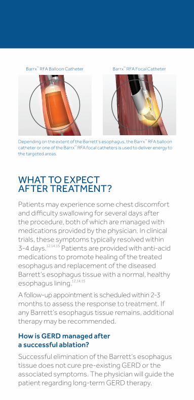
Barrx™ RFA Balloon Catheter Barrx™ RFA Focal Catheter
WHAT TO EXPECT AFTER TREATMENT?
Patients may experience some chest discomfort and difficulty swallowing for several days after the procedure, both of which are managed with medications provided by the physician. In clinical trials, these symptoms typically resolved within 3-4 days.12,14,15 Patients are provided with anti-acid medications to promote healing of the treated esophagus and replacement of the diseased Barrett’s esophagus tissue with a normal, healthy esophagus lining.12,14,15
A follow-up appointment is scheduled within 2-3 months to assess the response to treatment. If any Barrett’s esophagus tissue remains, additional therapy may be recommended.
How is GERD managed after a successful ablation?
Successful elimination of the Barrett’s esophagus tissue does not cure pre- existing GERD or the associated symptoms. The physician will guide the patient regarding long-term GERD therapy.
Depending on the extent of the Barrett’s esophagus, the Barrx™ RFA balloon catheter or one of the Barrx™ RFA focal catheters is used to deliver energy to the targeted areas.
†This is a global website. It is not specific to Canada.
© 2017 Medtronic. All rights reserved. Medtronic, Medtronic logo and Further, Together are trademarks of Medtronic. All other brands are trademarks of a Medtronic company.
Currently licensed under Covidien LLC. Manufactured by: Covidien LLC, 15 Hampshire Street, Mansfield, MA, 02048 USA. CA-ET-0060-E Rev. 04/2017 (US160245)
8455 Trans-Canada HighwaySaint-Laurent, Quebec, H4S 1Z1 877.664.8926 [t] 800.567.1939 [f]
medtronic.com†
Caution: Federal law restricts this device to sale by or on the order of a licensed healthcare practitioner. Rx only.
Risk Information: The following are transient side effects that may be expected after treatment: chest pain, difficulty swallowing, painful swallowing, throat pain, and/or fever. Complications observed at a very low frequency include: mucosal laceration, minor and major acute bleeding, stricture, perforation, cardiac arrhythmia, pleural effusion, aspiration, and infection. Potential complications that have not been observed include: death. Please consult a physician for further information.
*Important Reminder: This information is intended only to provide general information and not as a definitive basis for diagnosis or treatment in any particular case. It is very important that you consult your doctor about your specific condition, contraindications, and possible complications.
References: 1. Canadian Digestive Health Foundation, Understanding Barrett’s Esophagus, www.cshf.ca. 2. Strategic Market Assessment: Barrx GI”, Dymedex Market Development Consulting, Oct 2014 3. Haggitt RC. Barrett’s esophagus, dysplasia, and adenocarcinoma. Hum Pathol. 1994 Oct;25(10):982-93. 4. Noffsinger AE. Defining Cancer Risk in Barrett’s Esophagus: A Pathologist’s Perspective. Gastrointest Cancer Res. 2008 Nov-Dec; 2(6):308-310. 5. Sharma P, Falk GW, Weston AP, Reker D, Johnston M, Sampliner RE. Dysplasia and cancer in a large multicenter cohort of patients with Barrett’s esophagus. Clin Gastroenterol Hepatol. 2006;4(5):566-572. Epub 2006 Apr 17. 6. Wani S, Puli SR, Shaheen NJ, et al. Esophageal adenocarcinoma in Barrett’s esophagus after endoscopic ablative therapy: a meta-analysis and systematic review. Am J Gastroenterol. 2009 Feb;104(2):502-13. 7. de Jonge PJ, van Blankenstein M, Looman CW, et al. Risk of malignant progression in patients with Barrett’s oesophagus: a Dutch nationwide cohort study. Gut. 2010 Aug;59(8):1030-6. 8. Wani S, Falk GW, Post J, et al. Patients with nondysplastic Barrett’s esophagus have low risks for developing dysplasia or esophageal adenocarcinoma. Gastroenterology. 2011 Oct;141(4):1179-86. 9. Jung KW, Talley NJ, Romero Y, et al. Epidemiology and natural history of intestinal metaplasia of the gastroesophageal junction and Barrett’s esophagus: a population-based study. Am J Gastroenterol. 2011 Aug;106(8):1447-55. 10. American Gastroenterological Association Medical Position Statement on the Management of Barrett’s Esophagus. Gastroenterology. 2011;140:1084-1091. 11. McGahan JP, van Raalte VA. Tumor Ablation, 2005, Section I, 3-16, doI:10.1007/0-387-28674-8_1. 12. Fleischer DE, Overholt BF, Sharma VK, et al. Endoscopic ablation of Barrett’s esophagus: a multicenter study with 2.5-year follow-up. Gastrointest Endosc. 2008; 68:867-76. 13. Data on file, covering the period of April 2005 through February 2012. 14. Sharma VK, Kim HJ, Das A, et al. A prospective pilot trial of ablation of Barrett’s esophagus with low-grade dysplasia using stepwise circumferential and focal ablation (HALO system). Endoscopy. 2008;40:380-387. 15. Gondrie JJ, Pouw RE, Sondermeijer CM, et al. Stepwise circumferential and focal ablation of Barrett’s esophagus with high-grade dysplasia: results of first prospective series of 11 patients. Endoscopy. 2008;40:359-369. 16. Shaheen NJ, Sharma P, Overholt BF, et al. Radiofrequency ablation in Barrett’s esophagus with dysplasia. N Engl J Med. 2009;360:2277-2288.


![Barrett’s esophagus and new therapeutic modalitiesThe prevalence of Barrett’s esophagus in the adult population is 0.4–1.6% [1,3,12,13]. Assum-ing a US adult population in 2007](https://static.fdocuments.in/doc/165x107/5f4d5b4d6dfbad3c763bb443/barrettas-esophagus-and-new-therapeutic-modalities-the-prevalence-of-barrettas.jpg)