
Bagaria et al, nt eurorehabilitation ng 215, 2:3 oJ ... · PDF fileReliance Foundation...
4
Bagaria et al., Int J Neurorehabilitation Eng 2015, 2:3 DOI: 10.4172/2376-0281.1000175 Open Access Review Article Volume 2 • Issue 3 • 1000175 Int J Neurorehabilitation ISSN: 2376-0281 IJN, an open access journal *Corresponding author: Shalini Bagaria, Department of Orthopedics, Sir HN Reliance Foundation Hospital, Mumbai, India, Tel: +919999642211; E-mail: [email protected] Received May 29, 2015; Accepted July 14, 2015; Published July 20, 2015 Citation: Bagaria V, Shah S, Chaudhary K, Shah P, Bagaria S (2015) Technical Note: 3D Printing and Developing Patient Optimized Rehabilitation Tools (Port) - A Technological Leap. Int J Neurorehabilitation 2: 175. doi:10.4172/2376- 0281.1000175 Copyright: © 2015 Bagaria V, et al. This is an open-access article distributed under the terms of the Creative Commons Attribution License, which permits unrestricted use, distribution, and reproduction in any medium, provided the original author and source are credited. Keywords: 3D PORT; 3D printing; Orthoses; Additive manufactur- ing; Prosthesis; Implant manufacturing; Customized prosthesis Introduction ree dimensional printing, also known by a variety of other names most commonly additive manufacturing or medical rapid prototyping, is a valuable addition to the armamentarium of the clinicians and rehabilitation specialists who are therapists, bio-engineers and clinicians looking to push boundaries in neurology and orthopedic rehabilitation research. 3D printing makes a physical three-dimensional model of any body part/device or structure from the printable files derived either from a 3D scanner or through CT Images. In the process a thin plastic thread filament is heated and squeezed through a nozzle. A computer generated rendering sequence is created in a layered fashion, with the bottom layer being created first and subsequently layers are added over one another [1]. 3D PORT process e group of technology that uses the 3D printing for rehabilitation is oſten called as 3D patient optimized rehabilitation tools or 3D PORT. e steps involved are: 1. Scanning 2. Processing 3. Designing and Deliberation 4. 3D Printing 5. Clinical Application Scanning 3D PORT technology involves scanning the patient part to procure the patient data. e hardware typically is an inexpensive portable device that scans the exoskeleton. A 3D scanner typically creates point cloud of geometric samples form the surface of a given object, which in this case is the body part of the subject. ese point clouds are then extrapolated using soſtware to generate the actual shape of the object also called as a reconstruction. In effect these scanners akin to cameras with a cone like field of vision, however while a camera collects the information on colour and surfaces in its field, a 3D scanner also collects distance information about the surface at each point in the picture that helps recreate the topography accurately. To accurately reconstruct the object a common reference point Abstract There has been a gradual shift in the approach towards managing the occupational and physical health of patients in last few decades. While the earlier approach was standardized and involves using time tested solutions for all patients, the new era is moving towards customized medicine in which the focus is on an individual and how the solutions can be tailor made for his maximal benefit. Clinicians and therapists worldwide are looking for technological advancements that help them realize their goal to ensure optimal outcomes that are patient specific. 3D patient optimized rehabilitation tools also known as 3D PORT is based on additive manufacturing of customized devices using 3D printers. This review describes the basics of the use of technology and also on the future trends and possible clinical applications of the same. Technical Note: 3D Printing and Developing Patient Optimized Rehabilitation Tools (Port) - A Technological Leap Vaibhav Bagaria, Smit Shah, Kshitij Chaudhary, Priya Shah, Shalini Bagaria* Sir HN Reliance Foundation Hospital, Girgaum, Mumbai, India Figure 1: Scanning process where body part of patient is scanned. or system is needed and this is called as ‘alignment references’ or registration. Over the years various generations of 3D scanners have come in play. ey can be broadly classified as either contact or non- contact scanners. As the name suggest the contact scanners probe the subject through the physical touch, the non-contact scanners emit different types of radiation or light and capture its reflection or radiation passing through the object to capture the topography of an object or environment. e possible radiation or emission for this purpose includes: Light, Ultrasound, Infrared or X-rays (Figure 1). Processing e hand held scanners use the process of triangulation technique. Typically a laser dot or a laser line is projected towards the object that is being scanned and the receptive sensors (usually a position sensitive device or charge coupled device) then captures the data that measures I n t e r n a t i o n a l J o u r n a l o f N e u r o r e h a b i l i t a t i o n ISSN: 2376-0281 International Journal of Neurorehabilitation
Transcript of Bagaria et al, nt eurorehabilitation ng 215, 2:3 oJ ... · PDF fileReliance Foundation...

Bagaria et al., Int J Neurorehabilitation Eng 2015, 2:3 DOI: 10.4172/2376-0281.1000175
Open AccessReview Article
Volume 2 • Issue 3 • 1000175Int J NeurorehabilitationISSN: 2376-0281 IJN, an open access journal
*Corresponding author: Shalini Bagaria, Department of Orthopedics, Sir HNReliance Foundation Hospital, Mumbai, India, Tel: +919999642211; E-mail:[email protected]
Received May 29, 2015; Accepted July 14, 2015; Published July 20, 2015
Citation: Bagaria V, Shah S, Chaudhary K, Shah P, Bagaria S (2015) TechnicalNote: 3D Printing and Developing Patient Optimized Rehabilitation Tools (Port)- A Technological Leap. Int J Neurorehabilitation 2: 175. doi:10.4172/2376-0281.1000175
Copyright: © 2015 Bagaria V, et al. This is an open-access article distributed under the terms of the Creative Commons Attribution License, which permits unrestricted use, distribution, and reproduction in any medium, provided the original author and source are credited.
Keywords: 3D PORT; 3D printing; Orthoses; Additive manufactur-ing; Prosthesis; Implant manufacturing; Customized prosthesis
Introduction Three dimensional printing, also known by a variety of other names
most commonly additive manufacturing or medical rapid prototyping, is a valuable addition to the armamentarium of the clinicians and rehabilitation specialists who are therapists, bio-engineers and clinicians looking to push boundaries in neurology and orthopedic rehabilitation research. 3D printing makes a physical three-dimensional model of any body part/device or structure from the printable files derived either from a 3D scanner or through CT Images. In the process a thin plastic thread filament is heated and squeezed through a nozzle. A computer generated rendering sequence is created in a layered fashion, with the bottom layer being created first and subsequently layers are added over one another [1].
3D PORT process
The group of technology that uses the 3D printing for rehabilitation is often called as 3D patient optimized rehabilitation tools or 3D PORT. The steps involved are:
1. Scanning
2. Processing
3. Designing and Deliberation
4. 3D Printing
5. Clinical Application
Scanning
3D PORT technology involves scanning the patient part to procure the patient data. The hardware typically is an inexpensive portable device that scans the exoskeleton. A 3D scanner typically creates point cloud of geometric samples form the surface of a given object, which in this case is the body part of the subject. These point clouds are then extrapolated using software to generate the actual shape of the object also called as a reconstruction. In effect these scanners akin to cameras with a cone like field of vision, however while a camera collects the information on colour and surfaces in its field, a 3D scanner also collects distance information about the surface at each point in the picture that helps recreate the topography accurately.
To accurately reconstruct the object a common reference point
AbstractThere has been a gradual shift in the approach towards managing the occupational and physical health of patients
in last few decades. While the earlier approach was standardized and involves using time tested solutions for all patients, the new era is moving towards customized medicine in which the focus is on an individual and how the solutions can be tailor made for his maximal benefit. Clinicians and therapists worldwide are looking for technological advancements that help them realize their goal to ensure optimal outcomes that are patient specific. 3D patient optimized rehabilitation tools also known as 3D PORT is based on additive manufacturing of customized devices using 3D printers. This review describes the basics of the use of technology and also on the future trends and possible clinical applications of the same.
Technical Note: 3D Printing and Developing Patient Optimized Rehabilitation Tools (Port) - A Technological LeapVaibhav Bagaria, Smit Shah, Kshitij Chaudhary, Priya Shah, Shalini Bagaria*Sir HN Reliance Foundation Hospital, Girgaum, Mumbai, India
Figure 1: Scanning process where body part of patient is scanned.
or system is needed and this is called as ‘alignment references’ or registration. Over the years various generations of 3D scanners have come in play. They can be broadly classified as either contact or non-contact scanners. As the name suggest the contact scanners probe the subject through the physical touch, the non-contact scanners emit different types of radiation or light and capture its reflection or radiation passing through the object to capture the topography of an object or environment. The possible radiation or emission for this purpose includes: Light, Ultrasound, Infrared or X-rays (Figure 1).
Processing
The hand held scanners use the process of triangulation technique. Typically a laser dot or a laser line is projected towards the object that is being scanned and the receptive sensors (usually a position sensitive device or charge coupled device) then captures the data that measures
Inter
natio
nal J
ournal of Neurorehabilitation
ISSN: 2376-0281
International
Journal of Neurorehabilitation
Citation: Bagaria V, Shah S, Chaudhary K, Shah P, Bagaria S (2015) Technical Note: 3D Printing and Developing Patient Optimized Rehabilitation Tools (Port) - A Technological Leap. Int J Neurorehabilitation 2: 175. doi:10.4172/2376-0281.1000175
Page 2 of 4
Volume 2 • Issue 3 • 1000175Int J NeurorehabilitationISSN: 2376-0281 IJN, an open access journal
the distance to surface in reference to an internal coordinate. The data which is recorded as data points within the three dimensional space is converted into triangular mesh that is then converted into a Computer aided model (CAD Model).
Designing and deliberation
This involves parameterizing the exoskeleton into CAD (Computer aided designing model). Based on the measurement of user’s part dimension a linked dimension CAD model is created. The CAD data obtained from these 3D Scanners are converted to the editable forms typically the ones that are used by software engineers to design the object. A typical CAD file is meant to embody the ‘design intent’ based on critical feature and end product expectations. For e.g., the data for amputated arm prosthesis will include data from the patient upper limb both amputated and normal limb scan data and also the design for prosthesis that is envisaged. While many organizations and clinics have developed their own software, some off the shelf software including Geomagic, Image ware, Rhino 3D, CATIA, AutoCAD and Revit (Figure 2).
3D printing
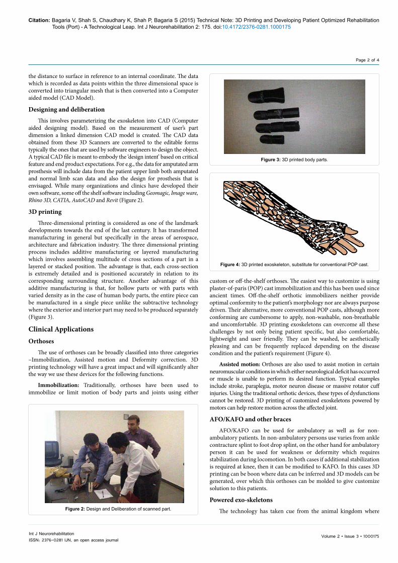
Three-dimensional printing is considered as one of the landmark developments towards the end of the last century. It has transformed manufacturing in general but specifically in the areas of aerospace, architecture and fabrication industry. The three dimensional printing process includes additive manufacturing or layered manufacturing which involves assembling multitude of cross sections of a part in a layered or stacked position. The advantage is that, each cross-section is extremely detailed and is positioned accurately in relation to its corresponding surrounding structure. Another advantage of this additive manufacturing is that, for hollow parts or with parts with varied density as in the case of human body parts, the entire piece can be manufactured in a single piece unlike the subtractive technology where the exterior and interior part may need to be produced separately (Figure 3).
Clinical ApplicationsOrthoses
The use of orthoses can be broadly classified into three categories –Immobilization, Assisted motion and Deformity correction. 3D printing technology will have a great impact and will significantly alter the way we use these devices for the following functions.
Immobilization: Traditionally, orthoses have been used to immobilize or limit motion of body parts and joints using either
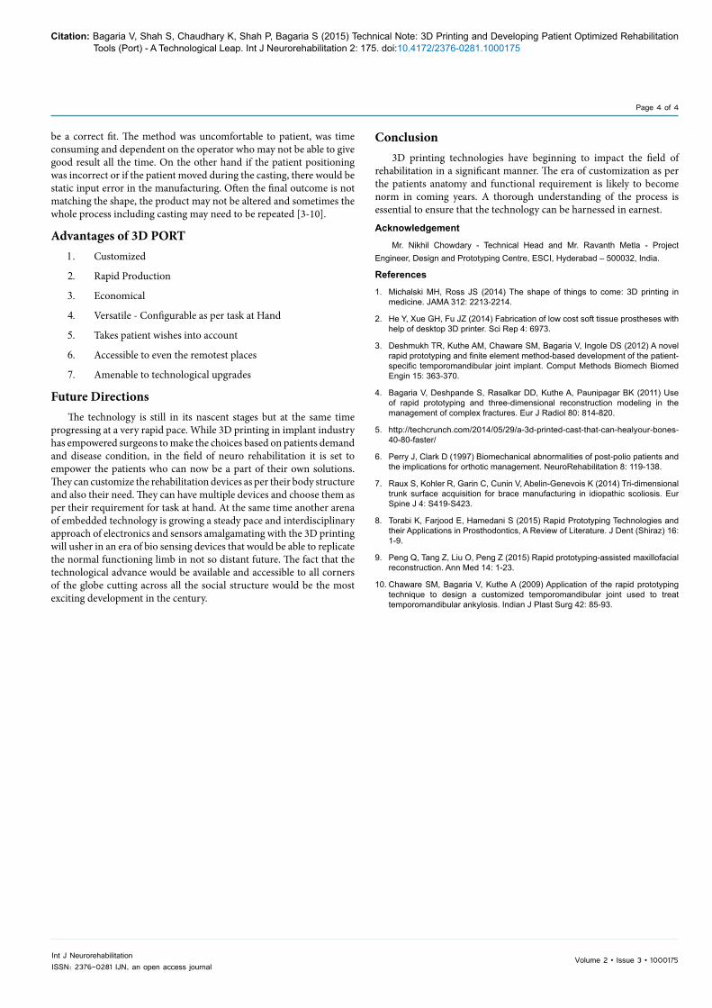
custom or off-the-shelf orthoses. The easiest way to customize is using plaster-of-paris (POP) cast immobilization and this has been used since ancient times. Off-the-shelf orthotic immobilizers neither provide optimal conformity to the patient’s morphology nor are always purpose driven. Their alternative, more conventional POP casts, although more conforming are cumbersome to apply, non-washable, non-breathable and uncomfortable. 3D printing exoskeletons can overcome all these challenges by not only being patient specific, but also comfortable, lightweight and user friendly. They can be washed, be aesthetically pleasing and can be frequently replaced depending on the disease condition and the patient’s requirement (Figure 4).
Assisted motion: Orthoses are also used to assist motion in certain neuromuscular conditions in which either neurological deficit has occurred or muscle is unable to perform its desired function. Typical examples include stroke, paraplegia, motor neuron disease or massive rotator cuff injuries. Using the traditional orthotic devices, these types of dysfunctions cannot be restored. 3D printing of customized exoskeletons powered by motors can help restore motion across the affected joint.
AFO/KAFO and other braces
AFO/KAFO can be used for ambulatory as well as for non-ambulatory patients. In non-ambulatory persons use varies from ankle contracture splint to foot drop splint, on the other hand for ambulatory person it can be used for weakness or deformity which requires stabilization during locomotion. In both cases if additional stabilization is required at knee, then it can be modified to KAFO. In this cases 3D printing can be boon where data can be inferred and 3D models can be generated, over which this orthoses can be molded to give customize solution to this patients.
Powered exo-skeletons
The technology has taken cue from the animal kingdom where
Figure 2: Design and Deliberation of scanned part.
Figure 3: 3D printed body parts.
Figure 4: 3D printed exoskeleton, substitute for conventional POP cast.

Citation: Bagaria V, Shah S, Chaudhary K, Shah P, Bagaria S (2015) Technical Note: 3D Printing and Developing Patient Optimized Rehabilitation Tools (Port) - A Technological Leap. Int J Neurorehabilitation 2: 175. doi:10.4172/2376-0281.1000175
Page 3 of 4
Volume 2 • Issue 3 • 1000175Int J NeurorehabilitationISSN: 2376-0281 IJN, an open access journal
in the outer structure known as exo-skeleton is now used in certain extraordinary situations. Also called as wearable 3D printed robots, powered armor or exo-suit. The exoskeletons have an outer framework powered by a system of motors and hydraulics. The system increases the endurance and strength of wearer. While these were originally developed for military use in combat situations, the use have extended into rehabilitation front mainly stroke and spinal injury patients.
Deformity correction
Spinal deformity, like scoliosis, is frequently treated with external brace. Typically a cast is made out of the torso of the patient with scoliosis. The brace is molded on this cast to make a custom fitting brace that can correct and maintain the deformity. This process is not only expensive and time-consuming, but also the brace can be restrictive, non-breathable, and uncomfortable. 3D body scanning technology can create a three dimensional shape of the patient and CAD software can be used to create a patient specific brace that can be 3D printed in any design. Honeycombing can be incorporated in the design to make the brace light weight and breathable. Reinforced struts in the design can provide additional strength to weak areas without increasing the overall weight of the brace. This will improve patient compliance that has traditionally been a problem with scoliosis patients. Similar technology can revolutionize the manufacturing of customized seating support orthosis for patients with neuromuscular scoliosis (e.g., cerebral palsy) (Figure 5).
Prostheses
These are the devices that artificially replaces the missing body part either physically, functionally or both. While the earliest written reference to such devices date back to 1579 when ‘Amboise Pare’ while working as a military surgeon wrote about the artificial limbs fitted to amputees. The earlier generations used mainly metals in manufacturing these however over time more and more plastic, carbon and silicon began to be used. It is believed that there are between 10 and 15 million amputees currently across the globe. The conventional process used to be similar to one described above. Briefly, the cast mold was derived from the plaster cast set over the amputated part this was then sent to the workshop where in the prosthetic technician would manufacture the part. The process would take several weeks and several to and fro between the amputee and technician. With 3D printing all the process can be done without the inconvenience and shorter turnaround time. Not only these are patient specific and conforming to the anatomy it can also be function specific. There are several open source printable file format available currently that can be downloaded from the net and printed according to the need [2].
Trans femoral/above knee prosthesis: This prosthesis is typically used for rehabilitation of above knee trans femoral amputee.
Conventionally trans femoral above knee amputation prosthesis are produced by polypropylene technology as recommended by ICRC (International Committee of Red Cross) involving several steps like 1) Measurements 2) Socket manufacturing 3) Socket alignment 4) Adjustment of length 5) Wielding of cylinders 6) Ankle foot alignment. Total contact trans femoral prosthesis is also manufactured using same process and steps involved (Figure 6).
Transtibial/below knee: This prosthesis being typically employed for rehabilitating transtibial below knee amputee. Conventionally manufactured under guidelines from ICR involving same steps as listed above. Difference here is socket which can be modified as PTB-SC (supra-condylar) or PTB-SCSP (supra-condylar, suprapatellar) (Figure 7).
Comparing 3D Port with the Old ProcessThe old process was a multi-level time consuming process that also
proved expensive. For e.g., in case an orthopedic surgeon required a custom TLSO brace for one of his patients, the typical procedure would consist of first getting a plaster cast as a mold of his body shape and the problem. The patient would need to be patient and wait covered in a plaster till the time it sets. It would then be cut off and the results would be physically mailed to manufacturer. The manufacture would subsequently measure them and then make a brace based on the parameters derived from the physical mold hoping that it would
Figure 5: Planning for scoliosis brace (TLSO- Thoracolumbar Spinal Orthoses).
Figure 6: Planning for prostheses of above knee amputation stump.
Figure 7: Planning for prostheses of below knee amputee patient.
Citation: Bagaria V, Shah S, Chaudhary K, Shah P, Bagaria S (2015) Technical Note: 3D Printing and Developing Patient Optimized Rehabilitation Tools (Port) - A Technological Leap. Int J Neurorehabilitation 2: 175. doi:10.4172/2376-0281.1000175
Page 4 of 4
Volume 2 • Issue 3 • 1000175Int J NeurorehabilitationISSN: 2376-0281 IJN, an open access journal
be a correct fit. The method was uncomfortable to patient, was time consuming and dependent on the operator who may not be able to give good result all the time. On the other hand if the patient positioning was incorrect or if the patient moved during the casting, there would be static input error in the manufacturing. Often the final outcome is not matching the shape, the product may not be altered and sometimes the whole process including casting may need to be repeated [3-10].
Advantages of 3D PORT1. Customized
2. Rapid Production
3. Economical
4. Versatile - Configurable as per task at Hand
5. Takes patient wishes into account
6. Accessible to even the remotest places
7. Amenable to technological upgrades
Future DirectionsThe technology is still in its nascent stages but at the same time
progressing at a very rapid pace. While 3D printing in implant industry has empowered surgeons to make the choices based on patients demand and disease condition, in the field of neuro rehabilitation it is set to empower the patients who can now be a part of their own solutions. They can customize the rehabilitation devices as per their body structure and also their need. They can have multiple devices and choose them as per their requirement for task at hand. At the same time another arena of embedded technology is growing a steady pace and interdisciplinary approach of electronics and sensors amalgamating with the 3D printing will usher in an era of bio sensing devices that would be able to replicate the normal functioning limb in not so distant future. The fact that the technological advance would be available and accessible to all corners of the globe cutting across all the social structure would be the most exciting development in the century.
Conclusion3D printing technologies have beginning to impact the field of
rehabilitation in a significant manner. The era of customization as per the patients anatomy and functional requirement is likely to become norm in coming years. A thorough understanding of the process is essential to ensure that the technology can be harnessed in earnest.
Acknowledgement
Mr. Nikhil Chowdary - Technical Head and Mr. Ravanth Metla - Project Engineer, Design and Prototyping Centre, ESCI, Hyderabad – 500032, India.
References
1. Michalski MH, Ross JS (2014) The shape of things to come: 3D printing in medicine. JAMA 312: 2213-2214.
2. He Y, Xue GH, Fu JZ (2014) Fabrication of low cost soft tissue prostheses with help of desktop 3D printer. Sci Rep 4: 6973.
3. Deshmukh TR, Kuthe AM, Chaware SM, Bagaria V, Ingole DS (2012) A novel rapid prototyping and finite element method-based development of the patient-specific temporomandibular joint implant. Comput Methods Biomech Biomed Engin 15: 363-370.
4. Bagaria V, Deshpande S, Rasalkar DD, Kuthe A, Paunipagar BK (2011) Use of rapid prototyping and three-dimensional reconstruction modeling in the management of complex fractures. Eur J Radiol 80: 814-820.
5. http://techcrunch.com/2014/05/29/a-3d-printed-cast-that-can-healyour-bones-40-80-faster/
6. Perry J, Clark D (1997) Biomechanical abnormalities of post-polio patients and the implications for orthotic management. NeuroRehabilitation 8: 119-138.
7. Raux S, Kohler R, Garin C, Cunin V, Abelin-Genevois K (2014) Tri-dimensional trunk surface acquisition for brace manufacturing in idiopathic scoliosis. Eur Spine J 4: S419-S423.
8. Torabi K, Farjood E, Hamedani S (2015) Rapid Prototyping Technologies and their Applications in Prosthodontics, A Review of Literature. J Dent (Shiraz) 16: 1-9.
9. Peng Q, Tang Z, Liu O, Peng Z (2015) Rapid prototyping-assisted maxillofacial reconstruction. Ann Med 14: 1-23.
10. Chaware SM, Bagaria V, Kuthe A (2009) Application of the rapid prototyping technique to design a customized temporomandibular joint used to treat temporomandibular ankylosis. Indian J Plast Surg 42: 85-93.