Bacterial Structure (Lab 3) - fac.ksu.edu.sa fileSporogenesis Members of the anaerobic genera...
15
Bacterial Structure (Lab 5) Rana Alqusumi
Transcript of Bacterial Structure (Lab 3) - fac.ksu.edu.sa fileSporogenesis Members of the anaerobic genera...

Bacterial Structure (Lab 5)Rana Alqusumi
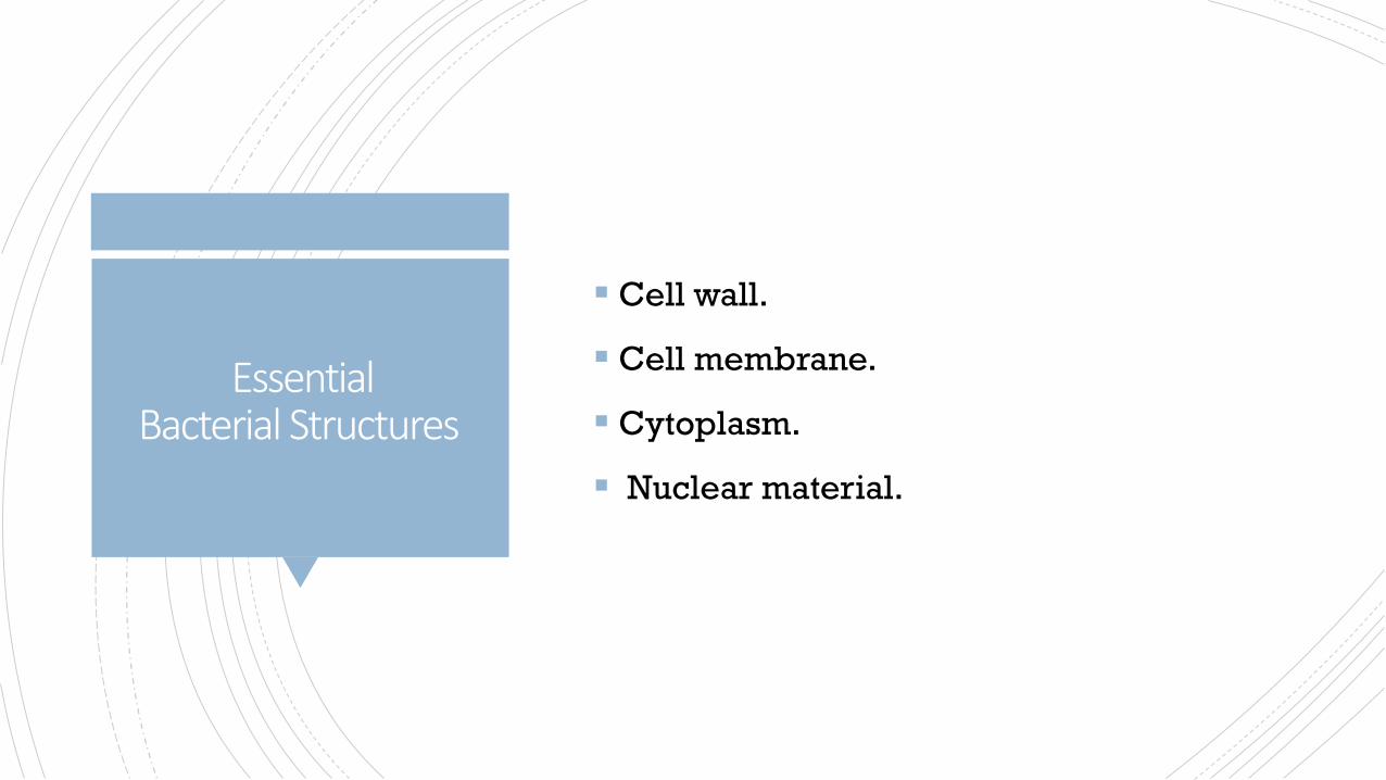
EssentialBacterial Structures
Cell wall.
Cell membrane.
Cytoplasm.
Nuclear material.
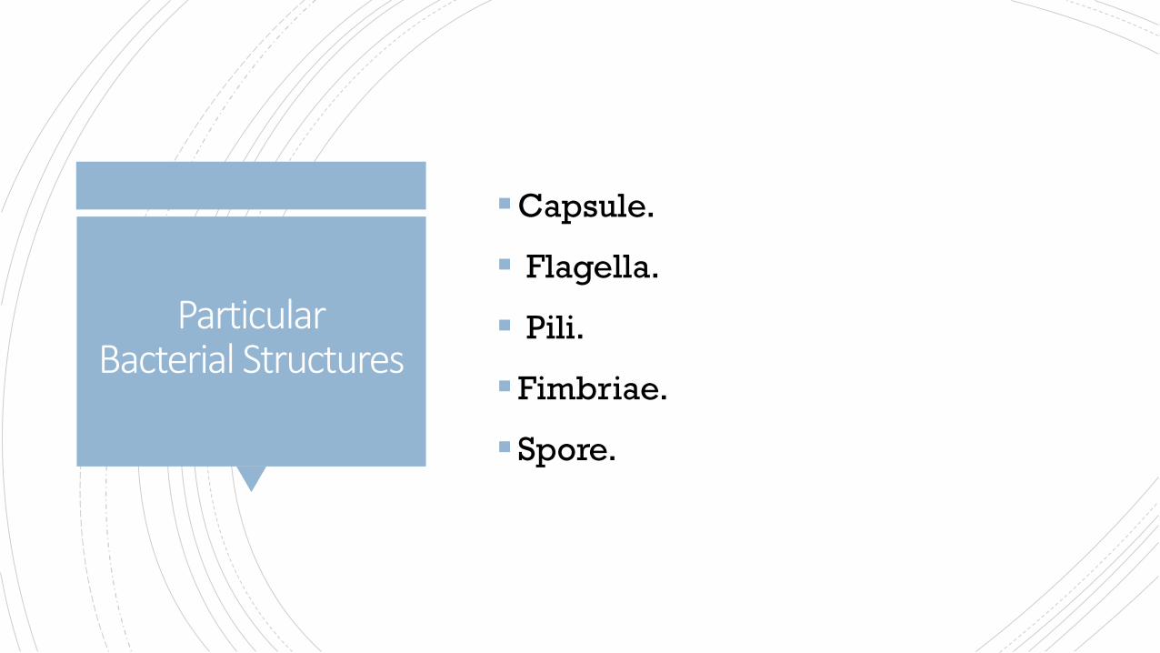
ParticularBacterial Structures
Capsule.
Flagella.
Pili.
Fimbriae.
Spore.

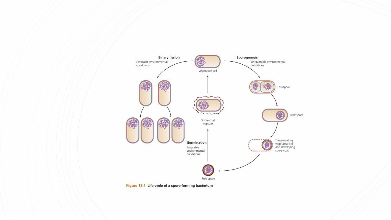
Sporogenesis
Members of the anaerobic genera Clostridium and the
aerobic genus Bacillus are examples of organisms that
have the capacity to exist either as metabolically active
vegetative cells or as highly resistant, metabolically
inactive cell types called spores.
When environmental conditions become unfavorable
for continuing vegetative cellular activities, particularly
with the exhaustion of a nutritional carbon source, these
cells have the capacity to undergo sporogenesis and
give rise to a new intracellular structure called the
endospore, which is surrounded by impervious layers
called spore coats.
As conditions continue to worsen, the endospore is
released from the degenerating vegetative cell and
becomes an independent cell called a free spore.
Spore Stain(Schaeffer-Fulton Method)
Because of the chemical composition of spore layers,
the spore is resistant to the damaging effects of
excessive heat, freezing, radiation, desiccation, and
chemical agents, as well as to the commonly employed
microbiological stains. With the return of favorable
environmental conditions, the free spore may revert to a
metabolically active and less resistant vegetative cell
through germination (see Figure 13.1).
It should be emphasized that sporogenesis and
germination are not means of reproduction but merely
mechanisms that ensure cell survival under all
environmental conditions.
Spore Stain(Schaeffer-Fulton Method)
The spore stain uses two different reagents:
1-Primary Stain(Malachite Green ):
unlike most vegetative cell types that stain by common
procedures, the free spore, because of its impervious coats, will
not accept the primary stain easily. For further penetration, the
application of heat is required. After the primary stain is applied
and the smear is heated, both the vegetative cell and spore will
appear green.

Spore Stain(Schaeffer-Fulton Method)
The spore stain uses two different reagents:
Decolorizing Agent(Water):
once the spore accepts the malachite green, it cannot be
decolorized by tap water, which removes only the excess
primary stain. The spore remains green. On the other
hand, the stain does not demonstrate a strong affinity for
vegetative cell components; the water removes it, and
these cells will be colorless.
Spore Stain(Schaeffer-Fulton Method)
The spore stain uses two different reagents:
2-Counterstain(Safranin):
This contrasting red stain is used as the second reagent to
color the decolorized vegetative cells, which will absorb
the counterstain and appear red. The spores retain the
green of the primary stain. A micrograph of spore-stained
cells appears in Figure 13.2.

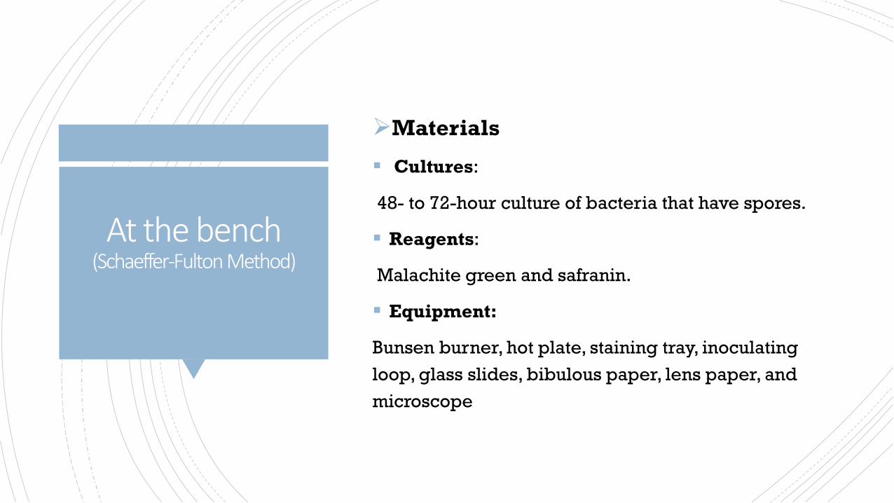
Materials
Cultures:
48- to 72-hour culture of bacteria that have spores.
Reagents:
Malachite green and safranin.
Equipment:
Bunsen burner, hot plate, staining tray, inoculating
loop, glass slides, bibulous paper, lens paper, and
microscope
At the bench (Schaeffer-Fulton Method)
At the bench (Schaeffer-Fulton Method)
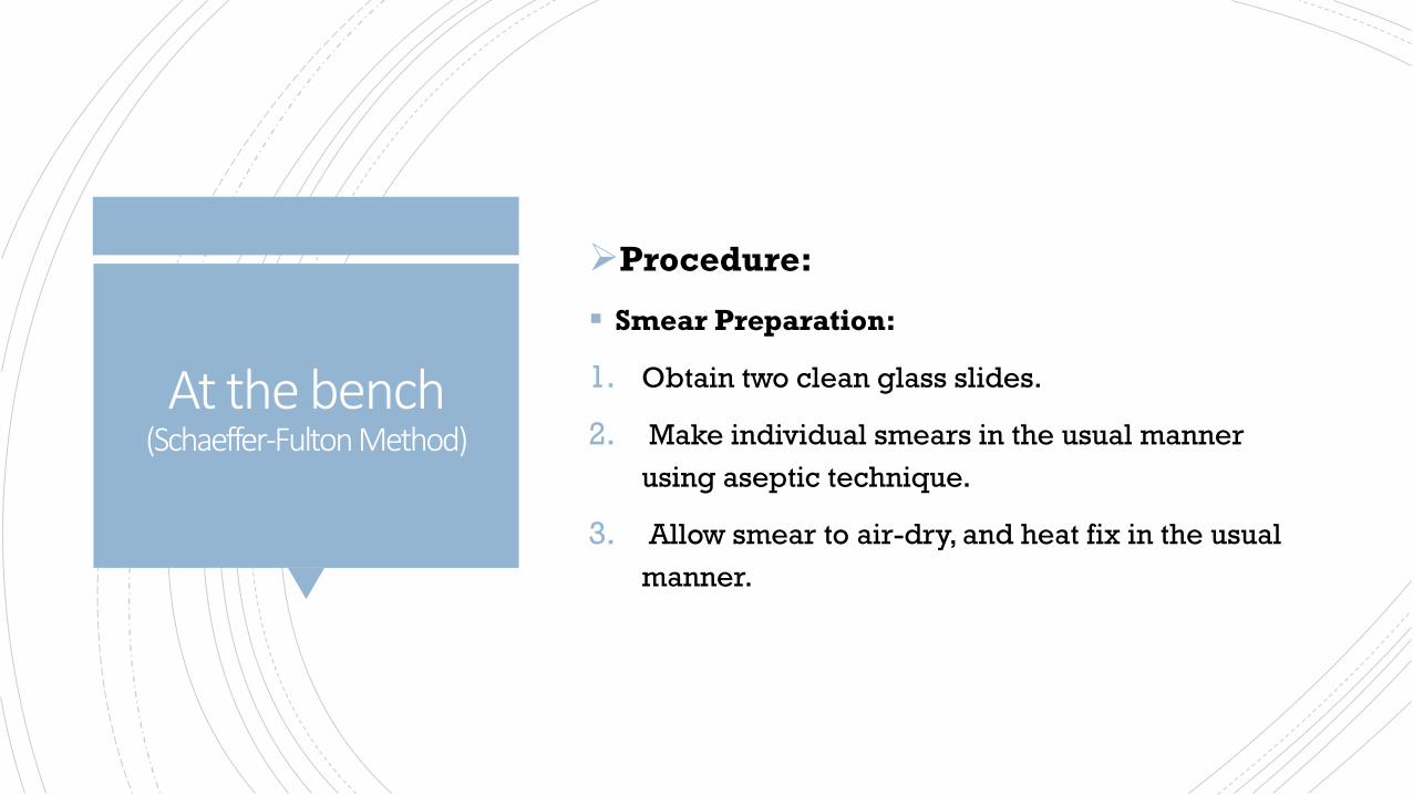
Procedure:
Smear Preparation:
1. Obtain two clean glass slides.
2. Make individual smears in the usual manner
using aseptic technique.
3. Allow smear to air-dry, and heat fix in the usual
manner.
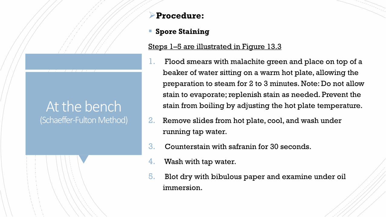
Procedure:
Spore Staining
Steps 1–5 are illustrated in Figure 13.3
1. Flood smears with malachite green and place on top of a
beaker of water sitting on a warm hot plate, allowing the
preparation to steam for 2 to 3 minutes. Note: Do not allow
stain to evaporate; replenish stain as needed. Prevent the
stain from boiling by adjusting the hot plate temperature.
2. Remove slides from hot plate, cool, and wash under
running tap water.
3. Counterstain with safranin for 30 seconds.
4. Wash with tap water.
5. Blot dry with bibulous paper and examine under oil
immersion.
At the bench (Schaeffer-Fulton Method)

Reference
James G. Cappuccino, Natalie Sherman. 2014.
Microbiology a laboratory manual. 10th ed.
https://fac.ksu.edu.sa/sites/default/files/362mic-
lab3.pdf