Bacterial infections in cirrhosis: A position statement based … · Bacterial infections in...
15
Bacterial infections in cirrhosis: A position statement based on the EASL Special Conference 2013 Rajiv Jalan 1 , Javier Fernandez 2 , Reiner Wiest 3 , Bernd Schnabl 4 , Richard Moreau 5 , Paolo Angeli 6 , Vanessa Stadlbauer 7 , Thierry Gustot 8 , Mauro Bernardi 9 , Rafael Canton 10 , Agustin Albillos 11 , Frank Lammert 12 , Alexander Wilmer 13 , Rajeshwar Mookerjee 1 , Jordi Vila 14 , Rita Garcia-Martinez 2 , Julia Wendon 15 , José Such 16 , Juan Cordoba 17 , Arun Sanyal 18 , Guadalupe Garcia-Tsao 19 , Vicente Arroyo 2 , Andrew Burroughs 20 , Pere Ginès 2,⇑ 1 Liver Failure Group, UCL Institute for Liver and Digestive Health, Royal Free Hospital, UK; 2 Liver Unit, Hospital Clinic, University of Barcelona, IDIBAPS, CIBERehd, Instituto de Salud Carlos III, Madrid, Spain; 3 Department of Gastroenterology, UVCM, Inselspital, 3010 Bern, Switzerland; 4 Department of Medicine, University of California San Diego, La Jolla, CA, USA; 5 INSERM U773, Centre de Recherche Biomédicale Bichat-Beaujon CRB3, UMRS 773, Université Paris-Diderot Paris, Service d’Hépatologie, Hôpital Beaujon, APHP, Clichy, France; 6 Unit of Hepatic Emergencies and Liver Transplantation, Department of Medicine, University of Padova, Italy; 7 Department of Internal Medicine, Division of Gastroenterology and Hepatology, Medical University of Graz, Austria; 8 Department of Gastroenterology and Hepato-Pancreatology, Erasme Hospital, Laboratory of Experimental Gastroenterology, Université Libre de Bruxelles, Brussels, Belgium; 9 Department of Medical and Surgical Sciences, University of Bologna, Italy; 10 Department of Microbiology, Hospital Universitario Ramón y Cajal and Intituto Ramon y Cajal de Investigación Sanitaria (IRYCIS), Madrid, Spain; 11 Gastroenterology Service, University Hospital Ramon y Cajal, Madrid, Spain; 12 Department of Medicine II, Saarland University Medical Center, Homburg, Germany; 13 Medical Intensive Care Unit, University Hospital Gasthuisberg, Leuven, Belgium; 14 Department of Microbiology, Hospital Clínic, School of Medicine, University of Barcelona, Barcelona, Spain; 15 Institute of Liver Studies and Critical Care, Kings College London, Kings College Hospital, UK; 16 Department of Clinical Medicine, Miguel Hernández University, Alicante, CIBERehd, Instituto de Salud Carlos III, Madrid, Spain; 17 Liver Unit, Department of Internal Medicine, Hospital Universitari Vall d’Hebron, Institut de Recerca (VHIR), Universitat Autònoma de Barcelona, Barcelona, CIBERehd, Instituto de Salud Carlos III, Madrid, Spain; 18 Charles Caravati Professor of Medicine, Department of Internal Medicine, Virginia Commonwealth University School of Medicine, Richmond, VA, USA; 19 Department of Medicine, Section of Digestive Diseases, Yale University School of Medicine, New Haven, CT, USA; 20 The Royal Free Shelia Sherlock Liver Centre and University Department of Surgery, University College London and Royal Free Hospital, UK We are dedicating this paper to Drs. Joan Cordoba and Andrew Burroughs in recognition of their excellent work as Doctors and Scientists. They will be remembered by many as a good friends and colleagues. Joan, Andrew may you rest in peace. Summary Bacterial infections are very common and represent one of the most important reasons of progression of liver failure, develop- ment of liver-related complications, and mortality in patients with cirrhosis. In fact, bacterial infections may be a triggering fac- tor for the occurrence of gastrointestinal bleeding, hypervolemic hyponatremia, hepatic encephalopathy, kidney failure, and devel- opment of acute-on-chronic liver failure. Moreover, infections are a very common cause of repeated hospitalizations, impaired health-related quality of life, and increased healthcare costs in cirrhosis. Bacterial infections develop as a consequence of immune dysfunction that occurs progressively during the course of cirrhosis. In a significant proportion of patients, infections are caused by gram-negative bacteria from intestinal origin, yet gram-positive bacteria are a frequent cause of infection, particu- larly in hospitalized patients. In recent years, infections caused by multidrug-resistant bacteria are becoming an important clinical problem in many countries. The reduction of the negative clinical impact of infections in patients with cirrhosis may be achieved by a combination of pro- phylactic measures, such as administration of antibiotics, to reduce the occurrence of infections in high-risk groups together with early identification and management of infection once it has developed. Investigation on the mechanisms of altered gut microflora, translocation of bacteria, and immune dysfunction may help develop more effective and safe methods of prevention compared to those that are currently available. Moreover, research on biomarkers of early infection may be useful in early diagnosis and treatment of infections. The current manuscript reports an in-depth review and a position statement on bacterial infections in cirrhosis. Ó 2014 European Association for the Study of the Liver. Published by Elsevier B.V. All rights reserved. Journal of Hepatology 2014 vol. 60 j 1310–1324 Keywords: Cirrhosis; Bacterial infection; Multiresistant bacteria; Diagnosis. Received 26 September 2013; received in revised form 30 December 2013; accepted 26 January 2014 ⇑ Corresponding author. Address: Liver Unit, Hospital Clinic, University of Barcelona, IDIBAPS, CIBERehd, Instituto de Salud Carlos III, Madrid, Spain. E-mail address: [email protected] (P. Ginès). Position Paper guide.medlive.cn
Transcript of Bacterial infections in cirrhosis: A position statement based … · Bacterial infections in...
Position Paper
Bacterial infections in cirrhosis: A position statement basedon the EASL Special Conference 2013
Rajiv Jalan1, Javier Fernandez2, Reiner Wiest3, Bernd Schnabl4, Richard Moreau5, Paolo Angeli6,Vanessa Stadlbauer7, Thierry Gustot8, Mauro Bernardi9, Rafael Canton10, Agustin Albillos11,
Frank Lammert12, Alexander Wilmer13, Rajeshwar Mookerjee1, Jordi Vila14, Rita Garcia-Martinez2,Julia Wendon15, José Such16, Juan Cordoba17, Arun Sanyal18, Guadalupe Garcia-Tsao19,
Vicente Arroyo2, Andrew Burroughs20, Pere Ginès2,⇑
1Liver Failure Group, UCL Institute for Liver and Digestive Health, Royal Free Hospital, UK; 2Liver Unit, Hospital Clinic,University of Barcelona, IDIBAPS, CIBERehd, Instituto de Salud Carlos III, Madrid, Spain; 3Department of Gastroenterology, UVCM,
Inselspital, 3010 Bern, Switzerland; 4Department of Medicine, University of California San Diego, La Jolla, CA, USA; 5INSERM U773,Centre de Recherche Biomédicale Bichat-Beaujon CRB3, UMRS 773, Université Paris-Diderot Paris, Service d’Hépatologie, Hôpital Beaujon,
APHP, Clichy, France; 6Unit of Hepatic Emergencies and Liver Transplantation, Department of Medicine, University of Padova, Italy; 7
Department of Internal Medicine, Division of Gastroenterology and Hepatology, Medical University of Graz, Austria; 8Department ofGastroenterology and Hepato-Pancreatology, Erasme Hospital, Laboratory of Experimental Gastroenterology, Université Libre de Bruxelles,
Brussels, Belgium; 9Department of Medical and Surgical Sciences, University of Bologna, Italy; 10Department of Microbiology,Hospital Universitario Ramón y Cajal and Intituto Ramon y Cajal de Investigación Sanitaria (IRYCIS), Madrid, Spain; 11GastroenterologyService, University Hospital Ramon y Cajal, Madrid, Spain; 12Department of Medicine II, Saarland University Medical Center, Homburg,
Germany; 13Medical Intensive Care Unit, University Hospital Gasthuisberg, Leuven, Belgium; 14Department of Microbiology, Hospital Clínic,School of Medicine, University of Barcelona, Barcelona, Spain; 15Institute of Liver Studies and Critical Care, Kings College London, KingsCollege Hospital, UK; 16Department of Clinical Medicine, Miguel Hernández University, Alicante, CIBERehd, Instituto de Salud Carlos III,
Madrid, Spain; 17Liver Unit, Department of Internal Medicine, Hospital Universitari Vall d’Hebron, Institut de Recerca (VHIR), UniversitatAutònoma de Barcelona, Barcelona, CIBERehd, Instituto de Salud Carlos III, Madrid, Spain; 18Charles Caravati Professor of Medicine,
Department of Internal Medicine, Virginia Commonwealth University School of Medicine, Richmond, VA, USA; 19Department of Medicine,Section of Digestive Diseases, Yale University School of Medicine, New Haven, CT, USA; 20The Royal Free Shelia Sherlock Liver Centre
and University Department of Surgery, University College London and Royal Free Hospital, UK
We are dedicating this paper to Drs. Joan Cordoba and Andrew Burroughs in recognition of their excellent work as Doctors and Scientists.They will be remembered by many as a good friends and colleagues. Joan, Andrew may you rest in peace.
Summary
Bacterial infections are very common and represent one of themost important reasons of progression of liver failure, develop-ment of liver-related complications, and mortality in patientswith cirrhosis. In fact, bacterial infections may be a triggering fac-tor for the occurrence of gastrointestinal bleeding, hypervolemichyponatremia, hepatic encephalopathy, kidney failure, and devel-opment of acute-on-chronic liver failure. Moreover, infections area very common cause of repeated hospitalizations, impairedhealth-related quality of life, and increased healthcare costs incirrhosis. Bacterial infections develop as a consequence ofimmune dysfunction that occurs progressively during the courseof cirrhosis. In a significant proportion of patients, infections arecaused by gram-negative bacteria from intestinal origin, yet
Journal of Hepatology 20
Keywords: Cirrhosis; Bacterial infection; Multiresistant bacteria; Diagnosis.Received 26 September 2013; received in revised form 30 December 2013; accepted26 January 2014⇑ Corresponding author. Address: Liver Unit, Hospital Clinic, University ofBarcelona, IDIBAPS, CIBERehd, Instituto de Salud Carlos III, Madrid, Spain.E-mail address: [email protected] (P. Ginès).
gram-positive bacteria are a frequent cause of infection, particu-larly in hospitalized patients. In recent years, infections caused bymultidrug-resistant bacteria are becoming an important clinicalproblem in many countries.The reduction of the negative clinical impact of infections inpatients with cirrhosis may be achieved by a combination of pro-phylactic measures, such as administration of antibiotics, toreduce the occurrence of infections in high-risk groups togetherwith early identification and management of infection once ithas developed. Investigation on the mechanisms of altered gutmicroflora, translocation of bacteria, and immune dysfunctionmay help develop more effective and safe methods of preventioncompared to those that are currently available. Moreover, researchon biomarkers of early infection may be useful in early diagnosisand treatment of infections.The current manuscript reports an in-depth review and a positionstatement on bacterial infections in cirrhosis.� 2014 European Association for the Study of the Liver. Publishedby Elsevier B.V. All rights reserved.
14 vol. 60 j 1310–1324
guide.medlive.cn

JOURNAL OF HEPATOLOGY
Introduction
Bacterial infections are very common in patients with cirrhosis
80706050403020100
1 month 1 year
Per
cent
age
(%)
Before 20002000-2009
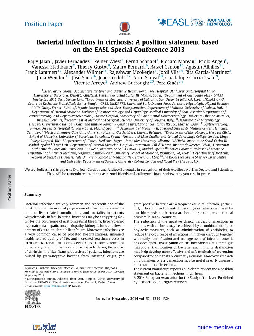
Fig. 1. Mortality rate caused by bacterial infections in cirrhosis in the lastdecades. 1-month and 1-year mortality rates were higher before than after 2000,although differences were not statistically significant. Modified from [6].
and currently represent one of the most common causes ofadmission to hospital in these patients and a major challengefor physicians caring for patients with liver diseases. Despitethe recent improvements in the knowledge of pathogenesis, pre-vention, and management, bacterial infections still represent amajor cause of morbidity and mortality among patients with cir-rhosis. On this background, the European Association for theStudy of the Liver (EASL) decided to hold a Special Conferenceon Bacterial Infections in cirrhosis in May 2013 in Barcelona.The conference gathered a large number of clinical and basicscientists as well as clinicians with special interest on the topicwho had presentations and extensive discussions on the mainareas of the field. The current manuscript represents a positionstatement that summarizes the different areas that werediscussed during the Conference and includes expert opinionson important aspects of the management of bacterial infectionsin cirrhosis.
Key Points
• The incidence and severity of infection in cirrhosis is greater than in the population without cirrhosis
• Infection with multiresistant organisms is common in cirrhosis and its occurrence is associated with higher mortality rates than in patients without cirrhosis
• The end-organ damaging effect of bacterial infection is greater in patients with cirrhosis due to altered sensitivity, which often culminates in acute-on-chronic liver failure
• Delays in the diagnosis and start of treatment results in higher mortality particularly in hypotensive patients with cirrhosis
• In patients with spontaneous bacterial peritonitis, the addition of albumin to antibiotics reduces mortality
• Primary prophylaxis of spontaneous bacterial peritonitis with norfloxacin is indicated in patients with variceal bleeding, severely decompensated cirrhosis, and those with ascites protein concentration of <15 g/L
• In patients with variceal bleeding, intravenous administration of 3rd generation cephalasporins improves survival
• Administration of norfloxacin to prevent recurrence of spontaneous bacterial peritonitis reduces mortality in cirrhosis
• Research into the mechanisms associated with increased risk of infection in cirrhosis, better use of current therapeutic strategies, development of rapid and accurate diagnostic tools, and development of new strategies to modulate the gut-liver interaction are urgently needed
Journal of Hepatology 2014
Clinical aspects of bacterial infections in cirrhosis and the
problem of multiresistant bacteriaPatients with cirrhosis have increased risk of developing bacterialinfections [1,2]. Infections are present at admission or developduring hospitalization in 25–35% of patients [3,4], an incidencethat is 4–5 fold higher than that observed in the general popula-tion. Spontaneous bacterial peritonitis (SBP) and urinary tractinfections are the most frequent infections followed by pneumo-nia, skin and soft tissue infections, and bacteremia. Clinical fac-tors associated with an increased risk of infection are poor liverfunction, variceal bleeding, low ascitic fluid protein levels, priorSBP and hospitalization [1,2]. Severity of infection is also higherin patients with cirrhosis who are more likely to die from sepsisthan individuals without cirrhosis. Bacterial infection increases3.75 fold the probability of death of patients with decompensatedcirrhosis, reaching 30% at 1 month and 63% at 1-year (Fig. 1) [5,6].
Enterobacteriaceae and non-enterococcal streptococci causethe majority of spontaneous infections in cirrhosis. As a conse-quence, b-lactams and quinolones have been widely used in theirtreatment and prevention [1,2]. This feature and the increasinglevel of invasiveness to which patients with cirrhosis are cur-rently submitted have induced important changes in the epide-miology of bacterial infections in cirrhosis. Spontaneous andsecondary infections caused by non-classical pathogens or multi-drug resistant (MR) bacteria are nowadays increasingly reportedin this population [1,4].
Infections by multiresistant bacteria in the general population andcirrhosis
MR bacteria are pathogens resistant to 3 or more of the main anti-biotic families, including b-lactams [7]. The main MR bacteria areextended-spectrum b-lactamase-producing Enterobacteriaceae(ESBL), non-fermentable gram-negative bacilli such as Pseudomo-nas aeruginosa, Stenotrophomonas maltophilia or Acinetobacter bau-manii, methicillin-resistant Staphylococcus aureus (MRSA) andvancomycin-susceptible or resistant enterococci (VSE, VRE). Infec-tions caused by these bacteria have increased in the general popu-lation mainly due to the dispersion of the so-called high-risk clonesnot only in the hospitals but also in the community. These clonesare specific bacteria able to acquire several resistance mechanismsand virulence determinants. Moreover, they efficiently colonizedifferent human niches, including the gastrointestinal tract [8].
vol. 60 j 1310–1324 1311
guide.medlive.cn

Table 1. Prevalence and clinical impact of different multiresistant bacterial infections in cirrhosis in several geographical areas.
Country, yr [Ref.] Prevalence of multiresistant bacteria Clinical impact Korea, 2006 [10] 29% ESBL-producing Enterobacteriaceae* No impactSpain, 2008 [11] 6% ESBL-producing Enterobacteriaceae No impactItaly, 2008 [12] 8% ESBL-producing Enterobacteriaceae Higher initial treatment failure
No impact on mortalityKorea, 2009 [13] 15% ESBL-producing Enterobacteriaceae Independent predictor of 30-day mortalityKorea, 2009 [14, 15] 4-7.5% ESBL-producing Enterobacteriaceae Higher initial treatment failure
Higher hospital and 30-day mortality Turkey, 2009 [16] 2% ESBL-producing Enterobacteriaceae
1% Pseudomonas aeruginosaNo data
France, 2009 [17] 8% MRSA**5% VSE#
4% ESBL-producing Enterobacteriaceae
No data
Germany, 2009 [18] 10% VSE Higher initial treatment failureHigher hospital mortality
Italy, 2010 [9] 20% ESBL-producing Enterobacteriaceae7% MRSA
Higher hospital mortality
Spain, 2012 [19] 6% ESBL-producing Enterobacteriaceae2% Pseudomonas aeruginosa2% Acinetobacter baumannii1% VSE
Independent predictor of mortality at 30 days
Spain, 2012 [4] 8-9% ESBL-producing Enterobacteriaceae3% Pseudomonas aeruginosa3-4% MRSA3-7% VSE
Lower infection resolutionHigher risk of septic shockHigher hospital mortality
USA, 2012 [20] 9% VRE&
6.5% ESBL-producing Enterobacteriaceae5% MRSA
No data
⁄ESBL, extended-spectrum b-lactamase-producing Enterobacteriaceae (bacteria with chromosomal b-lactamases are also included).⁄⁄MRSA, methicillin-resistant Staphylococcus aureus.#VSE, vancomycin-susceptible enterococci.&VRE, vancomycin-resistant enterococci.
Position Paper
Multiple studies from very different geographical areas haverecently reported an increased prevalence of infections causedby MR bacteria in cirrhosis [4,9–20]. The site of acquisition ofinfection determines the risk of MR bacterial infection withhigher rates of MR bacteria in infections acquired in the health-care environment: 23–39% in nosocomial infections, 14–41% inhealthcare-associated (HCA) episodes and 0–16% in infectionsacquired in the community [4,9].
Epidemiological pattern of MR bacteria differs markedlyamong geographical areas and even among hospitals. Regularassessment is therefore recommended [1,21]. ESBL-producingEnterobacteriaceae are predominant in South Europe and Asia[4,9–19], while MRSA and VRE are frequently isolated in centersfrom USA or South America [20].
Carbapenemase-producing K. pneumoniae are also beingincreasingly isolated in some centers in Italy [22]. Table 1 showsthe different prevalence of MR bacteria in infections in patientswith cirrhosis in different countries.
Clinical outcome of nosocomial and multiresistant bacterialinfections in cirrhosis
Early studies reported conflicting results regarding the prognosisof nosocomial infections in cirrhosis. Some showed very highmortality rates (59–67%) while others reported figures similarto those observed in community-acquired infections (Table 1)[13,23,24]. Recent data from Spain clearly support the formercontention [4]. The study included 669 infections from 2 series
1312 Journal of Hepatology 2014
(2005–2007 and 2010–2011). Hospital mortality rate of nosoco-mial infections (25–48% respectively) was significantly higherthan that observed in health-care associated (9–23% respectively)and community-acquired episodes (7–21% respectively). Thesame study also demonstrated that infections caused by MR bac-teria are more common among nosocomial infections, have apoorer prognosis than those caused by susceptible bacteria withhigher rates of treatment failure and associated septic shock (26%vs. 10% respectively) and higher hospital mortality (25% vs. 12%respectively) [4].
Pathogenesis of bacterial infections
The following part focuses on the interaction of gut microbiota,intestinal permeability, bacterial translocation (BT) and immunedeficiency which may be acquired or conferred by genetic sus-ceptibility, acting in concert as pathophysiological culprit formost bacterial infections seen in cirrhosis (Fig. 2) [25]. BT occursin healthy conditions but is increased in cirrhosis and hence,should be called pathological BT.
Gut microbiota
The host needs to keep gut bacteria under very tight control toprevent pathological BT for which the immune system of thegut-associated lymphatic tissue plays a crucial role. On the otherhand, intestinal bacteria contribute to symbiosis by educating
vol. 60 j 1310–1324
guide.medlive.cn

Ascites proteinseverity of
cirrhosisbleeding
NOD2TLR2FXR?
Barrier-failure
CirrhosisAID
Clinicalfactors
Geneticfactors
Micro-biota
Innate + adaptiveimmune deficits +persistent activation
IBOdysbiosis
Secretory:AMPs, IgA,bile, etc. ?Mechanic:TJs
Fig. 2. Pathogenesis of bacterial infections in cirrhosis. The risk of bacterialinfection in cirrhosis is due to multiple factors that include liver dysfunction,portosystemic shunting, gut dysbiosis, increased bacterial translocation, cirrho-sis-associated immune dysfunction (AID) and genetic factors.
JOURNAL OF HEPATOLOGY
and maintaining the host immune system. A dysbalance of thisdelicate homeostasis between host and microbiome can lead todisease [3,26,27].
Alterations in the microbiome can either occur as quantitative(intestinal bacterial overgrowth – IBO) or qualitative (dysbiosis)changes. IBO is a common feature in patients with liver cirrhosisand occurs predominantly in the small intestine [28–30]. IBO ismultifactorial, and contributing factors include modulation ofgastric acid secretion, decrease in intestinal motility, lack of bileconstituents and antimicrobial peptides as well as portal hyper-tension [31–36]. Patients with cirrhosis and IBO more frequentlyhave SBP than patients without bacterial overgrowth [31]. Exper-imental IBO itself can result in microbial translocation and liverinflammation [37] emphasizing the importance of quantitativemicrobiome changes. Reducing the intestinal bacterial burdenwith antibiotics ameliorates experimental liver disease [38,39]and, decreases the liver disease severity [40,41] and infectiouscomplications in patients with advanced cirrhosis [42]. Qualita-tive changes of the human microbiome have been characterizedby deep pyrosequencing. Several studies described the microbialtaxonomy in patients with early and end-stage liver disease [43]due to chronic hepatitis B infection [44,45], alcohol [44,46], andNAFLD/NASH [47,48]. Whether these qualitative disturbancesresults in factious complications will need further investigation.
Intestinal barrier dysfunction
Increased intestinal permeability has been demonstrated bycomplementary methods and shown to be particularly presentin advanced stages of disease and septic complications. Tightjunctions (TJ) between epithelial cells limit paracellular perme-ation and thus translocation of bacterial products. Alterations inTJ proteins are present in cirrhosis and most likely loosen TJ-func-tion [49,50]. As for invasion of viable bacteria however, transcy-tosis appears to represent the major route but is poorly definedin cirrhosis. One of the key regulators modulating TJ and transcy-tosis is tumor necrosis factor-a, which is increased in thegut-associated lymphatic tissue in advanced cirrhosis [51,52].Secreted mediators that limit the direct contact of intestinal bac-
Journal of Hepatology 2014
teria to the epithelial surface and shown to be deficient in cirrho-sis include IgA [53], biliary lipids [54], and antimicrobial peptides[35]. Compromised Paneth cell antimicrobial host defense isobserved in experimental cirrhosis being associated withdecreased mucosal killing activity against invading bacteria[35]. Expression of the antimicrobial protein Reg3g, which main-tains a physical barrier between the epithelial cell surface andintestinal microbes [47], was suppressed in intestinal biopsiesfrom patients with chronic alcohol abuse [38].
Genetic predisposition to bacterial infections
Extracellular bacteria are recognized by membrane-bound Toll-like receptors (TLR) and intracellular Nod-like receptors (NLR),including NOD2 and NLRP3, which lead to activation of nuclearfactor NFKb and stimulate the release of antimicrobial peptides.TLR1 and TLR2 recognize tri-acylated lipoprotein from gram-positive bacteria, TLR4 detects lipopolysaccharide (LPS), andNOD2 senses muramyl dipeptide, a cell wall component ofgram-negative bacteria. Of note, the presence of genetic variationin the NOD2, NLRP3, and TLR4 genes has been demonstrated toconfer susceptibility to Crohn’s disease [55], graft-vs.-host dis-ease after bone marrow transplantation [56] and for mortalityin patients with sepsis [57]. Interestingly, TLR4-deficient micehave less severe fulminant hepatitis and ischemic-reperfusioninjury compared to normal mice [58,59]. In cirrhosis [60], carriersof NOD2 risk variants displayed a higher risk for SBP and death.This association was replicated for culture-positive SBP; an inter-esting finding in this study was that patients with NOD2 variantspresented more often with variceal bleeding and hepatocellularcarcinoma [61]. In addition, SBP was more frequent in patientswith cirrhosis who carry TLR2 risk variants, which might be par-ticularly important when gram-positive organisms, become amajor cause of SBP [62]. In multivariate analysis, the simulta-neous presence of variants in both NOD and TLR2 genes indicatesa particularly high risk for SBP (OR = 11) and is also associatedwith surrogate markers for abnormal intestinal permeabilityand BT. Finally, in retrospective analysis, an association betweena TLR4 polymorphism and increased infection rates in cirrhosisand more pronounced stimulation of cytokine expression wasdescribed [63]. These studies indicate that common gene variantslinked to impaired mucosal barrier function and BT representgenetic risk factors for SBP and other infections in patients withliver cirrhosis.
Immune dysfunction
Cirrhosis associated immune dysfunction (CAID) involves a stateof immunodeficiency, and in parallel a state of persistent activa-tion of the immune system cells with production of pro-inflam-matory cytokines [64–66]. Immunodeficiency affects both theinnate and the adaptive arm of the immune system with a myriadof defects. Except for monocytes, cirrhosis leads to reduced num-bers of circulating immune system cells, which is particularlyprofound for neutrophils, naïve Th- and Tc-cells, as well asCD27+ memory B-cells [64,67]. Coupled with their reduced num-bers, mononuclear phagocytic cells and neutrophils showreduced abilities of phagocytosis and mobilization, T and B cellsshow hypo-proliferation in response to mitogens and CD40/TLR9, respectively, and NK cells display low cytotoxic activity[64,67–70]. Additionally, cirrhosis results in reticuloendothelial
vol. 60 j 1310–1324 1313
guide.medlive.cn

Table 2. Definition of organ failures in patients with cirrhosis. (A) Organ/systems whose functions are assessed by the Chronic Liver Failure (CLIF)-Sequential Assessmentof Organ Failure (SOFA) scale (adapted from [74]). (B) Definition of the presence or absence of acute-on-chronic liver failure (ACLF) provided by the CANONIC study [76].Organ failures shown here are those defined in panel A.
The CLIF-SOFA scaleOrgan/system 0 1 2 3 4Liver
Bilirubin (mg/dl) <1.2 ≥1.2-<1.9 ≥2-<5.9 ≥6-<12 ≥12Kidney
Creatinine (mg/dl)
<1.2 ≥1.2-<1.9 ≥2-<3.5 ≥3.5-<5 ≥5
Cerebral (HE grade) No HE 1 2 3 4Coagulation INR <1.1 INR 1.1-1.25 INR 1.26-1.5 INR 1.51-2.5 INR >2.5 or
platelets ≤20x103/μlCirculation
MAP (mmHg) ≥70 <70 Dopamine ≤5ordobutamineorterlipressin
Dopamine >5-≤15orepinephrine ≤0.1ornorepinephrine ≤0.1
Dopamine >15orepinephrine >0.1ornorepinephrine >0.1
LungsPaO2/FiO2orSpO2/FiO2
>400
>512
≤400
>357-≤512
≤300
>214-≤357
≤200
>89-≤214
≤100
≤89
ACLF grade DefinitionNo • No organ failure
• Single organ failure (liver, coagulation, circulation, lungs) + creatinine <1.5 mg/dl + no hepatic encephalopathy• Single cerebral failure + creatinine <1.5 mg/dl
1 • Single kidney failure• Single organ failure (liver, coagulation, circulation, lungs) + creatinine 1.5-≤1.9 mg/dl
and/or grade 1-2 hepatic encephalopathy• Single cerebral failure + creatinine 1.5-≤1.9 mg/dl
2 • 2 organ failures3 • 3 organ failures or more
B
A
The CLIF-SOFA score includes sub-scores ranging from 0 to 4 for each of six components (liver, kidneys, brain, coagulation, circulation, and lungs) with higher scoresindicating more severe organ impairment. Aggregated scores range from 0 to 24 and provide information on overall severity. The use of dobutamine or terlipressin, at anydose, is sufficient to count a score of 2 for circulation. Doses of catecholamines shown in the Table are lg/kg.min.The highlighted area in yellow shows the diagnostic criteria for organ failures [This table appears in colour on the web].HE, hepatic encephalopathy; INR, International Normalized Ratio; MAP, mean arterial pressure; PaO2, partial pressure of arterial oxygen; FIO2, fraction of inspired oxygen;SpO2, pulse oximetric saturation.
Position Paper
dysfunction, due to reduced number of liver reticuloendothelialmononuclear cells in liver and porto-systemic shunting, whichlower the liver ability to clear intestinal bacteria, as well asdecreased hepatic synthesis of molecules of the innate immuneresponse, such as complement components and secreted-patternrecognition receptors [71]. These defects coexist with an inducedexpression of activation molecules on the surface of immune cellsand the increased synthesis of pro-inflammatory cytokines, espe-cially by monocytes [72,73].
CAID has a multifactorial pathogenesis, which includes con-tinuous immune system cells stimulation by microbial- and dam-age-associated molecular patterns (MAMPs, DAMPs), decreasedhepatic synthesis of trophic factors, hypersplenism and splenicpooling of immune system cells, and the etiological factors of cir-rhosis such as alcohol or virus. Furthermore, the continuousinteraction of gut bacteria with stimulation of the immune sys-tem may lead to exhaustion of the immune response and
1314 Journal of Hepatology 2014
‘immune paralysis’, which might further increase the risk of bac-terial infections [65,74].
Consequences of bacterial infections
Bacterial infections are a common cause of acute decompensa-tion of cirrhosis [4]. Among patients with cirrhosis and acute bac-terial infections some have ‘‘mere’’ decompensated cirrhosiswhile others exhibit decompensated cirrhosis associated withnewly developed liver and/or extra-hepatic organ failure(s) [2].Patients with cirrhosis and ‘‘acute’’ organ failure(s) are at highrisk of short-term death [74,75]. These patients are consideredto have acute-on-chronic liver failure (ACLF) [74]. A large pro-spective observational study called CANONIC study was recentlyperformed with the aim of establishing the diagnostic criteria ofACLF [76]. This study used the CLIF-Sequential Organ Failure
vol. 60 j 1310–1324
guide.medlive.cn

Circulatory dysfunction
Excessive inflammatory response
Splanchnic vasodilation and cardiac dysfunction
triggered by infection
Rapid deterioration of cardiovascular functionand organ perfusion
Kidneys
Type-1 HRS
Intra-renal vasodilators imbalance
Other organs
Direct organ damage(endothelial dysfunction, oxidative stress...)
Liver Jaundice, coagulopathyBrain Encephalopathy
Adrenal insufficiencyAdrenal glandsGut Increased translocation of
bacteria and endotoxins
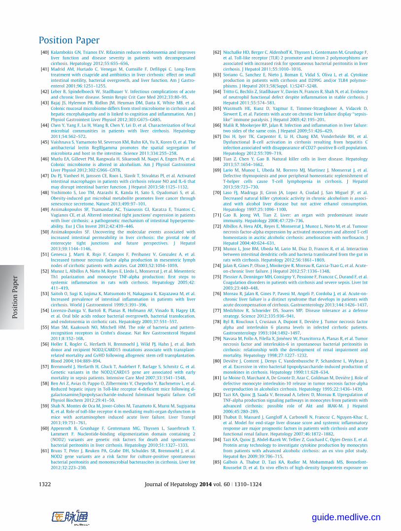
Fig. 3. Pathophysiological basis of acute on chronic liver failure and end-organ dysfunction in cirrhosis precipitated by infection.
JOURNAL OF HEPATOLOGY
Assessment (SOFA) score to recognize organ failures. The CANO-NIC study provided a robust definition of ACLF into three ACLFgrades, with increasing risk of short-term death from grade 1(22%) to grade 3 (77%) [76]. Table 2A and B describe the scoringsystem used and also the diagnostic criteria. Bacterial infectionwas the most common precipitating event of ACLF (33%) [76].Among patients with bacterial infection, ACLF was more commonin patients with SBP or pneumonia than in those with infectionsat other sites [9,76]. The pathophysiology and manifestations ofinfection-induced organ failure is incompletely understood [2];the following mechanisms have been suggested (Fig. 3).
Inflammation
Bacterial components (e.g., lipopolysaccharide (LPS) a Gram-neg-ative bacteria byproduct) may cause an excessive pro-inflamma-tory response of the host immune system resulting in tissuedamage (a process called immunopathology) and organ failure[77]. An excessive production of pro-inflammatory moleculeshas been shown to occur in vivo and ex vivo in patients [78–87]and animals [88–90] with cirrhosis. Among infected patients, sys-temic inflammation is more marked in patients with ACLF than inthose without [76]. The susceptibility to LPS-induced liver injury(assessed by the degree of hepatocyte apoptosis and necrosis) ishigher in animals with cirrhosis than in normal animals[89,90]. This higher susceptibility in cirrhosis decreases whenhigh-density lipoprotein administration is used to neutralizeLPS [90,91]. However, infection-induced organ failure may notbe entirely explained by an increased production of pro-inflam-matory molecules (see below).
Several mechanisms related to bacterial infections may leadto cell necrosis [77]. Dying cells are known to release nuclear
Journal of Hepatology 2014
molecules (called danger-associated molecular patterns, DAMPs),which are able to stimulate the innate immune system throughdifferent receptors and trigger inflammation [77]. The role ofDAMPs in ACLF-associated inflammation requires to beinvestigated.
Organ damage
Infection-induced tissue damage may depend not only on theintensity of the inflammatory response per se but also on theintrinsic capacity of host organs to tolerate (i.e., endure) theeffects of the inflammatory response [77]. The capacity of toler-ance of each organ depends on inducible mechanisms such asanti-apoptotic pathways, among others [77]. In the context ofGram-negative infections, normal livers are protected againstLPS-induced, TNF-a-mediated apoptosis because of simultaneousinduction of nuclear factor-jB (NF-jB)-dependent anti-apoptoticmolecules [89]. In contrast, cirrhotic livers are abnormally sus-ceptible to LPS-induced, TNF-a-mediated apoptosis because NF-jB-target anti-apoptotic molecules cannot be properly induced[89]. Therefore, in cirrhosis, infection-induced liver failure maybe related not only to an excessive pro-inflammatory responsebut also to a decrease in the hepatic capacity of tolerance. Therole of the alteration of tolerance mechanisms in thedevelopment of infection-induced extra-hepatic organ failuresshould be investigated in patients and animals with ACLF.
Kidney failure
Bacterial infections [79,92] are well-established triggers of kid-ney failure in cirrhosis. Patients with SBP without shock whoexhibit the highest pro-inflammatory response are those who
vol. 60 j 1310–1324 1315
guide.medlive.cn

Position Paper
are at risk of developing kidney failure [79]. Of note, in thesepatients, kidney failure frequently develops while resolution ofinfection has been obtained by antibiotic therapy [93] suggestingthat organ failure does not result from intrinsic virulence (i.e., tis-sue damage directly caused by bacteria) but rather extrinsic vir-ulence (i.e., caused by the excessive inflammatory response of thehost) or sepsis-related alterations in hemodynamics [93]. Apotential role for alterations of tolerance mechanisms in thedevelopment of kidney failure [94] has not yet been investigated.Brain failure
Bacterial infections are common triggers of hepatic encephalopa-thy [95]. Clinical and experimental data show that infections mayresult in brain oedema in patients with cirrhosis. It is currentlyuncertain whether this water accumulation is predominantlyintracellular or extracellular. There is some evidence that bothmechanisms combine to cause brain edema and hepatic enceph-alopathy (HE) [96].
Coagulation failure
Among infected patients with cirrhosis, disseminated intravascu-lar coagulation (DIC, which can be activated by pro-inflammatorycytokines) is more frequent in patients with ACLF than in thosewithout [75]. Thrombi in the microvasculature of a vital organmay play a role in tissue hypoxia [77]. Of note, activation of coag-ulation may stimulate inflammation [77].
Variceal bleeding
Variceal hemorrhage is a well-established risk factor for bacterialinfection in patients with cirrhosis [97]. Moreover, it has beensuggested that conversely bacterial infection might increase therisk of variceal hemorrhage [98]. This hypothesis needs to be con-firmed by future studies.
Adrenal insufficiency
Patients with cirrhosis and septic shock (most often classified asACLF grade 3 patients) may have high incidence of relative adre-nal insufficiency (RAI, 51–77%) [99]. The presence of RAI seems tobe associated with poor liver function, kidney failure, refractoryshock and hospital mortality [100]. RAI could result in decreasedcorticosteroid-related anti-inflammatory mechanisms and conse-quently unrestricted infection-induced production of pro-inflam-matory molecules. In addition, under stress conditions, defectivecorticosteroid production could be associated with decreasedcapacity of tolerance of vital organs [77,78].
Recent but limited data suggest that RAI can also occur innon-critically ill patients with cirrhosis. The reported prevalenceof this entity ranges between 7% and 49% depending on the meth-odology used for RAI diagnosis [99]. Since serum total cortisoloverestimates the prevalence of RAI in cirrhosis due to low trans-cortin and albumin concentrations, free cortisol levels have beensuggested as the preferred method for the diagnosis of RAI in thispopulation [101,102]. Delta total cortisol values, a dynamic diag-nostic criteria of RAI not affected by changes in transcortin oralbumin levels, can also be used for its diagnosis [103]. In a recentstudy involving non-critically ill patients with cirrhosis, RAI wasassociated with greater impairment of circulatory and renal func-
1316 Journal of Hepatology 2014
tion, higher probability of severe sepsis and type-1 HRS andhigher short-term mortality [103]. By contrast, another studyreported a higher risk of death in patients with high free cortisollevels [102].
Early diagnosis and biomarkers
Early diagnosis of bacterial infections is a crucial step in the man-agement of patients with cirrhosis. Since the presentation and theinitial course of a bacterial infection in some patients with cirrho-sis may be subtle and not very specific, clinical suspicion is impor-tant. Indeed, all hospitalized patients with cirrhosis should beconsidered as potentially infected until proven otherwise. There-fore, a complete work-up should be carried out at admission andon clinical deterioration of a hospitalized patient in order to detecta possible infection [1,4]. In addition, a close microbiological sur-veillance, is needed in patients who are at risk for the develop-ment of infections caused by MR organisms [1,22,104,105]. It iswell known that bacterial infections can induce systemic inflam-matory response (SIRS). On the basis of conventional criteria[106], SIRS has been described in 57–70% of infected patients withcirrhosis [81,107], but these data may underestimate the rate ofSIRS since these patients may have a low heart rate due to theuse of beta blockers and may present an apparently normal whiteblood cell count due to hypersplenism. On the other hand, SIRSmay be diagnosed in patients with cirrhosis in the absence of bac-terial infection since the hyperdynamic circulation, hepaticencephalopathy, tense ascites and hypersplenism may alter heartand respiratory rate, temperature and white-cell count. SIRS hasbeen described in 10–30% of patients with decompensated cirrho-sis without bacterial infection [83,107]. The evident lack of sensi-tivity and specificity of the conventional parameters for thedefinition of SIRS [105,106] makes it difficult to diagnose sepsisin these patients. Thus, new tools for the diagnosis of bacterialinfections are clearly needed. In this context, it should be men-tioned that a recent study reported that persistently high levelsof CRP in patients with decompensated cirrhosis are associatedwith increased short-term mortality. The predictive value of CRPwas independent from relevant predictive factors, such as MELDscore, and was better than that of SIRS diagnosed by conventionalcriteria [108]. Although not demonstrated, it is likely that the highlevels of CRP may indicate a systemic inflammatory reactionlinked to hidden bacterial infections and/or persistent bacterialtranslocation.
New tools for early detection of the presence and of the severity ofbacterial infections
From a pathophysiological perspective, pathological BT, alteredhost response to injury, and impairment in the ability of theinnate immune system to adequately fight off infections areimportant [1]. Therefore gut permeability, gut flora, bacterialproducts, acute phase proteins, the function of innate immunecells, cellular receptors and cellular products and moleculesinvolved in endotoxin presentation and removal are likely targetsfor such biomarkers.
Markers of gut barrier dysfunction/bacterial translocationIncreased intestinal permeability is predictive for infection aftervariceal haemorrhage [107] and calprotectin levels have been
vol. 60 j 1310–1324
guide.medlive.cn
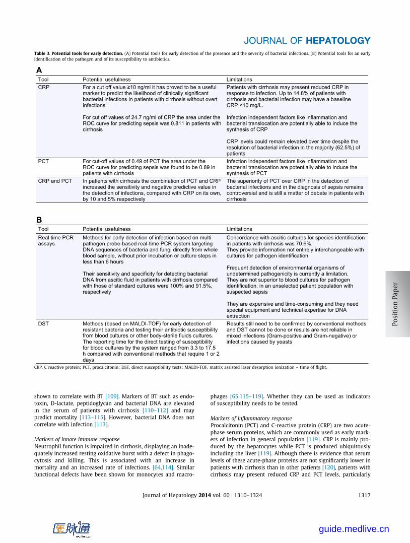
Table 3. Potential tools for early detection. (A) Potential tools for early detection of the presence and the severity of bacterial infections. (B) Potential tools for an earlyidentification of the pathogen and of its susceptibility to antibiotics.
Tool Potential usefulness LimitationsCRP For a cut off value ≥10 ng/ml it has proved to be a useful
marker to predict the likelihood of clinically significant bacterial infections in patients with cirrhosis without overt infections
For cut off values of 24.7 ng/ml of CRP the area under the ROC curve for predicting sepsis was 0.811 in patients with cirrhosis
Patients with cirrhosis may present reduced CRP in response to infection. Up to 14.8% of patients with cirrhosis and bacterial infection may have a baseline CRP <10 mg/L.
bacterial translocation are potentially able to induce the Infection independent factors like inflammation and
Infection independent factors like inflammation and
synthesis of CRP
CRP levels could remain elevated over time despite the resolution of bacterial infection in the majority (62.5%) of patients
PCT For cut-off values of 0.49 of PCT the area under the ROC curve for predicting sepsis was found to be 0.89 in patients with cirrhosis
bacterial translocation are potentially able to induce the synthesis of PCT
CRP and PCT In patients with cirrhosis the combination of PCT and CRP increased the sensitivity and negative predictive value in the detection of infections, compared with CRP on its own, by 10 and 5% respectively
The superiority of PCT over CRP in the detection of bacterial infections and in the diagnosis of sepsis remains controversial and is still a matter of debate in patients with cirrhosis
Tool Potential usefulness LimitationsReal time PCR assays
Methods for early detection of infection based on multi-pathogen probe-based real-time PCR system targeting DNA sequences of bacteria and fungi directly from whole blood sample, without prior incubation or culture steps in less than 6 hours
Their sensitivity and speciDNA from ascitic fluid in patients with cirrhosis compared
ficity for detecting bacterial
with those of standard cultures were 100% and 91.5%, respectively
Concordance with ascitic cultures for species identification in patients with cirrhosis was 70.6%.They provide information not entirely interchangeable with cultures for pathogen identification
Frequent detection of environmental organisms of undetermined pathogenicity is currently a limitation. They are not superior to blood cultures for pathogen identification, in an unselected patient population with suspected sepsis
They are expensive and time-consuming and they need special equipment and technical expertise for DNA extraction
DST Methods (based on MALDI-TOF) for early detection of resistant bacteria and testing their antibiotic susceptibility from blood cultures or other body-sterile fluids cultures.The reporting time for the direct testing of susceptibility for blood cultures by the system ranged from 3.3 to 17.5 h compared with conventional methods that require 1 or 2 days
Results still need to be confirmed by conventional methods and DST cannot be done or results are not reliable in mixed infections (Gram-positive and Gram-negative) or infections caused by yeasts
B
A
CRP, C reactive protein; PCT, procalcitonin; DST, direct susceptibility tests; MALDI-TOF, matrix assisted laser desorption ionization – time of flight.
JOURNAL OF HEPATOLOGY
shown to correlate with BT [109]. Markers of BT such as endo-toxin, D-lactate, peptidoglycan and bacterial DNA are elevatedin the serum of patients with cirrhosis [110–112] and maypredict mortality [113–115]. However, bacterial DNA does notcorrelate with infection [113].
Markers of innate immune responseNeutrophil function is impaired in cirrhosis, displaying an inade-quately increased resting oxidative burst with a defect in phago-cytosis and killing. This is associated with an increase inmortality and an increased rate of infections. [64,114]. Similarfunctional defects have been shown for monocytes and macro-
Journal of Hepatology 2014
phages [65,115–119]. Whether they can be used as indicatorsof susceptibility needs to be tested.
Markers of inflammatory responseProcalcitonin (PCT) and C-reactive protein (CRP) are two acute-phase serum proteins, which are commonly used as early mark-ers of infection in general population [119]. CRP is mainly pro-duced by the hepatocytes while PCT is produced ubiquitouslyincluding the liver [119]. Although there is evidence that serumlevels of these acute-phase proteins are not significantly lower inpatients with cirrhosis than in other patients [120], patients withcirrhosis may present reduced CRP and PCT levels, particularly
vol. 60 j 1310–1324 1317
guide.medlive.cn

Table 4. Recommended empirical antibiotic treatment for community-acquired and nosocomial bacterial infections in cirrhosis.
Type of infection Community-acquired infections Nosocomial infections*SBP, SBE and spontaneous bacteremia
Cefotaxime or ceftriaxone or amoxicillin/clavulanic acid
Piperacillin/tazobactamΔ
or meropenem§ ± glycopeptide#
Urinary infections Uncomplicated:
or cotrimoxazole ciprofloxacin
If sepsis: cefotaximeor ceftriaxone or amoxicillin/clavulanic acid
Uncomplicated:nitrofurantoin or fosfomycin
If sepsis: piperacillin/tazobactamΔ
or meropenem§ ± glycopeptide#
Pneumonia** Amoxicillin/clavulanic acid or ceftriaxone + macrolide
levofloxacinmoxifloxacin
oror
Piperacillin/tazobactammeropenem/ceftazidime + ciprofloxacin ±
Δ
orglycopeptide# should be added in patients with risk factors for MRSA¶
Cellulitis Amoxicillin/clavulanic acid or ceftriaxone + oxacillin
Meropenem/ceftazidime& + oxacillinor glycopeptides#
SBP, spontaneous bacterial peritonitis; SBE, spontaneous bacterial empyema; MRSA, methicillin-resistant Staphylococcus aureus.Dosages of antibiotics have not been formally investigated or defined in cirrhotic population and it is advisable to follow standard recommended dosages.⁄Recommended empirical treatment also for health-care associate (HCA) urinary infections and pneumonia. Empirical antibiotic treatment of HCA spontaneous infectionsand cellulitis will be decided on the basis of the severity of infection (patients with severe sepsis should receive the schedule proposed for nosocomial infections) and on thelocal prevalence of multiresistant bacteria in HCA infections.DIn areas with a low prevalence of multiresistant bacteria.§To cover extended-spectrum b-lactamase (ESBL)-producing Enterobacteriaceae.#IV vancomycin or teicoplanin in areas with a high prevalence MRSA and vancomycin-susceptible enterococci (VSE). Glycopeptides must be replaced by IV linezolid in areaswith a high prevalence of vancomycin-resistant enterococci (VRE).⁄⁄Liver disease is considered as severe comorbidity for community-acquired pneumonia in guidelines.&Antibiotics active against Pseudomonas aeruginosa.–Ventilator-associated pneumonia, previous antibiotic therapy, nasal MRSA carriage.
Position Paper
CRP, in response to infection [121]. Despite these potential limi-tations, the predictive power of CRP and PCT for detecting infec-tion has been found to be similar in patients with and withoutcirrhosis (Table 3A) [122–124]. However, as mentioned beforeit should be emphasised that CRP levels could remain elevatedover time despite the resolution of bacterial infection in a largepercent of patients with cirrhosis [108]. As far as the definitionof the severity of the infection, elevated serum levels of PCTand CRP are correlated with the presence, course, and outcomeof sepsis in patients with cirrhosis (Table 3A) [26] as in the gen-eral population [125]. Finally, randomized-controlled trials haveshown a benefit from the use of PCT algorithms to guide decisionsabout the initiation and/or discontinuation of antibiotic therapyin patients with some types of infections in the intensive careunit [126] but its usefulness in patients with cirrhosis has yetto be investigated.
The role of other acute phase proteins (lipopolysaccharidebinding protein, sCD14) in the early diagnosis as well as in thedefinition of infections in patients with cirrhosis is still unclear,even thought they are useful predictors of mortality in cirrhosis[127] mainly when they are used in combination with CRP andPCT [123]. Recently, mid region-proadrenomedullin has beenshown to provide potentially differential information in infectedpatients with cirrhosis compared to CRP [128].
New tools for early identification of the pathogenWith regard to the identification of the pathogen, real time PCRassays [129,130] were shown to have potential utility comparedwith standard culture techniques for the diagnosis of SBP inpatients with cirrhosis [131]. However, they provide informationnot entirely interchangeable with cultures for pathogen identifi-
1318 Journal of Hepatology 2014
cation [131] (Table 3B). In addition, these molecular assays areexpensive and time-consuming and they need special equipmentand technical expertise for DNA extraction. Considering all theselimitations they may not be suitable as a replacement of culturesfor routine use in clinical practice. Recently, the application of adirect susceptibility testing (DST) based on a Matrix AssistedLaser Desorption Ionization – Time of Flight (MALDI-TOF) frompositive blood cultures has been proposed for early detection ofresistant bacteria and their antibiotic susceptibility (Table 3B)[132,133].
Treatment of bacterial infections in cirrhosis
Antibiotic treatment
Early diagnosis and prompt initiation of adequate antibiotic ther-apy is essential in the management of patients with cirrhosis andbacterial infections [1,134] as delays and inappropriate therapy isassociated with increased mortality [135]. The choice of initialempirical antibiotics should be based on the type, severity andorigin of infection (community-acquired, nosocomial or healthcare-associated; HCA) and on the local epidemiological dataabout antibiotic resistance. In general, third-generation cephalo-sporins continue to be the gold-standard antibiotic treatment ofmany of the infections acquired in the community [136,137].By contrast, the empirical treatment of nosocomial and health-care associated infections should be tailored according to thelocal epidemiological pattern of MR bacteria (Table 4) [1,138].The failure of response to empirical antibiotics is due to inappro-priate choice of initial antibiotics, MR bacterial infections anddelayed start of appropriate antibiotics [135,138]. If the causative
vol. 60 j 1310–1324
guide.medlive.cn

Table 5. Current indications of antibiotic prophylaxis in cirrhosis.
Indication Antibiotic and dosePreserved liver function: norfloxacin 400 mg/12h PO for 7 daysGastrointestinal bleedingPatients with advanced cirrhosis (at least 2 of the following: ascites, jaundice, hepatic encephalopathy and malnutrition): IV ceftriaxone 1 g/d during 7 days
Primary prophylaxis of SBP in patients with low protein ascites (<15 g/L)
advanced cirrhosis:Norfloxacin 400 mg/d PO or ciprofloxacin 500 mg/d until liver transplantation or death in patients with
Norfloxacin 400 mg/d PO until liver transplantation, death, resolution of ascites or improvement in
- Child-Pugh score ≥9 points with serum bilirubin ≥3 mg/dland/or- Renal dysfunction (serum creatinine ≥1.2 mg/dl, BUN ≥25 mg/dl and/or serum sodium ≤130 mEq/L)
Secondary prophylaxis of SBPliver function to a compensated status
JOURNAL OF HEPATOLOGY
organism is identified (about 50% of cases), antibiotic regimenshould be narrowed to decrease the likelihood of emergence ofantibiotic resistance. Duration of antimicrobial treatment hasnot been formally investigated or defined in cirrhosis, exceptfor SBP with a minimum of 5 days [139]. In SBP, response to anti-microbials, arbitrarily defined by a P25% reduction of asciticpolymorphonuclear count, should be assessed by follow-up par-acentesis 48 h after initial diagnosis [138]. In the case of failureof response, initial antibiotics should be changed.
Intravenous albumin
In patients with cirrhosis and SBP without shock treated with cef-otaxime, an open-label randomized clinical trial (RCT) showedthat the IV administration of 20% albumin reduced the incidenceof renal failure and decreased mortality rates from 29% to 10%[93]. The mechanisms by which albumin improved hemodynam-ics could be related to its oncotic properties but also to the immu-nomodulation, antioxidant and endothelium stabilizationcapacity [140]. This effect was not observed in patients withlow risk of mortality (total bilirubin <4 mg/dl and creatinine<1 mg/dl) [141,142]. In a recent randomized study, the adminis-tration of albumin in unselected patients with cirrhosis and non-SBP infections was not associated with improved overall survivalbut albumin administration was an independent predictor of sur-vival after adjustment for other prognostic factors [143]. A largestudy is currently being planned.
Management of severe sepsis and septic shock
At this time, due to lack of data in cirrhosis, current guidelinesdefined in the general population should be followed [144].Although therapeutic goals for severe sepsis and septic shock inpatients with cirrhosis have not been defined, a prompt (withinthe 6 first hours) protocolized resuscitation of sepsis-inducedhypoperfusion with pre-defined targets (central venous pressure8–12 mmHg, urine output P0.5 ml.kg.hr and superior vena cavaor mixed venous saturation 70% or 65% respectively) and normal-ization of increased lactate levels is recommended. Studies spe-cifically investigating the ideal target level of mean arterialpressure in these patients have not been performed. However,it appears reasonable to state that arterial pressure should beincreased to a level close to the baseline of each patient, if known.If not known, it should be at least of 65 mmHg. The balancebetween fluid therapy (crystalloids and albumin) and vasopressoradministration in the hemodynamic support of cirrhotic patients
Journal of Hepatology 2014
is undefined. A strict monitoring of patients’ responsiveness tofluid replacement (i.e., pulse pressure variation and stroke vol-ume variation in sedated patients) is necessary to avoid fluidoverload, peripheral edema and abdominal compartmentsyndrome.
Current guidelines only recommend stress dose steroids inpatients with vasopressor-unresponsive septic shock in the gen-eral population [145]. Data in patients with cirrhosis is scantand controversial [146,147]. A large double-blind European RCTis currently underway to address this topic.
Prophylaxis of bacterial infections
Since most episodes of spontaneous bacterial infections in cirrho-sis are the result from the translocation of enteric gram-negativebacilli, prophylactic agents should be effective at decreasing theconcentration of these bacteria in the gut while preserving theprotective anaerobic flora. Norfloxacin, a poorly absorbable quin-olone that eliminates gram-negative bacilli selectively from theintestinal flora, has been used in the prophylaxis of bacterialinfections in cirrhosis. However, given the risk of developingresistant organisms, the use of prophylactic antibiotics must bestrictly restricted to patients at high risk of bacterial infectionssuch as patients with upper gastrointestinal bleeding, patientswith advanced cirrhosis and low total protein content in asciticfluid and patients with a previous history of SBP (Table 5)[135,136,138,148,149].
Gastrointestinal bleeding
Forty-five to 66% cirrhotic patients with upper gastrointestinalbleeding develop bacterial infection within the first 5–7 days ofthe bleeding episode [150,151]. The administration of oral or sys-temic antibiotics (penicillins, cephalosporins, and quinolones)decreases the incidence of bacterial infections to 10–20%, [152–154] improves control bleeding, prevents rebleeding andimproves survival [152]. Oral norfloxacin (400 mg/12 h for7 days) is the gold standard prophylaxis in patients with pre-served liver function. Nevertheless, patients with advanced cir-rhosis (at least two of the following: ascites, severemalnutrition, encephalopathy or jaundice) should receive IV cef-triaxone (1 g/day for 7 days) [1]. In a RCT, the probability ofdeveloping proven infection was significantly lower in patientsreceiving ceftriaxone iv than in those receiving norfloxacin peros (11% vs. 26%, p = 0.03) [152]. In patients with recent infectionwith extended-spectrum b-lactamase-producing Enterobacteria-
vol. 60 j 1310–1324 1319
guide.medlive.cn

Position Paper
ceae (3–6 months), antibiotics such as oral nitrofurantoin orertapenem should be used. Antibiotic prophylaxis should ideallybe given before or immediately after endoscopy [153].Primary prophylaxis in advanced cirrhosis
Patients with low protein ascites (10–15 g/L), liver failure (serumbilirubin >3.2 mg/dl) and low platelet count (<98,000 �mm3)have a high risk of developing the first episode of SBP[1,3,135,150,155]. A RCT evaluated the impact of primary pro-phylaxis with norfloxacin in cirrhotic patients at high risk ofdeveloping SBP and hepatorenal syndrome. Patients with lowprotein ascites (<15 g/L) and advanced liver failure (Child-Pughscore P9 points with serum bilirubin P3 mg/dl) or impairedrenal function (serum creatinine P1.2 mg/dl, BUN P25 mg/dlor serum sodium 6130 mEq/L) were randomized to receive nor-floxacin (400 mg/d for 1 year) or placebo. Norfloxacin reducedthe 1-year probability of developing SBP (7% vs. 61%) and hepato-renal syndrome (28% vs. 41%, p = 0.02) and improved 3-monthsurvival (94% vs. 62%) [154]. Long-term norfloxacin administra-tion is therefore indicated in this specific subpopulation ofpatients with advanced cirrhosis, particularly if they are awaitingliver transplantation. Oral ciprofloxacin 500 mg/d is an alterna-tive option to norfloxacin [1].
Secondary prophylaxis
Patients who recover from a previous episode of SBP are at a veryhigh risk of SBP recurrence in the absence of antibiotic prophy-laxis. Long-term norfloxacin administration (400 mg/d PO) iseffective in the prevention of SBP recurrence. Long-term norflox-acin prophylaxis was shown to decrease the overall probability ofSBP recurrence at 1 year from 68% in the placebo group to 20%and from 60% to 3% respectively if analysis was restricted toSBP caused by gram-negative bacilli [155]. After an episode ofSBP, liver transplantation must be considered [1].
Infections caused by multiresistant bacteria. The need for alternativestrategies
Prolonged antibiotic administration leads to the emergence ofresistant bacteria. Epidemiological studies have demonstratedthat long-term norfloxacin prophylaxis increases the risk of infec-tions caused by quinolone-resistant, trimethoprim-sulfamethox-azole-resistant and ESBL-producing strains in cirrhosis [4,20].Long-term norfloxacin prophylaxis increases 2.7 fold the risk ofdeveloping MR bacterial infections and almost 4 fold the risk ofinfections caused by ESBL-producing Enterobacteriaceae [3,4,20].
Rifaximin, an antibiotic with broad-spectrum antimicrobialactivity that eliminates intestinal flora non-selectively [156],has been suggested as a potential alternative to norfloxacin inthe prophylaxis of bacterial infections in cirrhosis. Its administra-tion in patients with hepatic encephalopathy is not associatedwith the development of infections by MR bacteria [157]. Threemain characteristics of rifaximin can explain this finding: (1) itreaches high fecal concentrations but is virtually non-absorbed(bioavailability in blood after oral administration <0.4%); (2) itreduces the expression of bacterial virulence factors and compro-mises plasmid transfer, an important mechanism of multiresis-tance; (3) despite high gut concentrations and its broadspectrum of activity, rifaximin produces minimal alterations in
1320 Journal of Hepatology 2014
the intestinal microflora (1 log reduction in intestinal coliformsper gram of stool after 2 weeks of treatment) [156–158]. Acase-control study has recently found a significant benefit for rif-aximin for prophylaxis of SBP when used in patients withencephalopathy [157]. Risk of Clostridium difficile was notincreased [157]. Despite these data, real efficacy and safety of rif-aximin in the prevention of spontaneous bacterial infections incirrhosis remains to be explored. Nevertheless, it should beemphasized that there are no studies comparing rifaximin vs.norfloxacin in the prevention of SBP.
Non-antibiotic strategies have been studied as a potentialalternative to quinolones in the prophylaxis of bacterial infec-tions in cirrhosis but evidence published so far is still limited[159–161,54,162]. Although these strategies seem to preventbacterial translocation and SBP in experimental models, none ofthem has been compared with norfloxacin in the prevention ofSBP in RCT in patients with cirrhosis.
Future research and conclusions
It has become clear that cirrhotic patients are susceptible to bac-terial infections due to a variety of possible pathogenic mecha-nisms as highlighted. In a recent study, the occurrence ofinfection in a cirrhotic patient was suggested to represent a crit-ical step in the progression of cirrhosis [6]. Gut dysbiosis,increased bacterial translocation and cirrhosis-associatedimmune dysfunction play important roles. The main researchefforts will be the generation of models and possible biomarkersto identify high-risk patients and the associated mechanisms toallow preventative strategies. The cirrhotic patient with superim-posed infection is more susceptible to its effects with increasedrisk of end-organ dysfunction and mortality. Future researchshould try to dissect the associated mechanisms and devise strat-egies to reduce this end-organ sensitivity while maintainingimmune competence. The diagnosis of infection in cirrhosis is dif-ficult because of the co-incident systemic inflammatory responsethat may exist due to the disease process itself rendering currentmarkers ineffective resulting in delayed diagnosis. Developmentof biomarkers that can be used early will result in a reductionin the morbidity and mortality. Infection with multi-drug resis-tant bacteria is increasing possibly due to reduced immune sur-veillance and inappropriate use of broad-spectrum antibiotics.Newer tools to detect the kind of infection will limit the use ofbroad-spectrum antibiotics and possibly reduce the incidence ofmulti-resistant bacterial infections. Better selection of patientsfor antibiotic prophylaxis and development of non-antibioticstrategies will be the key to improving the outcome of patients.Management of the acute episode of infection and use of albuminhas already yielded good results in spontaneous bacterial perito-nitis, and trials will be needed for other infections. It is clear thata concerted program of activity is necessary to address thesepending questions.
Conflict of interest
RJ received research funding from Vital Therapies, has served onScientific Advisory Board for Conatus Pharma, received lecturefees from Gambro, has on-going research collaboration withGambro, Grifols and is the PI of an Industry sponsored study
vol. 60 j 1310–1324
guide.medlive.cn

JOURNAL OF HEPATOLOGY
(Sequana Medical). He is also the inventor of a drug, L-ornithinephenylacetate, which UCL has licensed to Ocera Therapeutics.
PG has received a research grant and lecture fees from GrifolsInternational and a research grant from Sequana Medical AG.
Acknowledgement
This paper was developed following an EASL sponsored meetingon ‘Bacterial infections in Cirrhosis’ held in Barcelona on May23rd and 24th 2013.The authors would like to thank Nicki van Berckel, Hospital Clinic,University of Barcelona Medical School for her excellent supportand help with manuscript preparation and coordination betweenthe many authors.Some of the results reported in the paper were funded by publicgrants awarded to P.G. Fondo de Investigación Sanitaria (FIS PI08/0126 and FIS PI12/00330).
References
[1] Fernandez J, Gustot T. Management of bacterial infections in cirrhosis. JHepatol 2012;56:S1–S12.
[2] Gustot T, Durand F, Lebrec D, Vincent JL, Moreau R. Severe sepsis incirrhosis. Hepatology 2009;50:2022–2033.
[3] Fernandez J, Navasa M, Gomez J, Colmenero J, Vila J, Arroyo V, et al.Bacterial infections in cirrhosis: epidemiological changes with invasiveprocedures and norfloxacin prophylaxis. Hepatology 2002;35:140–148.
[4] Fernández J, Acevedo J, Castro M, Garcia O, de Lope CR, Roca D, et al.Prevalence and risk factors of infections by multiresistant bacteria incirrhosis: a prospective study. Hepatology 2012;55:1551–1561.
[5] Foreman MG, Mannino DM, Moss M. Cirrhosis as a risk factor for sepsis anddeath: analysis of the National Hospital Discharge Survey. Chest 2003;124:1016–1020.
[6] Arvaniti V, D’Amico G, Fede G, Manousou P, Tsochatzis E, Pleguezuelo M,et al. Infections in patients with cirrhosis increase mortality four-fold andshould be used in determining prognosis. Gastroenterology 2010;139:1246–1256.
[7] Magiorakos AP, Srinivasan A, Carey RB, Carmeli Y, Falagas ME, Giske CG,et al. Multidrug-resistant, extensively drug-resistant and pandrug-resistantbacteria: an international expert proposal for interim standard definitionsfor acquired resistance. Clin Microbiol Infect 2012;18:268–281.
[8] Woodford N, Turton JF, Livermore DM. Multiresistant Gram-negativebacteria: the role of high-risk clones in the dissemination of antibioticresistance. FEMS Microbiol Rev 2011;35:736–755.
[9] Merli M, Lucidi C, Giannelli V, Giusto M, Riggio O, Falcone M, et al. Cirrhoticpatients are at risk for health care-associated bacterial infections. ClinGastroenterol Hepatol 2010;8:979–985.
[10] Song JY, Jung SJ, Park CW, Sohn JW, Kim WJ, Kim MJ, et al. Prognosticsignificance of infection acquisition sites in spontaneous bacterial perito-nitis: nosocomial vs. community acquired. J Korean Med Sci 2006;21:666–671.
[11] Cereto F, Herranz X, Moreno E, Andreu A, Vergara M, Fontanals D, et al. Roleof host and bacterial virulence factors in Escherichia coli spontaneousbacterial peritonitis. Eur J Gastroenterol hepatol 2008;20:924–929.
[12] Angeloni S, Leboffe C, Parente A, Venditti M, Giordano A, Merli M, et al.Efficacy of current guidelines for the treatment of spontaneous bacterialperitonitis in the clinical practice. World J Gastroenterol2008;14:2757–2762.
[13] Cheong HS, Kang C, Lee JA, Moon SY, Joung MK, Chung DR, et al. Clinicalsignificance and outcome of nosocomial acquisition of spontaneousbacterial peritonitis in patients with liver cirrhosis. CID 2009;48:1230–1236.
[14] Song KH, Jeon JH, Park WB, Park SW, Kim HB, Oh MD, et al. Clinicaloutcomes of spontaneous bacterial peritonitis due to extended spectrumbeta-lactamase-producing Escherichia coli and Klebsiella species: a retro-spective matched case-control study. BMC Infect Dis 2009;9:41–46.
[15] Heo J, Seo YS, Yim HJ, Hahn T, Park SH, Ahn SH, et al. Clinical features andprognosis of spontaneous bacterial peritonitis in Korean patients with livercirrhosis: a multicenter retrospective study. Gut Liver 2009;3:197–204.
[16] Yakar T, Güçlü M, Serin E, Alis�kan H, Husamettin E. A recent evaluation ofempirical cephalosporin treatment and antibiotic resistance of changing
Journal of Hepatology 2014
bacterial profiles in spontaneous bacterial peritonitis. Dig Dis Sci2009;55:1149–1154.
[17] Piroth L, Pechinot A, Minello A, Jaulhac B, Patry I, Hadou T, et al. Bacterialepidemiology and antimicrobial resistance in ascitic fluid: a 2-yearretrospective study. Scand J Infect Dis 2009;37:2–8.
[18] Umgelter A, Reindl W, Miedaner M, Schmid RM, Huber W. Failure ofcurrent antibiotic first-line regimens and mortality in hospitalized patientswith spontaneous bacterial peritonitis. Infection 2009;37:2–8.
[19] Ariza X, Castellote J, Lora-Tamayo J, Girbau A, Salord S, Rota R, et al. Riskfactors for resistance to ceftriaxone and its impact on mortality incommunity, healthcare and nosocomial spontaneous bacterial peritonitis.J Hepatol 2012;56:825–832.
[20] Tandon P, Delisle A, Topal JE, Garcia-Tsao G. High prevalence of antibiotic-resistant bacterial infections among patients with cirrhosis at a US livercenter. Clin Gastroenterol Hepatol 2012;10:1291–1298.
[21] Acevedo J, Silva A, Prado V, Fernandez J. The new epidemiology ofnosocomial bacterial infections in cirrhosis: therapeutical implications.Hepatol Int 2013;7:72–79.
[22] Piano S, Romano A, Rosi S, Gatta A, Angeli P. Spontaneous bacterialperitonitis due to carbapenemase-producing Klebsiella pneumoniae: thelast therapeutic challenge. Eur J Gastroenterol Hepatol 2012;24:1234–1237.
[23] Campillo B, Richardet JP, Kheo T, Dupeyron C. Nosocomial spontaneousbacterial peritonitis and bacteremia in cirrhotic patients: impact of isolatetype on prognosis and characteristics of infection. CID 2002;35:1–10.
[24] Bert F, Andreu M, Durand F, Degos F, Galdbart JO, Moreau R, et al.Nosocomial and community-acquired spontaneous bacterial peritonitis:comparative microbiology and therapeutic implications. Eur J Clin Micro-biol Infect Dis 2003;22:10–15.
[25] Berg RD, Garlington AW. Translocation of certain indigenous bacteria fromthe gastrointestinal tract to the mesenteric lymph nodes and other organsin a gnotobiotic mouse model. Infect Immun 1979;23:403–411.
[26] Hooper LV, Littman DR, Macpherson AJ. Interactions between the micro-biota and the immune system. Science 2012;336:1268–1273.
[27] Littman DR, Pamer EG. Role of the commensal microbiota in normaland pathogenic host immune responses. Cell Host Microbe 2011;20:311–323.
[28] Bauer TM, Steinbruckner B, Brinkmann FE, Ditzen AK, Schwacha H, AponteJJ, et al. Small intestinal bacterial overgrowth in patients with cirrhosis:prevalence and relation with spontaneous bacterial peritonitis. Am JGastroenterol 2001;96:2962–2967.
[29] Morencos FC, las Heras CG, Martin RL, Lopez Arias MJ, Ledesma F, Pons RF.Small bowel bacterial overgrowth in patients with alcoholic cirrhosis. DigDis Sci 1995;40:1252–1256.
[30] Pande C, Kumar A, Sarin SK. Small-intestinal bacterial overgrowth incirrhosis is related to the severity of liver disease. Aliment Pharmacol Ther2009;29:1273–1281.
[31] Chang CS, Chen GH, Lien HC, Yeh HZ. Small intestine dysmotility andbacterial overgrowth in cirrhotic patients with spontaneous bacterialperitonitis. Hepatology 1998;28:1187–1190.
[32] Zhao G, Downes M, Yu RT, Shelton JM, Richardson JA, Repa JJ, et al.Regulation of antibacterial defense in the small intestine by the nuclear bileacid receptor. Proc Natl Acad Sci U S A 2006;103:3920–3925.
[33] Perez-Paramo M, Munoz J, Albillos A, Freile I, Portero F, Santos M, et al.Effect of propranolol on the factors promoting bacterial translocation incirrhotic rats with ascites. Hepatology 2000;31:43–48.
[34] Shindo M, Ken A, Okuno T. Varying incidence of cirrhosis and hepatocel-lular carcinoma in patients with chronic hepatitis C responding differentlyto interferon therapy. Cancer 1999;85:1943–1950.
[35] Teltschik Z, Wiest R, Beisner J, Nuding S, Hofmann C, Schoelmerich J, et al.Intestinal bacterial translocation in rats with cirrhosis is related tocompromised Paneth cell antimicrobial host defense. Hepatology2012;55:1154–1163.
[36] Yan AW, Fouts DE, Brandl J, Starkel P, Torralba M, Schott E, et al. Entericdysbiosis associated with a mouse model of alcoholic liver disease.Hepatology 2011;53:96–105.
[37] Lichtman SN, Sartor RB, Keku J, Schwab JH. Hepatic inflammation in ratswith experimental small intestinal bacterial overgrowth. Gastroenterology1990;98:414–423.
[38] Seki E, De Minicis S, Osterreicher CH, Kluwe J, Osawa Y, Brenner DA, et al.TLR4 enhances TGF-beta signaling and hepatic fibrosis. Nat Med2007;13:1324–1332.
[39] Adachi Y, Bradford BU, Gao W, Bojes HK, Thurman RG. Inactivation ofKupffer cells prevents early alcohol-induced liver injury. Hepatology1994;20:453–460.
vol. 60 j 1310–1324 1321
guide.medlive.cn
Position Paper
[40] Kalambokis GN, Tsianos EV. Rifaximin reduces endotoxemia and improvesliver function and disease severity in patients with decompensatedcirrhosis. Hepatology 2012;55:655–656.
[41] Madrid AM, Hurtado C, Venegas M, Cumsille F, Defilippi C. Long-Termtreatment with cisapride and antibiotics in liver cirrhosis: effect on smallintestinal motility, bacterial overgrowth, and liver function. Am J Gastro-enterol 2001;96:1251–1255.
[42] Leber B, Spindelboeck W, Stadlbauer V. Infectious complications of acuteand chronic liver disease. Semin Respir Crit Care Med 2012;33:80–95.
[43] Bajaj JS, Hylemon PB, Ridlon JM, Heuman DM, Daita K, White MB, et al.Colonic mucosal microbiome differs from stool microbiome in cirrhosis andhepatic encephalopathy and is linked to cognition and inflammation. Am JPhysiol Gastrointest Liver Physiol 2012;303:G675–G685.
[44] Chen Y, Yang F, Lu H, Wang B, Chen Y, Lei D, et al. Characterization of fecalmicrobial communities in patients with liver cirrhosis. Hepatology2011;54:562–572.
[45] Vaishnava S, Yamamoto M, Severson KM, Ruhn KA, Yu X, Koren O, et al. Theantibacterial lectin RegIIIgamma promotes the spatial segregation ofmicrobiota and host in the intestine. Science 2011;334:255–258.
[46] Mutlu EA, Gillevet PM, Rangwala H, Sikaroodi M, Naqvi A, Engen PA, et al.Colonic microbiome is altered in alcoholism. Am J Physiol GastrointestLiver Physiol 2012;302:G966–G978.
[47] Du PJ, Vanheel H, Janssen CE, Roos L, Slavik T, Stivaktas PI, et al. Activatedintestinal macrophages in patients with cirrhosis release NO and IL-6 thatmay disrupt intestinal barrier function. J Hepatol 2013;58:1125–1132.
[48] Yoshimoto S, Loo TM, Atarashi K, Kanda H, Sato S, Oyadomari S, et al.Obesity-induced gut microbial metabolite promotes liver cancer throughsenescence secretome. Nature 2013;499:97–101.
[49] Assimakopoulos SF, Tsamandas AC, Tsiaoussis GI, Karatza E, Triantos C,Vagianos CE, et al. Altered intestinal tight junctions’ expression in patientswith liver cirrhosis: a pathogenetic mechanism of intestinal hyperperme-ability. Eur J Clin Invest 2012;42:439–446.
[50] Assimakopoulos SF. Uncovering the molecular events associated withincreased intestinal permeability in liver cirrhosis: the pivotal role ofenterocyte tight junctions and future perspectives. J Hepatol2013;59:1144–1146.
[51] Genesca J, Marti R, Rojo F, Campos F, Peribanez V, Gonzalez A, et al.Increased tumour necrosis factor alpha production in mesenteric lymphnodes of cirrhotic patients with ascites. Gut 2003;52:1054–1059.
[52] Munoz L, Albillos A, Nieto M, Reyes E, Lledo L, Monserrat J, et al. MesentericTh1 polarization and monocyte TNF-alpha production: first steps tosystemic inflammation in rats with cirrhosis. Hepatology 2005;42:411–419.
[53] Saitoh O, Sugi K, Lojima K, Matsumoto H, Nakagawa K, Kayazawa M, et al.Increased prevalence of intestinal inflammation in patients with livercirrhosis. World J Gastroenterol 1999;5:391–396.
[54] Lorenzo-Zuniga V, Bartoli R, Planas R, Hofmann AF, Vinado B, Hagey LR,et al. Oral bile acids reduce bacterial overgrowth, bacterial translocation,and endotoxemia in cirrhotic rats. Hepatology 2003;37:551–557.
[55] Man SM, Kaakoush NO, Mitchell HM. The role of bacteria and pattern-recognition receptors in Crohn’s disease. Nat Rev Gastroenterol Hepatol2011;8:152–168.
[56] Holler E, Rogler G, Herfarth H, Brenmoehl J, Wild PJ, Hahn J, et al. Bothdonor and recipient NOD2/CARD15 mutations associate with transplant-related mortality and GvHD following allogeneic stem cell transplantation.Blood 2004;104:889–894.
[57] Brenmoehl J, Herfarth H, Gluck T, Audebert F, Barlage S, Schmitz G, et al.Genetic variants in the NOD2/CARD15 gene are associated with earlymortality in sepsis patients. Intensive Care Med 2007;33:1541–1548.
[58] Ben Ari Z, Avias O, Pappo O, Zilbermints V, Cheporko Y, Bachmetov L, et al.Reduced hepatic injury in Toll-like receptor 4-deficient mice following d-galactosamine/lipopolysaccharide-induced fulminant hepatic failure. CellPhysiol Biochem 2012;29:41–50.
[59] Shah N, Montes de Oca M, Jover-Cobos M, Tanamoto K, Muroi M, SugiyamaK, et al. Role of toll-like receptor 4 in mediating multi-organ dysfunction inmice with acetaminophen induced acute liver failure. Liver Transpl2013;19:751–761.
[60] Appenrodt B, Grunhage F, Gentemann MG, Thyssen L, Sauerbruch T,Lammert F. Nucleotide-binding oligomerization domain containing 2(NOD2) variants are genetic risk factors for death and spontaneousbacterial peritonitis in liver cirrhosis. Hepatology 2010;51:1327–1333.
[61] Bruns T, Peter J, Reuken PA, Grabe DH, Schuldes SR, Brenmoehl J, et al.NOD2 gene variants are a risk factor for culture-positive spontaneousbacterial peritonitis and monomicrobial bacterascites in cirrhosis. Liver Int2012;32:223–230.
1322 Journal of Hepatology 2014
[62] Nischalke HD, Berger C, Aldenhoff K, Thyssen L, Gentemann M, Grunhage F,et al. Toll-like receptor (TLR) 2 promoter and intron 2 polymorphisms areassociated with increased risk for spontaneous bacterial peritonitis in livercirrhosis. J Hepatol 2011;55:1010–1016.
[63] Soriano G, Sanchez E, Nieto J, Roman E, Vidal S, Oliva L, et al. Cytokineproduction in patients with cirrhosis and D299G and/or TLR4 polymor-phisms. J Hepatol 2013;58(Suppl. 1):S247–S248.
[64] Tritto G, Bechlis Z, Stadlbauer V, Davies N, Frances R, Shah N, et al. Evidenceof neutrophil functional defect despite inflammation in stable cirrhosis. JHepatol 2011;55:574–581.
[65] Wasmuth HE, Kunz D, Yagmur E, Timmer-Stranghoner A, Vidacek D,Siewert E, et al. Patients with acute on chronic liver failure display ‘‘sepsis-like’’ immune paralysis. J Hepatol 2005;42:195–201.
[66] Malik R, Mookerjee RP, Jalan R. Infection and inflammation in liver failure:two sides of the same coin. J Hepatol 2009;51:426–429.
[67] Doi H, Iyer TK, Carpenter E, Li H, Chang KM, Vonderheide RH, et al.Dysfunctional B-cell activation in cirrhosis resulting from hepatitis Cinfection associated with disappearance of CD27-positive B-cell population.Hepatology 2012;55:709–719.
[68] Tian Z, Chen Y, Gao B. Natural killer cells in liver disease. Hepatology2013;57:1654–1662.
[69] Lario M, Munoz L, Ubeda M, Borrero MJ, Martinez J, Monserrat J, et al.Defective thymopoiesis and poor peripheral homeostatic replenishment ofT-helper cells cause T-cell lymphopenia in cirrhosis. J Hepatol2013;59:723–730.
[70] Laso FJ, Madruga JI, Giron JA, Lopez A, Ciudad J, San Miguel JF, et al.Decreased natural killer cytotoxic activity in chronic alcoholism is associ-ated with alcohol liver disease but not active ethanol consumption.Hepatology 1997;25:1096–1100.
[71] Gao B, Jeong WI, Tian Z. Liver: an organ with predominant innateimmunity. Hepatology 2008;47:729–736.
[72] Albillos A, Hera ADL, Reyes E, Monserrat J, Munoz L, Nieto M, et al. Tumournecrosis factor-alpha expression by activated monocytes and altered T-cellhomeostasis in ascitic alcoholic cirrhosis: amelioration with norfloxacin. JHepatol 2004;40:624–631.
[73] Munoz L, Jose BM, Ubeda M, Lario M, Diaz D, Frances R, et al. Interactionbetween intestinal dendritic cells and bacteria translocated from the gut inrats with cirrhosis. Hepatology 2012;56:1861–1869.
[74] Jalan R, Gines P, Olson J, Mookerjee R, Moreau R, Garcia-Tsao G, et al. Acute-on-chronic liver failure. J Hepatol 2012;57:1336–1348.
[75] Plessier A, Denninger MH, Consigny Y, Pessione F, Francoz C, Durand F, et al.Coagulation disorders in patients with cirrhosis and severe sepsis. Liver Int2003;23:440–448.
[76] Moreau R, Jalan R, Gines P, Pavesi M, Angeli P, Cordoba J, et al. Acute-on-chronic liver failure is a distinct syndrome that develops in patients withacute decompensation of cirrhosis. Gastroenterology 2013;144:1426–1437.
[77] Medzhitov R, Schneider DS, Soares MP. Disease tolerance as a defensestrategy. Science 2012;335:936–941.
[78] Byl B, Roucloux I, Crusiaux A, Dupont E, Devière J. Tumor necrosis factoralpha and interleukin 6 plasma levels in infected cirrhotic patients.Gastroenterology 1993;104:1492–1497.
[79] Navasa M, Follo A, Filella X, Jiménez W, Francitorra A, Planas R, et al. Tumornecrosis factor and interleukin-6 in spontaneous bacterial peritonitis incirrhosis: relationship with the development of renal impairment andmortality. Hepatology 1998;27:1227–1232.
[80] Devière J, Content J, Denys C, Vandenbussche P, Schandene L, Wybran J,et al. Excessive in vitro bacterial lipopolysaccharide-induced production ofmonokines in cirrhosis. Hepatology 1990;11:628–634.
[81] Le Moine O, Marchant A, De Groote D, Azar C, Goldman M, Devière J. Role ofdefective monocyte interleukin-10 release in tumor necrosis factor-alphaoverproduction in alcoholics cirrhosis. Hepatology 1995;22:1436–1439.
[82] Tazi KA, Quioc JJ, Saada V, Bezeaud A, Lebrec D, Moreau R. Upregulation ofTNF-alpha production signaling pathways in monocytes from patients withadvanced cirrhosis: possible role of Akt and IRAK-M. J Hepatol2006;45:280–289.
[83] Thabut D, Massard J, Gangloff A, Carbonell N, Francoz C, Nguyen-Khac E,et al. Model for end-stage liver disease score and systemic inflammatoryresponse are major prognostic factors in patients with cirrhosis and acutefunctional renal failure. Hepatology 2007;46:1872–1882.
[84] Tazi KA, Quioc JJ, Abdel-Razek W, Tellier Z, Guichard C, Ogier-Denis E, et al.Protein array technology to investigate cytokine production by monocytesfrom patients with advanced alcoholic cirrhosis: an ex vivo pilot study.Hepatol Res 2009;39:706–715.
[85] Galbois A, Thabut D, Tazi KA, Rudler M, Mohammadi MS, Bonnefont-Rousselot D, et al. Ex vivo effects of high-density lipoprotein exposure on
vol. 60 j 1310–1324
guide.medlive.cn
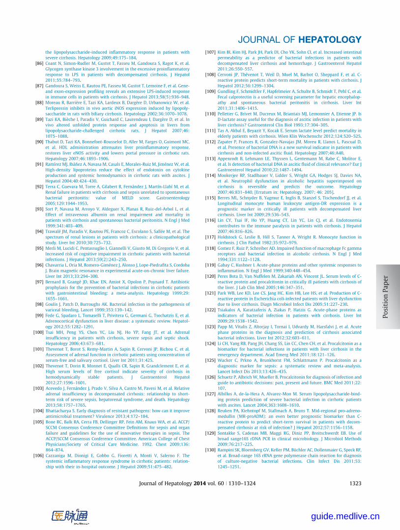
JOURNAL OF HEPATOLOGY
the lipopolysaccharide-induced inflammatory response in patients withsevere cirrhosis. Hepatology 2009;49:175–184.[86] Coant N, Simon-Rudler M, Gustot T, Fasseu M, Gandoura S, Ragot K, et al.Glycogen synthase kinase 3 involvement in the excessive proinflammatoryresponse to LPS in patients with decompensated cirrhosis. J Hepatol2011;55:784–793.
[87] Gandoura S, Weiss E, Rautou PE, Fasseu M, Gustot T, Lemoine F, et al. Gene-and exon-expression profiling reveals an extensive LPS-induced responsein immune cells in patients with cirrhosis. J Hepatol 2013;58(5):936–948.
[88] Moreau R, Barrière E, Tazi KA, Lardeux B, Dargère D, Urbanowicz W, et al.Terlipressin inhibits in vivo aortic iNOS expression induced by lipopoly-saccharide in rats with biliary cirrhosis. Hepatology 2002;36:1070–1078.
[89] Tazi KA, Bièche I, Paradis V, Guichard C, Laurendeau I, Dargère D, et al. Invivo altered unfolded protein response and apoptosis in livers fromlipopolysaccharide-challenged cirrhotic rats. J Hepatol 2007;46:1075–1088.
[90] Thabut D, Tazi KA, Bonnefont-Rousselot D, Aller M, Farges O, Guimont MC,et al. HDL administration attenuates liver proinflammatory response,restores liver eNOS activity and lowers portal pressure in cirrhotic rats.Hepatology 2007;46:1893–1906.
[91] Ramírez MJ, Ibáñez A, Navasa M, Casals E, Morales-Ruiz M, Jiménez W, et al.High-density lipoproteins reduce the effect of endotoxin on cytokineproduction and systemic hemodynamics in cirrhotic rats with ascites. JHepatol 2004;40:424–430.
[92] Terra C, Guevara M, Torre A, Gilabert R, Fernández J, Martín-Llahí M, et al.Renal failure in patients with cirrhosis and sepsis unrelated to spontaneousbacterial peritonitis: value of MELD score. Gastroenterology2005;129:1944–1953.
[93] Sort P, Navasa M, Arroyo V, Aldeguer X, Planas R, Ruiz-del-Arbol L, et al.Effect of intravenous albumin on renal impairment and mortality inpatients with cirrhosis and spontaneous bacterial peritonitis. N Engl J Med1999;341:403–409.
[94] Trawalé JM, Paradis V, Rautou PE, Francoz C, Escolano S, Sallée M, et al. Thespectrum of renal lesions in patients with cirrhosis: a clinicopathologicalstudy. Liver Int 2010;30:725–732.
[95] Merli M, Lucidi C, Pentassuglio I, Giannelli V, Giusto M, Di Gregorio V, et al.Increased risk of cognitive impairment in cirrhotic patients with bacterialinfections. J Hepatol 2013;59(2):243–250.
[96] Chavarria L, Oria M, Romero-Giménez J, Alonso J, Lope-Piedrafita S, CordobaJ. Brain magnetic resonance in experimental acute-on-chronic liver failure.Liver Int 2013;33:294–300.
[97] Bernard B, Grangé JD, Khac EN, Amiot X, Opolon P, Poynard T. Antibioticprophylaxis for the prevention of bacterial infections in cirrhotic patientswith gastrointestinal bleeding: a meta-analysis. Hepatology 1999;29:1655–1661.
[98] Goulis J, Patch D, Burroughs AK. Bacterial infection in the pathogenesis ofvariceal bleeding. Lancet 1999;353:139–142.
[99] Fede G, Spadaro L, Tomaselli T, Privitera G, Germani G, Tsochatzis E, et al.Adrenocortical dysfunction in liver disease: a systematic review. Hepatol-ogy 2012;55:1282–1291.
[100] Tsai MH, Peng YS, Chen YC, Liu NJ, Ho YP, Fang JT, et al. Adrenalinsufficiency in patients with cirrhosis, severe sepsis and septic shock.Hepatology 2006;43:673–681.
[101] Thevenot T, Borot S, Remy-Martin A, Sapin R, Cervoni JP, Richou C, et al.Assessment of adrenal function in cirrhotic patients using concentration ofserum-free and salivary cortisol. Liver Int 2011;31:425.
[102] Thevenot T, Dorin R, Monnet E, Qualls CR, Sapin R, Grandclement E, et al.High serum levels of free cortisol indicate severity of cirrhosis inhemodynamically stable patients. J Gastroenterol Hepatol2012;27:1596–1601.
[103] Acevedo J, Fernández J, Prado V, Silva A, Castro M, Pavesi M, et al. Relativeadrenal insufficiency in decompensated cirrhosis: relationship to short-term risk of severe sepsis, hepatorenal syndrome, and death. Hepatology2013;58:1757–1765.
[104] Bhattacharya S. Early diagnosis of resistant pathogens: how can it improveantimicrobial treatment? Virulence 2013;4:172–184.
[105] Bone RC, Balk RA, Cerra FB, Dellinger RP, Fein AM, Knaus WA, et al. ACCP/SCCM Consensus Conference Committee Definitions for sepsis and organfailure and guidelines for the use of innovative therapies in sepsis. TheACCP/SCCM Consensus Conference Committee. American College of ChestPhysicians/Society of Critical Care Medicine. 1992. Chest 2009;136:864–874.
[106] Cazzaniga M, Dionigi E, Gobbo G, Fioretti A, Monti V, Salerno F. Thesystemic inflammatory response syndrome in cirrhotic patients: relation-ship with their in-hospital outcome. J Hepatol 2009;51:475–482.
Journal of Hepatology 2014
[107] Kim BI, Kim HJ, Park JH, Park DI, Cho YK, Sohn CI, et al. Increased intestinalpermeability as a predictor of bacterial infections in patients withdecompensated liver cirrhosis and hemorrhage. J Gastroenterol Hepatol2011;26:550–557.
[108] Cervoni JP, Thévenot T, Weil D, Muel M, Barbot O, Sheppard F, et al. C-reactive protein predicts short-term mortality in patients with cirrhosis. JHepatol 2012;56:1299–1304.
[109] Gundling F, Schmidtler F, Hapfelmeier A, Schulte B, Schmidt T, Pehl C, et al.Fecal calprotectin is a useful screening parameter for hepatic encephalop-athy and spontaneous bacterial peritonitis in cirrhosis. Liver Int2011;31:1406–1415.
[110] Pelletier G, Brivet M, Ducreux M, Briantais MJ, Lemonnier A, Etienne JP. IsD-lactate assay useful for the diagnosis of ascitic infection in patients withliver cirrhosis? Gastroenterol Clin Biol 1993;17:304–305.
[111] Tas A, Akbal E, Beyazit Y, Kocak E. Serum lactate level predict mortality inelderly patients with cirrhosis. Wien Klin Wochenschr 2012;124:520–525.
[112] Zapater P, Frances R, Gonzalez-Navajas JM, Moreu R, Llanos L, Pascual D,et al. Presence of bacterial DNA is a new survival indicator in patients withcirrhosis and non-infected ascitic fluid. Hepatology 2007;46:A40.
[113] Appenrodt B, Lehmann LE, Thyssen L, Gentemann M, Rabe C, Molitor E,et al. Is detection of bacterial DNA in ascitic fluid of clinical relevance? Eur JGastroenterol Hepatol 2010;22:1487–1494.
[114] Mookerjee RP, Stadlbauer V, Lidder S, Wright GA, Hodges SJ, Davies NA,et al. Neutrophil dysfunction in alcoholic hepatitis superimposed oncirrhosis is reversible and predicts the outcome. Hepatology2007;46:831–840, [Erratum in: Hepatology. 2007; 46: 205].
[115] Berres ML, Schnyder B, Yagmur E, Inglis B, Stanzel S, Tischendorf JJ, et al.Longitudinal monocyte human leukocyte antigen-DR expression is aprognostic marker in critically ill patients with decompensated livercirrhosis. Liver Int 2009;29:536–543.
[116] Lin CY, Tsai IF, Ho YP, Huang CT, Lin YC, Lin CJ, et al. Endotoxemiacontributes to the immune paralysis in patients with cirrhosis. J Hepatol2007;46:816–826.
[117] Holdstock G, Leslie B, Hill S, Tanner A, Wright R. Monocyte function incirrhosis. J Clin Pathol 1982;35:972–979.
[118] Gomez F, Ruiz P, Schreiber AD. Impaired function of macrophage Fc gammareceptors and bacterial infection in alcoholic cirrhosis. N Engl J Med1994;331:1122–1128.
[119] Gabay C, Kushner I. Acute-phase proteins and other systemic responses toinflammation. N Engl J Med 1999;340:448–454.
[120] Peres Bota D, Van Nuffelen M, Zakariah AN, Vincent JL. Serum levels of C-reactive protein and procalcitonin in critically ill patients with cirrhosis ofthe liver. J Lab Clin Med 2005;146:347–351.
[121] Park WB, Lee KD, Lee CS, Jang HC, Kim HB, Lee HS, et al. Production of C-reactive protein in Escherichia coli-infected patients with liver dysfunctiondue to liver cirrhosis. Diagn Microbiol Infect Dis 2005;51:227–230.
[122] Tsiakalos A, Karatzaferis A, Ziakas P, Hatzis G. Acute-phase proteins asindicators of bacterial infection in patients with cirrhosis. Liver Int2009;29:1538–1542.
[123] Papp M, Vitalis Z, Altorjay I, Tornai I, Udvardy M, Harsfalvi J, et al. Acutephase proteins in the diagnosis and prediction of cirrhosis associatedbacterial infections. Liver Int 2012;32:603–611.
[124] Li CH, Yang RB, Pang JH, Chang SS, Lin CC, Chen CH, et al. Procalcitonin as abiomarker for bacterial infections in patients with liver cirrhosis in theemergency department. Acad Emerg Med 2011;18:121–126.
[125] Wacker C, Prkno A, Brunkhorst FM, Schlattmann P. Procalcitonin as adiagnostic marker for sepsis: a systematic review and meta-analysis.Lancet Infect Dis 2013;13:426–435.
[126] Schuetz P, Albrich W, Mueller B. Procalcitonin for diagnosis of infection andguide to antibiotic decisions: past, present and future. BMC Med 2011;22:107.
[127] Albillos A, de-la-Hera A, Alvarez-Mon M. Serum lipopolysaccharide-bind-ing protein prediction of severe bacterial infection in cirrhotic patientswith ascites. Lancet 2004;363:1608–1610.
[128] Reuken PA, Kiehntopf M, Stallmach A, Bruns T. Mid-regional pro-adreno-medullin (MR-proADM): an even better prognostic biomarker than C-reactive protein to predict short-term survival in patients with decom-pensated cirrhosis at risk of infection? J Hepatol 2012;57:1156–1158.
[129] Sontakke S, Cadenas MB, Maggi RG, Diniz PP, Breitschwerdt EB. Use ofbroad range16S rDNA PCR in clinical microbiology. J Microbiol Methods2009;76:217–225.
[130] Rampini SK, Bloemberg GV, Keller PM, Büchler AC, Dollenmaier G, Speck RF,et al. Broad-range 16S rRNA gene polymerase chain reaction for diagnosisof culture-negative bacterial infections. Clin Infect Dis 2011;53:1245–1251.
vol. 60 j 1310–1324 1323
guide.medlive.cn

Position Paper
[131] Hardick J, Won H, Jeng K, Hsieh YH, Gaydos CA, Rothman RE, et al.Identification of bacterial pathogens in ascitic fluids from patients withsuspected spontaneous bacterial peritonitis by use of broad-range PCR [16SPCR] coupled with high-resolution melt analysis. J Clin Microbiol2012;50:2428–2432.
[132] Pasqualini L, Mencacci A, Leli C, Montagna P, Cardaccia A, Cenci E, et al.Diagnostic performance of a multiple real-time PCR assay in patients withsuspected sepsis hospitalized in an internal medicine ward. J Clin Microbiol2012;50:1285–1288.
[133] Espinal P, Seifert H, Dijkshoorn L, Vila J, Roca I. Rapid and accurateidentification of genomic species from the Acinetobacter baumannii [Ab]group by MALDI-TOF MS. Clin Microbiol Infect 2012;18:1097–1103.
[134] Arabi YM, Dara SI, Memish Z, Abdulmajeed AA, Tamim HM, Al-Shirawi N,et al. Antimicrobial therapeutic determinants of outcomes from septicshock among patients with cirrhosis. Hepatology 2012;56:2305–2315.
[135] Ginès P, Angeli P, Lenz K, MØller S, Moore K, Moreau R, et al. EASL clinicalpractice guidelines on the management of ascites, spontaneous bacterialperitonitis, and hepatorenal syndrome in cirrhosis. J Hepatol 2010;53:397–417.
[136] Runyon BA. AASLD Practice Guidelines Committee. Management of adultpatients with ascites due to cirrhosis: update 2012. Hepatology2013;57:1651–1653, [http://www.aasld.org/practiceguidelines/Docu-ments/ascitesupdate2013.pdf].
[137] Fernandez J, Arroyo V. Bacterial infections in cirrhosis – a growing problemwith significant implications. Clin Liver Dis 2013;2:102–105.
[138] Rimola A, Garcia-Tsao G, Navasa M, Piddock LJ, Planas R, Bernard B, et al.Diagnosis, treatment and prophylaxis of spontaneous bacterial peritonitis:a consensus document. International ascites club. J Hepatol 2000;32:142–153.
[139] Runyon BA, McHutchison JG, Antillon MR, Akriviadis EA, Montano AA.Short-course vs. long-course antibiotic treatment of spontaneous bacterialperitonitis. A randomized controlled study of 100 patients. Gastroenterol-ogy 1991;100:1737–1742.
[140] Garcia-Martinez R, Caraceni P, Bernardi M, Gines P, Arroyo V, Jalan R.Albumin: pathophysiologic basis of its role in the treatment of cirrhosisand its complications. Hepatology 2013;58(5):1836–1846.
[141] Casas M, Soriano G, Ayala E, Guarner-Argente C, Ordas I, Merce J, et al.Intravenous albumin is not necessary in cirrhotic patients with spontane-ous bacterial peritonitis and low-risk mortality. J Hepatol 2007;76:S91.
[142] Fernandez J, Monteagudo J, Bargallo X, Jimenez W, Bosch J, Arroyo V,et al. A randomized unblinded pilot study comparing albumin vs.hydroxyethyl starch in spontaneous bacterial peritonitis. Hepatology2005;42:627–634.
[143] Guevara M, Terra C, Nazar A, Sola E, Fernandez J, Pavesi M, et al. Albuminfor bacterial infections other than spontaneous bacterial peritonitis incirrhosis. A randomized, controlled study. J Hepatol 2012;57:759–765.
[144] Dellinger RP, Levy MM, Rhodes A, Annane D, Gerlach H, Opal SM, et al.Surviving sepsis campaign: international guidelines for management ofsevere sepsis and septic shock: 2012. Crit Care Med 2013;41:580–637.
[145] Marik PE, Pastores SM, Annane D, Meduri GU, Sprung CL, Arlt W, et al.Recommendations for the diagnosis and management of corticosteroidinsufficiency in critically ill adult patients: consensus statements from aninternational task force by the American College of Critical Care Medicine.Crit Care Med 2008;36:1937–1949.
1324 Journal of Hepatology 2014
[146] Fernández J, Escorsell A, Zabalza M, Felipe V, Navasa M, Mas A, et al. Adrenalinsufficiency in patients with cirrhosis and septic shock: effect of treatmentwith hydrocortisone on survival. Hepatology 2006;44:1288–1295.
[147] Arabi YM, Aljumah A, Dabbagh O, Tamim HM, Rishu AH, Al-Abdulkareem A,et al. Low-dose hydrocortisone in patients with cirrhosis and septic shock:a randomized controlled trial. CMAJ 2010;182:1971–1977.
[148] Soriano G, Guarner C, Tomás A, Villanueva C, Torras X, González D, et al.Norfloxacin prevents bacterial infection in cirrhotics with gastrointestinalhaemorrhage. Gastroenterology 1992;103:1267–1272.
[149] Guarner C, Sola R, Soriano G, Novella MT, Vila MC, Sàbat M, et al. Risk of afirst community-acquired spontaneous bacterial peritonitis in cirrhoticswith low ascitic protein levels. Gastroenterology 1999;117:414–419.
[150] Blaise M, Pateron D, Trinchet JC, Levacher S, Beaugrand M, Pourriat JL.Systemic antibiotic therapy prevents bacterial infection in cirrhoticpatients with gastrointestinal haemorrhage. Hepatology 1994;20:34–38.
[151] Chavez-Tapia NC, Barrientos-Gutierrez T, Tellez-Avila FI, Soares-Weiser K,Uribe M. Antibiotic prophylaxis for cirrhotic patients with upper gastro-intestinal bleeding. Cochrane Database Syst Rev 2010;9:CD002907.
[152] Fernandez J, Ruiz del Arbol L, Gomez C, Durandez R, Serradilla R, Guarner C,et al. Norfloxacin vs. ceftriaxone in the prophylaxis of infections in patientswith advanced cirrhosis and hemorrhage. Gastroenterology 2006;131:1049–1056.
[153] de Franchis R. Baveno V faculty. Revising consensus in portal hypertension:report of the Baveno V consensus workshop on methodology of diagnosisand therapy in portal hypertension. J Hepatol 2010;53:762–768.
[154] Fernandez J, Navasa N, Planas R, Montoliu S, Monfort D, Soriano G, et al.Primary prophylaxis of spontaneous bacterial peritonitis delays hepatore-nal syndrome and improves survival in cirrhosis. Gastroenterology2007;133:818–824.
[155] Ginés P, Rimola A, Planas R, Vargas V, Marco F, Almela M, et al. Norfloxacinprevents spontaneous bacterial peritonitis recurrence in cirrhosis: resultsof a double-blind, placebo-controlled trial. Hepatology 1990;12:716–724.
[156] Koo HL, Dupont HL. Rifaximin: a unique gastrointestinal-selective antibi-otic for enteric diseases. Curr Opin Gastroenterol 2010;26:17–25.
[157] Bass NM, Mullen KD, Sanyal A, Poordad F, Neff G, Leevy CB, et al. Rifaximintreatment in hepatic encephalopathy. N Engl J Med 2010;362:1071–1081.
[158] Hanouneh MA, Hanouneh IA, Hashash JG, Law R, Esfeh JM, Lopez R, et al.The role of rifaximin in the primary prophylaxis of spontaneous bacterialperitonitis in patients with liver cirrhosis. J Clin Gastroenterol2012;46:709–715.
[159] Reiberger T, Ferlitsch A, Payer BA, Mandorfer M, Heinisch BB, Hayden H,et al. Non-selective betablocker therapy decreases intestinal permeabilityand serum levels of LBP and IL-6 in patients with cirrhosis. J Hepatol2013;58(5):911–921.
[160] Chiva M, Soriano G, Rochat I, Peralta C, Rochat F, Llovet T, et al. Effect ofLactobacillus johnsonii La1 and antioxidants on intestinal flora andbacterial translocation in rats with experimental cirrhosis. J Hepatol2002;37:456.
[161] Pardo A, Bartoli R, Lorenzo-Zuniga V, Planas R, Vinado B, Riba J, et al. Effectof cisapride on intestinal bacterial overgrowth and bacterial translocationin cirrhosis. Hepatology 2000;31:858–863.
[162] Bauer TM, Fernández J, Navasa M, Vila J, Rodés J. Failure of Lactobacillus spp.to prevent bacterial translocation in a rat model of experimental cirrhosis. JHepatol 2002;36:501–506.
vol. 60 j 1310–1324
guide.medlive.cn