Bacterial Identification Tests - University of Nevada, Las Vegas
33
Bacterial Identification Tests Some tests may be absent from this ppt presentation. These pictures are from students. If you see an error, please email me at [email protected] . Most of these pictures were given to me by Austin McDonald, from 351 Fall 2007. Thanks Austin!!
Transcript of Bacterial Identification Tests - University of Nevada, Las Vegas
Bacterial Identification Tests
Some tests may be absent from this ppt presentation. These pictures are from students. If you see an error, please email me at [email protected]. Most of these pictures were given to me by Austin McDonald,
from 351 Fall 2007. Thanks Austin!!
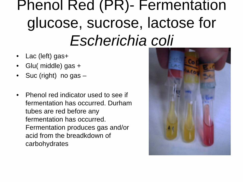
Phenol Red (PR)- Fermentation glucose, sucrose, lactose for
Escherichia coli• Lac (left) gas+• Glu( middle) gas + • Suc (right) no gas –
• Phenol red indicator used to see if fermentation has occurred. Durham tubes are red before any fermentation has occurred. Fermentation produces gas and/or acid from the breadkdown of carbohydrates
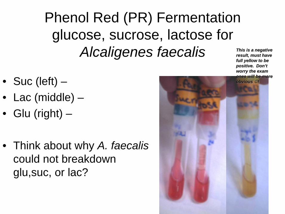
Phenol Red (PR) Fermentation glucose, sucrose, lactose for
Alcaligenes faecalis
• Suc (left) –• Lac (middle) –• Glu (right) –
• Think about why A. faecaliscould not breakdown glu,suc, or lac?
This is a negative This is a negative result, must have result, must have full yellow to be full yellow to be positive. Donpositive. Don’’t t worry the exam worry the exam ones will be more ones will be more obvious obvious ☺☺!!

Phenol Red (PR) Fermentation glucose, sucrose, lactose for Saccharomyces cerevisiae
• Lac (left) –• Glu (middle) gas• Suc (right) –
Why did S. cerevisiaeNOT change color?
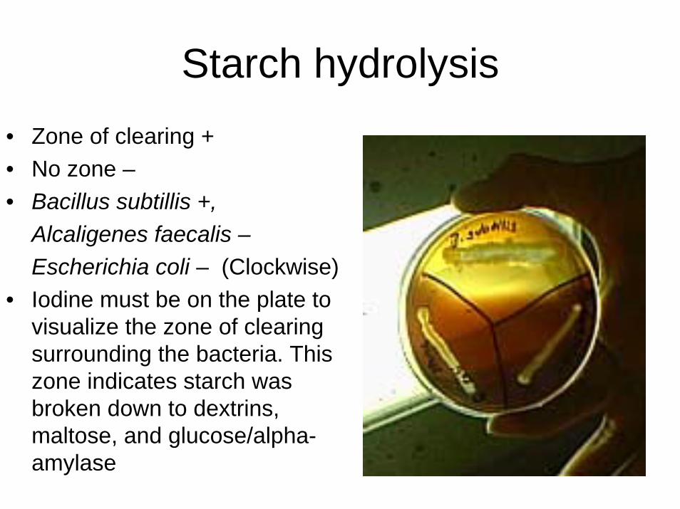
Starch hydrolysis• Zone of clearing +• No zone –• Bacillus subtillis +,
Alcaligenes faecalis –Escherichia coli – (Clockwise)
• Iodine must be on the plate to visualize the zone of clearing surrounding the bacteria. This zone indicates starch was broken down to dextrins, maltose, and glucose/alpha-amylase

Lipid Hydrolysis• Dark blue precipitant zone /
clearer blue zone +• No color change –• Bacillus subtilis &
Staphylococcus epidermidis +w / clearer blue zone around bacterial growth
• Spirit blue agar w/3%Bacto lipase reagent is used to see if triglycerides are hydrolyzed into glycerol and free fatty acids/lipase
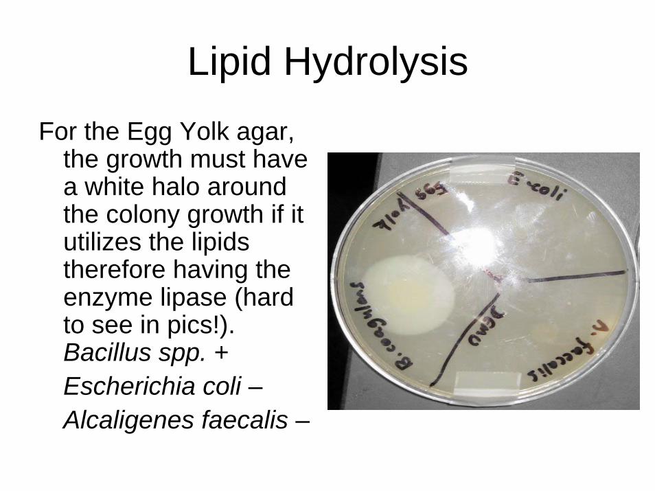
Lipid HydrolysisFor the Egg Yolk agar,
the growth must have a white halo around the colony growth if it utilizes the lipids therefore having the enzyme lipase (hard to see in pics!). Bacillus spp. +Escherichia coli –Alcaligenes faecalis –

Casein hydrolysis • Zone of clearing +• No zone –• Test used to see If
casein is degraded into amino acids for use as a carbon source/proteolytic enzymes
• Escherichia coli – , Alcaligenes faecalis –Bacillus subtilis +
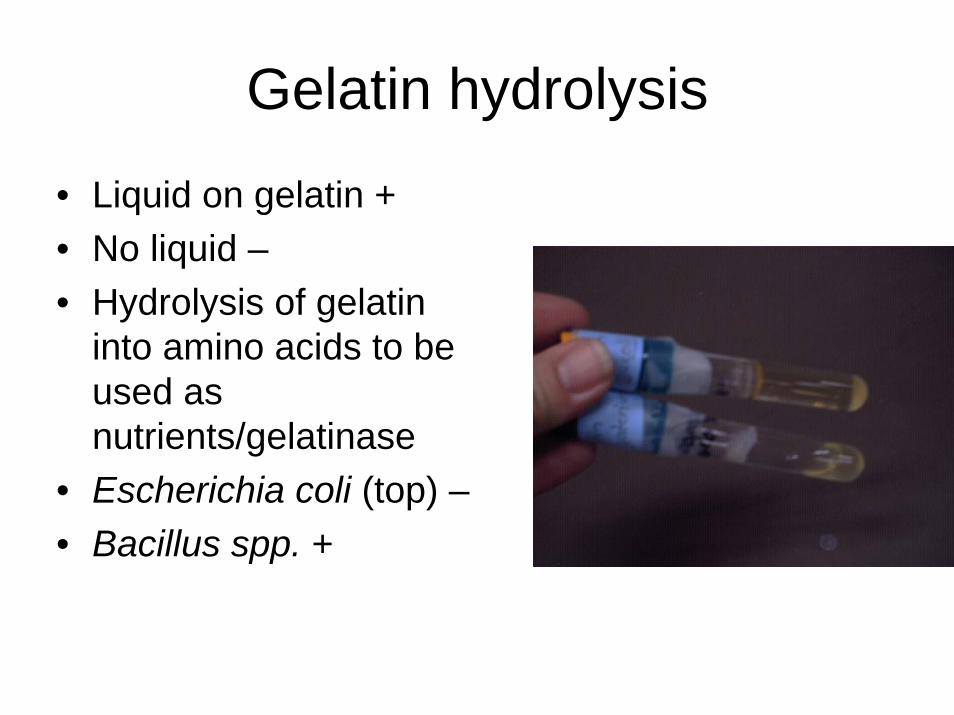
Gelatin hydrolysis
• Liquid on gelatin +• No liquid –• Hydrolysis of gelatin
into amino acids to be used as nutrients/gelatinase
• Escherichia coli (top) –• Bacillus spp. +

Catalase• Bubbles +• No bubbles –• Reagents 3% H2O2
Tests for the ability to break down toxic O 2 products/superoxide dismutase (catalyzes the destruction of superoxide) & catalase operoxidase (catalyzes the destruction of hydrogen peroxide)
2 O2-+ 2 H+ ---superstable dismutate O 2 + H2O2
2 H2O2 ---catalase 2 H2O + O2

Oxidase• Blue (30 sec) +• No color change –• Tests done on Oxidase strips• Tests for the oxidation of reduced cytochrome c to form
water and reduced cytochrome c / Cytochrome oxidase
Oxidized cyt C + reagent Wurster’s blue + red cyt C
clear dark purpleoxidized

Sulfur reduction test, Indole production, Motility (SIM) deeps
all 3 tests done w/SIM deeps just add Kovac’s reagent for Indole test
• Alcaligenes faecalis (left) -• Escherichia coli (middle) –• Proteus vulgaris (black
precipitate) +
• Reagent: Ferrous ammonium sulfate-indicator. H2S reacts w/ ferrous sulfate forming the black precipitate Sodium thiosulfate is reduced to sulfite/thiosulfate

Motility• Spreading growth +(Spreading growth looks like a
mascara brush in the deep)Escherichia coli (right)Proteus vulgaris (left)
• Linear growth –Staphylococcus epidermidis(middle)
• To test for the ability of bacterium to migrate in solid agar deep
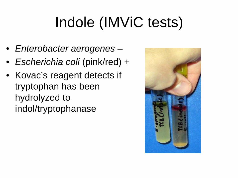
Indole (IMViC tests)
• Enterobacter aerogenes –• Escherichia coli (pink/red) +• Kovac’s reagent detects if
tryptophan has been hydrolyzed to indol/tryptophanase

Methyl Red (MR) (IMViC tests)
• Enterobacter aerogenes (left) –
• E. coli (bright red) +
• Reagent: Methyl red indicator identifies pH change due to mixed acid fermentation
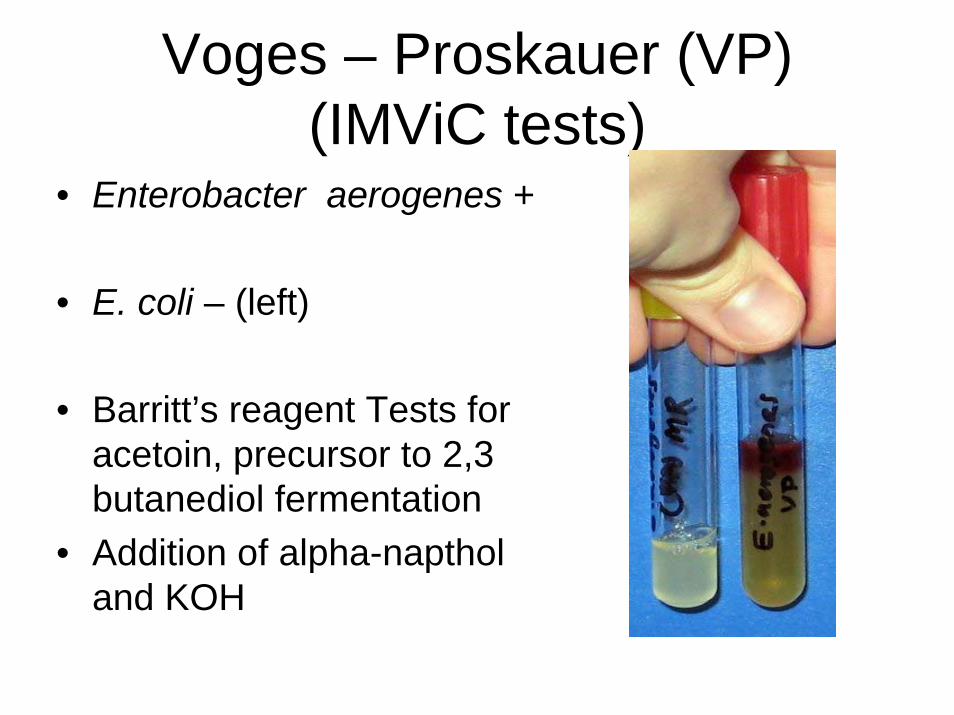
Voges – Proskauer (VP) (IMViC tests)
• Enterobacter aerogenes +
• E. coli – (left)
• Barritt’s reagent Tests for acetoin, precursor to 2,3 butanediol fermentation
• Addition of alpha-naptholand KOH
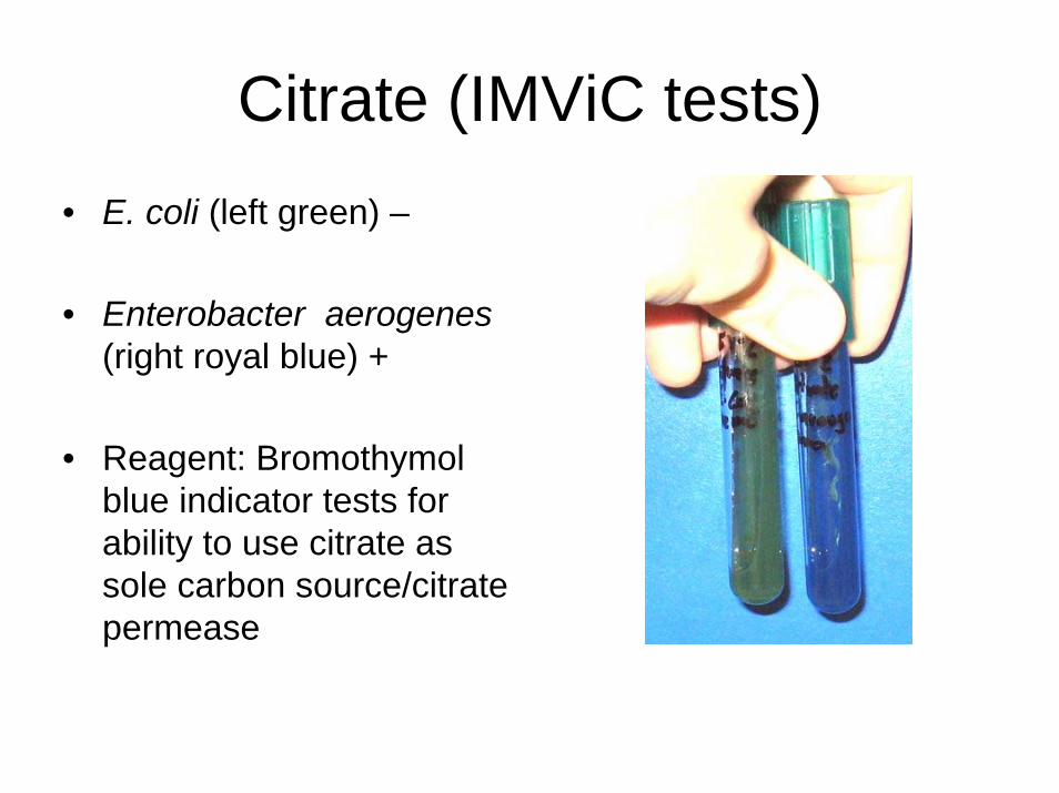
Citrate (IMViC tests)• E. coli (left green) –
• Enterobacter aerogenes (right royal blue) +
• Reagent: Bromothymol blue indicator tests for ability to use citrate as sole carbon source/citrate permease

Urease
Phenol Red a pH indicator turns tube bright pink because NH3 decreases the pH
CO(NH3)2 + 2 H2O –urease CO2+ H2O + 2 NH3
E. coli – (left)Proteus vulgaris +
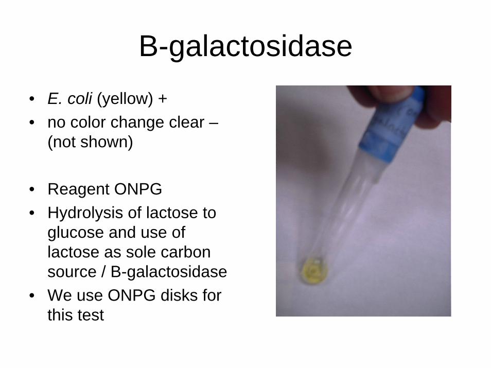
B-galactosidase• E. coli (yellow) +• no color change clear –
(not shown)
• Reagent ONPG • Hydrolysis of lactose to
glucose and use of lactose as sole carbon source / B-galactosidase
• We use ONPG disks for this test
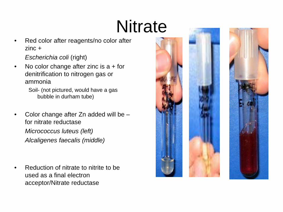
Nitrate• Red color after reagents/no color after
zinc + Escherichia coli (right)
• No color change after zinc is a + for denitrification to nitrogen gas or ammonia
Soil- (not pictured, would have a gas bubble in durham tube)
• Color change after Zn added will be –for nitrate reductaseMicrococcus luteus (left)Alcaligenes faecalis (middle)
• Reduction of nitrate to nitrite to be used as a final electron acceptor/Nitrate reductase

Coagulase • Results:+ clotting in thebottom of the broth • Reagents:Plasma• Reason/Enzymes Clots plasma to avoid attack by
host’s defenses/Coagulase
Staphylococcus aureus +; Staphylococcus epidermidis –
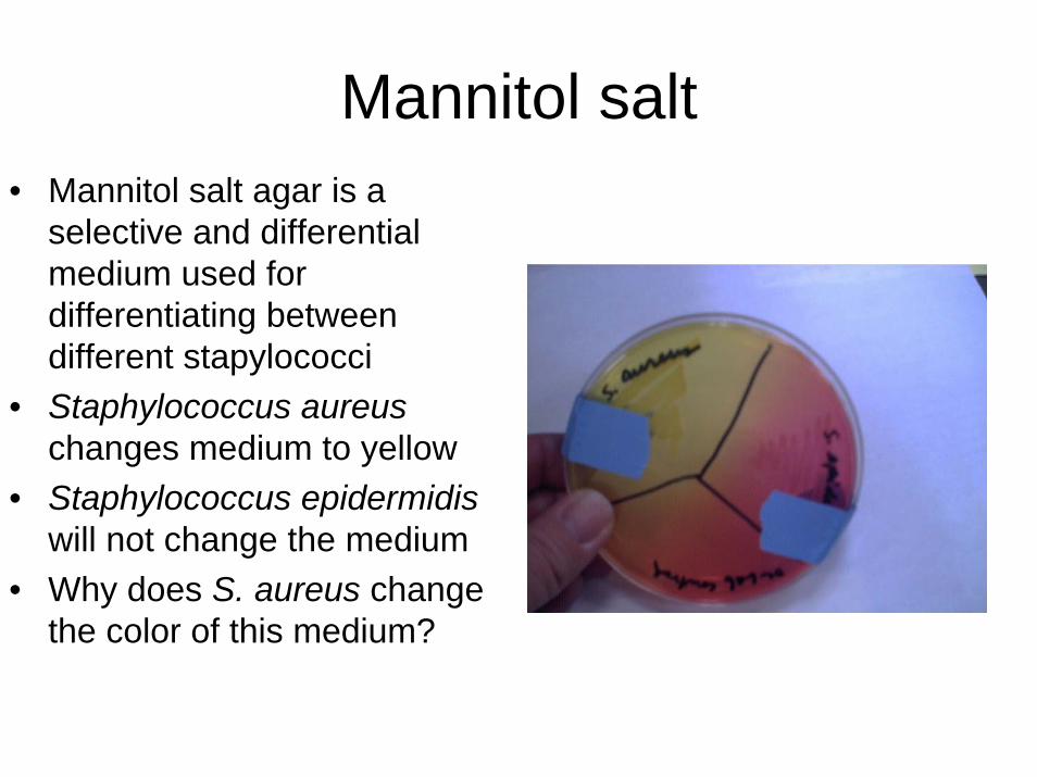
Mannitol salt• Mannitol salt agar is a
selective and differential medium used for differentiating between different stapylococci
• Staphylococcus aureuschanges medium to yellow
• Staphylococcus epidermidiswill not change the medium
• Why does S. aureus change the color of this medium?

Hemolysis• Check which bacteria are
capable of lysing red blood cells (RBCs) by using blood agar (sheep blood).
• α = partial lysis of red blood cells blood looks greenish
• β = complete lysis of blood clearing
• γ = no lysing• Clockwise starting from the left:
Staphylococcus aureus β, Staphylococcus epidermidis γ , teeth α
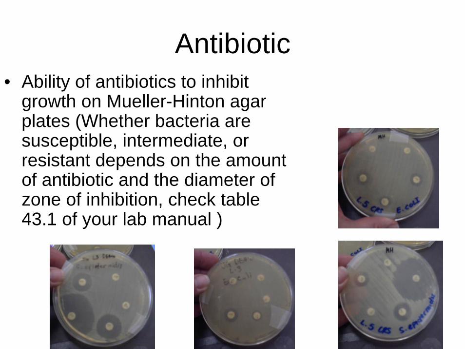
Antibiotic• Ability of antibiotics to inhibit
growth on Mueller-Hinton agar plates (Whether bacteria are susceptible, intermediate, or resistant depends on the amount of antibiotic and the diameter of zone of inhibition, check table 43.1 of your lab manual )

Normal Microbiota on the human body
• Both pictures show examples of normal microbiotathat grow on the ear,arm, palm, and feet.
• What bacteria do you think these colonies represent?
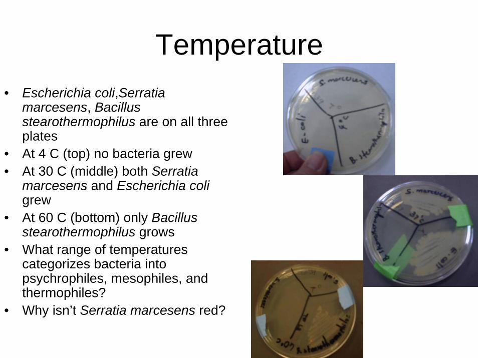
Temperature• Escherichia coli,Serratia
marcesens, Bacillus stearothermophilus are on all three plates
• At 4 C (top) no bacteria grew• At 30 C (middle) both Serratia
marcesens and Escherichia coligrew
• At 60 C (bottom) only Bacillus stearothermophilus grows
• What range of temperatures categorizes bacteria into psychrophiles, mesophiles, and thermophiles?
• Why isn’t Serratia marcesens red?

pH
• Bacterial tolerance to different pH varies much more than human tolerance.
• Can you remember the pH ranges for acidophiles, neutrophiles, and alkalophiles?
• No Demos – will not be a station
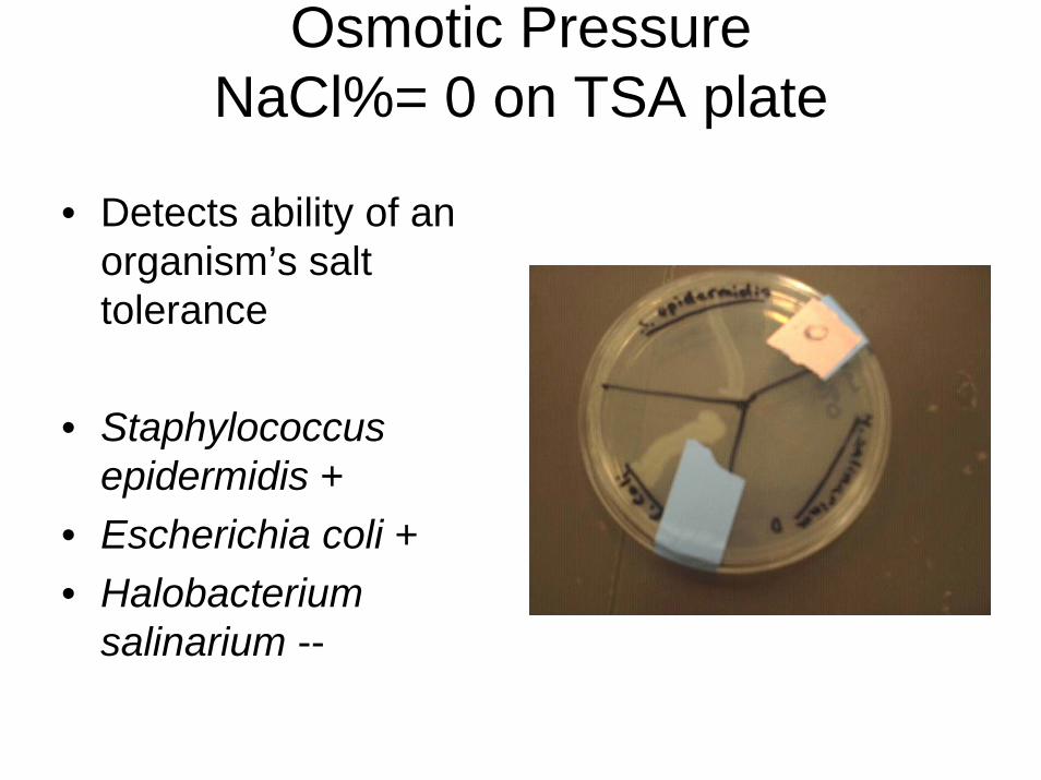
Osmotic Pressure NaCl%= 0 on TSA plate
• Detects ability of an organism’s salt tolerance
• Staphylococcus epidermidis +
• Escherichia coli +• Halobacterium
salinarium --
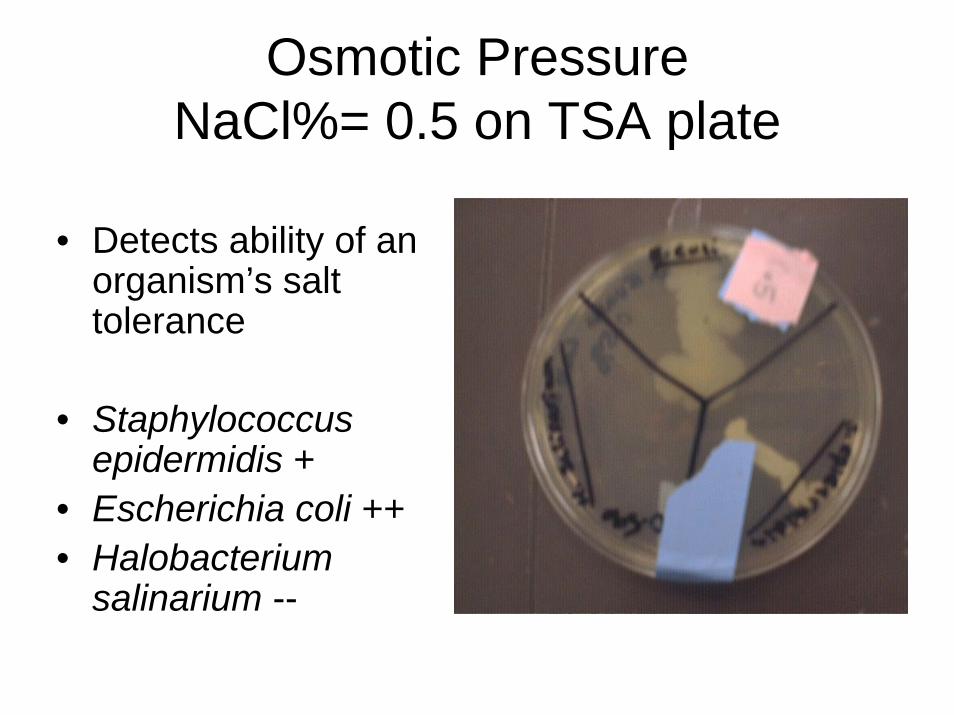
Osmotic Pressure NaCl%= 0.5 on TSA plate
• Detects ability of an organism’s salt tolerance
• Staphylococcus epidermidis +
• Escherichia coli ++• Halobacterium
salinarium --

Osmotic Pressure NaCl%= 5 on TSA plate
• Detects ability of an organism’s salt tolerance
• Staphylococcus epidermidis ++
• Escherichia coli ++• Halobacterium
salinarium --

Osmotic Pressure NaCl%= 10 on TSA plate
• Detects ability of an organism’s salt tolerance
• Staphylococcus epidermidis +
• Escherichia coli --• Halobacterium
salinarium --

Osmotic Pressure NaCl%= 20 on TSA plate
• Detects ability of an organism’s salt tolerance
• Staphylococcus epidermidis --
• Escherichia coli --• Halobacterium
salinarium ++

Compare how well the 3 bacteria grow on each plate of different NaCl conc. Is the
streak thickness the same on all of them?