Bacterial diversity and their adaptations in the shallow water
23
1 Author version: Cah. Biol. Mar., vol.53; 2012; 65-76 Bacterial diversity and their adaptations in the shallow water hydrothermal vent at D. João de Castro Seamount (DJCS), Azores, Portugal Chellandi MOHANDASS 1 , Raju RAJASABAPATHY 1 , Chinnarajan RAVINDRAN 1 , Ana COLACO 2 , Ricardo Serrao SANTOS 2 , Ram Murthi MEENA 1 1) Microbial Technology Lab, Biological Oceanography Division, National Institute of Oceanography (CSIR), Dona Paula, Goa-403 004, India. Phone: 91(0)832 2450414, Fax: 91(0) 832-2450606. E-mail: [email protected] 2) IMAR - Department of Oceanography and Fisheries, University of Azores, Cais de Sta Cruz 9901- 862, Horta, Portugal. Abstract: Bacterial diversity from the shallow vent of D. João de Castro Seamount (DJCS) Azores, Portugal and their adaptations to a nutrient rich environment was investigated from 2004 and 2005 cruise samples. Assessment of the qualitative composition of the heterotrophic bacterial diversity studied by various means like 16S rRNA, FAME and RFLP expressed the existence of consistent bacterial groups like Bacillus, Staphylococcus, Micrococcus, Halomonas, Pseudoalteromonas, Alcaligenes and Brevibacterium. In situ studies conducted in the laboratories with different metals, revealed that Fe, Mn and Pb reduces the bacterial growth period by 50% in some of the species like Staphylococcus succinus, Bacillus subtilis and Bacillus sp. About 55% of the organisms from the experimental site were able to hydrolyze carbohydrates, 27% of them were able to attack fatty acid and 45% were protein degraders. High temperatures (70 ºC) and neutral pH (7) were opted by most of the vent bacteria to produce their enzymes. Adaptive mechanisms to detoxify the metals by producing H 2 S gases, catalase and oxidase for decomposition of hydrogen peroxide and oxygen supply; extended survival in unfavourable conditions by sporulation were observed as a common characteristic features in most of the bacterial isolates of the shallow vent DJCS. Key words: Diversity • Enzymes • FAME • Metals • RFLP • 16S rRNA
Transcript of Bacterial diversity and their adaptations in the shallow water
1
Author version: Cah. Biol. Mar., vol.53; 2012; 65-76
Bacterial diversity and their adaptations in the shallow water hydrothermal vent at
D. João de Castro Seamount (DJCS), Azores, Portugal
Chellandi MOHANDASS1, Raju RAJASABAPATHY1, Chinnarajan RAVINDRAN1, Ana COLACO2,
Ricardo Serrao SANTOS2, Ram Murthi MEENA1
1) Microbial Technology Lab, Biological Oceanography Division, National Institute of Oceanography (CSIR), Dona Paula, Goa-403 004, India. Phone: 91(0)832 2450414, Fax: 91(0) 832-2450606. E-mail: [email protected]
2) IMAR - Department of Oceanography and Fisheries, University of Azores, Cais de Sta Cruz 9901-862, Horta, Portugal.
Abstract:
Bacterial diversity from the shallow vent of D. João de Castro Seamount (DJCS) Azores, Portugal and
their adaptations to a nutrient rich environment was investigated from 2004 and 2005 cruise samples.
Assessment of the qualitative composition of the heterotrophic bacterial diversity studied by various
means like 16S rRNA, FAME and RFLP expressed the existence of consistent bacterial groups like
Bacillus, Staphylococcus, Micrococcus, Halomonas, Pseudoalteromonas, Alcaligenes and
Brevibacterium. In situ studies conducted in the laboratories with different metals, revealed that Fe, Mn
and Pb reduces the bacterial growth period by 50% in some of the species like Staphylococcus succinus,
Bacillus subtilis and Bacillus sp. About 55% of the organisms from the experimental site were able to
hydrolyze carbohydrates, 27% of them were able to attack fatty acid and 45% were protein degraders.
High temperatures (70 ºC) and neutral pH (7) were opted by most of the vent bacteria to produce their
enzymes. Adaptive mechanisms to detoxify the metals by producing H2S gases, catalase and oxidase for
decomposition of hydrogen peroxide and oxygen supply; extended survival in unfavourable conditions
by sporulation were observed as a common characteristic features in most of the bacterial isolates of the
shallow vent DJCS.
Key words: Diversity • Enzymes • FAME • Metals • RFLP • 16S rRNA

2
Introduction
Microbial community of hydrothermal vent systems is necessary to understand the functioning of the
ecosystems and their impact to their surrounding environment (Sievert et al., 2000). Shallow water vents
are different from deep ones because primary production is based on photosynthesis (Tarasov et al.,
2005). Elements present in the shallow vents can be oxidized and used by many heterotrophic members
of the Archaea and bacteria as electron acceptors for the anaerobic degradation of organic matter
(Sievert et al., 2000). Vent associated microorganisms may be involved in microbially mediated
transformation and precipitation of elements. Hydrothermal vents offer a variety of habitats to
metabolically diverse microorganisms (Baross and Hoffman, 1985; Karl, 1995).
Cultivation based methods are not well suited for investigation of the general composition of
microbial communities, since only a small percentage of the microorganisms are cultivable (Amann et
al., 1995), however molecular tools such as 16S rRNA clone libraries made it possible to circumvent
this limitation. Microbial investigation on the hydrothermal vents mostly deals with the selective
isolation of chemosynthetic and thermophilic bacteria (Rusch et al., 2005). Although the trophic role of
the heterotrophic bacteria in the vent ecosystem is not completely understood, roles in detoxification
process by these microorganisms in association with polychaetes confirm the importance of these in the
vent system (Jeanthon and Prieur, 1990b).
The D. João de Castro Seamount (DJCS) located in the North Atlantic, between the Azorean
islands São Miguel and Terceira (38°13.3′N, 26°36.2′W), lies on the Terceira Rift (Vogt and Jung,
2004). The area of the DJCS shallower than 50 m is approximately 0.35 km2, with 0.011 km2 showing
hydrothermal activity. Venting on the seamount can be divided in two main types; ‘white’ and ‘yellow’
vents, according to the bottom colour around the fluid exits. The venting fluids from the white zone
(WZ) are significantly richer than those from the yellow zone (YZ) in dissolved sulphide [81.6 µM
(WZ)/1.24 µM (YZ)]; H2 [3183.5 µM (WZ)/84.4 µM (YZ)]; CH4 [29.8 µM (WZ)/12.8 µM (YZ)] and
the trace metal Pb [3.6 nM (WZ)/1.9 nM (YZ)], but poor in Fe [8.8 µM (WZ)/89.2 µM (YZ)] and Mn
concentrations [0.5 µM (WZ)/ 6.8 µM (YZ)]. The fluids from the yellow zone are warmer (61.3 ºC) than
the ones from the white zone (35.8 ºC) (Cardigos et al., 2005). These venting fluids have much higher
trace metal and gas concentrations than the ambient Atlantic seawater (Colaco et al., 2006). The Atlantic
seawater has 36.5 µM H2 (Herr and Barger, 1978), 5.48 µM CH4 (Conrad and Seiler, 1988) and 930
pmol of dissolved total sulphide (Andreae et al., 1991).

3
Hydrothermal vents are geological formations that release altered seawater which has been
heated, often to temperatures exceeding 400 °C, by subterranean magma pockets as it circulates through
the crust, mobilizing metals from basalt and acquiring elevated levels of sulphide from the reduction of
sulphate by ferrous iron dissolved in the hot fluid (Vincent et al., 1994). When the superheated sea water
mixes with cold oceanic sea water, the positively charged cations of metals, i.e. copper and zinc, leached
out of the basalt precipitate as sulphides (Malahoff, 1985). Hydrothermal vents host unique communities
of animals exposed to high concentrations of heavy metals and are covered with a community of metal-
resistant bacteria (Jeanthon and Prieur, 1990a). Bacterial symbionts present with other animals in the
vent utilize the metals like sulphide as an energy source to fix carbon (Vincent et al., 1994). Further, the
basis of this association between bacteria and higher organisms in the hydrothermal vent is to detoxify
invertebrate surfaces by symbiotic microorganisms (Alayse-Danet et al., 1987; Holmstrom and
Kjelleberg, 1999; Jeanthon and Prieur, 1990b). Although the presence of large amounts of metals is a
major feature of hydrothermal vent environments, relatively little is known about relationships between
the metals and organisms living at these sites. Heterotrophic bacteria are generally ignored in the vent
area and only the specialists like chemosynthetic and thermophilic bacteria are given more important.
Just a few studies on the diversity and adaptation of microorganisms have been carried out on the
DJCS (filamentous bacteria from the white area at DJCS by Cardigos et al., 2005; assemblage of benthic
diatoms and culturable heterotrophs by Raghukumar et al., 2008). The goal of the present study is to
confirm the abundance of genetic diversity of the heterotrophic microbial population in metal rich
environments and its adaptability for its better endurance.
Materials and Methods
Samples of water, sediment, bacterial and algal mats were collected in sterile containers from the DJCS
(Fig. 1) during cruises on the research vessels R.V. Arquipelago (2004) and Águas Vivas (2005).
Samples were collected by SCUBA diving. Pieces of algae, bacteria, and sediments were suspended in
50% sterile seawater, and then 100 µl was plated with solid nutrient agar media (HiMedia M001,
Mumbai, India). Samples were stored in the dark at 4 ºC on board and transported with ice packs to
University of Azores, Horta, Portugal. Colonies appeared on the nutrient agar plates from various
sampled sources like sediment, water, algal and bacterial mats were isolated and given numbers SV1 –
SV23.

4
Isolation, purification and identification
Isolates obtained from the seawater nutrient agar plates were selected based on size, shape, and colour.
Various morphological and biochemical tests like gram staining, spore staining, motility, H2S
production, catalase, oxidase, amylase, lipase and caseinase production were performed. All the tests
have been carried out as per the standard microbiological protocol suggested by the supplier (HiMedia
pvt ltd, Mumbai, India).
Bacterial identity based on Fatty acid Methyl Ester (FAME)
Approximately 30 – 40 mg of cells grown in Trypticase Soy Broth Agar (TSBA) media for one day was
collected and the fatty acid preparation, extraction, purification, and methylation were accomplished
using standard procedures (Sasser, 1990). The FAME profiles generated were compared against an
inbuilt Sherlock TSBA Library version 4.0 B (MIDI Inc., DE, USA) and matched to a library of 700+
bacterial species representing over 180 genera. Cellular fatty acid composition analysis was done at
Regional centre of Kochi, National Institute of Oceanography. Bacterial fatty acids were identified by
comparison of the retention times of their methyl esters on the SE30 column.
Genomic DNA isolation
The genomic DNA was prepared from all the isolates by K- Cetyl Trimethyl Ammonium Bromide
(CTAB) method (Sambrook and Russel, 2001) with some modifications. Sodium Dodecyl Sulphate
(SDS) was replaced with CTAB in the extraction buffer; Ammonium acetate and proteinase K were
eliminated from the original CTAB method since phenol was repeatedly used to remove proteins.
Overnight grown bacterial cultures in LB broth (HiMedia M1245, Mumbai) were centrifuged at 8000
rpm for 10 min and the bacterial pellets were lyophilized. Approximately 500 mg of sample (dry weight)
was re-suspended in 1.5 ml of extraction buffer (100 mM Tris HCl, 100 mM Na2EDTA, 1.5 M NaCl and
1% CTAB). The extraction buffer was preheated at 60 oC for 60 min prior to treatment of the samples.
After the addition of extraction buffer the samples were well mixed with sterile toothpicks and incubated
at 65 oC for 60 min. The samples were then centrifuged at 12000 rpm for 10 min. The supernatant was
collected and added with 500 µl of phenol: chloroform: iso-amyl alcohol mixture (25:24:1). The samples
were then mixed for 30 min and centrifuged at 12000 rpm for 10 min. The same step was repeated once
again and the supernatant was collected in a fresh tube. To the supernatant 500 µl of chloroform was
added and centrifuged at 12000 rpm for 10 min. Nucleic acids in the supernatant fraction were

5
precipitated by adding 100% chilled ethyl alcohol. This was centrifuged and washed with 70% ethyl
alcohol. The dried pellets were re-suspended in sterile double distilled water or TE buffer. The isolated
DNA was run on 0.8% (w/v) Tris-Acetate-EDTA (TAE) agarose gel electrophoresis with ethidium
bromide staining and visualized under UV.
Amplification of 16S rDNA
PCR amplification of 16S rRNA gene was done with the bacterial consensus primers 27F [5’-AGA GTT
TGA TCC TGG CTC AG-3’] and 1492R [5’-GGT TAC CTT GTT ACG ACT T-3’] universal forward
and reverse 16S rDNA primers (Lane, 1991). The PCR of the genomic DNA of 22 isolates were
conducted in a final volume of 25µl. The reaction mixture contained 1x PCR buffer (Sigma, USA), 2.5
mM MgCl2, 200 µM DNTP’s, 1 U of Taq DNA polymerase, 25 pmol of each forward and reverse
oligonucleotide primers and approximately 20 ng of genomic DNA. The amplification profile consisted
of an initial denaturation at 94 oC for 3 min, followed by 35 cycles at 94 oC for 1 min, 55 oC for 1 min
and 72 oC for 1 min. This was followed by a final extension step of 72 oC for 5 min. The samples were
held at 4 oC until further analyse.
PCR-RFLP
RFLP marker was used to identify the variability among the bacterial groups from the shallow vent
strains. The DNA obtained from all 22 bacteria were amplified using 16S primers and the PCR products
were digested with Alu I with appropriate buffers (Fermentas) according to the manufacturer’s protocol.
The 20 µl of above reaction mixture was incubated at 37 oC for 3 h. Restriction fragments were
separated using 2.5% TAE-agarose gel at 80 V for 1 hour and visualized with UV light after staining
with ethidium bromide (0.5 µg ml−1).
Sequencing
Sequencing of all the isolates was performed with an automatic sequencer Applied bio system. Foster
City, USA. Dona-Paula, Goa. India.
Accession Numbers
The sequences were submitted to GenBank and accession numbers were assigned from GU143784
(SV1) to GU143805 (SV23).

6
BLAST Search and Phylogenetic Analysis
The PINTAIL program (Ashelford et al., 2005) was used to check chimera formations. No anomalies
were detected. The partial 16S rRNA gene sequences of the isolated strains were compared with those
available in the public databases. Identification to the species level was determined by a 16S rDNA
sequence similarity of 99% with that of the prototype sequence in GenBank. Sequence alignment and
comparison was performed using the multiple sequence alignment program Clustal X 1.81 (Thompson
et al., 1997). Sequences were edited manually to remove the gaps. Neighbor-joining method was
employed to construct the Phylogenetic tree using MEGA4 software (Tamura et al., 2007) and the
maximum likelihood method was adopted for calculating the evolutionary distance (Tamura et al.,
2004).
Metal studies
The growth preferences of bacterial cultures isolated from the DJCS vent site for Fe, Mn and Pb were
tested. Zobell marine broth (M385 HiMedia, Mumbai) supplemented with one µM of filter sterilized
FeCl3, MnCl2 and one nM of Pb(CH3COO)2 H2O were inoculated to the bacterial cultures having initial
optical density (OD) of 0.01 – 0.02 at 600 nm. Medium without metal supplement was used as control.
OD of the cultures was measured every 24 h till 8th day. From the growth curve of each isolate in each
media, organisms and its preferences were recorded.
Combination of metals, pH and temperature on amylase production
Known amount of bacterial cells inoculated in to nutrient broth (HiMedia M088, Mumbai) with 1%
starch, subsequently added with 1 µM Fe and Mn, and 1 nM of Pb. Inoculated flasks were incubated in a
rotary shaker for 5 days. Media were centrifuged at 10,000 rpm for 10 minutes at 4 ºC. The supernatants
were assayed for amylase activity (Medda and Chandra, 1980). Nutrient broth with 1% starch without
metal was used as blank. Effect of pH on the vent strains were studied at various pH from 3 – 9, by
incubating 0.1 ml enzyme solution with 1 ml buffer and 1 ml of 1% starch. The various buffers used to
obtain proper pH were citrate buffer for 3 – 6 pH; phosphate buffer for 7 & 8; carbonate - bicarbonate
buffer for 9. Temperature effects on enzyme activity from 30 – 90 ºC were determined by the standard
procedure (Medda & Chandra, 1980).

7
Results
Bacterial diversity by 16S rRNA of the shallow vent
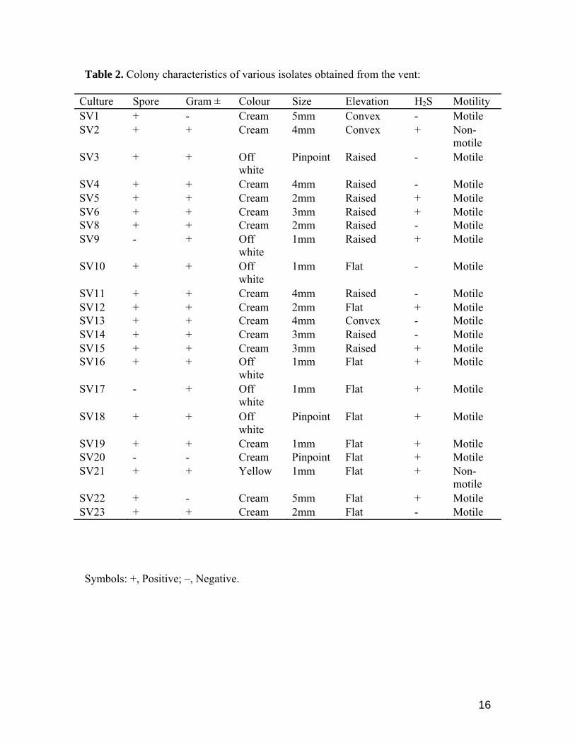
Shallow vent bacteria of the DJCS identified by 16S rRNA revealed seven major genera from this site.
They belong to Bacillus, Staphylococcus, Micrococcus, Halomonas, Pseudomonas, Alcaligenes and
Brevibacterium. Among the genera Bacillus, nine belongs to B. subtilis, one B. licheniformis and one B.
flexus. Staphylococcus arlettae and S. succinus, Micrococcus lutes, Alcaligenes faecalis and
Brevibacterium casei were some of the other bacteria retrieved from these vents (Fig. 2). Sequences
obtained from the vent strains were shown 99 to 100% homology with the GenBank (Table 1).
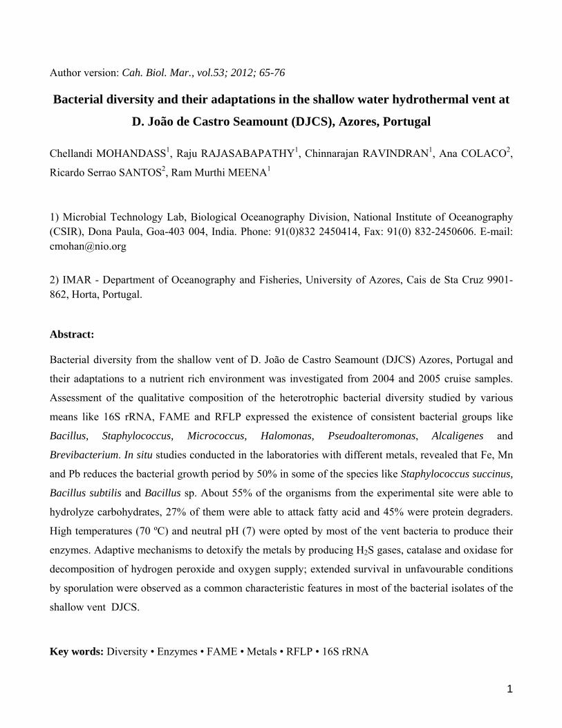
Eighty six percent of the strains isolated from the shallow vent belonged to Gram-positive and
spore producers. Most of the colonies were circular and creamy or dull white and only one is reported to
be yellow in colour. Ninety percent of the strains were motile and the remaining was non motile (Table
2).
Bacterial strains isolated from the shallow vents of DJCS identified by FAME analysis resulted
that, many of them belongs to Bacillus spp (15). Brevibacterium casei, Staphylococcus cohinii, S.
arlettae, Yersinia, Halomonas and Micrococcus were some of the other type of bacteria identified by
FAME (Table 3). The bacterial groups corresponds to the gas chromatographic profile of a bacterial
species in the Sherlock TSBA Library version 4.0 B (Microbial ID, MIDI Inc.) with a similarity index of
0.3 – 0.8. High similarity index were noticed with Yersinia, 0.822; Micrococcus, 0.847 while the lowest
index have been seen in SV17, Bacillus with 0.255 (Table 3).
High similarities of identity were noticed in 16S rRNA and FAME tests. Differences were
observed in a few identities like Alcaligenes by 16S rRNA (SV1) while it was identified as being
Yersinia in FAME (Table 3); this analysis did indicate Alcaligenes as a second option with the similarity
index of 0.528 (data not shown).
Influence of metals on bacterial growth, diversity and its hydrolyses
Bacterial survival in the hydrothermal vents depends on the effective uptake and detoxification of the
materials present over there. The shallow vent of Azores presents high content of H2S, CH4, H2, Pb, Co,
Fe, Ba and Mn (Cardigos et al., 2005). Systems for the detoxification of metals required enzymes for its
reduction. Twenty two bacterial cultures isolated from the site were investigated adding Fe, Pb and Mn

8
in to their growth media. Around 90% of them enhanced their growth in the presence of these metals.
Only 10% of them were able to grow in a better way without metals (Table 1). Different organisms
preferred different metals for their growth. Seventy seven percent of the organisms grow well with Fe.
Strains like SV14, SV19, SV3, SV11 and SV15 obtained their biomass earlier in the Fe enriched media
when comparing to the media with no metals (Zobell broth). Staphylococcus succinus (accession no.
GU143799) has reduced its growth time by 50% with Fe (Table 1). Eighteen percent of the vent
organisms enhanced their growth in the presence of Mn. Bacillus subtilis (GU143798) reduced its
growth time from 7 days to 3 days in presence of Mn to attain a close equivalent biomass (0.706 –
0.603). Overall growth studies with various metals indicated that most of the bacteria preferred Fe for
their growth (Table 1).
The various shallow vent bacteria isolates were tested for enzyme production in their respective
media. All the isolates produced catalase. Degradative enzymes like amylase, lipase and caseinase were
produced by 55, 27 and 45% of the isolates respectively (Table 4). The high percentage (55%) of
amylase producing strains (12 out of 22) conducted to further investigations on these enzymes under
metal influence. Among the 12 strains exposed to the three different metals (Fe, Pb and Mn) in to their
enzyme production medium (Medda & Chandra. 1980) only 4 strains had their activity augmented. B.
subtilis (GU143788) yielded a high amylase activity with Fe (49.5 U/ml) (Fig. 3) subsequently the
strains Bacillus sp (GU143797), B. licheniformis (GU143794) given around 12 U/ml and B. subtilis
(GU143793) reported to produce only 2 U/ml more than the media without metal. Mn almost equalizes
the enzyme production with no metals for Bacillus sp (GU143797) and Pb did not support the enzyme
activity in a noticeable way (Fig. 3). Characterization studies showed that pH 7 and temperature 70 ºC to
be ideal for most of the strains. B. subtilis (GU143793) showed its maximum activity of 47.2 U/ml
among the tested strains (Fig. 4 & 5).
Homogeneity of the bacteria by RFLP
Ten RFLP patterns were detected from the shallow vent strains using with Alu-I endonucleases (Fig. 6).
Restriction fragments shorter than 100 bp were disregarded for better clarity. Nine cultures, of B.
subtilis gave an identical RFLP pattern. Strains SV13 and 14 appear to be very close. SV6 and SV15
showed a strong similarity to Bacillus fluxes. SV1, SV2, SV9, SV17, SV20, SV21 and SV22 were not
similar and appear to be seven different patterns of expressions (Fig. 6).

9
Discussion
Exploration of heterotrophic bacteria from DJCS vent region using 16S rRNA sequencing analysis
expressed the dominance of Bacillus spp. More than 15 strains of 22 tested belong to the genus Bacillus.
Seventy seven percent of the sequenced isolates were found to be Firmicutes phylum; 13.63% were
Proteobacteria and the rest were Actinobacteria. Maximal occurrence of Bacillus sp, from the
experimental site may be due to their ability to form spores, thus enabling them to withstand a wide
range of environmental conditions like temperature 35.8 – 61.3 ºC and the pH 5.01 – 5.09 prevailing
over the DJCS site. The presence of the heterotrophic bacteria in low temperature diffuse fluids of the
DJCS suggests that they were originated from the sub sea floor hydrothermal habitat where temperatures
and the pH were within their growth range.
The heterotrophic bacterial strains identified (Gugliandolo and Maugeri, 1997) by classical
taxonomy from the shallow water hydrothermal vent of Eolian Island Italy belong to the genera:
Flavobacterium, Acinetobacter, Vibrio, Pseudomonas, Alcaligenes, Micrococcus, Arthrobacter, and
Coryneforms. While comparing the heterotrophic diversity of the DJCS with other shallow vents like
Eolian Island, these Azorean shallow vents were also showed similar groups of bacteria and did not
varied much from others.
Bacteria which belongs to heterogeneous genera that often contains several FAME biotypes eg.,
Bacillus, Micrococcus and Arthrobacter, will not be identified with the existing program of TSBA 4.0
software, therefore additional technique will be needed to identify them at species level (Heyrman et al.,
1999). This could be the reason that some of the bacteria identified by FAME did not match with 16S
rRNA in DJCS samples.
RFLP patterns analysed from the DJCS samples did not expressed its similarity when relating
species with enzyme production and metal preferences. The bacteria which were showing attraction
towards a particular metal sometimes did not produce the same type of enzyme i.e., SV13 and SV14
preferred Fe mostly for its growth, but the bacterium SV13 produced only two enzymes (catalase and
oxidase) and SV14 produced 4 enzymes (Table 2). Probably both organisms belong to same species but
the strain may be different (Fig. 2). In some of the metal preference and enzyme synthesis investigations
by different bacteria showed the same pattern like with SV6 and SV15. It can be said that, these
organisms belong to the same strain (Fig. 2).

10
Enzymes play a vital role in breaking down the materials to simple and less toxic substance,
helping the organisms on their system. Catalase is a common enzyme found nearly in all living
organisms that are exposed to oxygen. Its function is to catalyze the decomposing of hydrogen peroxide
to water and oxygen (Chelikani et al., 2004). Hydrogen peroxide is a harmful by product of many
normal metabolic processes. This has to be quickly converted to other less dangerous substances. Our
results can be interpreted in a way that all the 22 bacterial isolates from the DJCS tested for catalase
production showed positive indication and these enzymes may help in the detoxification of the toxic
materials produced from various metabolic processes and help them to better survive on the shallow
vent region.
Neutral pH was found to be optimal for amylase production by Bacillus thermooleovorans and
B. coagulans (Medda and Chandra, 1980), B. licheniformis (Krishnan and Chandra, 1983) and B. brevis
(Tsvetkov and Emanuilova, 1989). Our results were also expressed in the same manner that minimum
activity obtained by strain B. subtilis (GU143793) 3.4 U/ml at pH 3 raised up to 47.2 U/ml at pH 7. Most
of the strains when increasing the pH presented the same behaviour of enhancement of the enzyme
activities. This extends up to neutral pH. Afterwards the activities were not increasing. From the
Shallow vent strains recorded high enzyme activities at 70 ºC, which were higher or similar to what was
described for other Bacillus alpha amylases (Bolton et al., 1997; Igarashi et al., 1998). Even though the
sampling sites were showing pH of 5.01 to 5.09, neutral pH has been favoured for high enzyme
synthesis. This differentiates the adaption and preference of pH over the bacteria on shallow vents.
Bacteria inhabiting the hydrothermal vents are well adapted to flourish in the diverse
environment despite the presence of metals that would normally inhibit the growth of most bacteria.
Only a small fraction (10%) of the bacteria Staphylococcus arlettae and Halomonas did not grow well in
the presence of metals. The enriched growth of the bacterium Staphylococcus succinus in a shorter time
with Fe expresses the significant adaptation of bacteria to the metal rich waters over DJCS.
Microorganisms are known to employ a variety of mechanism to adapt to the presence of heavy metals.
One of these is the production of hydrogen sulphide, which may act as a detoxification process through
precipitation of metal sulphides (Prieur and Jeanthon, 1987). Among the 22 retrieved organisms from
the DJCS, 13 organisms were able to produce H2S. This is a good indication and expresses elevated
adaptation to the shallow vent condition. Probably this could be one of the essential mechanisms adapted
by the shallow vent bacteria for its survival over DJCS. In addition, most of the heterotrophic bacteria

11
from the DJCS were able to produce no less than one of the physiological enzymes like protease, lipase
and amylases. These bacterial enzymes may play a vital role in mineralization of organic material in this
nutrient rich environment. As suggested by Maugeri (2009), microorganisms inhabiting shallow
hydrothermal vents possess nutritional requirements and overall metabolic pathways ideally suited to
such ecosystem that represents a clear example of the close connection between geosphere and
biosphere. Cole (1977) stated that there was no significant inhibition of amylase activity by Pb but the
quantity of amylase formed was reduced to 26% of control levels. Our results show the high inhibition
of enzyme activity when we introduce Pb in to the enzyme production media. Enzyme productions were
decreased by more than five times; even it shows ten times lower in B. subtilis GU143793 (Fig. 3).
In conclusion the DJCS vent is richly diversified by gram positive spore producing bacteria,
specially dominated with Bacillus followed by Staphylococcus, Pseudoalteromonas, Alcaligenes,
Brevibacterium, Halomonas and Micrococcus. Most of the bacterial communities which live on the
shallow vent of DJCS were well adapted to Fe, Mn and Pb for their growth and metabolism. High
indication of catalases and H2S productions from the vent bacteria expresses its detoxification potentials
over the toxic substances for their healthy survival. Producing more than two to three physiological
enzymes by a single bacterium from the vents might have been the unique identity for their better
survival and endurance. High temperature, neutrally active and metal induced amylases reported in the
DJCS vent may be of better prospects in biotechnological applications.
Acknowledgements
This work was done under the Indo-Portuguese project in Science and Technology (GRICES/DST-
India). We acknowledge GRICES, Portugal and Department of Science and Technology, India for
funding the trips to the DJCS site for sampling. Thanks to the crew members of R.V. Arquipelago and
Águas Vivas for their support. IMAR-DOP/UAç research activities are additionally supported through
the pluri-annual and programmatic funding schemes of FCT (Portugal) and Azorean Regional
Directorate for Science and Technology (DRCT, Azores, Portugal) as Research Unit #531 and Associate
Laboratory #9 (ISR-Lisboa).

12
References
Alayse-Danet A.M., Desbruyeres D. & Gaill F. 1987. The possible nutritional or detoxification role of the epibiotic bacteria of Alvinellid polychaetes: review of current data. Symbiosis 4: 51–62.
Amann R.I., Ludwig W. & Schleifer K.H. 1995. Phylogenetic identification and in-situ detection of individual microbial cells without cultivation. Microbiological Reviews, 59: 143–169.
Andreae T.W., Cutter G.A., Hussain N., Radford-knoery J. & Andreae M.O. 1991. Hydrogen Sulfide and Radon in and over the Western North Atlantic Ocean. Journal of Geophysical Research, 96(D10): 18753–18760.
Ashelford K.E., Chuzhanova N.A., Fry J.C., Jones A.J., & Weightman A.J. 2005. At least 1 in 20 16S rRNA sequence records currently held in public repositories is estimated to contain substantial anomalies. Applied and Environmental Microbiology, 71, 7724–7736.
Baross J.A. & Hoffman S.E. 1985. Submarine hydrothermal vents and associated gradient environments as sites for the origin and evolution of life. Origins of Life and Evolution of the Biosphere, 15: 327–345.
Bolton D.J., Kelly C.T. & Fogarty W.M. 1997. Purification and characterization of the α-amylase of Bacillus flavothermus. Enzyme and Microbial Technology, 20: 340–343.
Cardigos F., Colaco A., Dando P.R., Avila S.P., Sarradin P.M., Tempera F., Conceicao P., Pascoal A. & Santos R.S. 2005. Shallow water hydrothermal vent field fluids and communities of the D. João de Castro Seamount (Azores). Chemical Geology, 224: 153–168.
Chelikani P., Fita I. & Loewen P.C. 2004. Diversity of structures and properties among catalases. Cellular and Molecular Life Sciences, 61(2): 192–208.
Colaco A., Raghukumar C., Mohandass C., Cardigos F. & Santos R.S. 2006. Effect of shallow-water venting in Azores on a few marine biota. Cahiers de Biologie Marine, 47(4): 359–364.
Cole M.A. 1977. Lead inhibition of enzyme synthesis in soil. Applied and Environmental Microbiology, 33(2): 262–268.
Conrad R. & Seiler W. 1998. Methane and hydrogen in seawater (Atlantic Ocean). Deep-Sea Research, 35(12): 1903–1917.
Felsenstein J. 1985. Confidence limits on phylogenies: An approach using the bootstrap. Evolution, 39: 783–791.
Gugliandolo C. & Maugeri T.L. 1997. Temporal variations in heterotrophic mesophilic bacteria from a marine shallow hydrothermal vent off the Island of Vulcano (Eolian Islands, Italy). Microbial Ecology, 36: 13–22.
Herr F. & Barger W. 1978. Molecular Hydrogen in the near surface atmosphere and dissolved in waters of the Tropical North Atlantic. Journal of Geophysical Research, 83(C12): 6199–6205.

13
Heyrman J., Mergaert J., Denys R. & Swings J. 1999. The use of fatty acid methyl ester analysis (FAME) for the identification of heterotrophic bacteria present on three mural paintings showing severe damage by microorganisms. FEMS Microbiology Letters, 181(1): 55–62.
Holmstrom C. & Kjelleberg S. 1999. Marine Pseudoalteromonas species are associated with higher organisms and produce biologically active extracellular agents. FEMS Microbiology Ecology, 30: 285–293.
Igarashi K., Hatada Y., Hagihara H., Saeki K., Takaiwa M., Uemura T., Ara K., Ozaki K., Kawai S., Kobayashi T. & Ito S. 1998. Enzymatic properties of a novel liquefying α-amylase from an alkaliphilic Bacillus isolate and entire nucleotide and amino acid sequences. Applied and Environmental Microbiology, 64: 3282–3289.
Jeanthon C. & Prieur D. 1990a. Resistance to heavy metals of heterotrophic bacteria isolated from the deep sea hydrothermal vent polychaete, Alvinella pompejana. Progress in Oceanography, 24: 81–88.
Jeanthon C. & Prieur D. 1990b. Susceptibility to heavy metals and characterization of heterotrophic bacteria isolated from two hydrothermal vent polychaete annelids, Alvinella pompejana and Alvinella caudata. Applied and Environmental Microbiology, 56: 3308–3314.
Karl D.M. 1995. Ecology of free-living, hydrothermal vent microbial communities. In: Microbiology of deep-sea hydrothermal vents (Karl D.M. ed), pp. 35–124. CRC Press Inc: Boca Raton Florida.
Krishnan T. & Chandra A.K. 1983. Purification and characterization of α-amylase from Bacillus licheniformis CUMC305. Applied and Environmental Microbiology, 46(2): 430–437.
Lane D.J. 1991. 16S/23S rRNA sequencing. In: Nucleic acid techniques in bacterial systematics (Stackebrandt E. and Goodfellow M. eds), pp. 115–175. John Wiley & Sons Inc: New York.
Malahoff A. 1985. Hydrothermal vents and polymetallic sulphides of the Galapagos and Gorda/Juan de Fuca ridge systems and of submarine volcanoes. In: The hydrothermal vents of the Eastern Pacific: An overview (Jones M.L. ed), pp. 6: 19–41. Bulletin of the Biological Society of Washington.
Maugeri T.L., Lentini V., Gugliandolo C., Italiano F., Cousin S. & Stackebrandt E. 2009. Bacterial and archaeal populations at two shallow hydrothermal vents off Panarea Island (Eolian Islands, Italy). Extremophiles, 13: 199–212.
Medda S. & Chandra A.K. 1980. New strains of Bacillus licheniformis and Bacillus coagulans producing thermo stable a-amylase active at alkaline pH. Journal of Applied Bacteriology, 48: 47–48.
Prieur, D. & Jeanthon C. 1987. Preliminary study of heterotrophic bacteria isolated from two deep sea hydrothermal vent invertebrates; Alvinella pompejana (Polychaete) and Bathymodiolus thermophilus (Bivalve). Symbiosis, 4: 87–98.

14
Raghukumar C., Mohandass C., Cardicos F., Costa P.M.D., Santos R.S. & Colaco A. 2008. Assemblage of benthic diatoms and culturable heterotrophs in shallow water hydrothermal vent of the D. João de Castro Seamount, Azores in the Atlantic Ocean. Current Science, 95(12): 1715–1723.
Rusch A., Walpersdorf E., DeBeer D., Gurrieri S. & Amend J.P. 2005. Microbial communities near the oxic/anoxic interface in the hydrothermal system of Vulcano Island, Italy. Chemical Geology, 224: 169–182.
Sambrook J. & Russel D.W. 2001. Molecular cloning: A laboratory manual. 3rd edn. CSH laboratory press, cold spring harbor, Newyork.
Sasser M. 1990. Identification of bacteria through fatty acid analysis In: Methods in Phytobacteriology (Klement Z., Rudolph K. and Sands D.C. eds), pp. 199–204. Akademiai Kiado: Budapest, Hungary.
Sievert S.M., Ziebis W., Kuever J., & Sahm K. 2000. Relative abundance of Archaea and Bacteria along a thermal gradient of a shallow water hydrothermal vent quantified by rRNA slot-blot hybridization. Microbiology, 146: 1287–1293.
Tamura K., Dudley J., Nei M. & Kumar S. 2007. MEGA4: Molecular Evolutionary Genetics Analysis (MEGA) software version 4.0. Molecular Biology and Evolution, 24: 1596–1599.
Tamura K., Nei M. & Kumar S. 2004. Prospects for inferring very large phylogenies by using the neighbor-joining method. Proceedings of the National Academy of Sciences, USA, 101: 11030–11035.
Tarasov V.G., Gebruk A.V., Mironov A.N. & Moskalev L.I. 2005. Deep-sea and shallow-water hydrothermal vent communities: Two different phenomena? Chemical Geology, 224: 5–39.
Thompson J.D., Gibson T.J., Plewniak F., Jeanmougin F. & Higgins D.G. 1997. The ClustalX windows interface: flexible strategies for multiple sequence alignment aided by quality analysis tools. Nucleic Acids Research, 24: 4876–4882.
Tsvetkov V.T. & Emanuilova E.I. 1989. Purification properties of heat stable α-amylase from Bacillus brevis. Applied Microbiology and Biotechnology, 31: 246–248.
Vincent P., Pignet P., Talmont F., Bozzi L., Fournet B., Guezennec J., Jeanthon C. & Prieur D. 1994. Production and characterization of an exopolysaccharide excreted by a deep-sea hydrothermal vent bacterium isolated from the polychaete annelid Alvinella pompejana. Applied and Environmental Microbiology, 60: 4134–4141.
Vogt P. & Jung W. 2004. The Terceira Rift as hyperslow, hotspot dominated oblique spreading axis: a comparison with other slow spreading boundaries. Earth and Planetary Science letters, 218: 77–90.

15
Table 1. 16S rRNA diversity and their growth with various metals in comparison with
normal Zobell broth
Strain No.
16S rRNA Identity Metals Preferred
Maximum biomass in the normal media in days (OD at 600nm)
Maximum biomass in days Enriched with metals (OD at 600nm)
Accession number
% Homology
SV1 Alcaligenes faecalis Fe 6(0.689) 7(1.125) GU143784 99 SV2 Brevibacterium
casei 6(0.701) 5(0.984) GU143785 99
SV3 Bacillus subtilis 7(0.521) 5(0.603) GU143786 100 SV4 Bacillus subtilis 6(0.744) 5(1.042) GU143787 100 SV6 Bacillus flexus 3(0.674) 5(1.188) GU143789 100 SV8 Bacillus subtilis 7(0.551) 5(1.000) GU143790 99 SV10 Bacillus subtilis 6 (0.666) 6(0.972) GU143792 99 SV11 Bacillus subtilis 5(0.680) 4(0.645) GU143793 100 SV13 Bacillus sp. 6(0.564) 5(1.094) GU143795 99 SV14 Bacillus sp. 4(0.613) 3(1.033) GU143796 100 SV15 Bacillus sp. 6 (0.674) 4(0.918) GU143797 100 SV17 Staphylococcus
succinus 6(0.721) 3(1.075) GU143799 100
SV18 Bacillus subtilis 6(0.811) 6(1.625) GU143800 99 SV19 Bacillus subtilis 6(0.741) 4(0.829) GU143801 99 SV21 Micrococcus luteus 7(0.833) 6(1.137) GU143803 99 SV22 Pseudoalteromonas
issachenkonii 5(0.764) 6(1.067) GU143804 99
SV23 Bacillus subtilis 6(0.596) 7(0.885) GU143805 99 SV5 Bacillus subtilis Mn 6 (0.698) 6(0.892) GU143788 100 SV13 Bacillus sp. 6(0.564) 3(0.584) GU143795 99 SV16 Bacillus subtilis 7(0.706) 3(0.603) GU143798 100 SV22 Pseudoalteromonas
issachenkonii 5(0.764) 6(0.841) GU143804 99
SV12 Bacillus licheniformis
Pb 6 (0.841) 5(0.557) GU143794 99
SV15 Bacillus sp. 6 (0.674) 5(0.808) GU143797 100 SV9 Staphylococcus
arlettae Did not prepare any metals
6(0.629) GU143791 99
SV20 Halomonas sp. 4(0.874) GU143802 99
16
Table 2. Colony characteristics of various isolates obtained from the vent:
Culture Spore Gram ± Colour Size Elevation H2S Motility SV1 + - Cream 5mm Convex - Motile SV2 + + Cream 4mm Convex + Non-
motile SV3 + + Off
white Pinpoint Raised - Motile
SV4 + + Cream 4mm Raised - Motile SV5 + + Cream 2mm Raised + Motile SV6 + + Cream 3mm Raised + Motile SV8 + + Cream 2mm Raised - Motile SV9 - + Off
white 1mm Raised + Motile
SV10 + + Off white
1mm Flat - Motile
SV11 + + Cream 4mm Raised - Motile SV12 + + Cream 2mm Flat + Motile SV13 + + Cream 4mm Convex - Motile SV14 + + Cream 3mm Raised - Motile SV15 + + Cream 3mm Raised + Motile SV16 + + Off
white 1mm Flat + Motile
SV17 - + Off white
1mm Flat + Motile
SV18 + + Off white
Pinpoint Flat + Motile
SV19 + + Cream 1mm Flat + Motile SV20 - - Cream Pinpoint Flat + Motile SV21 + + Yellow 1mm Flat + Non-
motile SV22 + - Cream 5mm Flat + Motile SV23 + + Cream 2mm Flat - Motile
Symbols: +, Positive; –, Negative.

17
Table 3. Bacterial identity by FAME
Strain number Identity Similarity Index SV1 Yersinia pseudotuberculosis 0.822 SV2 Brevibacterium casei 0.605 SV3 Bacillus subtilis 0.746 SV4 Bacillus sphaericus 0.344 SV5 Bacillus subtilis 0.770 SV6 Bacillus GC group 22 0.552 SV8 Bacillus subtilis 0.303 SV9 Staphylococcus arlettae 0.661 SV10 Yersinia pseudotuberculosis 0.770 SV11 Bacillus subtilis 0.443 SV12 Bacillus sphaericus 0.403 SV13 Bacillus sphaericus 0.408 SV14 Bacillus sphaericus 0.517 SV15 Bacillus flexus 0.524 SV16 Bacillus subtilis 0.459 SV17 Staphylococcus cohinii 0.743 SV18 Bacillus subtilis 0.255 SV19 Bacillus subtilis 0.763 SV20 Halomonas aquamarina 0.767 SV21 Micrococcus lysae 0.847 SV22 Bacillus lentimorbus 0.353 SV23 Bacillus subtilis 0.413

18
Table 4. Various shallow vent bacteria tested for its enzyme production in their
respective media.
Culture Catalase Oxidase Amyalse Lipase Caseinase SV1 + + + + - SV2 + + + - + SV3 + - - - - SV4 + + + - + SV5 + + + + + SV6 + + - - - SV8 + + - + - SV9 + - - - - SV10 + + + - + SV11 + + + - - SV12 + + + - - SV13 + + + - - SV14 + + - - + SV15 + + + - - SV16 + + - + + SV17 + - - - - SV18 + + + - - SV19 + - + + + SV20 + + - - - SV21 + + - - + SV22 + + - - + SV23 + + + + +
Symbols: +, Production; –, No Production.

19
Fig.1.
20
Fig. 2. *Bacillus subtilis SV8 [GU143790]
*Bacillus subtilis SV10 [GU143792]
*Bacillus subtilis SV16 [GU143798]
*Bacillus subtilis SV23 [GU143805]
Bacillus subtilis XJRML-1 [EF466142]
Bacillus sp. zh161 [EU526087]
Bacillus subtilis 47-B-41 [EU927413]
Bacillus subtilis HAZ2 [AY162127]
Bacillus subtilis W1 [EU709742]
*Bacillus subtilis SV19 [GU143801]
*Bacillus subtilis SV3 [GU143786]
Bacillus subtilis BHP6 [AY162131]
Bacillus subtilis GA1-5 [AY162126]
Bacillus subtilis subsp. spizizenii PDA [DQ219358]
*Bacillus subtilis SV18 [GU143800]
*Bacillus subtilis SV4 [GU143787]
*Bacillus subtilis SV5 [GU143788]
Bacillus sp. Ni36 [DQ643186]
Bacillus sp. GCNB5 [DQ834373]
Bacillus licheniformis KL-164 [AF391127]
Bacillus subtilis JM1C1 [EU221332]
Bacillus subtilis KU201-7 [EU557030]
*Bacillus licheniformis SV12 [GU143794]
Bacillus subtilis IDCC1105 [AY995572]
Bacillus sp. Z1 [EU236733]
Bacillus subtilis PUL-A [EU144043]
*Bacillus subtilis SV11 [GU143793]
*Bacillus flexus SV6 [GU143789]
Bacillus sp. G1DM-53 [DQ416791]
Bacillus flexus XJU-3 [EF157300]
Bacillus sp. cp-h20 [EU584536]
*Bacillus sp. SV15 [GU143797]
Bacillus sp. BY134(B)Ydz-qz [EU070387]
Bacillus sp. B218Ydz-dh [EU070369]
*Bacillus sp. SV13 [GU143795]
Bacillus sp. R-27341 [AM910198]
*Bacillus sp. SV14 [GU143796]
Bacillus flexus KSC-SF9C [DQ870687]
Staphylococcus arlettae B2S4 [EU221364]
Staphylococcus sp. Cobs2Tis23 [EU246837]
*Staphylococcus arlettae SV9 [GU143791]
*Staphylococcus succinus SV17 [GU143799]
Staphylococcus sp. TM6-3 [DQ279390]
Staphylococcus succinus [AF004219]
Staphylococcus succinus [AF004220]
*Brevibacterium casei SV2 [GU143785]
Brevibacterium casei yp154 [EU652099]
Brevibacterium casei 60 [EU086802]
Brevibacterium casei DSM20657 [AJ251418]
Antartic Bacterium R-9183 [AJ441006]
*Micrococcus luteus SV21 [GU143803]
Micrococcus sp. UFLA 11-LS [EF194088]
Micrococcus sp. 0946011 [EF522836]
*Alcaligenes faecalis SV1 [GU143784]
Uncultured Bacterium clone RA7C9 [AF407379]
Uncultured Bacterium clone RB13C11 [AF407414]
Uncultured beta Proteobacterium clone 20F1 [DQ366010]
Pseudoalteromonas sp. P11-B-12 [EU016154]
Pseudoalteromonas sp. BSi20394 [DQ492762]
Pseudoalteromonas sp. BSi20306 [DQ492730]
Halomonas sp. TG39 [DQ994160]
Halomonas sp. SMT L20 [AB085655]
*Halomonas sp. SV20 [GU143802]
Halomonas sp. SMT L4 [AB085654]
Leptolinea tardivitalis [AB109438]
5541
100
56100
70
100
76
59
100
99
89
100
100
86
100
100
100
100
64
100
98100
57
62
68
100
50
42
0.05

21
Fig. 3.
0.0
10.0
20.0
30.0
40.0
50.0
60.0
B.s
ubtil
is(G
U14
3788
)
B. s
ubtil
is(G
U14
3793
)
B. l
iche
nifo
rmis
(GU
1437
94)
Bac
illus
sp.
(GU
1437
97)
Enzy
me
activ
ity U
/ml
ironleadManganeseNo metals
Fig. 4.
0
10
20
30
40
50
60
3 4 5 6 7 8 9
pH Tested
Enzy
me
activ
ity U
/m
B.subtilis (GU143788)
B. subtilis (GU143793)
B. licheniformis (GU143794)
Bacillus sp. (GU143797)

22
Fig. 5.
0
10
20
30
40
50
60
B.subtilis(GU143788) B. subtilis(GU143793) B. licheniformis(GU143794) Bacillus sp(GU143797)
Enzy
me
activ
ity U
/ml
30 40 50 60
70 80 90
Fig. 6.

23
Legends to the Figures
Fig. 1. Sampling site of the shallow-water hydrothermal vent of the D. João de Castro
Seamount (DJCS) in the Azores Islands, Portugal. The close up square in the picture is
the enlargement of the DJCS vent with depth profile. The site lies between Terceira and
São Miguel
Fig. 2. Neighbor-Joining tree based on evolutionary distances computed using Maximum
Composite Likelihood method (Tamura et al., 2004) representing relationship between
the 16S rDNA sequence of 21 bacterial isolates from DJCS. Bootstrap values generated
from 500 replicates are shown at the nodes (Felsenstein, 1985); Bootstrap values above
40% are shown. Leptollnea tartivitalls AB109438 is used to assign an out group.
Sequence <1000 bp is not included (SV22). Scale bar represents the 5% nucleotide
substitution. Star indicates the bacteria isolated from DJCS
Fig. 3. Enzyme amylase activity on the selected vent strains while introducing the metals
Fe, Pb and Mn in comparison with metal free enzyme production media
Fig. 4. pH effect on amylase activity on the selected vent strains
Fig. 5. Temperature effect on amylase activity on the selected vent strains
Fig. 6. Restriction patterns of PCR amplified 16S rRNA genes from the DJCS bacterial
isolates digested with Alu I. Ladder used - 100 bp (Genei- Bangalore, India). Same
number indicates similarity over the banding pattern.