Bacterial and Fungal Endophthalmitis · Keratitis related Corneal infection 0–10 Fungi...
17
Bacterial and Fungal Endophthalmitis Marlene L. Durand Departments of Medicine and Ophthalmology, Harvard Medical School, and Infectious Disease Service, Massachusetts Eye and Ear Infirmary, and Department of Medicine, Massachusetts General Hospital, Boston, Massachusetts, USA SUMMARY ..................................................................................... 597 INTRODUCTION ............................................................................... 598 PATHOGENESIS ............................................................................... 598 CLINICAL FEATURES ......................................................................... 599 DIAGNOSIS .................................................................................... 600 Sampling and Culture Techniques ........................................................ 600 Molecular Diagnostic Techniques ......................................................... 601 POSTOPERATIVE ENDOPHTHALMITIS ...................................................... 602 Acute Postcataract Endophthalmitis ....................................................... 602 Chronic Postcataract Endophthalmitis .................................................... 603 Postvitrectomy Endophthalmitis ........................................................... 604 Postkeratoplasty Endophthalmitis ......................................................... 604 POSTINJECTION ENDOPHTHALMITIS ...................................................... 604 POSTTRAUMATIC ENDOPHTHALMITIS .................................................... 605 BLEB-RELATED ENDOPHTHALMITIS ........................................................ 606 KERATITIS-RELATED ENDOPHTHALMITIS ................................................. 606 ENDOGENOUS ENDOPHTHALMITIS ........................................................ 607 Endogenous Bacterial Endophthalmitis ................................................... 607 Endogenous Fungal Endophthalmitis ..................................................... 608 VISUAL OUTCOMES AND MICROBIOLOGY ................................................ 609 PREVENTION .................................................................................. 609 CONCLUSION .................................................................................. 610 REFERENCES ................................................................................... 610 AUTHOR BIO ................................................................................... 613 SUMMARY Endophthalmitis is a severe eye infection that may result in perma- nent loss of useful vision in the affected eye. Most cases are exogenous and oc- cur as a complication of cataract surgery, an intravitreal injection, or penetrating ocular trauma. Endogenous endophthalmitis results from hematogenous seeding of the eye by bacteria or fungi, but bacteremia or fungemia may be transient and patients may present without symptoms of systemic infection. Nearly all en- dophthalmitis patients present with decreased vision, and some also have eye pain. Eye examination usually reveals a hypopyon and intraocular inflammation. Diagnosis is clinical, supported by cultures of the vitreous and/or aqueous or by blood cultures in some endogenous cases. Molecular diagnostic techniques have been used in research laboratories for pathogen identification in endophthalmitis and offer the possibility of rapid diagnosis, including in culture-negative cases. Intravitreal injection of antibiotics is the most important component of treat- ment; some cases also benefit from surgical debridement of the vitreous by a vitrectomy. The visual outcome depends partly on the pathogen: coagulase- negative staphylococcal endophthalmitis has a better prognosis than does strep- tococcal endophthalmitis, for example. Endophthalmitis is a medical emergency, and prompt diagnosis and treatment are essential for saving vision. KEYWORDS endophthalmitis, bacterial endophthalmitis, fungal endophthalmitis, endophthalmitis prophylaxis, Candida endophthalmitis, bleb-related endophthalmitis, keratitis-related endophthalmitis, posttraumatic endophthalmitis, postoperative endophthalmitis Published 29 March 2017 Citation Durand ML. 2017. Bacterial and fungal endophthalmitis. Clin Microbiol Rev 30:597– 613. https://doi.org/10.1128/CMR.00113-16. Copyright © 2017 American Society for Microbiology. All Rights Reserved. Address correspondence to [email protected]. REVIEW crossm July 2017 Volume 30 Issue 3 cmr.asm.org 597 Clinical Microbiology Reviews on May 19, 2020 by guest http://cmr.asm.org/ Downloaded from
Transcript of Bacterial and Fungal Endophthalmitis · Keratitis related Corneal infection 0–10 Fungi...
Bacterial and Fungal Endophthalmitis
Marlene L. DurandDepartments of Medicine and Ophthalmology, Harvard Medical School, and Infectious Disease Service,Massachusetts Eye and Ear Infirmary, and Department of Medicine, Massachusetts General Hospital, Boston,Massachusetts, USA
SUMMARY . . . . . . . . . . . . . . . . . . . . . . . . . . . . . . . . . . . . . . . . . . . . . . . . . . . . . . . . . . . . . . . . . . . . . . . . . . . . . . . . . . . . . 597INTRODUCTION . . . . . . . . . . . . . . . . . . . . . . . . . . . . . . . . . . . . . . . . . . . . . . . . . . . . . . . . . . . . . . . . . . . . . . . . . . . . . . . 598PATHOGENESIS . . . . . . . . . . . . . . . . . . . . . . . . . . . . . . . . . . . . . . . . . . . . . . . . . . . . . . . . . . . . . . . . . . . . . . . . . . . . . . . 598CLINICAL FEATURES . . . . . . . . . . . . . . . . . . . . . . . . . . . . . . . . . . . . . . . . . . . . . . . . . . . . . . . . . . . . . . . . . . . . . . . . . 599DIAGNOSIS . . . . . . . . . . . . . . . . . . . . . . . . . . . . . . . . . . . . . . . . . . . . . . . . . . . . . . . . . . . . . . . . . . . . . . . . . . . . . . . . . . . . 600
Sampling and Culture Techniques . . . . . . . . . . . . . . . . . . . . . . . . . . . . . . . . . . . . . . . . . . . . . . . . . . . . . . . . 600Molecular Diagnostic Techniques . . . . . . . . . . . . . . . . . . . . . . . . . . . . . . . . . . . . . . . . . . . . . . . . . . . . . . . . . 601
POSTOPERATIVE ENDOPHTHALMITIS . . . . . . . . . . . . . . . . . . . . . . . . . . . . . . . . . . . . . . . . . . . . . . . . . . . . . . 602Acute Postcataract Endophthalmitis . . . . . . . . . . . . . . . . . . . . . . . . . . . . . . . . . . . . . . . . . . . . . . . . . . . . . . . 602Chronic Postcataract Endophthalmitis . . . . . . . . . . . . . . . . . . . . . . . . . . . . . . . . . . . . . . . . . . . . . . . . . . . . 603Postvitrectomy Endophthalmitis . . . . . . . . . . . . . . . . . . . . . . . . . . . . . . . . . . . . . . . . . . . . . . . . . . . . . . . . . . . 604Postkeratoplasty Endophthalmitis . . . . . . . . . . . . . . . . . . . . . . . . . . . . . . . . . . . . . . . . . . . . . . . . . . . . . . . . . 604
POSTINJECTION ENDOPHTHALMITIS . . . . . . . . . . . . . . . . . . . . . . . . . . . . . . . . . . . . . . . . . . . . . . . . . . . . . . 604POSTTRAUMATIC ENDOPHTHALMITIS . . . . . . . . . . . . . . . . . . . . . . . . . . . . . . . . . . . . . . . . . . . . . . . . . . . . 605BLEB-RELATED ENDOPHTHALMITIS . . . . . . . . . . . . . . . . . . . . . . . . . . . . . . . . . . . . . . . . . . . . . . . . . . . . . . . . 606KERATITIS-RELATED ENDOPHTHALMITIS . . . . . . . . . . . . . . . . . . . . . . . . . . . . . . . . . . . . . . . . . . . . . . . . . 606ENDOGENOUS ENDOPHTHALMITIS . . . . . . . . . . . . . . . . . . . . . . . . . . . . . . . . . . . . . . . . . . . . . . . . . . . . . . . . 607
Endogenous Bacterial Endophthalmitis . . . . . . . . . . . . . . . . . . . . . . . . . . . . . . . . . . . . . . . . . . . . . . . . . . . 607Endogenous Fungal Endophthalmitis . . . . . . . . . . . . . . . . . . . . . . . . . . . . . . . . . . . . . . . . . . . . . . . . . . . . . 608
VISUAL OUTCOMES AND MICROBIOLOGY . . . . . . . . . . . . . . . . . . . . . . . . . . . . . . . . . . . . . . . . . . . . . . . . 609PREVENTION . . . . . . . . . . . . . . . . . . . . . . . . . . . . . . . . . . . . . . . . . . . . . . . . . . . . . . . . . . . . . . . . . . . . . . . . . . . . . . . . . . 609CONCLUSION . . . . . . . . . . . . . . . . . . . . . . . . . . . . . . . . . . . . . . . . . . . . . . . . . . . . . . . . . . . . . . . . . . . . . . . . . . . . . . . . . . 610REFERENCES . . . . . . . . . . . . . . . . . . . . . . . . . . . . . . . . . . . . . . . . . . . . . . . . . . . . . . . . . . . . . . . . . . . . . . . . . . . . . . . . . . . 610AUTHOR BIO . . . . . . . . . . . . . . . . . . . . . . . . . . . . . . . . . . . . . . . . . . . . . . . . . . . . . . . . . . . . . . . . . . . . . . . . . . . . . . . . . . . 613
SUMMARY Endophthalmitis is a severe eye infection that may result in perma-nent loss of useful vision in the affected eye. Most cases are exogenous and oc-cur as a complication of cataract surgery, an intravitreal injection, or penetratingocular trauma. Endogenous endophthalmitis results from hematogenous seedingof the eye by bacteria or fungi, but bacteremia or fungemia may be transientand patients may present without symptoms of systemic infection. Nearly all en-dophthalmitis patients present with decreased vision, and some also have eyepain. Eye examination usually reveals a hypopyon and intraocular inflammation.Diagnosis is clinical, supported by cultures of the vitreous and/or aqueous or byblood cultures in some endogenous cases. Molecular diagnostic techniques havebeen used in research laboratories for pathogen identification in endophthalmitisand offer the possibility of rapid diagnosis, including in culture-negative cases.Intravitreal injection of antibiotics is the most important component of treat-ment; some cases also benefit from surgical debridement of the vitreous by avitrectomy. The visual outcome depends partly on the pathogen: coagulase-negative staphylococcal endophthalmitis has a better prognosis than does strep-tococcal endophthalmitis, for example. Endophthalmitis is a medical emergency,and prompt diagnosis and treatment are essential for saving vision.
KEYWORDS endophthalmitis, bacterial endophthalmitis, fungal endophthalmitis,endophthalmitis prophylaxis, Candida endophthalmitis, bleb-related endophthalmitis,keratitis-related endophthalmitis, posttraumatic endophthalmitis, postoperativeendophthalmitis
Published 29 March 2017
Citation Durand ML. 2017. Bacterial and fungalendophthalmitis. Clin Microbiol Rev 30:597–613.https://doi.org/10.1128/CMR.00113-16.
Copyright © 2017 American Society forMicrobiology. All Rights Reserved.
Address correspondence [email protected].
REVIEW
crossm
July 2017 Volume 30 Issue 3 cmr.asm.org 597Clinical Microbiology Reviews
on May 19, 2020 by guest
http://cmr.asm
.org/D
ownloaded from
INTRODUCTION
Endophthalmitis is one of the most devastating eye infections and may lead toirreversible blindness in the infected eye within hours or days of symptom onset.
The term “endophthalmitis” refers to infection of the vitreous and/or aqueous bybacteria or fungi. Intraocular infections by viruses or parasites are usually consideredtypes of uveitis rather than endophthalmitis.
Endophthalmitis may be either exogenous, in which microbes on the ocular surfaceor from an external source are introduced into the eye, or endogenous, arising fromhematogenous seeding of pathogens during bacteremia or fungemia. Most cases ofendophthalmitis are exogenous. Exogenous endophthalmitis is further divided intoseveral categories, primarily by risk factor, such as postcataract, posttraumatic, and blebrelated. It is important to identify the category of endophthalmitis, as this influences thetypical presentation, microbiology, and visual outcome (Table 1).
Endophthalmitis is rare, and the incidence varies by category. The rate of endoph-thalmitis after cataract surgery is approximately 0.1%, for example, while the rate afterpenetrating eye trauma is 1 to 18%. Postoperative and posttraumatic endophthalmitisare the major types of endophthalmitis seen worldwide, with postoperative (primarilypostcataract) cases accounting for 40 to 80% and posttraumatic cases comprising 2 to15% of all endophthalmitis cases seen at centers in Brazil, England, Israel, Iran, India,Australia, and South Korea (1–7). Regional differences exist: posttraumatic endophthal-mitis accounted for 40 to 60% of all endophthalmitis cases treated in some centers inEgypt, India, and China (7–9). The time period included in a study also influences thefrequency of various types of endophthalmitis. The U.S. Food and Drug Administration(FDA) approved intravitreal anti-vascular endothelial growth factor (anti-VEGF) medi-cations to treat neovascular age-related macular degeneration in 2004, and since then,there has been a rapid increase in the use of these and other intravitreal injections.Some centers report that postinjection endophthalmitis is now more common thanpostoperative endophthalmitis (3, 10).
PATHOGENESIS
The source of pathogens in exogenous endophthalmitis is the ocular surface (e.g.,in postoperative, postinjection, keratitis-related, bleb-related, or device-related endo-phthalmitis) or the environment (e.g., in posttraumatic endophthalmitis). In endoge-nous endophthalmitis, the source of infection is either a transient focus (e.g., anindwelling central venous catheter) or an ongoing one (e.g., a liver abscess).
The likelihood that a patient will develop endophthalmitis depends on host factors,inoculum size, and pathogen factors. Bacteria that colonize the conjunctiva, such ascoagulase-negative staphylococci, may be cultured from the aqueous at the end ofsurgery in approximately one-third of cataract surgery cases (11, 12), yet only 1 in 500to 1 in 1,000 cataract surgeries result in endophthalmitis, probably because of theimmune system’s ability to clear small inocula. The constant turnover of the aqueousevery 100 min likely helps; communication with the vitreous, which does not regen-erate, during cataract surgery increases the risk of postoperative endophthalmitis 6-fold(13). Large numbers of pathogens introduced into the eye can overwhelm hostdefenses; outbreaks resulting from use of a contaminated solution during surgery, forexample, typically result in attack rates of 80 to 100%.
Pathogen factors also play a role in pathogenesis. Bacteremia or fungemia rarelyresults in endogenous endophthalmitis (incidence of �1%) unless the organism ishypermucoviscous (serotype K1 or K2) Klebsiella pneumoniae. That organism has beenassociated with liver abscess in East Asian centers, and as many as 7% of cases developendophthalmitis (14). Experimental models with mice have confirmed that eyes in-jected with Klebsiella with the hypermucoviscosity (HMV) phenotype have greaterretinal function loss and inflammation than eyes injected with HMV-negative strains(15). The magA (mucoviscosity-associated gene) region has been associated withproduction of capsular type K1, the predominate type causing liver abscess andmetastatic complications such as endophthalmitis. A bacteriophage that specifically
Durand Clinical Microbiology Reviews
July 2017 Volume 30 Issue 3 cmr.asm.org 598
on May 19, 2020 by guest
http://cmr.asm
.org/D
ownloaded from
infects K1 serotype Klebsiella strains has been isolated and may have future implicationsin diagnosis (16). In posttraumatic endophthalmitis, Bacillus cereus and other Bacillusspecies are major pathogens and cause a fulminant endophthalmitis with very poorvisual prognosis. Factors that play a role in this destruction include membrane-damagingtoxins such as hemolysins, sphingomyelinases, and phospholipases, virulence factorsregulated by the quorum sensing-dependent transcriptional regulator PlcR, rapidintraocular growth, and bacterial motility within the eye (17–20). Neutrophils them-selves cause damage to the retina, and chemokine CXCL1 and other factors contributeto neutrophil recruitment and the damaging inflammation seen during Bacillus endo-phthalmitis (21). In bleb-related endophthalmitis, Streptococcus pneumoniae is an im-portant pathogen and causes a severe endophthalmitis; the pneumococcal capsule,pneumolysin, and autolysin all appear to contribute to pathogenesis (22, 23).
Biofilms may play a role in cases of endophthalmitis related to implants such asglaucoma drainage devices, keratoprostheses (artificial corneas), and possibly intraoc-ular lenses (IOLs). Intraocular lenses are placed during cataract surgery, and the IOLmaterial may affect biofilm formation. A study comparing Staphylococcus epidermidisadhesion and biofilm formation on 4 types of IOLs (polymethylmethacrylate, silicone,and hydrophilic and hydrophobic acrylic) found that biofilm growth occurred on alltypes but that there was a significant difference between the types (hydrophilic acrylichad the least bacterial binding, and silicone had the most) (24). The role of biofilms inpostcataract endophthalmitis is still unclear, however, since biofilms apparently occuron IOLs in uninfected eyes: 19% of IOLs in eyes donated after death for cornealtransplant had bacterial biofilms (25).
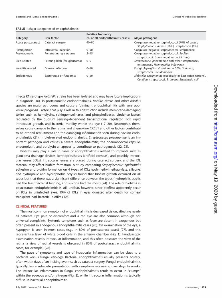
CLINICAL FEATURES
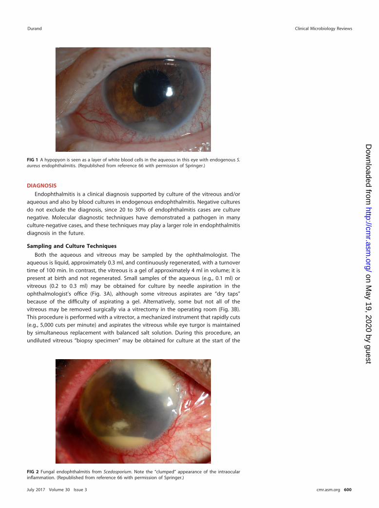
The most common symptom of endophthalmitis is decreased vision, affecting nearlyall patients. Eye pain or discomfort and a red eye are also common although notuniversal complaints. Systemic symptoms such as fever are absent in exogenous butoften present in endogenous endophthalmitis cases (26). On examination of the eye, ahypopyon is seen in most cases (e.g., in 80% of postcataract cases) (27), and thisrepresents a layer of white blood cells in the anterior chamber (Fig. 1). Funduscopicexamination reveals intraocular inflammation, and this often obscures the view of theretina (a view of retinal vessels is obscured in 80% of postcataract endophthalmitiscases, for example) (28).
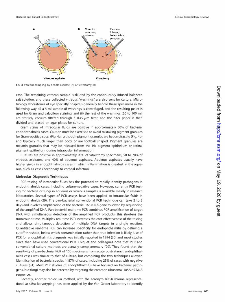
The pace of symptoms and type of intraocular inflammation can be clues to abacterial versus fungal etiology. Bacterial endophthalmitis usually presents acutely,often within days of an inciting event such as cataract surgery. Fungal endophthalmitistypically has a subacute presentation with symptoms worsening over days to weeks.The intraocular inflammation in fungal endophthalmitis tends to occur in “clumps”within the aqueous and/or vitreous (Fig. 2), while intraocular inflammation is typicallydiffuse in bacterial endophthalmitis.
TABLE 1 Major categories of endophthalmitis
Category Risk factorRelative frequency(% of all endophthalmitis cases) Major pathogens
Acute postcataract Cataract surgery 40–80 Coagulase-negative staphylococci (70% of cases),Staphylococcus aureus (10%), streptococci (9%)
Postinjection Intravitreal injection 0–50 Coagulase-negative staphylococci, streptococciPosttraumatic Penetrating eye trauma 2–15 Coagulase-negative staphylococci, Bacillus,
streptococci, Gram-negative bacilli, fungiBleb related Filtering bleb (for glaucoma) 0–5 Streptococcus pneumoniae and other streptococci,
enterococci, Haemophilus influenzaeKeratitis related Corneal infection 0–10 Fungi (Aspergillus, Fusarium) in 50%, S. aureus,
streptococci, PseudomonasEndogenous Bacteremia or fungemia 0–20 Klebsiella pneumoniae (especially in East Asian nations),
Candida, streptococci, S. aureus, Escherichia coli
Bacterial and Fungal Endophthalmitis Clinical Microbiology Reviews
July 2017 Volume 30 Issue 3 cmr.asm.org 599
on May 19, 2020 by guest
http://cmr.asm
.org/D
ownloaded from
DIAGNOSIS
Endophthalmitis is a clinical diagnosis supported by culture of the vitreous and/oraqueous and also by blood cultures in endogenous endophthalmitis. Negative culturesdo not exclude the diagnosis, since 20 to 30% of endophthalmitis cases are culturenegative. Molecular diagnostic techniques have demonstrated a pathogen in manyculture-negative cases, and these techniques may play a larger role in endophthalmitisdiagnosis in the future.
Sampling and Culture Techniques
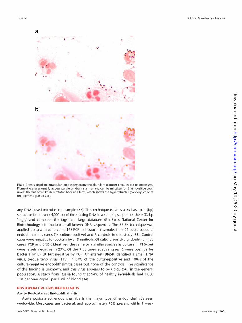
Both the aqueous and vitreous may be sampled by the ophthalmologist. Theaqueous is liquid, approximately 0.3 ml, and continuously regenerated, with a turnovertime of 100 min. In contrast, the vitreous is a gel of approximately 4 ml in volume; it ispresent at birth and not regenerated. Small samples of the aqueous (e.g., 0.1 ml) orvitreous (0.2 to 0.3 ml) may be obtained for culture by needle aspiration in theophthalmologist’s office (Fig. 3A), although some vitreous aspirates are “dry taps”because of the difficulty of aspirating a gel. Alternatively, some but not all of thevitreous may be removed surgically via a vitrectomy in the operating room (Fig. 3B).This procedure is performed with a vitrector, a mechanized instrument that rapidly cuts(e.g., 5,000 cuts per minute) and aspirates the vitreous while eye turgor is maintainedby simultaneous replacement with balanced salt solution. During this procedure, anundiluted vitreous “biopsy specimen” may be obtained for culture at the start of the
FIG 1 A hypopyon is seen as a layer of white blood cells in the aqueous in this eye with endogenous S.aureus endophthalmitis. (Republished from reference 66 with permission of Springer.)
FIG 2 Fungal endophthalmitis from Scedosporium. Note the “clumped” appearance of the intraocularinflammation. (Republished from reference 66 with permission of Springer.)
Durand Clinical Microbiology Reviews
July 2017 Volume 30 Issue 3 cmr.asm.org 600
on May 19, 2020 by guest
http://cmr.asm
.org/D
ownloaded from
case. The remaining vitreous sample is diluted by the continuously infused balancedsalt solution, and these collected vitreous “washings” are also sent for culture. Micro-biology laboratories of eye specialty hospitals generally handle these specimens in thefollowing way: (i) a 5-ml sample of washings is centrifuged, and the resulting pellet isused for Gram and calcofluor staining, and (ii) the rest of the washings (50 to 100 ml)are sterilely vacuum filtered through a 0.45-�m filter, and the filter paper is thendivided and placed on agar plates for culture.
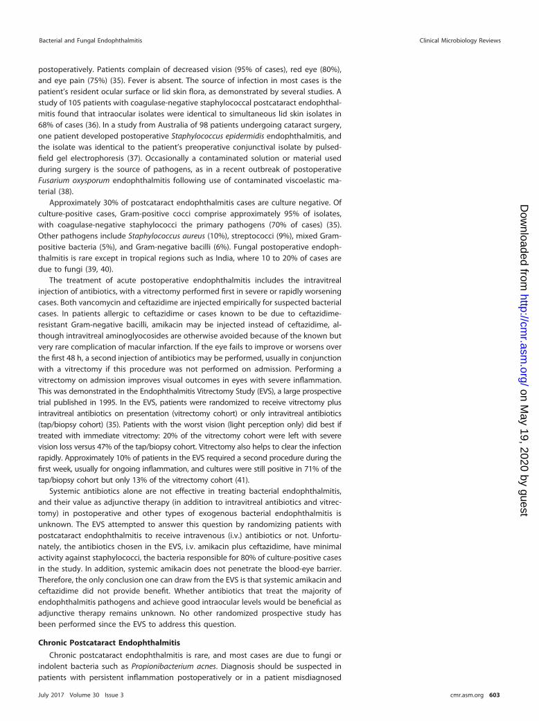
Gram stains of intraocular fluids are positive in approximately 50% of bacterialendophthalmitis cases. Caution must be exercised to avoid mistaking pigment granulesfor Gram-positive cocci (Fig. 4a), although pigment granules are hyperrefractile (Fig. 4b)and typically much larger than cocci or are football shaped. Pigment granules aremelanin granules that may be released from the iris pigment epithelium or retinalpigment epithelium during intraocular inflammation.
Cultures are positive in approximately 90% of vitrectomy specimens, 50 to 70% ofvitreous aspirates, and 40% of aqueous aspirates. Aqueous aspirates usually havehigher yields in endophthalmitis cases in which inflammation is greatest in the aque-ous, such as cases secondary to corneal infection.
Molecular Diagnostic Techniques
PCR testing of intraocular fluids has the potential to rapidly identify pathogens inendophthalmitis cases, including culture-negative cases. However, currently PCR test-ing for bacteria or fungi in aqueous or vitreous samples is available mainly in researchlaboratories. Several types of PCR assays have been applied to intraocular fluids inendophthalmitis (29). The pan-bacterial conventional PCR technique can take 2 to 3days and involves amplification of the bacterial 16S rRNA gene followed by sequencingof the amplified DNA. Pan-bacterial real-time PCR combines PCR amplification of targetDNA with simultaneous detection of the amplified PCR products; this shortens theturnaround time. Multiplex real-time PCR increases the cost-effectiveness of the testingand allows simultaneous detection of multiple DNA targets in a single reaction.Quantitative real-time PCR can increase specificity for endophthalmitis by defining acutoff threshold, below which contamination rather than true infection is likely. Use ofPCR for endophthalmitis diagnosis was initially reported in 1994 (30) and most studiessince then have used conventional PCR. Chiquet and colleagues note that PCR andconventional culture methods are actually complementary (29). They found that thesensitivity of pan-bacterial PCR of 100 specimens from acute postcataract endophthal-mitis cases was similar to that of culture, but combining the two techniques allowedidentification of bacterial species in 87% of cases, including 25% of cases with negativecultures (31). Most PCR studies of endophthalmitis have focused on bacterial patho-gens, but fungi may also be detected by targeting the common ribosomal 18S/28S DNAsequence.
Recently, another molecular method, with the acronym BRiSK (biome representa-tional in silico karyotyping) has been applied by the Van Gelder laboratory to identify
FIG 3 Vitreous sampling by needle aspirate (A) or vitrectomy (B).
Bacterial and Fungal Endophthalmitis Clinical Microbiology Reviews
July 2017 Volume 30 Issue 3 cmr.asm.org 601
on May 19, 2020 by guest
http://cmr.asm
.org/D
ownloaded from
any DNA-based microbe in a sample (32). This technique isolates a 33-base-pair (bp)sequence from every 4,000 bp of the starting DNA in a sample, sequences these 33-bp“tags,” and compares the tags to a large database (GenBank, National Center forBiotechnology Information) of all known DNA sequences. The BRiSK technique wasapplied along with culture and 16S PCR to intraocular samples from 21 postproceduralendophthalmitis cases (14 culture positive) and 7 controls in one study (33). Controlcases were negative for bacteria by all 3 methods. Of culture-positive endophthalmitiscases, PCR and BRiSK identified the same or a similar species as culture in 71% butwere falsely negative in 29%. Of the 7 culture-negative cases, 2 were positive forbacteria by BRiSK but negative by PCR. Of interest, BRiSK identified a small DNAvirus, torque teno virus (TTV), in 57% of the culture-positive and 100% of theculture-negative endophthalmitis cases but none of the controls. The significanceof this finding is unknown, and this virus appears to be ubiquitous in the generalpopulation. A study from Russia found that 94% of healthy individuals had 1,000TTV genome copies per 1 ml of blood (34).
POSTOPERATIVE ENDOPHTHALMITISAcute Postcataract Endophthalmitis
Acute postcataract endophthalmitis is the major type of endophthalmitis seenworldwide. Most cases are bacterial, and approximately 75% present within 1 week
FIG 4 Gram stain of an intraocular sample demonstrating abundant pigment granules but no organisms.Pigment granules usually appear purple on Gram stain (a) and can be mistaken for Gram-positive cocciunless the fine-focus knob is rotated back and forth, which shows the hyperrefractile (coppery) color ofthe pigment granules (b).
Durand Clinical Microbiology Reviews
July 2017 Volume 30 Issue 3 cmr.asm.org 602
on May 19, 2020 by guest
http://cmr.asm
.org/D
ownloaded from
postoperatively. Patients complain of decreased vision (95% of cases), red eye (80%),and eye pain (75%) (35). Fever is absent. The source of infection in most cases is thepatient’s resident ocular surface or lid skin flora, as demonstrated by several studies. Astudy of 105 patients with coagulase-negative staphylococcal postcataract endophthal-mitis found that intraocular isolates were identical to simultaneous lid skin isolates in68% of cases (36). In a study from Australia of 98 patients undergoing cataract surgery,one patient developed postoperative Staphylococcus epidermidis endophthalmitis, andthe isolate was identical to the patient’s preoperative conjunctival isolate by pulsed-field gel electrophoresis (37). Occasionally a contaminated solution or material usedduring surgery is the source of pathogens, as in a recent outbreak of postoperativeFusarium oxysporum endophthalmitis following use of contaminated viscoelastic ma-terial (38).
Approximately 30% of postcataract endophthalmitis cases are culture negative. Ofculture-positive cases, Gram-positive cocci comprise approximately 95% of isolates,with coagulase-negative staphylococci the primary pathogens (70% of cases) (35).Other pathogens include Staphylococcus aureus (10%), streptococci (9%), mixed Gram-positive bacteria (5%), and Gram-negative bacilli (6%). Fungal postoperative endoph-thalmitis is rare except in tropical regions such as India, where 10 to 20% of cases aredue to fungi (39, 40).
The treatment of acute postoperative endophthalmitis includes the intravitrealinjection of antibiotics, with a vitrectomy performed first in severe or rapidly worseningcases. Both vancomycin and ceftazidime are injected empirically for suspected bacterialcases. In patients allergic to ceftazidime or cases known to be due to ceftazidime-resistant Gram-negative bacilli, amikacin may be injected instead of ceftazidime, al-though intravitreal aminoglyocosides are otherwise avoided because of the known butvery rare complication of macular infarction. If the eye fails to improve or worsens overthe first 48 h, a second injection of antibiotics may be performed, usually in conjunctionwith a vitrectomy if this procedure was not performed on admission. Performing avitrectomy on admission improves visual outcomes in eyes with severe inflammation.This was demonstrated in the Endophthalmitis Vitrectomy Study (EVS), a large prospectivetrial published in 1995. In the EVS, patients were randomized to receive vitrectomy plusintravitreal antibiotics on presentation (vitrectomy cohort) or only intravitreal antibiotics(tap/biopsy cohort) (35). Patients with the worst vision (light perception only) did best iftreated with immediate vitrectomy: 20% of the vitrectomy cohort were left with severevision loss versus 47% of the tap/biopsy cohort. Vitrectomy also helps to clear the infectionrapidly. Approximately 10% of patients in the EVS required a second procedure during thefirst week, usually for ongoing inflammation, and cultures were still positive in 71% of thetap/biopsy cohort but only 13% of the vitrectomy cohort (41).
Systemic antibiotics alone are not effective in treating bacterial endophthalmitis,and their value as adjunctive therapy (in addition to intravitreal antibiotics and vitrec-tomy) in postoperative and other types of exogenous bacterial endophthalmitis isunknown. The EVS attempted to answer this question by randomizing patients withpostcataract endophthalmitis to receive intravenous (i.v.) antibiotics or not. Unfortu-nately, the antibiotics chosen in the EVS, i.v. amikacin plus ceftazidime, have minimalactivity against staphylococci, the bacteria responsible for 80% of culture-positive casesin the study. In addition, systemic amikacin does not penetrate the blood-eye barrier.Therefore, the only conclusion one can draw from the EVS is that systemic amikacin andceftazidime did not provide benefit. Whether antibiotics that treat the majority ofendophthalmitis pathogens and achieve good intraocular levels would be beneficial asadjunctive therapy remains unknown. No other randomized prospective study hasbeen performed since the EVS to address this question.
Chronic Postcataract Endophthalmitis
Chronic postcataract endophthalmitis is rare, and most cases are due to fungi orindolent bacteria such as Propionibacterium acnes. Diagnosis should be suspected inpatients with persistent inflammation postoperatively or in a patient misdiagnosed
Bacterial and Fungal Endophthalmitis Clinical Microbiology Reviews
July 2017 Volume 30 Issue 3 cmr.asm.org 603
on May 19, 2020 by guest
http://cmr.asm
.org/D
ownloaded from
with “uveitis” postoperatively even if onset of inflammation is weeks later. A pattern ofapparent response to topical corticosteroids followed by relapse each time corticoste-roids are tapered is common, particularly in chronic bacterial postcataract endophthal-mitis. A clue to diagnosis in chronic fungal endophthalmitis is the “clumped” appear-ance of the intraocular inflammation, while a clue to chronic P. acnes endophthalmitisis the presence of a white plaque on the posterior lens capsule. Diagnosis by needleaspirate alone is often negative, and vitrectomy may be required. In P. acnes endoph-thalmitis, aspirate of the white capsular plaque is often the sample most likely to yielda positive culture.
Treatment of postcataract fungal endophthalmitis nearly always requires removal ofthe IOL in addition to an intraocular injection of an antifungal agent (either ampho-tericin or voriconazole), vitrectomy, and a systemic azole. For systemic therapy, flu-conazole is given for susceptible Candida species and voriconazole for susceptiblemolds and fluconazole-resistant but voriconazole-susceptible Candida species. Treat-ment of chronic P. acnes endophthalmitis does not include systemic therapy but doesusually require a combination of intraocular antibiotic injections plus surgery (vitrec-tomy, capsulectomy, and exchange or removal of the IOL), since intraocular injectionsalone result in relapse in 70% of cases (42).
Postvitrectomy Endophthalmitis
Postvitrectomy endophthalmitis is less common than postcataract endophthalmitis,but the microbiology is similar, with the majority of cases caused by coagulase-negativestaphylococci. Vitrectomies are performed for various retinal conditions (e.g., retinaltears or detachment, vitreous hemorrhage), and most recent studies report anincidence of endophthalmitis of 0.02 to 0.06% (43). Methods for diagnosing andtreating postvitrectomy endophthalmitis are the same as those for postcataractendophthalmitis.
Postkeratoplasty Endophthalmitis
Postkeratoplasty endophthalmitis refers to endophthalmitis following cornealtransplant (keratoplasty). Endophthalmitis occurs in approximately 0.2% of kerato-plasty cases in the acute postoperative period but in up to 0.7% if later cases areincluded (44, 45). The microbiology varies; a study from the United Kingdom ofpostkeratoplasty endophthalmitis, including cases developing long after surgery,found that 31% of cases were due to fungi (mostly Candida) and the remainder dueto Pseudomonas, streptococci, staphylococci, and mycobacterial species (45). Areview of 31 culture-positive cases in the literature reported similar results, withCandida causing 33% of cases, streptococci 27%, staphylococci 20%, enterococci10%, and Pseudomonas 10% (46). The rim of the donor cornea, left over after thecentral core is removed for transplantation, is often sent for surveillance culture, butthe value of doing this has been controversial. However, this information might beimportant for Candida: an eye that receives a cornea whose donor rim culturesubsequently grows Candida has a 3% chance of developing Candida endophthal-mitis (46). It is unknown whether such eyes should be treated prophylactically withan antifungal agent or just followed closely by the ophthalmologist. Treatment ofpostkeratoplasty endophthalmitis may require replacement of the infected corneain addition to intracameral (into the aqueous) and/or intravitreal antibiotics andvitrectomy (as needed).
POSTINJECTION ENDOPHTHALMITIS
The use of intravitreal injections has rapidly increased since FDA approval ofanti-VEGF injections in 2004, as noted above. A study utilizing a Medicare databasereported that intravitreal injections increased from 83,000 in 2004 to 2.4 million in 2012(47). Most injections involve use of anti-VEGF agents, given primarily for neovascularage-related macular degeneration (AMD) but also given for diabetic retinopathy andother indications. The risk of endophthalmitis is approximately 0.05% per injection (48),
Durand Clinical Microbiology Reviews
July 2017 Volume 30 Issue 3 cmr.asm.org 604
on May 19, 2020 by guest
http://cmr.asm
.org/D
ownloaded from
and since anti-VEGF injections are repeated at regular intervals (usually monthly) forAMD, the risk is cumulative. Other types of intravitreal injections, such as corticosteroidsfor inflammatory eye conditions, are also being used with increasing frequency. Aretrospective study based on a U.S. medical claims database found a higher rate ofendophthalmitis after corticosteroid than after anti-VEGF injections (0.13% versus0.02%) (49), but this study has been questioned because it did not compare onlyculture-positive cases (culture results were not available). Sterile inflammation mimick-ing endophthalmitis may occur with higher frequency after corticosteroid than afteranti-VEGF injections.
Patients with postinjection endophthalmitis typically present within 5 days after theinjection, and the most common symptom is decreased vision. A British review of 47patients with postinjection endophthalmitis found that 96% had decreased vision, 73%had eye pain/photophobia, and 49% had eye redness (50). The microbiology ofpostinjection endophthalmitis includes coagulase-negative staphylococci (65%), viri-dans streptococci (30%), S. aureus (0 to 5%), and others (0 to 4%) (48, 50, 51). This issimilar to the microbiology of postcataract endophthalmitis except that the incidenceof viridans streptococci is over 3-fold higher than in postcataract endophthalmitis, andthis may be due to the fact that intravitreal injections are usually performed in the officerather than the operating room and without the use of masks. Oral flora, includingviridans streptococci, may be aerosolized by speaking, and using masks or observing astrict “no talking” policy during injections decreases the rate of streptococcal endoph-thalmitis. A study of 25 centers in France that used masks for injections (someperformed in operating rooms) reported a very low (0.007%) incidence of endophthal-mitis, with streptococci causing only 4% of cases (52). A center in the United Statesdecreased the postinjection endophthalmitis rate 2-fold (from 0.02% to 0.01%) andendophthalmitis due to oral pathogens 7-fold (from 0.015% to 0.002%) by instituting ano-talking policy during office-based injections (53).
Rarely, a postinjection endophthalmitis case may be part of an outbreak due to useof contaminated solutions. The onset of symptoms is usually rapid in bacterial cases butmay be delayed in fungal cases. All 12 patients who developed streptococcal endoph-thalmitis after injection of contaminated anti-VEGF solutions presented 1 to 6 dayspostinjection, while 14 patients who developed Bipolaris hawaiiensis endophthalmitisfollowing injection of contaminated triamcinolone developed symptoms a median of83 days later (54, 55). Outcomes in outbreaks are typically poor: an outbreak ofEscherichia coli and Citrobacter endophthalmitis related to a counterfeit anti-VEGFsolution left 14% of eyes blind (no light perception) (56).
Treatment of postinjection endophthalmitis is the same as for postoperative endo-phthalmitis, described above, and visual outcomes depend partly on the pathogen:streptococci are particularly virulent in the eye. A review of 197 cases reported in 43publications found that very poor outcomes (�20/400 vision) were seen in 13% ofcoagulase-negative staphylococcal cases and 31% of culture-negative cases but in 94%of streptococcal cases (48).
POSTTRAUMATIC ENDOPHTHALMITIS
Penetrating eye trauma (open globe injury) occurs in 2 to 3.8/100,000 population inthe United States. Posttraumatic endophthalmitis occurs in 0.9 to 18% of adults and 5to 54% of children with such injuries (57, 58). Risk factors include delay in treatment ofthe eye trauma, rural setting of the injury, presence of an intraocular foreign body, lenscapsule disruption, and a lacerating injury rather than blunt trauma with globe rupture.Endophthalmitis symptoms include decreased vision and eye pain. On examination, ahypopyon and intraocular inflammation are usually seen, and an eye wound withpurulent drainage may sometimes be seen. Specific findings may suggest the patho-gen. Bacillus infections are fulminant and sometimes associated with a ring cornealabscess, while Clostridium endophthalmitis cases may have gas bubbles in the anteriorchamber and a green-brown hypopyon (59). Major causes of posttraumatic endoph-thalmitis include coagulase-negative staphylococci, Bacillus species, streptococci, Gram-
Bacterial and Fungal Endophthalmitis Clinical Microbiology Reviews
July 2017 Volume 30 Issue 3 cmr.asm.org 605
on May 19, 2020 by guest
http://cmr.asm
.org/D
ownloaded from
negative bacilli (e.g., Pseudomonas and Klebsiella), and fungi. Treatment includes re-moval of any retained intraocular foreign object, intravitreal antibiotics, vitrectomy inmost cases, and often adjunctive topical and systemic antibiotics. Tetanus vaccine isindicated after open globe injuries if the patient’s last vaccination was �5 years earlier.Final visual acuity is variable and depends to a great extent on the pathogen, but onemulticenter study found that 41% of all cases achieved 20/40 or better, while 47% wereleft with minimal (light perception only) or no vision (60).
BLEB-RELATED ENDOPHTHALMITIS
Bleb-related endophthalmitis usually occurs suddenly but months to years followingglaucoma surgery in which a “bleb” is created. A filtering bleb is a defect in the scleracovered by conjunctiva that improves aqueous resorption into the systemic circulation.However, following this surgery, only a thin barrier of conjunctiva separates theaqueous from the outside world in the location of the bleb, and this carries an ongoingrisk of developing endophthalmitis. A large study from Japan reported an endophthal-mitis risk of 1% over 5 years (61), and other studies have reported higher rates. Blebsthat are leaking (aqueous) increase the endophthalmitis risk by nearly 5-fold (61). Aninfection of the bleb, or blebitis, often precedes endophthalmitis. Blebitis should bepromptly treated to prevent progression. Streptococci, primarily viridans streptococcibut also S. pneumoniae, are the pathogens in over one-third of bleb-related endoph-thalmitis cases; other major pathogens include S. aureus, Haemophilus influenzae, andenterococci. Treatment of endophthalmitis is with intracameral and/or intravitrealantibiotic injections, vitrectomy in severe cases, plus topical antibiotics in most cases;systemic antibiotics such as quinolones are also sometimes given as adjunctive therapy.Visual outcome is poor for cases due to pathogens that are virulent in the eye, such asenterococci and any type of streptococci.
KERATITIS-RELATED ENDOPHTHALMITIS
Keratitis means corneal infection, and most cases are treated with topical anti-biotics. Progression to endophthalmitis is uncommon and usually occurs by exten-sion of the infection through the cornea into the aqueous. A majority of cases ofkeratitis-related endophthalmitis are due to molds. A series from Florida of nearly10,000 keratitis cases found that only 0.5% progressed to endophthalmitis; fungalkeratitis (keratomycosis) was a significant risk factor for such progression (62). Overhalf the endophthalmitis cases in that series were due to molds (53%), whileGram-positive bacteria (27%) and Gram-negative bacilli (20%) comprised the re-maining cases. A series from New Jersey reported a lower rate of fungal endoph-thalmitis (17%), but this reflects the much lower incidence of keratomycosis incolder, less humid climates (63). The risk of keratomycosis progressing to endoph-thalmitis varies, but one center reported that 6% of Fusarium keratitis cases seenduring an outbreak developed endophthalmitis (64).
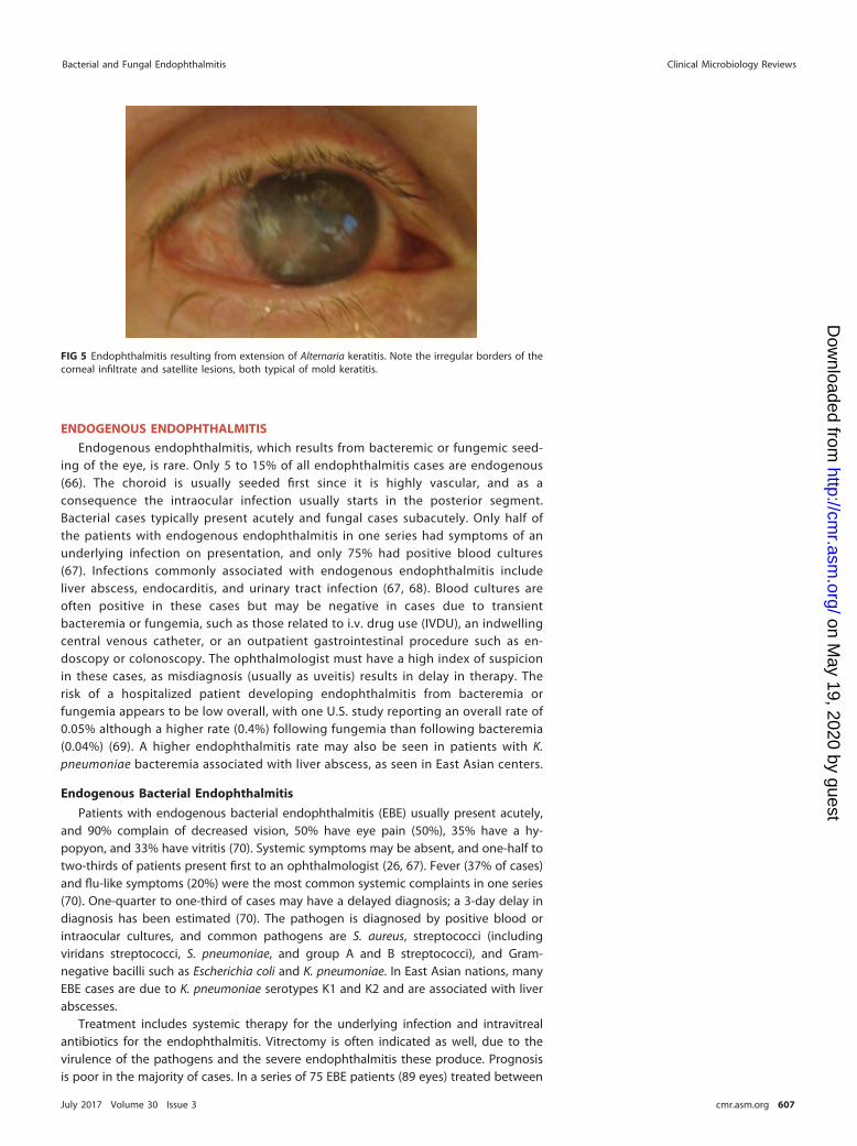
In cases of keratomycosis-related endophthalmitis, Fusarium and Aspergillus are themost common etiologies (65). Keratomycosis may be difficult to diagnose, ascultures of corneal scrapings may be falsely negative. The appearance of the cornealinfiltrate often suggests the diagnosis. Unlike bacteria, molds often produce cornealinfiltrates with fuzzy or feathery borders and satellite lesions (Fig. 5). Noninvasivetechniques such as optical coherence tomography can support the diagnosis ofkeratomycosis. The finding of frond-like projections extending from the back of thecornea into the aqueous, or of thick clumped material in the aqueous, is concerningfor keratomycosis-related endophthalmitis. Treatment of keratitis-related endoph-thalmitis includes intracameral and/or intravitreal antibiotics, topical antibiotics,adjunctive systemic antifungal antibiotics for keratomycosis-related cases, andvitrectomy as needed. Corneal transplant to debulk the infection may be required,particularly in cases due to molds.
Durand Clinical Microbiology Reviews
July 2017 Volume 30 Issue 3 cmr.asm.org 606
on May 19, 2020 by guest
http://cmr.asm
.org/D
ownloaded from
ENDOGENOUS ENDOPHTHALMITIS
Endogenous endophthalmitis, which results from bacteremic or fungemic seed-ing of the eye, is rare. Only 5 to 15% of all endophthalmitis cases are endogenous(66). The choroid is usually seeded first since it is highly vascular, and as aconsequence the intraocular infection usually starts in the posterior segment.Bacterial cases typically present acutely and fungal cases subacutely. Only half ofthe patients with endogenous endophthalmitis in one series had symptoms of anunderlying infection on presentation, and only 75% had positive blood cultures(67). Infections commonly associated with endogenous endophthalmitis includeliver abscess, endocarditis, and urinary tract infection (67, 68). Blood cultures areoften positive in these cases but may be negative in cases due to transientbacteremia or fungemia, such as those related to i.v. drug use (IVDU), an indwellingcentral venous catheter, or an outpatient gastrointestinal procedure such as en-doscopy or colonoscopy. The ophthalmologist must have a high index of suspicionin these cases, as misdiagnosis (usually as uveitis) results in delay in therapy. Therisk of a hospitalized patient developing endophthalmitis from bacteremia orfungemia appears to be low overall, with one U.S. study reporting an overall rate of0.05% although a higher rate (0.4%) following fungemia than following bacteremia(0.04%) (69). A higher endophthalmitis rate may also be seen in patients with K.pneumoniae bacteremia associated with liver abscess, as seen in East Asian centers.
Endogenous Bacterial Endophthalmitis
Patients with endogenous bacterial endophthalmitis (EBE) usually present acutely,and 90% complain of decreased vision, 50% have eye pain (50%), 35% have a hy-popyon, and 33% have vitritis (70). Systemic symptoms may be absent, and one-half totwo-thirds of patients present first to an ophthalmologist (26, 67). Fever (37% of cases)and flu-like symptoms (20%) were the most common systemic complaints in one series(70). One-quarter to one-third of cases may have a delayed diagnosis; a 3-day delay indiagnosis has been estimated (70). The pathogen is diagnosed by positive blood orintraocular cultures, and common pathogens are S. aureus, streptococci (includingviridans streptococci, S. pneumoniae, and group A and B streptococci), and Gram-negative bacilli such as Escherichia coli and K. pneumoniae. In East Asian nations, manyEBE cases are due to K. pneumoniae serotypes K1 and K2 and are associated with liverabscesses.
Treatment includes systemic therapy for the underlying infection and intravitrealantibiotics for the endophthalmitis. Vitrectomy is often indicated as well, due to thevirulence of the pathogens and the severe endophthalmitis these produce. Prognosisis poor in the majority of cases. In a series of 75 EBE patients (89 eyes) treated between
FIG 5 Endophthalmitis resulting from extension of Alternaria keratitis. Note the irregular borders of thecorneal infiltrate and satellite lesions, both typical of mold keratitis.
Bacterial and Fungal Endophthalmitis Clinical Microbiology Reviews
July 2017 Volume 30 Issue 3 cmr.asm.org 607
on May 19, 2020 by guest
http://cmr.asm
.org/D
ownloaded from
2001 and 2012, only 41% of eyes recovered 20/200 vision or better, while 19% of eyeswere enucleated or eviscerated (70). Vitrectomy was associated with a better visualprognosis and a lower rate of evisceration or enucleation.
Endogenous Fungal Endophthalmitis
Candida is the most common cause of endogenous fungal endophthalmitis (EFE).The initial manifestation is usually chorioretinitis, manifested as fluffy white chorioreti-nal lesions. This may be clinically silent, with symptoms developing only after devel-opment of significant vitritis. Of note, Candida chorioretinitis is counted as a type ofendophthalmitis in some studies, while others distinguish chorioretinitis from endoph-thalmitis, meaning cases with significant vitritis; the latter convention will be followedhere.
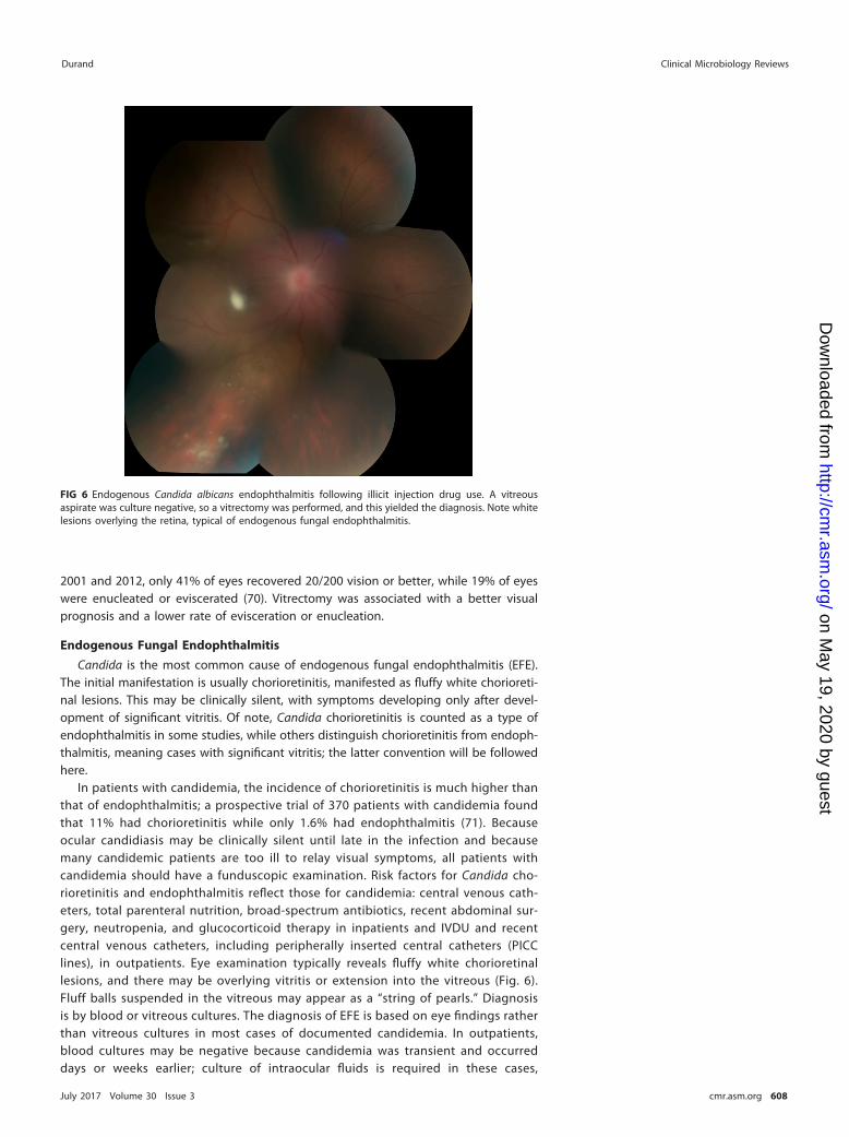
In patients with candidemia, the incidence of chorioretinitis is much higher thanthat of endophthalmitis; a prospective trial of 370 patients with candidemia foundthat 11% had chorioretinitis while only 1.6% had endophthalmitis (71). Becauseocular candidiasis may be clinically silent until late in the infection and becausemany candidemic patients are too ill to relay visual symptoms, all patients withcandidemia should have a funduscopic examination. Risk factors for Candida cho-rioretinitis and endophthalmitis reflect those for candidemia: central venous cath-eters, total parenteral nutrition, broad-spectrum antibiotics, recent abdominal sur-gery, neutropenia, and glucocorticoid therapy in inpatients and IVDU and recentcentral venous catheters, including peripherally inserted central catheters (PICClines), in outpatients. Eye examination typically reveals fluffy white chorioretinallesions, and there may be overlying vitritis or extension into the vitreous (Fig. 6).Fluff balls suspended in the vitreous may appear as a “string of pearls.” Diagnosisis by blood or vitreous cultures. The diagnosis of EFE is based on eye findings ratherthan vitreous cultures in most cases of documented candidemia. In outpatients,blood cultures may be negative because candidemia was transient and occurreddays or weeks earlier; culture of intraocular fluids is required in these cases,
FIG 6 Endogenous Candida albicans endophthalmitis following illicit injection drug use. A vitreousaspirate was culture negative, so a vitrectomy was performed, and this yielded the diagnosis. Note whitelesions overlying the retina, typical of endogenous fungal endophthalmitis.
Durand Clinical Microbiology Reviews
July 2017 Volume 30 Issue 3 cmr.asm.org 608
on May 19, 2020 by guest
http://cmr.asm
.org/D
ownloaded from
including vitrectomy if initial aspirate cultures are negative. Candida albicans is thepredominant species of EFE, but all Candida species have been described.
Treatment of candidemia has been discussed in recent Infectious Disease Societyof America guidelines, which also discuss treatment of Candida chorioretinitis andendophthalmitis (72). Treatment with systemic agents is usually adequate for casesof chorioretinitis that do not have macula-threatening lesions. Macula-threateningchorioretinitis and cases of endophthalmitis require intravitreal antifungal injec-tions (amphotericin or voriconazole) in addition to systemic therapy. Endophthal-mitis cases with significant vitritis usually require vitrectomy as well. For systemicagents, fluconazole is recommended for fluconazole-susceptible Candida, voricona-zole for fluconazole-resistant but voriconazole-susceptible isolates, and liposomalamphotericin, with or without 5-flucytosine, for azole-resistant strains. Fluconazoleor voriconazole is preferred to amphotericin for susceptible isolates because azolesare less toxic and produce higher levels in the vitreous. Voriconazole, for example,achieves vitreous levels that are approximately 40% of serum levels even inuninflamed eyes. Systemic echinocandins do not reach adequate concentrations inthe vitreous to treat endophthalmitis. Echinocandins may achieve reasonable levelsin the choroid, but their role in treating chorioretinitis alone is unknown becausethere are few data in humans. One study measured micafungin levels in a patientgiven i.v. micafungin prior to a scheduled enucleation and found reasonable levelsin the choroid (34% of plasma levels) but very poor levels in the retina or vitreous(7% and 0.9% of plasma levels, respectively) (73).
Endogenous mold endophthalmitis is rare and seen primarily in immunocompro-mised patients, such as patients with hematologic malignancies or transplant recipi-ents, or patients with IVDU. Aspergillus and Fusarium are the major pathogens, althoughScedosporium and other fungi are also seen. Therapy includes systemic antifungaltherapy plus intravitreal amphotericin or voriconazole and usually vitrectomy.
VISUAL OUTCOMES AND MICROBIOLOGY
Some loss of vision is common after endophthalmitis, but the final visual acuityusually cannot be determined for weeks to months later. Unless the eye has no lightperception, vision may improve once the acute inflammation has resolved. Anyvision is worth saving, so every effort should be made to save even light perceptionvision.
Visual outcomes in endophthalmitis are related to a number of factors, includingpresenting visual acuity and the promptness of appropriate therapy, but thepathogen involved is a major factor in nearly all cases. In postcataract endophthal-mitis, the EVS found that a “good” final visual acuity of 20/100 or better occurredin approximately 80% of culture-negative or coagulase-negative staphylococcalcases but in only 50% due to S. aureus, 30% due to streptococci (of any type), and56% due to Gram-negative bacilli (86). Considering outcomes by pathogen regard-less of endophthalmitis category, a group in Florida reported that very poor visualacuity (20/400 or worse) occurred in 75% of cases due to streptococci (with nodifference in outcomes between viridans streptococci, S. pneumoniae, and beta-hemolytic streptococci), 93% of enterococcal cases, 64% of Bacillus cases, 69% of H.influenzae cases, 70% of Serratia cases, and 92% of Pseudomonas cases (74–79).These cases were seen over a 10-year period at a tertiary eye hospital where themost severe endophthalmitis cases are likely to be referred. However, there is hopeeven in endophthalmitis cases due to bacteria associated with a poor visualprognosis. Bacillus produces a fulminant endophthalmitis, but 18% of cases at thateye hospital had a final acuity of 20/60 or better (76).
PREVENTION
There are almost no published randomized controlled trials evaluating theefficacy of various proposed measures to prevent endophthalmitis, so the optimalmethods are largely unknown (80). Prophylaxis for eye surgery with topical
Bacterial and Fungal Endophthalmitis Clinical Microbiology Reviews
July 2017 Volume 30 Issue 3 cmr.asm.org 609
on May 19, 2020 by guest
http://cmr.asm
.org/D
ownloaded from
povidone-iodine preoperatively and topical antibiotics postoperatively is routinelygiven, but these measures have not been evaluated by a randomized controlledtrial. Intracameral antibiotics are being used with increasing frequency as prophy-laxis for cataract surgery. A randomized controlled trial in Europe found thatintracameral cefuroxime prophylaxis at the end of cataract surgery was associatedwith a postoperative endophthalmitis rate of 0.06%, versus 0.3% in control eyes(81). However, the study results were questioned in the United States because thecontrol group’s endophthalmitis rate was approximately 3-fold higher than thatseen at most U.S. centers. Rare cases of anaphylaxis to prophylactic intracameralcefuroxime have been reported (82), and use of prophylactic intracameral vanco-mycin has been associated with a rare but devastating complication of hemorrhagicocclusive retinal vasculitis (83, 84). For decreasing the risk of postinjection endo-phthalmitis, many centers have adopted the use of masks or observing a strictno-talking policy during injections. For patients presenting with penetrating eyetrauma, prompt surgical repair and 48 h of prophylactic broad-spectrum systemicantibiotics (e.g., i.v. vancomycin plus ceftazidime) have been associated with a verylow rate of posttraumatic endophthalmitis (57). Patients who have filtering blebs forglaucoma should be treated for blebitis without delay in order to prevent thisrelatively minor infection from leading to bleb-related endophthalmitis. I alsorecommend that patients with filtering blebs receive pneumococcal vaccination(80, 85). Endogenous bacterial endophthalmitis cannot usually be anticipated orprevented, but some cases of endogenous Candida endophthalmitis can be pre-vented by screening all candidemic patients with funduscopic examinations.
CONCLUSION
Endophthalmitis is a severe eye infection that requires rapid diagnosis andtreatment to save vision. Endophthalmitis may occur from pathogens introducedinto the eye from an external source (exogenous endophthalmitis) or via thebloodstream (endogenous endophthalmitis). Most cases of exogenous endophthal-mitis occur as a complication of cataract surgery, an intravitreal injection, orpenetrating ocular trauma. Endogenous endophthalmitis may develop as a result ofmetastatic spread of an extraocular focus of infection, such as endocarditis or a liverabscess, or from transient bacteremia or fungemia related to illicit injection druguse or an indwelling central venous catheter. Nearly all patients present withdecreased vision, and some also have eye pain. A hypopyon and intraocularinflammation are typical findings on eye examination. Diagnosis is clinical, sup-ported by cultures of vitreous and/or aqueous or also by blood cultures in endog-enous cases. Newer diagnostic techniques such as PCR offer the promise of rapididentification of intraocular pathogens, including in culture-negative cases. Prompttreatment with intravitreal antibiotics is essential; some cases also benefit from asurgical vitrectomy. The visual outcome depends partly on the pathogen. Cases dueto coagulase-negative staphylococci usually recover good vision, while half or moreof the eyes infected with streptococci (of any type), S. aureus, or Gram-negativebacilli are left with poor vision. Few randomized controlled trials have beenperformed to determine the most effective ways to prevent endophthalmitis, andmore such trials are needed.
REFERENCES1. Melo GB, Bispo PJM, Yu MCZ, Pignatari AC, Höfling-Lima AL. 2011.
Microbial profile and antibiotic susceptibility of culture-positive bac-terial endophthalmitis. Eye 25:382–388. https://doi.org/10.1038/eye.2010.236.
2. Gupta A, Orlans HO, Hornby SJ, Bowler ICJ. 2014. Microbiology andvisual outcomes of culture-positive bacterial endophthalmitis in Oxford,UK. Graefes Arch Clin Exp Ophthalmol 252:1825–1830. https://doi.org/10.1007/s00417-014-2658-7.
3. Kessner R, Golan S, Barak A. 2014. Changes in the etiology of endoph-
thalmitis from 2003 to 2010 in a large tertiary medical center. Eur JOphthalmol 24:918 –924. https://doi.org/10.5301/ejo.5000473.
4. Falavarjani KG, Nekoozadeh S, Modarres M, Parvaresh MM, Hashemi M,Soodi R, Alemzadeh SA. 2012. Isolates and antibiotic resistance ofculture-proven endophthalmitis cases presented to a referral center inTehran. Middle East Afr J Ophthalmol 19:361–363. https://doi.org/10.4103/0974-9233.102740.
5. Moloney TP, Park J. 2014. Microbiological isolates and antibiotic sensi-tivities in culture-proven endophthalmitis: a 15-year review. Br J Oph-
Durand Clinical Microbiology Reviews
July 2017 Volume 30 Issue 3 cmr.asm.org 610
on May 19, 2020 by guest
http://cmr.asm
.org/D
ownloaded from
thalmol 98:1492–1497. https://doi.org/10.1136/bjophthalmol-2014-305030.
6. Nam KY, Lee JE, Lee JE, Jeung WJ, Park JM, Park JM, Chung IY, Han YS,Yun IH, Kim HW, Byon IS. 2015. Clinical features of infectious endoph-thalmitis in South Korea: a five-year multicenter study. BMC Infect Dis15:177–183. https://doi.org/10.1186/s12879-015-0900-5.
7. Sharma S, Padhi TR, Basu S, Kar S, Roy A, Das T. 2014. Endophthalmitispatients seen in a tertiary eye care centre in Odisha: a clinicomicrobio-logical analysis. Indian J Med Res 139:91–98.
8. Gharamah AA, Moharram AM, Ismail MA, Al-Hussaini AK. 2012. Bacterialand fungal endophthalmitis in Upper Egypt: related species and riskfactors. Asian Pac J Trop Biomed 2:655– 659. https://doi.org/10.1016/S2221-1691(12)60115-4.
9. Duan F, Wu K, Liao J, Zheng Y, Yuan Z, Tan J, Lin X. 2016. Causativemicroorganisms of infectious endophthalmitis: a 5-year retrospectivestudy. J Ophthalmol https://doi.org/10.1155/2016/6764192.
10. Simunovic MP, Rush RB, Hunyor AP, Chang AA. 2012. Endophthalmitisfollowing intravitreal injection versus endophthalmitis following cat-aract surgery: clinical features, causative organisms and post-treatment outcomes. Br J Ophthalmol 96:862– 866. https://doi.org/10.1136/bjophthalmol-2011-301439.
11. Valdez-Garcia JE, Climent A, Chavez-Mondragon E, Lozano-Ramirez JF.2014. Anterior chamber bacterial contamination in cataract surgery.BMC Ophthalmol 14:57. https://doi.org/10.1186/1471-2415-14-57.
12. Srinivasan R, Tiroumal S, Kanungo R, Natarajan MK. 2002. Microbialcontamination of the anterior chamber during phacoemulsification. JCataract Refract Surg 28:2173–2176. https://doi.org/10.1016/S0886-3350(02)01493-1.
13. Lundstrom M, Wejde G, Stenevi U, Thorbum W, Montan P. 2007. Endo-phthalmitis after cataract surgery: a nationwide prospective study eval-uating incidence in relation to incision type and location. Ophthalmol-ogy 114:866 – 870. https://doi.org/10.1016/j.ophtha.2006.11.025.
14. Fang CT, Lai SY, Yi WC, Hsueh PR, Liu KL, Chang SC. 2007. Klebsiellapneumoniae genotype K1: an emerging pathogen that causes septicocular or central nervous system complications from pyogenic liverabscess. Clin Infect Dise 45:284 –293. https://doi.org/10.1086/519262.
15. Wiskur BJ, Hunt JJ, Callegan MC. 2008. Hypermucoviscosity as a viru-lence factor in experimental Klebsiella pneumoniae endophthalmitis.Invest Ophthalmol Vis Sci 49:4931– 4938. https://doi.org/10.1167/iovs.08-2276.
16. Lin TL, Hsieh PF, Huang YT, Lee WC, Tsai YT, Su PA, Pan YJ, Hsu CR, WuMC, Wang JT. 2014. Isolation of a bacteriophage and its depolymerasespecific for K1 capsule of Klebsiella pneumoniae: implication in typingand treatment. J Infect Dis 210:1734 –1744. https://doi.org/10.1093/infdis/jiu332.
17. Parkunan SM, Callegan MC. 2016. The pathogenesis of bacterial endo-phthalmitis, p 17– 47. In Durand ML, Miller JW, Young LH (ed), Endoph-thalmitis. Springer International Publishing, Basel, Switzerland.
18. Callegan MC, Kane ST, Cochran DC, Ramadan RT, Chodosh J, McLean C,Stroman DW. 2006. Virulence factor profiles and antimicrobial suscepti-bilities of ocular Bacillus isolates. Cur Eye Res 31:693–702. https://doi.org/10.1080/02713680600850963.
19. Callegan MC, Kane ST, Cochran DC, Gilmore MS, Gominet M, Lereclus D.2003. Relationship of plcR-regulated factors to Bacillus endophthalmitisvirulence. Infect Immun 71:3116 –3124. https://doi.org/10.1128/IAI.71.6.3116-3124.2003.
20. Callegan MC, Kane ST, Cochran DC, Novosad B, Gilmore MS, Gominet M,Lereclus D. 2005. Bacillus endophthalmitis: roles of bacterial toxins andmotility during infection. Invest Ophthalmol Vis Sci 46:3233–3238.https://doi.org/10.1167/iovs.05-0410.
21. Parkunan SM, Randall CB, Astley RA, Furtado GC, Lira SA, Callegan MC. 10June 2016. CXCL1, but not IL-6, significantly impacts intraocular inflam-mation during infection. J Leukoc Biol.
22. Ng EW, Costa JR, Samiy N, Ruoff KL, Connolly E, Cousins FV, D’Amico DJ.2002. Contribution of pneumolysin and autolysin to the pathogenesis ofexperimental pneumococcal endophthalmitis. Retina 22:622– 632.https://doi.org/10.1097/00006982-200210000-00014.
23. Sanders ME, Norcross EW, Robertson ZM, Moore QC 3rd, Fratkin J,Marquart ME. 2011. The Streptococcus pneumoniae capsule is requiredfor full virulence in pneumococcal endophthalmitis. Invest OphthalmolVis Sci 52:865– 872. https://doi.org/10.1167/iovs.10-5513.
24. Baillif S, Ecochard R, Casoli E, Freney J, Burillon C, Kodjikian L. 2008.Adherence and kinetics of biofilm formation of Staphylococcus epider-midis to different types of intraocular lenses under dynamic flow con-
ditions. J Cataract Refract Surg 34:153–158. https://doi.org/10.1016/j.jcrs.2007.07.058.
25. Mazoteras P, Quiles MG, Martins Bispo PJ, Höfling-Lima AL, Pignatari AC,Casaroli-Marano RP. 2016. Analysis of intraocular lens biofilms and fluidsafter long-term uncomplicated cataract surgery. Am J Ophthalmol 169:46 –57. https://doi.org/10.1016/j.ajo.2016.06.012.
26. Jackson TL, Eykyn SJ, Graham EM, Stanford MR. 2003. Endogenousbacterial endophthalmitis: a 17-year prospective series and review of267 reported cases. Survey Ophthalmol 48:403– 423. https://doi.org/10.1016/S0039-6257(03)00054-7.
27. Lalwani GA, Flynn HW, Jr, Scott IU, Quinn CM, Berrocal AM, Davis JL,Murray TG, Smiddy WE, Miller D. 2008. Acute-onset endophthalmitisafter clear corneal cataract surgery (1996-2005). Clinical features, caus-ative organisms, and visual acuity outcomes. Ophthalmology 115:473– 476.
28. Taban M, Behrens A, Newcomb RL, Nobe MY, Saedi G, Sweet PM,McDonnell PJ. 2005. Acute endophthalmitis following cataract surgery: asystematic review of the literature. Arch Ophthalmol 123:613– 620.https://doi.org/10.1001/archopht.123.5.613.
29. Chiquet C, Boisset S, Cornut P-L, Maurin M. 2016 The molecular diagno-sis of endophthalmitis, p 77–97. In Durand ML, Miller JW, Young LH (ed),Endophthalmitis. Springer International Publishing, Basel, Switzerland.
30. Hykin PG, Tobal K, McIntye G, Matheson MM, Towler HM, Lightman SL.1994. The diagnosis of delayed post-operative endophthalmitis by poly-merase chain reaction of bacterial DNA in vitreous samples. J MedMicrobiol 40:408 – 415. https://doi.org/10.1099/00222615-40-6-408.
31. Chiquet C, Cornut P-L, Benito Y, Thuret G, Maurin M, Lafontaine PO,Pechinot A, Palombi K, Lina G, Bron A, Denis P, Carricajo A, Creuzot C,Romanet JP, Vandenesch F, French Institutional Endophthalmitis StudyGroup. 2008. Eubacterial PCR for bacterial detection and identification in100 acute postcataract surgery endophthalmitis. Invest Ophthalmol VisSci 49:1971–1978. https://doi.org/10.1167/iovs.07-1377.
32. Hong BK, Lee CS, Van Gelder RN, Garg SJ. 2015. Emerging techniques forpathogen discovery in endophthalmitis. Curr Opin Ophthalmol 26:221–225. https://doi.org/10.1097/ICU.0000000000000145.
33. Lee AY, Akileswaran L, Tibbetts MD, Garg SJ, Van Gelder RN. 2015.Identification of torque teno virus in culture-negative endophthalmitisby representational deep DNA sequencing. Ophthalmology 122:524 –530. https://doi.org/10.1016/j.ophtha.2014.09.001.
34. Vasilyev EV, Trofimov DY, Tonevitsky AG, Ilinsky VV, Korostin DO, Re-brikov DV. 2009. Torque teno virus (TTV) distribution in healthy Russianpopulation. Virology J 6:134. https://doi.org/10.1186/1743-422X-6-134.
35. Endophthalmitis Vitrectomy Study Group. 1995. Results of the Endoph-thalmitis Vitrectomy Study: a randomized trial of immediate vitrectomyand of intravenous antibiotics for the treatment of postoperative bac-terial endophthalmitis. Arch Ophthalmol 113:1479 –1496. https://doi.org/10.1001/archopht.1995.01100120009001.
36. Bannerman TL, Rhoden DL, McAllister SK, Miller JM, Wilson LA. 1997. Thesource of coagulase-negative staphylococci in the Endophthalmitis Vit-rectomy Study. A comparison of eyelid and intraocular isolates usingpulsed-field gel electropheresis. Arch Ophthalmol 115:357–361.
37. Leong JK, Shah R, McCluskey PJ, Benn RA, Taylor RF. 2002. Bacterialcontamination of the anterior chamber during phacoemulsification cat-aract surgery. J Cataract Refract Surg 28:826 – 833. https://doi.org/10.1016/S0886-3350(01)01160-9.
38. Buchta V, Feuermannova A, Vasa M, Bašková L, Kutová R, Kubátová A,Vejsová M. 2014. Outbreak of fungal endophthalmitis due to Fusariumoxysporum following cataract surgery. Mycopathologia 177:115–121.https://doi.org/10.1007/s11046-013-9721-5.
39. Anand AR, Therese KL, Madhavan HN. 2000 Spectrum of aetiologicalagents of postoperative endophthalmitis and antibiotic susceptibility ofbacterial isolates. Indian J Ophthalmol 48:123–128.
40. Sharma S, Sahu SK, Dhillon V, Das S, Rath S. 2015. Reevaluating intra-cameral cefuroxime as a prophylaxis against endophthalmitis after cat-aract surgery in India. J Cataract Refract Surg 41:393–399. https://doi.org/10.1016/j.jcrs.2014.05.038.
41. Doft BH, Kelsey SF, Wisniewski SR. 1998. Additional procedures afterthe initial vitrectomy or tap-biopsy in the Endophthalmitis Vitrec-tomy Study. Ophthalmology 105:707. https://doi.org/10.1016/S0161-6420(98)94028-3.
42. Shirodkar AR, Pathengay A, Flynn HW, Albini TA, Berrocal AM, Davis JL,Lalwani GA, Murray TG, Smiddy WE, Miller D. 2012. Delayed versusacute-onset endophthalmitis after cataract surgery. Am J Ophthalmol153:391–398. https://doi.org/10.1016/j.ajo.2011.08.029.
Bacterial and Fungal Endophthalmitis Clinical Microbiology Reviews
July 2017 Volume 30 Issue 3 cmr.asm.org 611
on May 19, 2020 by guest
http://cmr.asm
.org/D
ownloaded from
43. Dave VP, Schwartz SG, Flynn HW, Jr. 2014. Endophthalmitis followingpars plana vitrectomy: a literature review of incidence, causative organ-isms, and treatment outcomes. Clin Ophthalmol 8:2183–2188. https://doi.org/10.2147/OPTH.S71293.
44. Taban M, Behrens A, Newcomb RL, Nobe MY, McDonnell PJ. 2005.Incidence of acute endophthalmitis following penetrating keratoplasty:a systematic review. Arch Ophthalmol 123:605– 609. https://doi.org/10.1001/archopht.123.5.605.
45. Chen JY, Jones MN, Srinivasan S, Neal TJ, Armitage WJ, Kaye SB, NHSBTOcular Tissue Advisory Group and Contributing Ophthalmologists. 2015.OTAG audit study 18. Endophthalmitis after penetrating keratoplasty.Ophthalmology 122:25–30.
46. Wilhelmus KR, Hassan SS. 2007 The prognostic role of donor corneo-scleral rim cultures in corneal transplantation. Ophthalmology 114:440 – 445. https://doi.org/10.1016/j.ophtha.2006.09.006.
47. Williams GA. 2014. IVT injections: health policy implications. Rev Oph-thalmol 21:62– 64.
48. Fileta JB, Scott IU, Flynn HW. 2014. Meta-analysis of infectious endoph-thalmitis after intravitreal injection of anti-vascular endothelial growthfactor agents. Ophthalmic Surg Lasers Imaging Retina 45:143–149.https://doi.org/10.3928/23258160-20140306-08.
49. VanderBeek BL, Bonaffini SG, Ma L. 2015. The association betweenintravitreal steroids and post-injection endophthalmitis rates. Ophthal-mology 122:2311–2315. https://doi.org/10.1016/j.ophtha.2015.07.005.
50. Lyall DAM, Tey A, Foot B, Roxburgh ST, Virdi M, Robertson C, MacEwenCJ. 2012. Post-intravitreal anti-VEGF endophthalmitis in the UnitedKingdom: incidence, features, risk factors, and outcomes. Eye (Lond)26:1517–1526. https://doi.org/10.1038/eye.2012.199.
51. McCannel CA. 2011. Meta-analysis of endophthalmitis after intravitrealinjection of anti-vascular endothelial growth factor agents. Retina 31:654 – 661. https://doi.org/10.1097/IAE.0b013e31820a67e4.
52. Dossarps D, Bron AM, Koehrer P, Aho-Glélé LS, Creuzot-Garcher C, FRCRnet (FRenCh Retina specialists net). 2015. Endophthalmitis after intrav-itreal injections: incidence, presentation, management, and visual out-come. Am J Ophthalmol 160:17–25. https://doi.org/10.1016/j.ajo.2015.04.013.
53. Garg SJ, Dollin M, Hsu J, Storey P, Vander JF. 2015. Effect of a strict“no-talking” policy during intravitreal injection on post-injection endo-phthalmitis. Ophthalmic Surg Lasers Imaging Retina 46:1028 –1034.https://doi.org/10.3928/23258160-20151027-07.
54. Goldberg RA, Flynn HW, Jr, Isom RF, Miller D, Gonzalez S. 2012. Anoutbreak of streptococcus endophthalmitis after intravitreal injection ofbevacizumab. Am J Ophthalmol 153:204 –208. https://doi.org/10.1016/j.ajo.2011.11.035.
55. Small KW, Chan CK, Silva-Garcia R, Walsh TJ. 2014. Onset of an outbreakof Bipolaris hawaiiensis fungal endophthalmitis after intravitreal injec-tions of triamcinolone. Ophthalmology 121:952–958. https://doi.org/10.1016/j.ophtha.2013.10.040.
56. Entezari M, Karimi S, Ahmadieh H, Mahmoudi AH, Parhizgar H, Yaseri M.2016. A large outbreak of fulminant bacterial endophthalmitis afterintravitreal injection of counterfeit bevacizumab. Graefes Arch Clin ExpOphthalmol 254:1851–1856. https://doi.org/10.1007/s00417-016-3426-7.
57. Andreoli CM, Andreoli MT, Kloek CE, Ahuero AE, Vavvas D, Durand ML.2009. Low rate of endophthalmitis in a large series of open globeinjuries. Am J Ophthalmol 147:601– 608. https://doi.org/10.1016/j.ajo.2008.10.023.
58. Li XL, Zarbin MA, Bhagat N. 2015. Pediatric open globe injury: a reviewof the literature. J Emerg Trauma Shock 8:216 –223. https://doi.org/10.4103/0974-2700.166663.
59. Bhagat N, Li X, Zarbin MA. 2016. Post-traumatic endophthalmitis, p151–170. In Durand ML, Miller JW, Young LH (ed), Endophthalmitis.Springer International, Basel, Switzerland.
60. Cornut PL, el Youssef B, Bron A, Thuret G, Gain P, Burillon C, Romanet JP,Vandenesch F, Maurin M, Creuzot-Garcher C, Chiquet C, French Institu-tional Endophthalmitis Study (FRIENDS) Group. 2013. A multicentreprospective study of post-traumatic endophthalmitis. Acta Ophthalmol91:475– 482. https://doi.org/10.1111/j.1755-3768.2011.02349.x.
61. Yamamoto T, Sawada A, Mayama C, Araie M, Ohkubo S, Sugiyama K,Kuwayama Y, Collaborative Bleb-Related Infection Incidence and Treat-ment Study Group. 2014. The 5 year incidence of bleb-related infectionand its risk factors following filtering surgeries with adjunctive mitomy-cin C: collaborative bleb-related infection incidence and treatment study2. Ophthalmology 121:1001–1006. https://doi.org/10.1016/j.ophtha.2013.11.025.
62. Henry CR, Flynn HW, Jr, Miller D, Forster RK, Alfonso EC. 2012. infectiouskeratitis progressing to endophthalmitis: a 15-year study of microbiol-ogy, associated factors, and clinical outcomes. Ophthalmology 119:2443–2449. https://doi.org/10.1016/j.ophtha.2012.06.030.
63. Malihi M, Li X, Patel S, Eck T, Chu DS, Zarbin MA, Bhagat N. 14 July 2016Infectious keratitis-associated endophthalmitis: a 14-year study. Retina.Epub ahead of print.
64. Rosenberg KD, Flynn HW, Jr, Alfonso EC, Miller D. 2006 Jul-Aug. Fusariumendophthalmitis following keratitis associated with contact lenses. Oph-thalmic Surg Lasers Imaging 37:310 –313.
65. Shen YC, Wang CY, Tsai HY, Lee HN. 2010. Intracameral voriconazoleinjection in the treatment of fungal endophthalmitis resulting fromkeratitis. Am J Ophthalmol 149:916 –921. https://doi.org/10.1016/j.ajo.2010.01.024.
66. Durand ML. 2016. Endophthalmitis: an overview, p 1–16. In Durand ML,Miller JW, Young LH (ed), Endophthalmitis. Springer International Pub-lishing, Basel, Switzerland.
67. Okada AA, Johnson RP, Liles WC, D’Amico DJ, Baker AS. 1994. Endoge-nous bacterial endophthalmitis. Report of a ten-year retrospective study.Ophthalmology 101:832– 838.
68. Cho H, Shin YU, Siegel NH, Yu HG, Sobrin L, Patel A, Durand ML, MillerJW, Husain D 2016. Endogenous endophthalmitis in the American andKorean population: an 8-year retrospective study. Ocul Immunol In-flamm. 26:1– 8.
69. Vaziri K, Pershing S, Albini TA, Moshfeghi DM, Moshfeghi AA. 2015. Riskfactors predictive of endogenous endophthalmitis among hospitalizedpatients with hematogenous infections in the United States. Am JOphthalmol 159:498 –504. https://doi.org/10.1016/j.ajo.2014.11.032.
70. Jackson TL, Paraskevopoulos T, Georgalas I. 2014. Systematic review of342 cases of endogenous bacterial endophthalmitis. Surv Ophthalmol.59:627– 635. https://doi.org/10.1016/j.survophthal.2014.06.002.
71. Oude Lashof AM, Rothova A, Sobel JD, Ruhnke M, Pappas PG, Viscoli C,Schlamm HT, Oborska IT, Rex JH, Kullberg BJ. 2011. Ocular manifesta-tions of candidemia. Clin Infect Dis 53:262. https://doi.org/10.1093/cid/cir355.
72. Pappas PG, Kauffman CA, Andes DR, et al. 2016. Clinical practice guide-line for the management of candidiasis: 2016 update by the InfectiousDiseases Society of America. Clin Infect Dis 62:e1– e50. https://doi.org/10.1093/cid/civ933.
73. Mochizuki K, Sawada A, Suemonri S, Kawakami H, Niwa Y, Kondo Y,Ohkusu K, Yamada N, Ogura S, Yaguchi T, Nishimura K, Kishino S. 2013.Intraocular penetration of intravenous micafungin in inflamed humaneyes. Antimicrob Agents Chemother 57:4027– 4030. https://doi.org/10.1128/AAC.02300-12.
74. Kuriyan AE, Weiss KD, Flynn HW Jr, Smiddy WE, Berrocal AM, Albini TA,Miller D. 2014. Endophthalmitis caused by streptococcal species: clinicalsettings, microbiology, management, and outcomes. Am J Ophthalmol.157:774 –780. https://doi.org/10.1016/j.ajo.2013.12.026.
75. Kuriyan AE, Sridhar J, Flynn HW Jr, Smiddy WE, Albini TA, Berrocal AM,Forster RK, Belin PJ, Miller D. 2014. Endophthalmitis caused by Entero-coccus faecalis: clinical features, antibiotic sensitivities, and outcomes.Am J Ophthalmol 158:1018 –1023. https://doi.org/10.1016/j.ajo.2014.07.038.
76. Miller JJ, Scott IU, Flynn HW Jr, Smiddy WE, Murray TG, Berrocal A, MillerD. 2016. Endophthalmitis caused by Bacillus species. Am J Ophthalmol145:883– 888.
77. Yoder DM, Scott IU, Flynn HW Jr, Miller D. 2004. Endophthalmitis causedby Haemophilus influenzae. Ophthalmology 111:2023–2026. https://doi.org/10.1016/j.ophtha.2004.05.018.
78. Sridhar J, Kuriyan AE, Flynn HW Jr, Smiddy WE, Venincasa VD, MillerD. 2015. Endophthalmitis caused by Serratia marcescens: clinical fea-tures, antibiotic susceptibilities, and treatment outcomes. Retina 35:1095–1100. https://doi.org/10.1097/IAE.0000000000000509.
79. Sridhar J, Kuriyan AE, Flynn HW Jr, Miller D. 2015. Endophthalmitiscaused by Pseudomonas aeruginosa: clinical features, antibiotic suscep-tibilities, and treatment outcomes. Retina 35:1101–1106. https://doi.org/10.1097/IAE.0000000000000469.
80. Durand ML. 2016. Preventing endophthalmitis, p 261–278. In DurandML, Miller JW, Young LH (ed), Endophthalmitis. Springer International,Basel, Switzerland.
81. ECSRS Endophthalmitis Study Group. 2007. Prophylaxis of postoperativeendophthalmitis following cataract surgery: results of the ESCRS multi-center study and identification of risk factors. J Cataract Refract Surg33:978 –988. https://doi.org/10.1016/j.jcrs.2007.02.032.
Durand Clinical Microbiology Reviews
July 2017 Volume 30 Issue 3 cmr.asm.org 612
on May 19, 2020 by guest
http://cmr.asm
.org/D
ownloaded from
82. Moisseiev E, Levinger E. 2013. Anaphylactic reaction following intracam-eral cefuroxime injection during cataract surgery. J Cataract Refract Surg39:1432–1434. https://doi.org/10.1016/j.jcrs.2013.06.008.
83. Nicholson LB, Kim BT, Jardón J, Townsend-Pico W, Santos C, Mosh-feghi AA, Albini TA, Eliott D, Sobrin L. 2014. Severe bilateral ischemicretinal vasculitis following cataract surgery. Ophthalmic Surg LasersImaging Retina. 45:338 –342. https://doi.org/10.3928/23258160-20140605-01.
84. Witkin AJ, Shah AR, Engstrom RE, Kron-Gray MM, Baumal CR, JohnsonMW, Witkin DI, Leung J, Albini TA, Moshfeghi AA, Batlle IR, Sobrin L, Eliott
D. 2015. Postoperative hemorrhagic occlusive retinal vasculitis: expand-ing the clinical spectrum and possible association with vancomycin.Ophthalmology 122:1438 –1451. https://doi.org/10.1016/j.ophtha.2015.03.016.
85. Durand ML. 2003. Eye infections, p 222–250. In Betts RF, Chapman SW,Penn RL (ed), Reese and Betts’ a practical approach to infectious dis-eases. Lippincott, Williams and Wilkins, Philadelphia, PA.
86. Endophthalmitis Vitrectomy Study Group. 1996. Microbiologic factorsand visual outcome in the Endophthalmitis Vitrectomy Study. Am JOphthalmol 122:830 – 846.
Marlene L. Durand, M.D., graduated fromStanford University and Harvard MedicalSchool, completed her residency and infec-tious disease fellowship at MassachusettsGeneral Hospital (MGH), and then joined theMGH staff. She remains a member of theMGH Infectious Disease staff and is also theDirector of the Infectious Disease Service atMassachusetts Eye and Ear Infirmary. She isan Associate Professor of Medicine and anAssociate Professor of Ophthalmology atHarvard Medical School. Her clinical work treating patients with eye orears, nose, and throat (ENT) infections led to her research interest inidentifying optimal ways to diagnose and treat these infections. She hasenjoyed collaborating with her colleagues in infectious diseases, micro-biology, ophthalmology, and otolaryngology on clinical research topics.
Bacterial and Fungal Endophthalmitis Clinical Microbiology Reviews
July 2017 Volume 30 Issue 3 cmr.asm.org 613
on May 19, 2020 by guest
http://cmr.asm
.org/D
ownloaded from