Bacterial and Firefly Luciferase Genes in Transgenic Plants: … · 2012-01-10 · bacterial...
9
DEVELOPMENTAL GENETICS 11:224-232 (1990) Bacterial and Firefly Luciferase Genes in Transgenic Plants: Advantages and Disadvantages of a Reporter Gene CSABA KONCZ, WILLIAM H.R. LANGRIDGE, OLOF OLSSON, JEFF SCHELL, AND ALADAR A. SZALAY Institute of Plant Physiology (C.K.), Biological Research Center, Hungarian Acadamy of Sciences, H-6701, Hungary; Max-Planck-Znstitutfur Zuchtungsforschung (C.K., J.S.), Koln 30, Federal Republic of Germany; Plant Molecular Genetics and Plant Biotechnology Centre and Department of Cell Biology (W.H.R.L., A.A.S.),Medical Sciences Building, University of Alberta, Edmonton, Alberta, Canada; and Department of Plant Physiology (O.O.), University of Umea, Umea, Sweden ABSTRACT Genes encoding light-emitting luciferase were recently isolated from luminous marine bacteria and fireflies. Expression of lu- ciferase genes in diverse organisms is a unique way for studying gene expression by simple and sensitive measurement of light. Recent advances in application of luciferase reporter genes are re- viewed and documented by examples of in vivo visualization of their expression in transgenic plants. Key words: lux and luc reporter genes, light emission, gene expression, single photon imaging in vivo INTRODUCTION Light-emitting organisms attracted the attention of scientists throughout history from Caius Plinius (23- 79 A.D.) to E.N. Harvey [Harvey, 19571. The terms of luciferase and luciferin were first applied by Dubois in 1885 to describe light-emitting proteins and their sub- strates extracted from jelly fishes. Luciferases and lu- ciferins were purified and characterized from diverse species of fireflies, beetles, marine bacteria, molluscs, medusas, fishes, and earthworms. A schematic classi- fication of luciferins shows that most eukaryotic lu- ciferases use heterocyclic compounds as substrates while in marine bacteria and earthworms, photogenic substrates are aliphatic aldehydes [DeLuca, 1978; De- Luca and McElroy, 19861. therefore has been used widely as a bioluminescent indicator for metabolic assays (see below). The cata- lytic reaction is initiated by the formation of an en- zyme-bound luciferin-adenylate. This is followed by a change in protein conformation which provides a hy- drophobic active site for deprotonation and hydroper- oxide addition a t the C4 position of luciferin. Subse- quent decarboxylation and splitting of the linear peroxide leads to formation of COz, H20, AMP, and an excited, dianionic form of oxyluciferin. In excess of sub- strate the reaction produces a quick flash of light pro- portional to the quantity of the enzyme. After the flash, an extended low-light emission occurs indicating a slow-rate dissociation of the product. The quantum yield of firefly luciferase is 0.88, the highest among known luciferases. All fireflies use the same substrates but diverse spe- cies emit different colours of light varying from yellow (582 nm) to green (522 nm). Low pH and divalent cat- ions shift the light emission to red, indicating that the conformational change of the enzyme and alterations in its structure play an important role in determining the energy of the excited product and thus the colour of emitted light [DeLuca, 1976, 19781. Recently, lu- ciferase cDNAs were cloned from the Japanese firely [Masuda et al., 19891 and from beetles [Wood et al., 19891 which produce different colours of light when expressed in E. coli. The identification of amino acid exchanges between these enzymes should pinpoint pep- tide domains involved in enzyme-substrate interaction, as well as open the way to engineer novel luciferases. From an evolutionary point of view it is intriguing that the reaction leading to luciferin adenylation is analogous to those involved in the activation of amino FIREFLY LUCIFERASE Luciferase isolated from the North American firefly, Photinus pyralis (Photinus, 1uciferin:oxygen 4-oxy- doreductase, EC. 1.13.12.7; 62 kD) catalyzes the OxY- hived for publieation October 18,1989; accepted October 28,1989. dative decarboxylation of luciferin, a 8hydroxy- benzothiazole, to 0xyluciferin in the presence of ATP, Address reprint requests to Csaba Koncz, Max-Planck-Institut fiir Mg2' and 02. The enzyme is specific for ATP and Ziichtungsforschung, D-5000 K6ln 30, Federal Republic of Germany. O 1990 WILEY-LISS, INC.
Transcript of Bacterial and Firefly Luciferase Genes in Transgenic Plants: … · 2012-01-10 · bacterial...
DEVELOPMENTAL GENETICS 11:224-232 (1990)
Bacterial and Firefly Luciferase Genes in Transgenic Plants: Advantages and Disadvantages of a Reporter Gene CSABA KONCZ, WILLIAM H.R. LANGRIDGE, OLOF OLSSON, JEFF SCHELL, AND ALADAR A. SZALAY Institute of Plant Physiology (C.K.), Biological Research Center, Hungarian Acadamy of Sciences, H-6701, Hungary; Max-Planck-Znstitut fur Zuchtungsforschung (C.K., J.S.), Koln 30, Federal Republic of Germany; Plant Molecular Genetics and Plant Biotechnology Centre and Department of Cell Biology (W.H.R.L., A.A.S.), Medical Sciences Building, University of Alberta, Edmonton, Alberta, Canada; and Department of Plant Physiology (O.O.), University of Umea, Umea, Sweden
ABSTRACT Genes encoding light-emitting luciferase were recently isolated from luminous marine bacteria and fireflies. Expression of lu- ciferase genes in diverse organisms is a unique way for studying gene expression by simple and sensitive measurement of light. Recent advances in application of luciferase reporter genes are re- viewed and documented by examples of in vivo visualization of their expression in transgenic plants.
Key words: lux and luc reporter genes, light emission, gene expression, single photon imaging in vivo
INTRODUCTION Light-emitting organisms attracted the attention of
scientists throughout history from Caius Plinius (23- 79 A.D.) to E.N. Harvey [Harvey, 19571. The terms of luciferase and luciferin were first applied by Dubois in 1885 to describe light-emitting proteins and their sub- strates extracted from jelly fishes. Luciferases and lu- ciferins were purified and characterized from diverse species of fireflies, beetles, marine bacteria, molluscs, medusas, fishes, and earthworms. A schematic classi- fication of luciferins shows that most eukaryotic lu- ciferases use heterocyclic compounds as substrates while in marine bacteria and earthworms, photogenic substrates are aliphatic aldehydes [DeLuca, 1978; De- Luca and McElroy, 19861.
therefore has been used widely as a bioluminescent indicator for metabolic assays (see below). The cata- lytic reaction is initiated by the formation of an en- zyme-bound luciferin-adenylate. This is followed by a change in protein conformation which provides a hy- drophobic active site for deprotonation and hydroper- oxide addition a t the C4 position of luciferin. Subse- quent decarboxylation and splitting of the linear peroxide leads to formation of COz, H20, AMP, and an excited, dianionic form of oxyluciferin. In excess of sub- strate the reaction produces a quick flash of light pro- portional to the quantity of the enzyme. After the flash, an extended low-light emission occurs indicating a slow-rate dissociation of the product. The quantum yield of firefly luciferase is 0.88, the highest among known luciferases.
All fireflies use the same substrates but diverse spe- cies emit different colours of light varying from yellow (582 nm) to green (522 nm). Low pH and divalent cat- ions shift the light emission to red, indicating that the conformational change of the enzyme and alterations in its structure play an important role in determining the energy of the excited product and thus the colour of emitted light [DeLuca, 1976, 19781. Recently, lu- ciferase cDNAs were cloned from the Japanese firely [Masuda et al., 19891 and from beetles [Wood et al., 19891 which produce different colours of light when expressed in E. coli. The identification of amino acid exchanges between these enzymes should pinpoint pep- tide domains involved in enzyme-substrate interaction, as well as open the way to engineer novel luciferases.
From an evolutionary point of view i t is intriguing that the reaction leading to luciferin adenylation is analogous to those involved in the activation of amino FIREFLY LUCIFERASE
Luciferase isolated from the North American firefly, Photinus pyralis (Photinus, 1uciferin:oxygen 4-oxy- doreductase, EC. 1.13.12.7; 62 kD) catalyzes the OxY- h i v e d for publieation October 18,1989; accepted October 28,1989. dative decarboxylation of luciferin, a 8hydroxy- benzothiazole, to 0xyluciferin in the presence of ATP, Address reprint requests to Csaba Koncz, Max-Planck-Institut fiir Mg2' and 02. The enzyme is specific for ATP and Ziichtungsforschung, D-5000 K6ln 30, Federal Republic of Germany.
O 1990 WILEY-LISS, INC.
LUCIFERASE GENES IN TRANSGENIC PLANTS
acids and fatty acids. Furthermore, luciferase can also catalyze the addition of adenylated dihydroluciferin to CoA, in a reaction similar to that catalyzed by fatty acyl CoA synthases. Since this reaction is CoA-specific, it is probably not a coincidence that the enzyme shows a significant homology to other CoA-specific enzymes, such as plant 4-coumarate-CoA ligase [Schroeder, 19891.
In Photinus the luciferase is encoded by a transcript of about 1,800 nt, which is synthesized from a single copy luc gene containing six introns [deWet et al., 1985, 19871. Localization of luciferase protein in photocytes of firefly lantern, as well as in animal and plant cells, indicated that the enzyme is targeted to peroxisomes by a peptide signal (PTS) consisting of the last three C-terminal amino acids Lys-Ser-Leu [Keller et al., 1987; Gould et al., 1987, 1989; Gould and Subramani, 19881.
ANALOGIES BETWEEN EUKARYOTIC LUMINESCENCE SYSTEMS
Formation of a linear peroxide at various positions of eukaryotic luciferins appears to be a common event in most luciferase reactions producing C02 and H20. Al- though the excited oxyluciferin is always the primary emitter it is common that the energy is transmitted to a secondary emitter, i.e., protein-bound flavin chro- mophore. Luminescence systems are frequently regu- lated by the nerve net of organisms. When disturbed, cypridinas produce a blue luminescence by ejecting lu- ciferase and luciferin from separate glands into seawa- ter. A more complicated network, evolved in the antho- zoan coelenterate Renilla, involves four proteins. A luciferin sulfokinase catalyzes the synthesis of lu- ciferin from luciferyl sulfate and 3',5'-diphosphoade- nosine. Luciferin is oxydized to oxyluciferin and C02 by luciferase. The energy is transmitted to a luciferase- associated green fluorescent protein (GFP). The chro- mophore is enclosed in vesicles and emits green light only when a luciferin-binding protein (BP-LH,) is ac- tivated by binding of Ca2+ upon excitation of nerves [Cormier, 19781. In the hydromedusa Aequorea both luciferin and Ca2+ are bound by a single protein, ae- quorin, that catalyzes the formation of oxyluciferin. cDNA of blue light emitting aequorin has been isolated
. . from Aequorea victoria and expressed in E. coli [Inouye et al., 1986; Prasher et al., 19861.
BACTERIAL LUCIFERASES Luminous marine bacteria are ubiquitous and occur
either in planktonic forms or as symbionts within light organs of fishes and squids. Luciferases from Vibrio harveyi, V. fisheri, and Photobacterium phosphoreum were extensively characterized [Ziegler and Baldwin, 1981; Hastings and Nealson, 19771.
All bacterial luciferases are heterodimeric (ap), mixed function oxidases which catalyze the oxidation
of reduced FMN and long-chain aldehydes with molec- ular oxygen to yield FMN, H20, corresponding carbox- ylic acids, and blue-green light (490 nm). The reaction can be considered as a branch of an electron-transport pathway which shunts electrons to oxygen at the level of flavin. The enzymatic reaction is unusual because it results in long-life intermediates. A key intermediate is an enzyme-bound 4a-hydroperoxide (FMN-OOH) whose reaction with the aldehyde probably leads to for- mation of a hydroxy-flavin emitter in its singulet ex- cited state. Due to quick oxidation of free FMNH, and to long-time relaxation of the enzyme from its altered conformational state, only one catalytic cycle is possi- ble. Therefore the light production is strictly propor- tional to the amount of enzyme in excess of FMNH, and aldehyde substrate. The quantum yield of bacterial luciferase is 0.1, equivalent with 60 ATP per photon.
The aldehyde-binding site of the luciferase a-subunit contains an essential sulfhydryl group close to the ap- subunit interphase. The non-catalytic P-subunit is re- quired for proper folding and conformational change of the a-subunit during interaction with the flavin mol- ecule. Structural mutations in both enzyme subunits, as well as various flavin analogs, can alter the emis- sion spectra between 490 and 535 nm [Hastings, 1978; Kurfiirst et al., 1984; Lee et al., 1988; Aboukhair et al., 1985; Paquette et al., 1988; Chen and Baldwin, 19891. Ln Photobacterium strains a lumazine protein forms
a complex with luciferase and, by energy transfer from the flavin to the secondary emitter 6,7-dimethyl-8-(l1- D-ribityll-lumazine, blue light (475 nm) is emitted [Lee et al., 19891. In Vibrio fischeri strain Y-1 energy trans- fer occurs from excited flavin intermediates to a flavin- containing secondary emitter protein, termed yellow fluorescent protein (YFP), causes a yellow shift (534 nm) in the emission of light. In the presence of NAD(P)H-FMN oxydoreductase, an enzyme supplying reduced FMN in luminous bacteria, the addition of YFP does not only shift the colour but also increases the intensity of total light emitted three- to four-fold [Daubner et al., 19871. A further accessory enzyme of bacterial bioluminescence systems is a fatty acid reduc- tase which is co-induced with the luciferase and recy- cles fatty acid products to substrate aldehydes (usually converts tetradecanonic acid to tetradecanal).
Bacterial luciferase genes were identified by trans- poson insertional mutagenesis and by hybridization to mixed oligonucleotide probes of known peptide se- quences elas as et al., 1982; Cohn et al., 1983:~aldwin et al., 1984; Engebrecht and Silverman, 1984; Enge- brecht et al., 1983; Delong et al., 19871. LuCDABE genes are located in a single operon (R) in Vibrio harueyi, V. fischeri, and Photobacterium phosphoreum. L u C , D, and E encode fatty acid reductase (54 kD), acyl-transferase (34 kD), and acyl-protein synthase (42 kD) subunits of fatty acid reductase, respectively, while the a- and P-subunits of luciferase (40 and 37 kD) are synthesized from genes l u A and B [Cohn et al.,
226 KONCZ ET AL.
1985; Johnston et al., 1986; Haygood and Cohn, 1986; Miyamoto et al., 1985, 1987, 1988; Foran and Brown, 1988; Illarionov et al., 1988; Mancini et al., 19881. In Photobacteriumphosphoreum an additional gene, luxF, was identified between luxB and E and shown to result from a duplication of the luxB gene [Soly et al., 19881. Homology between luxA and luxB suggests that they also evolved by an earlier gene duplication event [Bald- win et al., 19791. Although overall homology exists be- tween lux genes of diverse species, protein complemen- tation studies between luciferase subunits of Vibrw and Photobacterium species demonstrated significant differences [Meighen and Bartlet, 19801. In Vibrio fis- cheri gene luxI, located immediately upstream of l a c , belongs to the 1uxCDABE operon. A gene located fur- ther upstream, luxR, constitutes an independent op- eron (L) which is transcribed in the opposite direction. Expression of lux operons is regulated a t the levels of both transcription and translation. lwR encodes a pos- itive regulatory protein while lux1 is responsible for the synthesis of an autoinducer, N-(P-ketocaproy1)-homo- serine lactone. lux gene expression correlates with the density of cell cultures. Initially the luxR protein is constitutively produced and the luxCDABE operon is expressed only a t a low level. At higher cell densities an increased concentration of luxR protein, bound to autoinducer, leads to further activation of the 1uxICD- ABE operon and to a burst of autoinducer synthesis and light production. Later on the concentration of luxR protein becomes limiting because the luxR pro- tein-autoinducer complex inhibits the translation of luxR transcript. lux operons are also regulated by cat- abolic repression because their promoters contain cAMP/CRP binding sites [Dunlap and Greenberg, 1985; Dunlap, 1989; Engebrecht and Silverman, 1987; Devine et al., 19881. At low 0, concentrations the syn- thesis of luciferase is limited in Vibrio harveyi and Photobacterium leiognathi. In contrast, the synthesis of luciferase is not influenced by oxygen in Vibrio fischeri and Photobacterium phosphoreum which results in ac- cumulation of luciferase at low 0, tensions. Low osmo- larity stimulates, whereas high iron concentrations re- press bacterial luminescence [Dunlap, 1985; Haygood and Nealson, 19851.
LUCIFERASE ASSAYS Light can be monitored visually, photographically,
or electronically at different sensitivities. A great va- riety of methods for detection and measurement of bi- oluminescence have been described [Van Dyke, 19851. Following pioneering work by several laboratories on the purification and immobilization of luminescent en- zymes, luciferases found a wide range of applications in most areas of life sciences. A particular advantage of luciferase assays is their ease, sensitivity, and effi- ciency. Practically any reaction which can be linked to measurement of ATP, NAD(P), FMN, fatty acids, or
aldehydes can be monitored by firefly and bacterial luciferases. Special features of other luminescent pro- teins, such as aequorin also allow one to measure Ca2+-mediated reactions. The range of in vitro assays extends from clinical, microbial detection of pathogens to biochemical assays of enzymes, cofactors, and sub- strates, to mutagenicity tests, to detection of steroid hormones and insect pheromones, and to the measure- ment of membrane transport and organellar functions [Weinhausen and De Luca, 1982; Campbell et al., 1985; Ulitzur, 1986; Kricka, 19881. Recent advances in selec- tive modification of a reactive sulfhydryl group of bac- terial luciferase and in the synthesis of firefly lucifer- ins derivatized a t the 6-position led to the general application of luciferases in immunoassays, protein im- muno-blotting, and non-radioactive nucleic acid hy- bridization [Baldwin et al., 1986; Haubner and Geiger, 19881.
LUCIFERASE REPORTER GENES It has been realized early that a number of sensitive
assays, such as determination of the concentration of O,, anaesthetics, antibiotics, mutagens, etc., can be carried out in vivo by expression of luciferases in living cells [Hastings and Nealson, 19771.
Cloning of luciferase genes opened the way to novel applications in molecular biology. Gene fusion is a gen- eral approach to study the temporal and spatial regu- lation of gene expression and to delineate regulatory DNA sequences both in procaryotic and eucaryotic or- ganisms. Reporter genes, such as P-galactosidase (lac), p-glucuronidase (gus), chloramphenicol acetyltrans- ferase (cat), and aminoglycoside phosphotransferase (aph(3')II), are fused to transcriptional regulatory ele- ments by construction of chimaeric genes which are then transformed into cells of target organisms. The expression of reporter gene fusions is followed in tran- sient assays or in stable transformants either by in vitro enzyme assays or by histological staining. Alter- natively, promoterless reporter genes are linked to the ends of transposable elements such that their insertion into genes will generate transcriptional or transla- tional gene fusions.
Engebrecht et al. [I9851 used the latter technique for isolation of gene fusions to the luxCDABE operon by mini-Mu transposon insertional mutagenesis in bacte- ria and demonstrated that light production provides a simple and sensitive in vivo indicator of gene expres- sion. A systematic development of luciferase gene con- structs and light assays followed this initial report. A similar transposon, Tn4431, and diverse plasmid con- structs were designed to visualize gene expression in Xanthomonas during its pathogenic invasion of plant tissues [Shaw et al., 1986, 1987, 19881. Expression of luxAB genes in E. coli was successfully visualized by exogenous addition of n-decanal demonstrating that volatile aldehyde substrates of bacterial luciferase are promptly taken up by living cells and that luxAB struc-
LUCIFERASE GENES IN TRANSGENIC PLANTS
tural genes are as effective reporters as the full-length lux operon [Baldwin et al., 19861. Subsequently expres- sion of luxAB genes was demonstrated in filamentous cyanobacteria [Schmetterer et al., 19861, in Bacillus subtilis [Karp, 19891, and in Pseudomonas, Agrobacte- rium, and Rhizobium [Legocki et al., 1986; Boivin et al., 19881. Various vectors were constructed for the study of promoters and transcription terminators in bacteria [Carmi et al., 1987; Peabody et al., 19891. In order to monitor gene activation in symbiotic bacteroids of rhizobia, luxAB genes were fused to a regulated pro- moter of nifD [O'Kane et al., 19881. This offered a good model system to adjust several methods for visualiza- tion of light-emitting tissues and cells inside the plant body (Fig. 1).
Expression and stability of firefly luciferase in E. coli has been explored by the synthesis of N-terminal Cro-luciferase fusion proteins by using partial luc cDNA clones. However, attempts to monitor light- emission mediated by the firefly enzymes in vivo failed because bacteria did not take up the luciferin substrate [deWet et al., 19851.
EXPRESSION OF LUCIFERASE GENES IN TRANSGENIC PLANTS
Application of luciferase genes in eucaryotes is still in its infancy, but will certainly lead to a burst of novel experimental systems soon. Among eukaryotic organ- isms the expression of luciferase genes was first dem- onstrated in plants. Linked to the promoter of the Cau- liflower Mosaic Virus 35s transcript, the firefly luciferase gene was introduced into carrot protoplasts by electroporation and into tobacco by Agrobacterium- mediated transformation. In addition to in vitro mea- surements of luciferase activity in transient assays, the luciferase-mediated light emission was visualized by autoradiography of cell suspensions and by contact ex- posure of a transgenic plant to a photographic emulsion [Ow et al., 19861.
To study the expression of bacterial luciferase genes in plants, luxA and l u B coding sequences were dis- sected from the lux operon and fused separately to 1' and 2' promoters of mannopine synthase genes. lux gene expression vectors were transformed into carrot and tobacco cells. Light emission and detection of lu-
. _ ciferase subunits showed that assembly of functional luciferase occurred in the cytoplasm of transformed plant cells [Koncz et al., 19871.
PROBLEMS These initial reports incited much debate about fa-
vourable and disadvantageous traits of both lu- ciferases. I t was initially thought that the bacterial system would not be useful for application in the eu- karyotic cells because expression of two genes is re- quired for the synthesis of an active enzyme. Although the stability of individually synthesized Luxa and
Luxp subunits has been confirmed in plants, there was some question regarding protease sensitivity of the cat- alytic a-subunit. Furthermore, inefficient in vitro as- sembly of separately folded luciferase subunits led to the assumption that coordinate folding of subunits dur- ing translation in bacteria would be essential. Since transcription and translation are coupled in bacteria, this model predicted poor assembly of bacterial lu- ciferase in eukaryotic systems. Other concerns pre- dicted that the concentration of FMN would be limiting because FMN is enzyme-bound or enclosed in cell com- partments in eucaryotes. In contrast, the firefly lu- ciferase substrate ATP is ubiquitous in eukaryotic cells. Comparisons of kinetic parameters and quantum yields of luciferases also favoured the firefly enzyme which needs only one ATP per emitted photon in con- trast to the requirement of 60 ATP for the bacterial enzyme. On the other hand, i t is known that luciferin is taken up inefficiently by living cells. In order to en- hance the uptake, cells have to be treated by DMSO, low pH, and high concentrations of luciferin, all of which reduce their viability. Furthermore, the trans- port of the firefly enzyme to peroxisomes further re- duces the availability of the substrate.
ADVANCES Recent developments in the use of both reporter
genes provided some answers to the initial questions. The firefly luciferase gene has been used successfully for the analysis of promoters [Ow et al., 19871, tran- scription terminator signals, and translational en- hancer elements in plants [Gallie et al., 19891. The luc gene was also exploited to optimize transient assays and to confirm stable transformation in plants [Ballas et al., 1988; Gupta et al., 1988; Ellis et al., 1989; Ko- mari, 19891, mammalian cells [deWet et al., 1987; Nguyen et al., 1988; Maxwell and Maxwell, 1988; Williams et al., 19891, Dictyostelium [Howard et al., 19881 and transgenic mice [DiLella et al., 19881. Inser- tion of the luc gene into a vaccinia virus genome illus- trated possible applications of the luc reporter gene for the monitoring of viral gene expression and virus dis- semination in cell cultures and in tissues of infected animals [Rodriguez et al., 19881. The sensitivity of the in vitro luciferase assay was estimated to be 100- to 1,000-fold higher than that of P-galactosidase or CAT. When luciferin was supplied in low pH buffer with DMSO, this sensitivity permitted photographic detec- tion of light in bacterial colonies as well as in animal and in plant tissues. Computer-enhanced video imag- ing of individual cells was also achieved [Wood and DeLuca, 1987; Gallie et al., 1989; Maly et al., 19881.
A step forward in the systematic development of bac- terial luciferase reporter genes involved a functional analysis of luxA and luxB coding sequences. It was demonstrated that, although the N-terminal domain of the a-subunit is required for enzyme activity, both 5'
228 KONCZ ET AL.
and 3' transcriptional and translational fusions to the luxA gene can be generated [Olsson et al., 19891. luxA and luxB genes introduced into bacteria on separate plasmids expressed active luciferase in an amount equal to that produced by cells carrying linked luxAB genes [Gupta et al., 1985; Olsson et al., 19881. In cells in which the P-subunit is present in excess, light produr- tion is correlated with the limiting concentration of the a-subunit, indicating that luxA alone can be employed as a reporter gene. A similar analysis showed that transcriptional and translational gene fusions can also be generated by using both 5' and 3' ends of the luxB gene [Sugihara and Baldwin, 19881.
These observations led to the construction of 1uxA-B and luxB-A gene fusions encoding functional mono- meric bacterial luciferases [Olsson et al., 1989; Boylan et al., 19891 which were expressed in bacterial, yeast, and plant cells. Analysis of the correlation between structure and activity of monomeric luciferases indi- cated that the length of the interconnecting peptide region plays an important role in determining proper folding of the fusion enzymes. Luciferases displaying different thermal stability and light emission proper- ties were crystallized for comparison of their structure to that of the native enzyme [Swanson et al., 1985; Escher et al., unpublished].
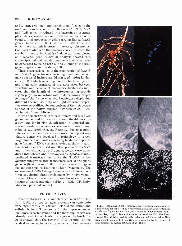
It was demonstrated that both binary and fused lux genes can be used for precise and reproducible in vitro assays and for in vivo visualization of temporal and spatial regulation of gene expression in plants [Lang- ridge et al., 19891 (Fig. 2). Recently, due to a great interest in the identification and isolation of plant reg- ulatory genes, we developed a technology to screen large numbers of plants expressing luciferase reporter gene fusions. T-DNA vectors carrying a t their integra- tion borders either fused lwAB or promoterless luxA and linked chimaeric luxB gene cassettes were intro- duced into tobacco and Arabidopsis by Agrobacterium- mediated transformation. Since the T-DNA is fre- quently integrated into transcribed loci of the plant genome [Koncz et al., 19891, transcriptional lux gene fusions can thus be isolated a t high frequencies. The expression of T-DNA-tagged genes can be followed con- tinuously during plant development by in vivo visual- ization of the expression of lux gene fusions in diverse tissues of transgenic plants (Fig. 2) [Redei GP, Univ. Missouri, personal comm.].
PROSPECTIVES The results described above clearly demonstrate that
both luciferase reporter gene systems can contribute very significantly to various fields of molecular and cellular biology. Some trends for the improvement of luciferase reporter genes and for their application are already predictable. Deletion analysis of the firefly luc gene showed that the removal of C terminal amino acids does not influence enzyme activity but converts
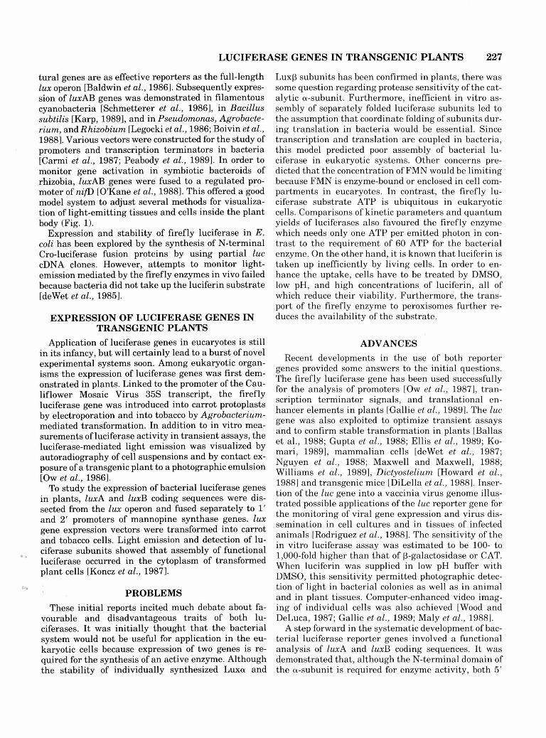
Fig. 1. Visualization of bioluminescence in soybean nodules and in single nodule cells infected by Bradyrhizobium japonicum containing a nifD-lwAB gene fusion. Top (left): Nodules under normal illumi- nation. Top (right): Bioluminescence recorded on IS0 400 .Ekta- chrome film. Middle: Nodule cells under normal illumination. Bot- tom: Visual image of light-emitting cells recorded by LDG low-light video microscope system [O'Kane et al., 19881.
LUCIFERASE GENES IN TRANSGENIC PLANTS
Fig. 2.
230 KONCZ ET AL.
Fig. 2. Visualization of lux gene expression in transgenic plants, organs, and tissues by a photon-counting video camera-photomulti- plier system. Panel A: Light image. Panel B: Recorded image of light emission. Top: Tobacco leaf expressing 1wA and luxB genes driven by 1' and 2' promoters of mannopine synthase genes (left) and leaf of a non-transformed tobacco plant (right). Middle: Stem section of a to- bacco plant expressing mas promoter-lux gene fusions. Bottom: Ex- pression of a lux gene fusion in a transgenic Arabidopsis thaliana plant.
the peroxisomal luciferase to a cytoplasmic enzyme; thus the need for the transport of luciferin to peroxi- somes can be overcome [Could et al., 19891. The perox- isomal targeting signals identified in luciferases can moreover be employed to transport other proteins into peroxisomes. Genes for beetle luciferases responsible for the emission of light of different colours could pro- vide tools for the simultaneous monitoring of the ex- pression of different genes in a single cell. Inactive mo- nomeric bacterial luciferases carrying recognition sites for specific proteases in between the fused subunits could be used for sensitive detection of proteases within cells or during fermentation in vivo. Both enzymes can be applied to measure thermal denaturation of proteins in diverse tissues during heat-shock. By addition of appropriate signal peptides, luciferases could probably be transported into cell organelles such as chloroplasts or mitochondria for monitoring ATP-consuming syn- thesis or electron transport. Production of luciferase- fused antibodies may play an important role for diverse aspects of immunology. Fusion of luciferases to chro- mosomal proteins can lead to unique approaches in cy- tology. The binary luxA-luxB system may find an im- mediate application for the visualization of cell fusion and fertilization events or help in hybrid seed produc- tion. Both binary and monocistronic luciferase genes can be employed to monitor virus-cell interactions and virus or virus-induced gene expression. Furthermore, luciferase gene fusions could be constructed to detect site-specific recombination or translocation events in a cell population. Expression of proteins associated with the bacterial luciferase may also provide in vivo assays for selection of fatty-acid- or riboflavin-overproducing organisms. This prospective list of applications can probably be extended, but our intention was simply to illustrate the potential of this reporter gene system.
REFERENCES Aboukhair NK, Ziegler MM, Baldwin TO (1985): Bacterial luciferase:
Demonstration of a catalytically competent altered conformational - - state follow?ng a single turnover. Biochemistry 24:3942-3947.
Baldwin TO, Ziegler MM, Powers DA (1979): Covalent structure of subunits of, bacterial luciferase: NH,-terminal sequence demon- strates subunit homology. Proc Natl Acad Sci USA 76:4887-4889.
Baldwin TO, Berends T, Bunch TA, Holzman TF, Rausch SK, Sha- mansky L, Treat ML, Ziegler MM (1984): Cloning of luciferase structural genes from Vibrw harveyi and expression of biolumines- cence in Escherichia coli. Biochemistry 23:3663-3667.
. .-
Baldwin TO, Holzman TF, Holzman R (1986): Active center-based immuno-assay approach using bacterial luciferase. In DeLuca M, McElroy WD (eds): "Bioluminescence and Chemiluminescence." Methods Enzymol 133:248-264.
Ballas N, Zakai N, Friedberg D, Loyter A (1988): Linear forms of plasmid DNA are superior to supercoiled structures as active tem- plates for gene expression in plant protoplasts. Plant Mol Biol 11: 517-527.
Belas R, Mileham A, Cohn D, Hilmen M, Simon M, Silverman M (1982): Bacterial bioluminescence: Isolation and expression of the luciferase genes from Vibrw harveyi. Science 218:791-792.
Boivin R, Chalifour F-P, Dion P (1988): Construction of a Tn5 deriv- ative encoding bioluminescence and its introduction in Pseudorno- nns, Agrobacteriurn and Rhizobium. Mol Gen Genet 213:50-55.
Boylan M, Pelletier J , Meighen EA (1989): Fused bacterial luciferase subunits catalyze light emission in eucaryotes and procaryotes. J Biol Chem 264:1915-1918.
Campbell AK, Hallett MB, Weeks I (1985): Chemiluminescence as an analytical tool in cell biology and medicine. In Glick D (ed): Meth- ods in Biochem Anal 31:317-416.
Carmi OA, Stewart GSAB, Ulitzur S, Kuhn J (1987): Use of bacterial luciferase to establish a promoter probe vehicle capable of non- destructive real-time analysis of gene expression in Bacillus ssp. J Baeteriol 169:2165-2170.
Chen LH, Baldwin TO (1989): Random and site-directed mutagenesis of bacterial luciferase: Investigation of the aldehyde binding site. Biochemistry 28:2684-2689.
Cohn DH, Ogden RC, Abelson JN, Baldwin TO, Nealson KH, Simon MI, Mileham AJ (1983): Cloning of the Vibrw hrveyi luciferase genes: Use of a synthetic oligonucleotide probe. Proc Natl Acad Sci USA 80:120-123.
Cohn DH, Mileham AJ, Simon MI, Nealson KH, Rausch SK, Bonam D, Baldwin TO (1985): Nucleotide sequence of the IuxA gene of Vibrio harveyi and the complete amino acid sequence of the ol sub- unit of bacterial luciferase. J Biol Chern 260:6139-6146.
Cormier MJ (1978): Applications of Renilla bioluminescence: An in- troduction. In DeLuca M (ed): "Bioluminescence and Chemilumi- nescence." Methods Enzymol 57:237-244.
Daubner SC, Astorga AH, Leisman GB, Baldwin TO (1987): Yellow light emission of Vibrw ficheri strain Y1: Purification and charac- terization of the energy-accepting yellow fluorescent protein. Proc Natl Acad Sci USA 84:8912-8916.
Delong EF, Steinhauer D, Israel A, Nealson KH (1987): Isolation of the lux genes from Photobacterium lewgnathi and expression in Escherichia coli. Gene 54:203-210.
DeLuca M (1976): Firefly luciferase. Adv Enzymol 44:37-68. DeLuca M (1978): "Bioluminescence and Chemiluminescence." Meth-
ods Enzymol 57. DeLuca M, McElroy WD (1986): "Bioluminescence and Chemilumi-
nescence." Methods Enzymol 133. Devine JH, Countryman C, Baldwin TO (1988): Nucleotide sequence
of the 1wR and lux1 genes and structure of the primary regulatory region of the lux regulon of Vibrio fischeri ATCC 7744. Biochemis- try 27:837-842.
deWet JR, Wood KV, Helsinki DR, DeLuca M (1985): Cloning of fire- fly luciferase cDNA and expression of active luciferase in Escher- ichia coli. Proc Natl Acad Sci USA 82:7870-7873.
deWet JR, Wood KV, DeLuca M, Helsinki DR, Subramani S (1987): Firefly luciferase gene: Structure and expression in mammalian cells. Mol Cell Biol 7:725-737.
DiLella AG, Hope DA, Chen H, Trumbauer M, Schwartz RJ, Smith RG (1988): Utility of firefly luciferase as a reporter gene for pro- moter activity in transgenic mice. Nucleic Acids Res 16:4159.
Dunlap PV (1985): Osmotic control of bioluminescence and growth in Photobacterium lewgnathi from ponyfish light organs. Arch Micro- biol 141:44-50.
Dunlap PV, Greenberg EP (1985): Control of Vibrw fscheri lumines- cence gene expression in Escherichia coli by cyclic AMP and cyclic AMP receptor protein. J Bacteriol 164:45-50.
Dunlap PV (1989): Regulation of luminescence by cyclic AMP in cya-
LUCIFERASE GENES IN TRANSGENIC PLANTS 231
like and crp-like mutants of Vibrio fischeri. J Bacteriol 171:1199- 1202.
Ellis D, Roberts D, Sutton B, Lazaroff W, Webb D, Flinn B (1989): Transformation of white spruce and other conifer species by Agro- bacterium tunaefaciens. Plant Cell Rep 8:16-20.
Engebrecht J, Nealson K, Silverman M (1983): Bacterial biolumines- cence: Isolation and genetic analysis of functions from Vibrio fis- cheri. Cell 32773-781.
Engebrecht J, Silverman M (1984): Identification of genes and gene products necessary for bacterial bioluminescence. Proc Natl Acad Sci USA 81:4154-4158.
Engebrecht J , Simon M, Silverman M (1985): Measuring gene expres- sion with light. Science 227:1345-1347.
Engebrecht J , Silverman M (1987): Nucleotide sequence of the regu- latory locus controlling expression of bacterial genes for biolumi- nescence. Nucleic Acids Res 15:10455-10467.
Foran DR, Brown WM (1988): Nucleotide sequence of the luxA and IuxB genes of bioluminescent marine bacterium Vibrw fischeri. Nu- cleic Acids Res 16:777.
Gallie DR, Lucas WJ, Walbot V (1989): Visualizing mRNA expression in plant protoplasts: Factors influencing efficient mRNA uptake and translation. Plant Cell 1:301-311.
Gould SJ, Keller GA, Subramani S (1987): Identification of a perox- isomal targeting signal a t the carboxy terminus of firefly lu- ciferase. J Cell Biol 105:2923-2931.
Gould SJ, Subramani S (1988): Firefly luciferase as a tool in molec- ular and cell biology. Anal Biochem 175:5-13.
Gould SJ, Keller GA, Hosken N, Wilkinson J , Subramani S (1989): A conserved tripeptide sorts proteins into peroxisomes. J Cell Biol 108:1657-1664.
Gupta SC, O'Brien D, Hastings JW (1985): Expression of the cloned subunits of bacterial luciferase from separate replicons. Biochem Biophys Res Commun 127:1007-1011.
Gupta PK, Dandekar AM, Durzan DJ (1988): Somatic proembryo for- mation and transient expression of a luciferase gene in douglas fir and loblolly pine protoplasts. Plant Sci 58:85-92.
Harvey EN (1957): "A History of Luminescence From Earliest Time Until 1900." Philadelphia: Philosophical Society of Pennsylvania.
Hastings WJ, Nealson KH (1977): Bacterial bioluminescence. Annu Rev Microbiol 31:549-595.
Hastings WJ (1978): Bacterial bioluminescence: An overview. In De- Luca M (ed): "Bioluminescence and Chemiluminescence." Methods Enzymol57:125-135.
Haubner R, Geiger R (1988): A sensitive, bioluminescent enhanced detection method for DNA dot-hybridization. Nucleic Acids Res 16: 1213.
Haygood MG, Nealson KH (1985): Mechanism of iron regulation of luminescence in Vibrw fischeri. J Bacteriol 162:209-216.
Haygood MG, Cob:, DM (1986): Luciferase genes cloned from the un- culturable luminous bacteroid symbiont of the Caribbean flash- light fish, Kryptophanaron alfredi. Gene 45203-209.
Howard PK, Ahern KG, Firtel RA (1988): Establishment of a tran- sient expression system for Dictyostelium discoideum. Nucleic Ac- ids Res 16:2613-2623.
Illarionov BA, Protopopova MV, Karginov VA, Mertvetsov NP, Gitel- son J I (1988): Nucleotide sequence of part of Photobacterium lei- ognathi lux region. Nucleic Acids Res 16:9855.
Inouye S, Sakaki Y, Goto T, Tsuji FI (1986): Expression of apoae- quorin complementary DNA in Escherichia coli. Biochemistry 25: 8425-8429.
Johnston TC, Thompson RB, Baldwin TO (1986): Nucleotide sequence of the luxB gene of Vibrio hurveyi and the complete amino acid sequence of the f3 subunit of bacterial luciferase. J Biol Chem 261: 4805-4811.
Karp M (1989): Expression of bacterial luciferase genes from Vibrw harveyi in Bacillus subtilis and in Escherichia coli. BBA 1007:84- 90.
Keller GA, Gould S, DeLuca M, Subramani S (1987): Firefly lu- ciferase is targeted to peroxisomes in mammalian cells. Proc Natl Acad Sci USA 84:3264-3268.
Komari T (1989): Transformation of callus cultures of nine plant spe- cies mediated by Agrobacterium. Plant Sci 60:223-229.
Koncz C, Olsson 0 , Langridge WHR, Schell J , Szalay AA (1987): Expression and assembly of functional bacterial luciferase in plants. Proc Natl Aoad Sci USA 84:131-135.
Koncz C, Martini N, Mayerhofer R, Kona-Kalman ZS, Korber H, Redei GP, Schell J (1989) High-frequency T-DNA-mediated gene tagging in plants. Proc Natl Acad Sci USA 86:8467-8471.
Kricka LJ (1988): Clinical and biochemical applications of luciferases and luciferins. Anal Biochem 175:14-21.
Kurfiirst M, Ghisla S, Hastings W (1984): Characterization and pos- tulated structure of the primary emitter in the bacterial luciferase reaction. Proc Natl Acad Sci USA 81:2990-2994.
Longridge WHR, Fitzgerald KJ, Kona C, Schell J, Sealay AA (1989) Dual promoter of Agrobacterium tumefaciens mannopine synthase genes is regulated by plant growth hormones. Proc Natl Acad Sci USA 86:3213-3223.
Lee J , O'Kane DJ, Gibson BG (1988): Dynamic fluorescence proper- ties of bacterial luciferase intermediates. Biochemistry 27:4862- 4870.
Lee J, O'Kane DJ, Gibson BG (1989): Bioluminescence spectral and fluorescence dynamics study of the interaction of lumazine protein with the intermediates of bacterial luciferase bioluminescence. Bio- chemistry 28:4263-4271.
Legocki RP, Legocki M, Baldwin TO, Szalay AA (1986): Biolumines- cence in soybean root nodules: Demonstration of a general approach to assay gene expression in vivo by using bacterial luciferase. Proc Natl Acad Sci USA 83:9080-9084.
Maly FE, Urwyler A, Rolli HP, Dahinden CA, DeWeck LA (1988): A single-photon imaging system for the simultaneous quantitation of luminescence emission from multiple samples. Anal Biochem 168: 462-469.
Mancini JA, Boylan M, Soly RR, Graham AF, Meighen EA (1988): Cloning and expression of the Photobacterium phosphoreum lumi- nescence system demonstrates a unique lux gene organization. J Biol Chem 263:14308-14314.
Masuda T, Tatsumi H, Nakano E (1989): Cloning and sequence anal- ysis of cDNA of a Japanese firefly, Luciola crmiata. Gene 77:265- 270.
Maxwell IH, Maxwell F (1988): Electroporation of mammalian cells with a firefly luciferase expression plasmid: Kinetics of transient expression differ markedly among cell types. DNA 7:557-562.
Meighen EA, Bartlet I(1980): Complication of subunits from different bacterial luciferases. J Biol Chem 255:11181-11187.
Miyamoto C, Graham AD, Boylan M, Evans JR, Hasel KW, Meighen EA, Graham AF (1985): Polycistronic mRNA codes for polypeptides of the Vibrw hurvey luminescence system. J Bacteriol 161:995- 1001.
Miyamoto C, Byers D, Graham AF, Meighen EA (1987): Expression of bioluminescence by Escherichia coli containing recombinant Vibrio hurvey DNA. J ~ac t e r io l 169:247-253.
-
Miyamoto C, Graham AF, Meighen EA (1988): Nucleotide sequence of the l w C gene and the upstream DNA from the bioluminescent system of Vibrio hurveyi. Nucleic Acids Res 16:1551-1562.
Nguyen VT, Morange M, Bensaude 0 (1988): Firefly luminescence assays using scintillation counters for quantitation in transfected mammalian cells. Anal Biochem 171:404-408.
O'Kane DJ, Lingle WL, Wampler JE, Legocki M, Legocki PR, Szalay AA (1988): Visualization of bioluminescence a s a marker of gene expression in Rhizobium-infected nodules. Plant Mol Biol 10:387- 399.
Olsson 0 , Koncz C, Szalay AA (1988): The use of the luxA gene of the bacterial luciferase operoe as a reporter gene. Mol Gen Genet 215: 1-9.
Olsson 0 , Escher A, Sandberg G, Schell J , Koncz C, Szalay AA (1989): Engineering of monomeric bacterial luciferases by fusion of luxA and IuxB genes in Vibrio harueyi. Gene 81:335-347.
Ow DW, Wood KV, DeLuca M, deWet JR, Helsinki DR, Howell SH (1986): Transient and stable expression of the firefly luciferase gene in plant cells and transgenic plants. Science 234:856-859.
232 KONCZ ET AL.
Ow DW, Jacobs J , Howell SH (1987): Functional regions of the Cau- liflower Mosaic Virus 35s RNA promoter determined by use of the firefly luciferase gene as a reporter of promoter activity. Proc Natl Acad Sci USA 84:4870-4874.
Paquette 0 , Fried A, Tu SC (1988): Delineation of bacterial luciferase aldehyde site by bifunctional labeling reagents. Arch Biochem Bic- phys 264:392-399.
Peabody DS, Andrews CL, Escudero KW, Devine JH, Baldwin TO, Bear DG (1989): A plasmid vector and quantitative techniques for the study of transcription termination in Escherichia coli using bac- terial luciferase. Gene 75:289-296.
Prasher DC, McCahn RO, Cormier MJ (1986): Isolation and expres- sion of a cDNA for aequorin, the Ca2+ -activated photoprotein from Aequorea victoria. In DeLuca M, McElroy WD (eds): "Biolumines- cence and Chemiluminescence." Methods Enzymol 133288-297.
Rodriguez JF, Rodriguez D, Rodriguez JR, McGowan EB, Estehan M (1988): Expression of the firefly luciferase gene in vaccinia virus: A highly sensitive gene marker to follow virus dissemination in tis- sues of infected animals. Proc Natl Acad Sci USA 85:1667-1671.
Schmetterer G, Wolk PC, Elhai J (1986): Expression of luciferases from Vibrw harvey and Vibrw Fcher i in filamentous cyanobacte- ria. J Bacteriol 167:411-414.
Schroeder J (1989): Protein sequence homology between plant 4 coumarate coenzyme A ligase and firefly luciferase. Nucleic Acids Res 17:460.
Shaw J J , Kado CI (1986): Development of a Vibrio bioluminescence gene-set to monitor phytopathogenic bacteria during the ongoing disease process in a nondis~pt ive manner. Biotechnology 4:560- 564.
Shaw J J , Rogowsky P, Close TJ, Kado CI (1987): Working with bac- terial bioluminescence. Plant Mol Biol Rep 5225-236.
Shaw J J , Settles LG, Kado CI (1988): Transposon Tn4431 mutagen- esis of Xanthomnas campestris pv. campestris: Characterization of
a non-pathogenic mutant and cloning of a locus for pathogenicity. Mol Plant Microb Interact 1:39-45.
Soly RR, Mancini JA, Ferri SR, Boylan M, Meighen EA (1988): A new lux gene in bioluminescent bacteria codes for a protein homologous to the bacterial luciferase subunits. Biochem Biophys Res Commun 155:351-358.
Sugihara J , Baldwin TO (1988): Effects of 3' end deletions from the Vibrw harveyi lwcB gene on luciferase subunit folding and enzyme assembly: Generation of temperature-sensitive polypeptide folding mutants. Biochemistry 27:2872-2880.
Swanson R, Weaver LH, Remington ST, Matthews BW, Baldwin TO (1985): Crystals of luciferase from Vibrin hnrueyi. J Biol Chem 260: 1287-1289.
Ulitzur S (1986): Determination of antibiotic activities with the aid of luminous bacteria. In DeLuca M, McElroy WD (eds): "Biolumines- cence and Chemiluminescence." Methods Enzymol 133:275-284.
VanDyke K (1985): "Bioluminescence and Chemiluminescence: In- struments and Applications." Boca Raton, Florida: CRC Press.
Weinhausen G, DeLuca M (1982): Bioluminescent assays of picomole levels of various metabolites using immobilized enzymes. Anal Bio- chem 127:380-388.
Williams TM, Burlein JE, Ogden S, Kricka LJ, Kant JA (1989): Ad- vantages of firefly luciferase as a reporter gene: Application to the interleukin-2 gene promoter. Anal Biochem 176:28-32.
Wood KV, DeLuca M (1987): Photographic detection of luminescence in Escherichia coli containing the gene for firefly luciferase. Anal Biochem 161:501-507.
Wood KV, Lam AY, Seliger HH, McElroy WD (1989): Complementary DNA encoding click beetle luciferases can elicit bioluminescence of different colours. Science 244700-702.
Ziegler MM, Baldwin TO (1981): Biochemistry of bacterial biolumi- nescence. Curr Top Bioenerg 12:65-113.


![The Evolution of the Bacterial Luciferase Gene …1]Luciferousenzymecassette.pdfSensors 2012, 12, 732-752; doi:10.3390/s120100732 sensors ISSN 1424-8220 Review The Evolution of the](https://static.fdocuments.in/doc/165x107/5affd64a7f8b9a6a2e8bc5a7/the-evolution-of-the-bacterial-luciferase-gene-1luciferousenzymecassettepdfsensors.jpg)