Bacteria, Fungus, or Virus?
61
Bacteria, Fungus, or Virus? An Update on Infections of the Skin Skin, Bones, Hearts, and Private Parts Kara N. Roman, MMS, PA-C Associate Director & Assistant Professor Midwestern University PA Program, Downers Grove, IL
Transcript of Bacteria, Fungus, or Virus?

Bacteria, Fungus, or Virus?An Update on Infections of the Skin
Skin, Bones, Hearts, and Private Parts
Kara N. Roman, MMS, PA-C
Associate Director & Assistant Professor
Midwestern University PA Program, Downers Grove, IL

Disclosures and Images
• No conflicts to disclose
• Images – all images obtained and used within the guidelines provided from DermNetNZ.org unless otherwise attributed
• Reproduction or copying of content or images prohibited
• Thank you
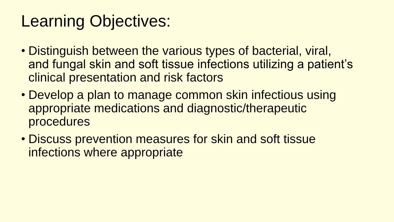
Learning Objectives:
• Distinguish between the various types of bacterial, viral, and fungal skin and soft tissue infections utilizing a patient’s clinical presentation and risk factors
• Develop a plan to manage common skin infectious using appropriate medications and diagnostic/therapeutic procedures
• Discuss prevention measures for skin and soft tissue infections where appropriate

Picture goes herePatient presents for evaluation of the following skin lesions.
Image source: https://dermnetnz.org/

Bacteria, Fungus, or Virus?
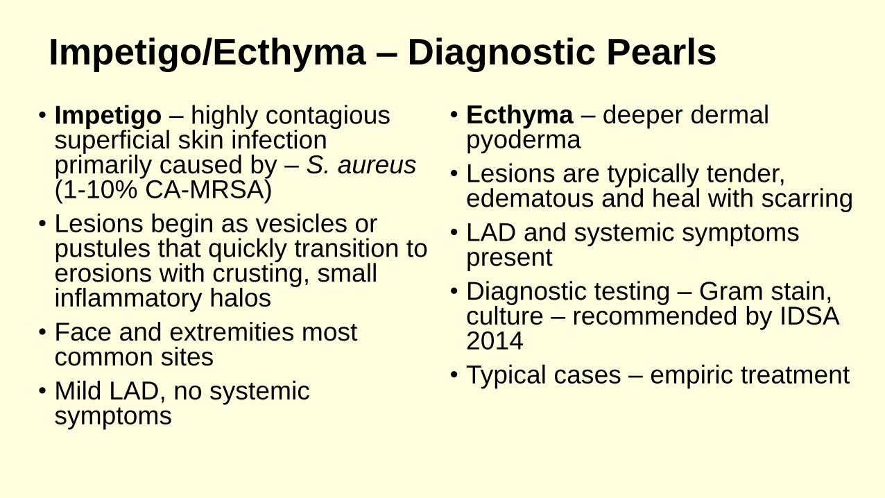
Impetigo/Ecthyma – Diagnostic Pearls
• Impetigo – highly contagious superficial skin infection primarily caused by – S. aureus (1-10% CA-MRSA)
• Lesions begin as vesicles or pustules that quickly transition to erosions with crusting, small inflammatory halos
• Face and extremities most common sites
• Mild LAD, no systemic symptoms
• Ecthyma – deeper dermal pyoderma
• Lesions are typically tender, edematous and heal with scarring
• LAD and systemic symptoms present
• Diagnostic testing – Gram stain, culture – recommended by IDSA 2014
• Typical cases – empiric treatment

Impetigo/Ecthyma – Treatment
• Practice Guidelines for Diagnosis and Management of Skin and Soft Tissue Infections: 2014 Update by IDSA https://www.idsociety.org/practice-guideline/skin-and-soft-tissue-infections/
• Topical treatment for limited impetigo• Mupirocin, Retapamulin – BID for 5 days
• Soak to remove crust
• Oral for numerous impetigo, in outbreaks, and for all ecthyma• CA-MRSA – Doxycycline, Clindamycin, TMP/SMX – 7 days
• MSSA - Dicloxacillin, Cephalexin – 7 days

Picture goes here Patient presents for evaluation of the following skin lesions.
Image source: https://dermnetnz.org/

Bacteria, Fungus, or Virus?

Purulent Infections – Diagnostic Pearls
• Folliculitis – single follicle
• Furuncle – deeper, yet still one follicle
• Carbuncle – larger, often with fever, LAD, fatigue
• Gram stain and culture are ideal
• Mild, moderate, severe classifications
• Most purulent infections = Staphylococcus aureus (MSSA and MRSA)

Purulent Infections – Treatment
• Incision and Drainage
• Warm, moist compresses
• Add on oral or parenteral antibiotics
Image source: https://www.idsociety.org/globalassets/idsa/practice-guidelines/practice-guidelines-for-the-diagnosis-and-management-of-skin-and-soft-tissue-infections-2014-update-by-the-infectious-diseases-society-of-america.pdf
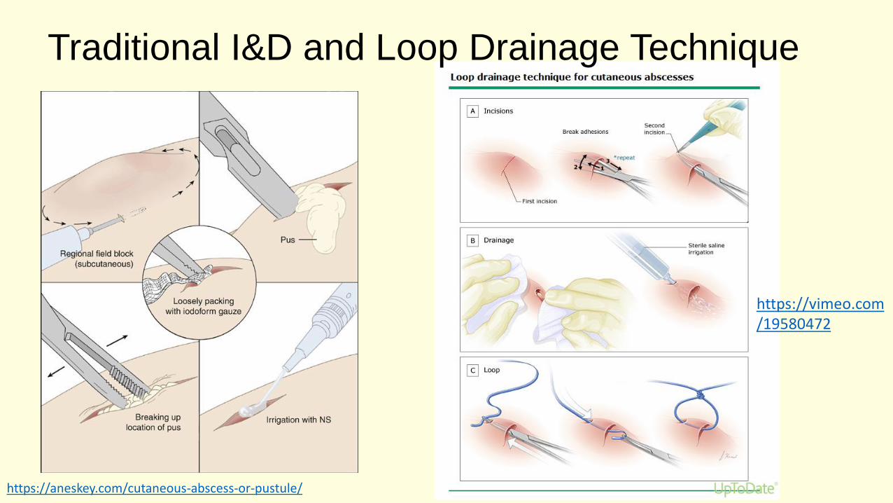
Traditional I&D and Loop Drainage Technique
https://aneskey.com/cutaneous-abscess-or-pustule/
https://vimeo.com/19580472

Addition of Antibiotics in Purulent Skin Infections per IDSA 2014
• Temp > 38 degrees C or < 36 degrees C
• Tachypnea > 24/min
• Tachycardia > 90 bpm
• WBC > 12,000 or < 4,000
• Immunosuppression
• Hypotension

Guidelines vs Actual Management of Skin and Soft Tissue Infections in the EDKamath RS, et al. OFID 2017
• 214 cases of SSTI in ED retrospectively analyzed at Michael E. DeBakey Veterans Affairs Medical Center, Houston
• Total number that were managed in accordance with IDSA 2014 guidelines in all 4 categories (site of treatment, choice of antibiotic, I&D of abscess, ordering cultures)
43/214 = 20.1%
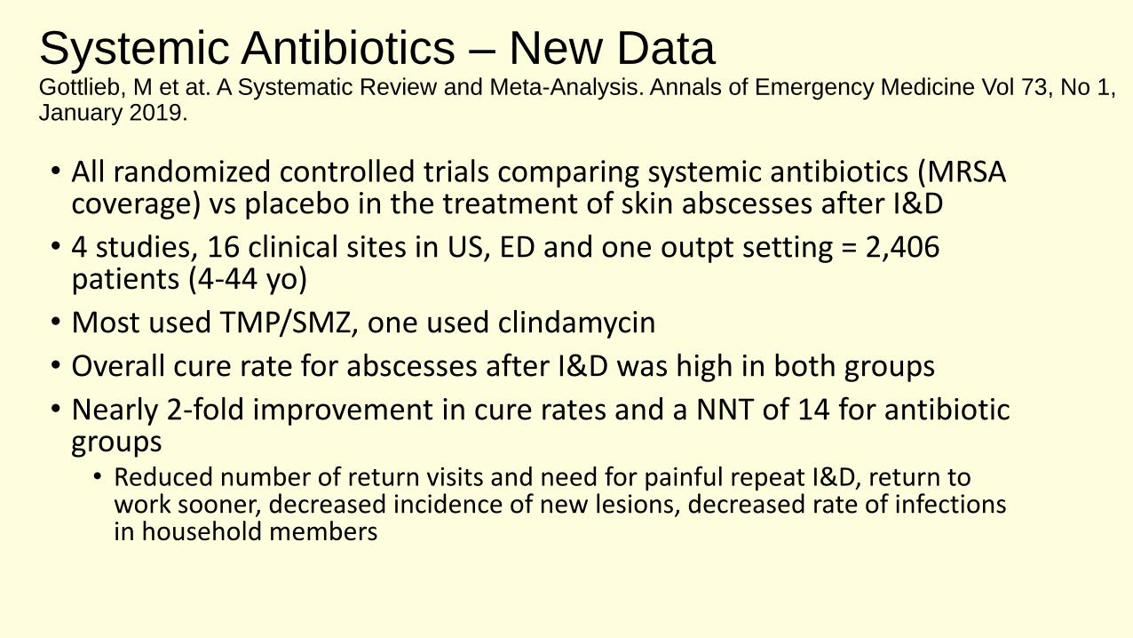
Systemic Antibiotics – New DataGottlieb, M et at. A Systematic Review and Meta-Analysis. Annals of Emergency Medicine Vol 73, No 1, January 2019.
• All randomized controlled trials comparing systemic antibiotics (MRSA coverage) vs placebo in the treatment of skin abscesses after I&D
• 4 studies, 16 clinical sites in US, ED and one outpt setting = 2,406 patients (4-44 yo)
• Most used TMP/SMZ, one used clindamycin
• Overall cure rate for abscesses after I&D was high in both groups
• Nearly 2-fold improvement in cure rates and a NNT of 14 for antibiotic groups
• Reduced number of return visits and need for painful repeat I&D, return to work sooner, decreased incidence of new lesions, decreased rate of infections in household members

To Pack or Not to Pack
NO
• 5 cm or less
• Immunocompetent pt
• Less pain
• No change in cure rate
• No change in secondary interventions
Maybe or YES
• Larger abscesses
• Consider the area
• Immunocompromised pts have not been studied
Source: List M et al. Treatment of Skin Abscesses: A Review of Wound Packing and Post-Procedural Antibiotics. South Dakota Medicine, March 2016.

Recurrent Skin Abscesses
• Consider 5 day decolonization regimen:• Twice daily intranasal mupirocin
• Daily chlorhexidine washes
• Daily decontamination of personal items such as towels, sheets, and cloths for recurrent S. aureus
• Evaluate adult patients for neutrophil disorder if recurrent abscesses began in childhood
• Search for local causes – pilonidal cyst, hidradenitis suppurativa, foreign body

Picture goes here A patient presents for evaluation of the following skin lesions.
Image source: https://dermnetnz.org/

Bacteria, Fungus, or Virus?

Cellulitis/Erysipelas – Diagnostic Pearls
• Erysipelas – bright red, more superficial, raised border, well-demarcated margin, preceded by flu-like symptoms, burning at site
• Cellulitis – deeper location to subcutaneous tissues, non-elevated, poorly defined margins,
• Usually caused by StreptococcusA, B, C, G
• Bedside ultrasound best option for differentiating abscess from cellulitis

Cellulitis/Erysipelas – Treatment
• Mild and typical with no evidence of purulence or trauma
• Oral dicloxacllin, cephalexin, clindamycin for 5 days
• Moderate infections with systemic signs
• IV ceftriaxone, clindamycin, cefazolin, penicillin
• Severe• Vancomycin plus piperacillin-
tazobactam or imipenem
• Additional corticosteroids• Prednisone 40 mg daily for 7 days
• Immobilization and elevation
• Treat tinea pedis
• Watch for worsening• Consider marking borders
• MRSA more likely• Penetrating trauma
• Illicit drug use
• Known nasal colonization
• Purulent drainage
• MRSA = Vancomycin

Recurrent Cellulitis
• Risk factors
• Tinea pedis
• Obesity
• Venous insufficiency
• Lymphedema
• Prophylactic abx if 3-4 episodes per year despite treating predisposing factors
• Pen VK or erythromycin orally twice daily for 4-52 weeks

Risk for Atypical Organisms
• Immunosuppression
• Animal or human bites
• Sea or freshwater exposure to broken skin
• Exposure to animals, fish, or reptiles
• IVD use

Cat/Dog Bites – Identify pts at high risk
• Immunocompromised
• Asplenic
• Advanced liver disease
• Pre-existing or resultant edema of the affected area
• Moderate to severe injuries, especially to hand or face
• Injuries that penetrate the periosteum or joint capsule
• Deep puncture wounds
• 3-5 days of preemptive therapy
• Augmentin 875 mg twice daily
• Alternatives• Cefuroxime plus
clindamycin/metronidazole
• Imipenem/meropenem
• Moxifloxacin
• Doxycycline
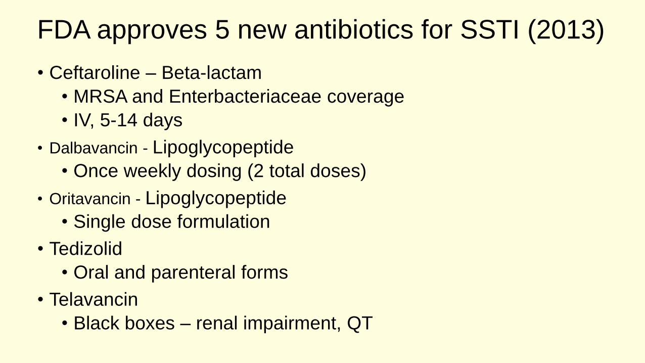
FDA approves 5 new antibiotics for SSTI (2013)
• Ceftaroline – Beta-lactam
• MRSA and Enterbacteriaceae coverage
• IV, 5-14 days
• Dalbavancin - Lipoglycopeptide
• Once weekly dosing (2 total doses)
• Oritavancin - Lipoglycopeptide
• Single dose formulation
• Tedizolid
• Oral and parenteral forms
• Telavancin
• Black boxes – renal impairment, QT

Bacterial Infections
• Impetigo/Ecthyma
• Furuncle/Carbuncle/Abscess
• Cellulitis/Erysipelas
• Staphylococcal scalded skin/Scarlet fever/TSS
• Lyme
• Secondary syphilis
• Erythrasma
• Infectious Diseases Society of America (IDSA). Guidelines for skin and soft tissue infections, 2014.https://www.idsociety.org/practice-guideline/skin-and-soft-tissue-infections/

Picture goes here Patient presents for evaluation of the following skin lesions.

Bacteria, Fungus, or Virus?

HPV – Diagnostic Pearls
• HPV 1, 2, 3, 4, 10, 27, 57
• Most common in children and young adults (nearly 50% affected), increasingly seen in patients with atopic dermatitis and decreased cell-mediated immunity
• Types – verruca vulgaris, plantaris, plana, filiform
• Clinical diagnosis
• Thrombosed capillaries
• Altered dermatoglyphics
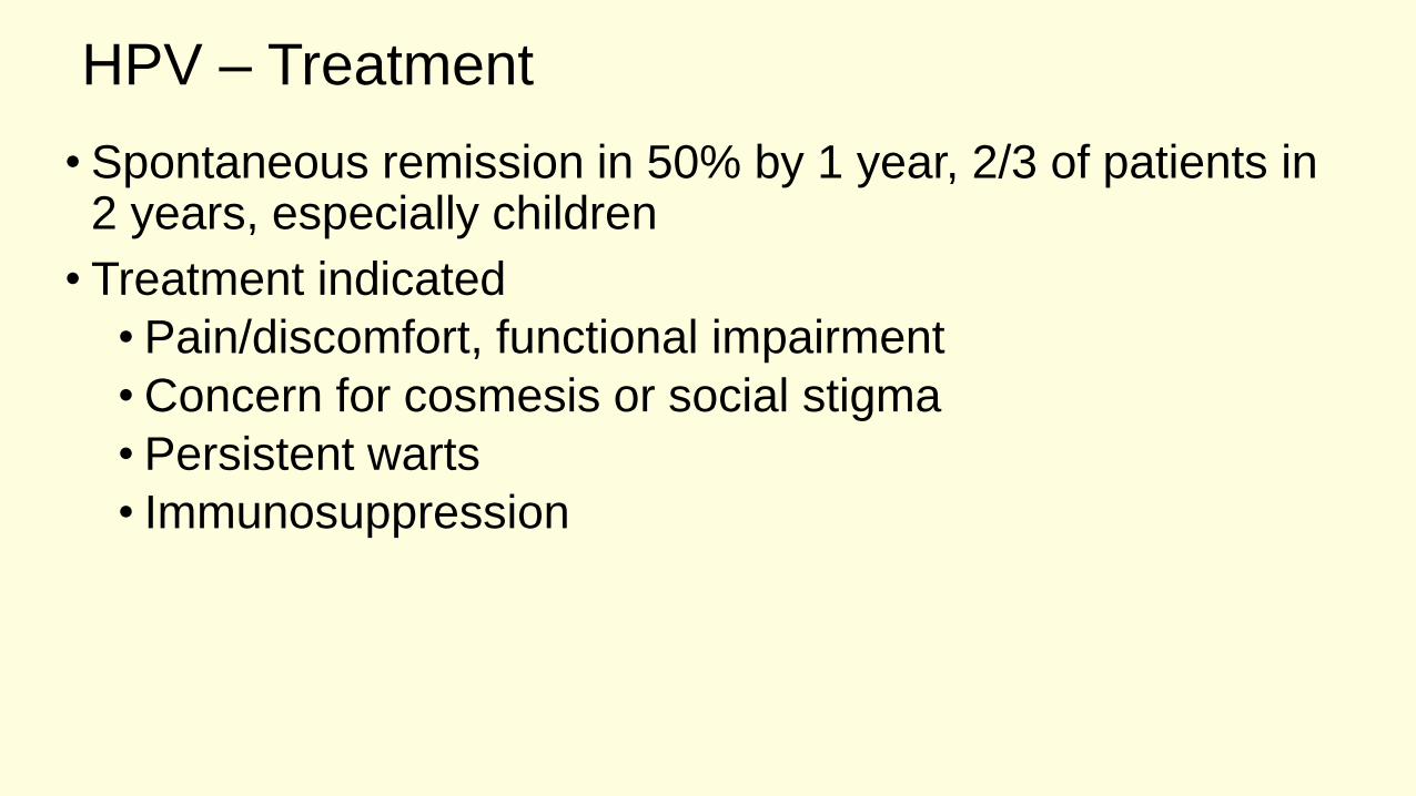
HPV – Treatment
• Spontaneous remission in 50% by 1 year, 2/3 of patients in 2 years, especially children
• Treatment indicated
• Pain/discomfort, functional impairment
• Concern for cosmesis or social stigma
• Persistent warts
• Immunosuppression
HPV- Treatment• Chemical or physical destruction
• Salicylic acid – irritation and exfoliation, paints or plasters, applied daily after paring with occlusion for 3-4 months
• Cryotherapy – every 2-3 weeks for 3 months, keep wart frozen for 15-30s
• Combo of both
• Enhancement of local immune response
• Imiquimod
• Contact or intralesional immunotherapy
• Antiproliferative therapy
• 5-FU
• Bleomycin
• Tretinoin

An Option for Recalcitrant and Extensive Warts?
• Intralesional or intramuscular HPV vaccine• Nofal A et al. Intralesional versus intramuscular bivalent human papillomavirus
vaccine in the treatment of recalcitrant common warts, J Am Acad Dermatol vol82, No 1, July 2019.
• Waldman, Abigail MD; Whiting, Dennis PA-C; Rani, Monica MD; Alam, Murad MD, MSCI, MBA, HPV Vaccine for Treatment of Recalcitrant Cutaneous Warts in Adults A Retrospective Cohort Study. Dermatologic Surgery: December 2019 - Volume 45 - Issue 12 - p 1739–1741

Picture goes here Patient presents for evaluation of the following skin lesions.
Image source: https://dermnetnz.org/

Bacteria, Fungus, or Virus?

Molluscum Contagiosum – Diagnostic Pearls
• Benign, self-limited disease
• Caused by a pox virus
• Most common ages 1-4
• More common in pts with atopic dermatitis and with swimming
• In adolescents and adults, consider STIs and immunocompromised states
• Clinical diagnosis
• Small, firm, pearly papules with a central depression
• Core may be expressed, producing a white cheesy material
• The lesions average 2 to 5 mm in size and are usually painless, but may become inflamed, red, and swollen
• Distribution typically face, truck, limbs
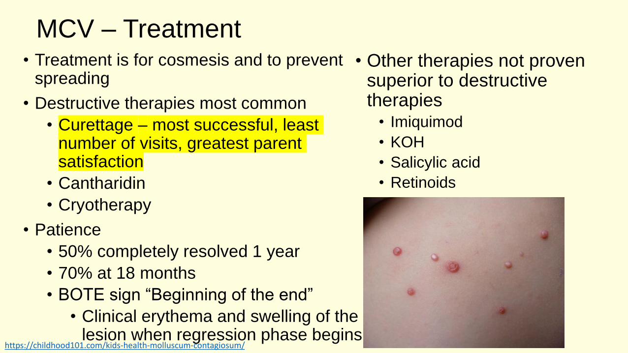
MCV – Treatment• Treatment is for cosmesis and to prevent
spreading
• Destructive therapies most common
• Curettage – most successful, least number of visits, greatest parent satisfaction
• Cantharidin
• Cryotherapy
• Patience
• 50% completely resolved 1 year
• 70% at 18 months
• BOTE sign “Beginning of the end”
• Clinical erythema and swelling of the lesion when regression phase begins
• Other therapies not proven superior to destructive therapies
• Imiquimod
• KOH
• Salicylic acid
• Retinoids
https://childhood101.com/kids-health-molluscum-contagiosum/

https://ars.els-cdn.com/content/image/1-s2.0-S0190962213004106-gr1_lrg.jpg https://www.dermatologytimes.com/article/verrica-develops-solution-common-warts
https://dermnetnz.org/topics/cantharidin/
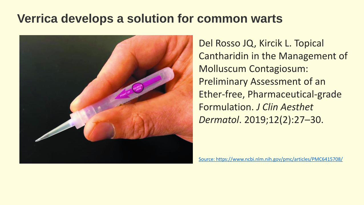
Verrica develops a solution for common warts
Del Rosso JQ, Kircik L. Topical Cantharidin in the Management of Molluscum Contagiosum: Preliminary Assessment of an Ether-free, Pharmaceutical-grade Formulation. J Clin AesthetDermatol. 2019;12(2):27–30.
Source: https://www.ncbi.nlm.nih.gov/pmc/articles/PMC6415708/

14-year-old male presents for evaluation of red circular rash on his body that he noticed after soccer practice yesterday
• Pt states he doesn’t feel sick, but the rash might be a little itchy
• He is unaware of any family or friends that are ill or have rashes
• The soccer coach will not let him play or practice until he gets “checked out”
Image source: https://dermnetnz.org/

Bacteria, Fungus, or Virus?

Pityriasis Rosea – Diagnostic Pearls• Self-limiting skin condition
• Reactivation of HHV-6/7
• Typically presents in 10-35-year-old pts
• Herald patch appears first on truck in 90% of cases (lasts for 2 weeks in isolation)
• Prodromal symptoms – malaise, nausea, headache, URI, concentration difficulty, ST, body aching
• Secondary eruption on trunk in Langer lines and to proximal extremities (lasts for up to 12 weeks)
• All lesions have scaling and are typically pruritic
• Some medications associated with PR-like eruption – ACEI, NSAID
Image source: https://dermnetnz.org/topics/pityriasis-rosea-images/
PR – Treatment
• Self-limited = watchful waiting, patience
• Treat options
• Oral antihistamines
• Oral or topical corticosteroids
• Acyclovir (maybe)
• Phototherapy also hastens resolution
• No benefit to macrolides
Viral Infections
• HPV
• Molluscum
• Viral exanthems
• Erythema infectiousum
• Roseola
• Herpangina/HFM
• Pityriasis rosea
• Herpes
• Simplex
• Zoster

6-year-old male presents for evaluation of this pruritic eczematous rash on his torso
• It developed over the past week as an initial small circular lesion that has spread
http://advancedskinmd.com/tinea-corporis-ringworm/
http://www.crestingthehill.com.au/2015_04_01_archive.html

Bacteria, Fungus, or Virus?

Tinea Corporis – Diagnostic Pearls
• Usually presents as annular, scaly plaques
• At risk groups• Contact sports
• Domestic animal contact
• Warm, humid climates
• DM, immunodeficiency
• Typical organisms• M. canis
• T. rubrum, mentagrophytes, tonsurans
• Diagnostic options• KOH
• Wood’s lamp
• Culture
• Biopsy
Tinea Corporis – Treatment
• Topical antifungals• Azoles
• Econazole
• Oxiconazole – comes in a lotion form for more hairy areas
• Allylamines• Terbinafine
• Naftifine
• Oral antifungals• Terbinafine
• Itraconazole
• Twice a day application of topicals for 4-6 weeks generally
• Keep skin cool and dry
• Avoid combination products with steroids and antifungals
• Topical nail lacquers modestly effective
• Efinaconazole
• Tavaborole

Majocchi’s granuloma
• Perifollicular lesions
• T. rubrum, T. mentagrophytes
• Systemic therapy needed
Image source: https://dermnetnz.org/topics/majocchi-granuloma/

Picture goes here Patient presents for evaluation of the following skin lesions.
https://dermnetnz.org/

Bacteria, Fungus, or Virus?
Onychomycosis – Diagnostic Pearls
• Most often occurs in adults• Nail injury increases risk
• T. rubrum most frequent dermatophyte
• Common clinical manifestations• Nail discoloration
• Subungual hyperkeratosis
• Onycholysis
• Nail plate splitting and destruction
• Diagnostic testing• KOH - screen
• Fungal culture & PCR – identify the organism before treatment

Onychomycosis – Treatment
• Mild to moderate disease (<50% nail involvement) with no matrix involvement
• Ciclopirox
• Efinaconazole
• Tavaborole
• Established and severe disease• Terbinafine – preferred
• 250 mg orally once a day for 6 weeks for fingernails and 12 weeks for toenails
• Measure transaminases (and maybe CBC) before initiating therapy, no repeat testing in healthy <65 yo pts
• Itraconazole• 200 mg per day for 12 weeks for
toenails
• 200 mg twice a day for 1 week with second “pulse” 3 weeks later for fingernails

Devices to Treat Nail Fungus
• Laser
• Drilling
• Photodynamic therapy
• Plasma therapy
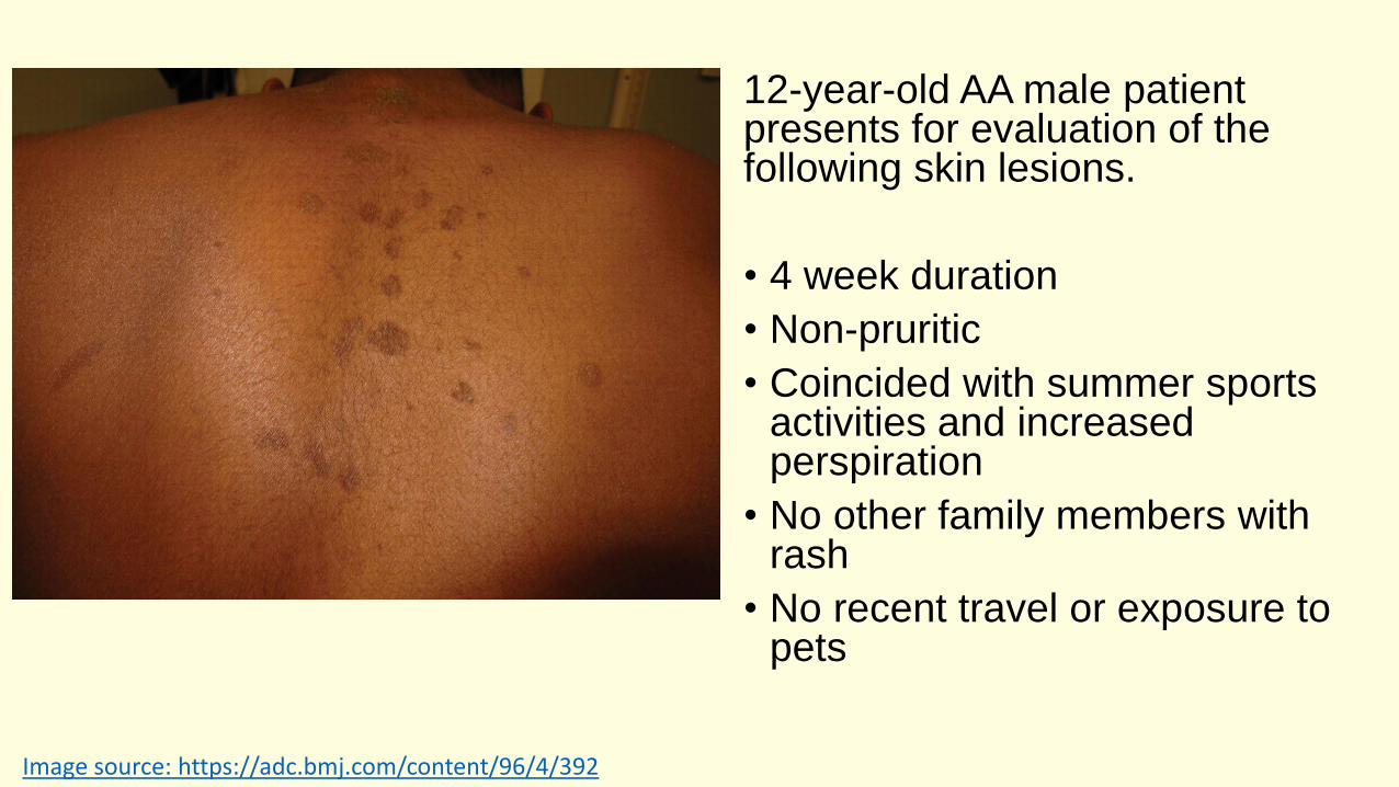
12-year-old AA male patient presents for evaluation of the following skin lesions.
• 4 week duration
• Non-pruritic
• Coincided with summer sports activities and increased perspiration
• No other family members with rash
• No recent travel or exposure to pets
Image source: https://adc.bmj.com/content/96/4/392

Bacteria, Fungus, or Virus?
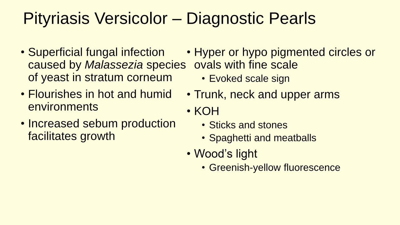
Pityriasis Versicolor – Diagnostic Pearls
• Superficial fungal infection caused by Malassezia species of yeast in stratum corneum
• Flourishes in hot and humid environments
• Increased sebum production facilitates growth
• Hyper or hypo pigmented circles or ovals with fine scale
• Evoked scale sign
• Trunk, neck and upper arms
• KOH• Sticks and stones
• Spaghetti and meatballs
• Wood’s light• Greenish-yellow fluorescence
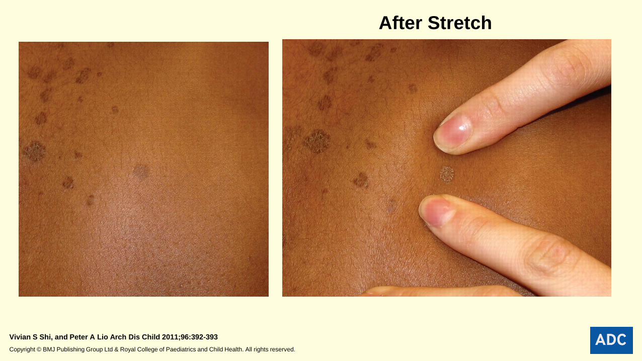
After Stretch
Vivian S Shi, and Peter A Lio Arch Dis Child 2011;96:392-393
Copyright © BMJ Publishing Group Ltd & Royal College of Paediatrics and Child Health. All rights reserved.

https://www.researchgate.net/figure/Patients-dorsum-with-pityriasis-versicolor-caused-by-Malassezia-fur-fur-with_fig3_323228320https://step1.medbullets.com/dermatology/112070/tinea-versicolor
Pityriasis Versicolor – Treatment
• Topical treatments effective and well-tolerated
• Shampoos and lotions – apply to affected area for 5-10 minutes then wash off, twice a day for 7-14 days, then once a month
• Zinc pyrithione
• Selenium sulfide
• Ketaconazole
• May take considerable time and maintenance
• Recalcitrant or recurring consider oral therapy
• Fluconazole 300 mg weekly for 2-4 weeks
• NOT terbinafine
• NOT ketaconazole

Fungal Infections
• Dermatophytoses• Tinea corporis
• Onychomycosis
• Pityriasis versicolor
• Candidiasis
• Majocchi granuloma (Pronunciation: mah-yok′ē)
• Sporotrichosis

Selected References/Resources
• Practice Guidelines for Diagnosis and Management of Skin and Soft Tissue Infections: 2014 Update by IDSA https://www.idsociety.org/practice-guideline/skin-and-soft-tissue-infections/
• Gupta, AK, et al. Fungal Skin Infections. Pediatrics in Review 2017;38;8.
• Lipner, SR, Scher, RK, Onychomycosis Treatment and Prevention of Recurrence, J Am Acad Dermatol, Vol 80, No 4, April 2019.
• Rush, J, Dinulos, JG, Childhood skin and soft tissue infections: new discoveries and guidelines regarding the management of bacterial soft tissue infections, molluscum contagiosum, and warts, CurrOpin Pediatr 201, 28:250-257.
• Ibrahim, F, Khan R, Pujalte, GA, Bacterial Skin Infections, Prim Care Clin Office Pract 42 (2015) 485-499.