Bacteria Dr. Cserynik Jan. 18, 2007 Slides by Bogdan Irimies PGY 4 EM Resident.
108
Bacteria Bacteria Dr. Cserynik Dr. Cserynik Jan. 18, 2007 Jan. 18, 2007 Slides by Bogdan Irimies Slides by Bogdan Irimies PGY 4 EM Resident PGY 4 EM Resident
-
Upload
emory-elliott -
Category
Documents
-
view
213 -
download
0
Transcript of Bacteria Dr. Cserynik Jan. 18, 2007 Slides by Bogdan Irimies PGY 4 EM Resident.

BacteriaBacteria
Dr. CserynikDr. CserynikJan. 18, 2007Jan. 18, 2007
Slides by Bogdan IrimiesSlides by Bogdan IrimiesPGY 4 EM ResidentPGY 4 EM Resident


Diptheria: Diptheria: Corynebacterium Corynebacterium diptheriaediptheriae
Epidemiology: humans are the only Epidemiology: humans are the only known reservoirsknown reservoirs Spread is via person-person contact thru Spread is via person-person contact thru
respiratory droplets or by direct contact respiratory droplets or by direct contact w/skin lesion exudatesw/skin lesion exudates
0-5 cases nationwide/year0-5 cases nationwide/year Usually seen unimmunized or under Usually seen unimmunized or under
immunized adults in urban and poor rural immunized adults in urban and poor rural areasareas

Diphtheria: Diphtheria: Corynebacterium Corynebacterium diphtheriadiphtheria
Etiology/pathophysiology:Etiology/pathophysiology: Gram + bacillius: korynee=club shaped Gram + bacillius: korynee=club shaped
bacteria; diphtheria=leather hide bacteria; diphtheria=leather hide looking pharyngeal membranelooking pharyngeal membrane
C. diphtheria C. diphtheria produces exotoxin that produces exotoxin that causes pharyngeal membrane exudates causes pharyngeal membrane exudates and systemic effects of infectionand systemic effects of infection
Causes skin, respiratory, cardiac Causes skin, respiratory, cardiac neurological manifestationsneurological manifestations
Diphtheria: Diphtheria: Corynebacterium Corynebacterium diphtheriadiphtheria
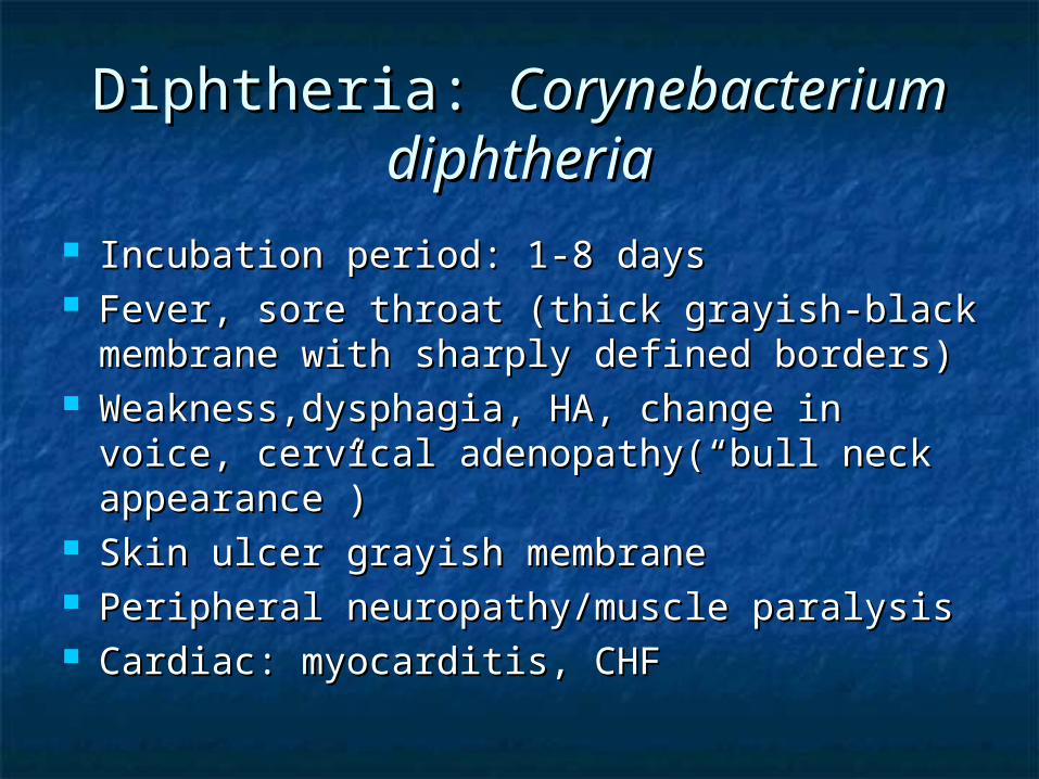
Incubation period: 1-8 daysIncubation period: 1-8 days Fever, sore throat (thick grayish-black Fever, sore throat (thick grayish-black
membrane with sharply defined borders)membrane with sharply defined borders) Weakness,dysphagia, HA, change in voice, Weakness,dysphagia, HA, change in voice,
cervical adenopathy(“bull neck cervical adenopathy(“bull neck appearance”)appearance”)
Skin ulcer grayish membraneSkin ulcer grayish membrane Peripheral neuropathy/muscle paralysisPeripheral neuropathy/muscle paralysis Cardiac: myocarditis, CHFCardiac: myocarditis, CHF

Diphtheria Membrane:Diphtheria Membrane:

Diphtheria: Diphtheria: Corynebacterium Corynebacterium diphtheriadiphtheria
Complications:Complications: Airway obstruction from Airway obstruction from
edema/membrane formationedema/membrane formation CHFCHF Cardiac conduction disturbancesCardiac conduction disturbances muscle paralysismuscle paralysis

Diphtheria: Diphtheria: Corynebacterium Corynebacterium diphtheriadiphtheria
Diagnosis: throat or nasopharyngeal Diagnosis: throat or nasopharyngeal swabs, cutaneous swabsswabs, cutaneous swabs
PCR for diphtheria genePCR for diphtheria gene Leukocytosis, mild Leukocytosis, mild
thrombocytopenia, proteinuriathrombocytopenia, proteinuria EKG: ST-T changes, AV block EKG: ST-T changes, AV block
dysrhythmiasdysrhythmias

Diphtheria: Diphtheria: Corynebacterium Corynebacterium diphtheria: D/Dx:diphtheria: D/Dx:
Strep/viral Strep/viral pharyngitispharyngitis
TonsillitisTonsillitis Vincent’s anginaVincent’s angina EpiglottitisEpiglottitis MonoMono
LaryngitisLaryngitis BronchitisBronchitis TracheitisTracheitis Monilial infxnMonilial infxn RhinitisRhinitis

Diphtheria: Diphtheria: Corynebacterium Corynebacterium diphtheria: Treatmentdiphtheria: Treatment
Place pt. in respiratory isolationPlace pt. in respiratory isolation Bronchodilators, fluidsBronchodilators, fluids Equine serum antitoxin is mainstay 20,000-Equine serum antitoxin is mainstay 20,000-
40,000 units IV40,000 units IV Erythromycin or procaine penicillin for 14 Erythromycin or procaine penicillin for 14
daysdays Close contacts should be observed for 7 Close contacts should be observed for 7
days, receive booster of diphtheria toxoid days, receive booster of diphtheria toxoid (Td) if >5 yrs. (Td) if >5 yrs.

Pertussis: EpidemiologyPertussis: Epidemiology
Localized respiratory illness Localized respiratory illness transmitted by respiratory dropletstransmitted by respiratory droplets
Avg incubation period 7-10 daysAvg incubation period 7-10 days Neither vaccination nor prior Neither vaccination nor prior
infection confer lifelong immunityinfection confer lifelong immunity

Pertussis: EtiologyPertussis: Etiology
Bordetella pertussis,Bordetella pertussis, gram neg. gram neg. coccobacillicoccobacilli
Preferentially adheres to ciliated Preferentially adheres to ciliated respiratory epithelial cellsrespiratory epithelial cells

Pertussis: Clinical Pertussis: Clinical
Arises in 3 distinct clinical stages:Arises in 3 distinct clinical stages: Catarrhal phase: begins after Catarrhal phase: begins after
incubation period, lasts 1-2 weeks, incubation period, lasts 1-2 weeks, infectivity is greatest during this infectivity is greatest during this phasephase Clinically indistinguishable from an URI: Clinically indistinguishable from an URI:
rhinorrhea, , low grade fever, malaise, rhinorrhea, , low grade fever, malaise, conjunctival injection, anorexiaconjunctival injection, anorexia

Pertussis: ClinicalPertussis: Clinical
Paroxysmal phase: fever subsides Paroxysmal phase: fever subsides and cough increases(2-4 wks.)and cough increases(2-4 wks.)
Staccato cough: pt. coughs Staccato cough: pt. coughs repeatedly in short exhalations repeatedly in short exhalations followed by a short inspiratory followed by a short inspiratory “whoop”“whoop”
Pt. may have post-tussive emesis, Pt. may have post-tussive emesis, syncope, brief apneasyncope, brief apnea

Pertussis: ClinicalPertussis: Clinical
Convalescent stage: residual cough Convalescent stage: residual cough that lasts from several weeks to that lasts from several weeks to monthsmonths
PE: low grade fever and tachypneaPE: low grade fever and tachypnea

Pertussis: DiagnosisPertussis: Diagnosis
Diagnosis should be entertained in Diagnosis should be entertained in anyone w/prolonged cough anyone w/prolonged cough w/paroxysmal whoops or posttussive w/paroxysmal whoops or posttussive emesisemesis
LeukocytosisLeukocytosis Lab confirmation by nasopharyngeal Lab confirmation by nasopharyngeal
swab or direct fluorescent antibodyswab or direct fluorescent antibody

Pertussis: D/DxPertussis: D/Dx
Viral URIViral URI Pneumonia Pneumonia BronchiolitisBronchiolitis CFCF TBTB
COPD Exac.COPD Exac. Foreign Body Foreign Body
AspirationAspiration

Pertussis: ComplicationsPertussis: Complications
Periorbital edemaPeriorbital edema Subconjunctival Subconjunctival
hemorrhagehemorrhage PetechiaePetechiae EpistaxisEpistaxis HemoptysisHemoptysis SQ EmphysemaSQ Emphysema
PTXPTX PneumomediastinuPneumomediastinu
mm Diaphragmatic Diaphragmatic
rupturerupture Hernia exac.Hernia exac. Rectal prolapseRectal prolapse

Pertussis:TreatmentPertussis:Treatment
O2, suctioning, hydrationO2, suctioning, hydration Antibiotics doesn’t appear to reduce the Antibiotics doesn’t appear to reduce the
severity of illness or duration especially if severity of illness or duration especially if started in paroxysmal phasestarted in paroxysmal phase
Erythromycin>Azithromax> BactrimErythromycin>Azithromax> Bactrim Postexposure prophylaxis w/erythromycin Postexposure prophylaxis w/erythromycin
is recommended for household contacts of is recommended for household contacts of pts. w/pertussis regardless of previous pts. w/pertussis regardless of previous vaccination statusvaccination status

Tetanus:Tetanus:

Tetanus: EpidemiologyTetanus: Epidemiology
Tetanus is a toxin mediated disease Tetanus is a toxin mediated disease characterized by uncontrolled skeletal characterized by uncontrolled skeletal muscle spasmsmuscle spasms
Avg. 43 cases reported to CDC/yearAvg. 43 cases reported to CDC/year Most common portals of entry are: Most common portals of entry are:
puncture wounds, lacerations, abrasionspuncture wounds, lacerations, abrasions Primary risk factors are inadequate Primary risk factors are inadequate
primary immunization and waning primary immunization and waning immunityimmunity

Tetanus: EtiologyTetanus: Etiology
Clostridium tetaniClostridium tetani gram positive, gram positive, spore forming, anaerobic bacillusspore forming, anaerobic bacillus
Found in soil, dust, fecesFound in soil, dust, feces Development of clinical tetanus Development of clinical tetanus
requires a portal of entry for infecting requires a portal of entry for infecting spores as well as tissue conditions spores as well as tissue conditions that promote germination and growth that promote germination and growth in an immunologically susceptible hostin an immunologically susceptible host

Tetanus: EtiologyTetanus: Etiology
C. tetani C. tetani produces a neurotoxin that produces a neurotoxin that causes clinical illnesscauses clinical illness C. tetani produces the neurotoxin C. tetani produces the neurotoxin
tetanospasmin(TS) at the site of tissue injurytetanospasmin(TS) at the site of tissue injury TS binds to the motor nerve ending and then TS binds to the motor nerve ending and then
moves by retrograde axonal transport to the moves by retrograde axonal transport to the CNSCNS
Preferentially binds to GABA and blocks Preferentially binds to GABA and blocks presynaptic release of GABA resulting in presynaptic release of GABA resulting in muscle spasm muscle spasm

Tetanus: ClinicalTetanus: Clinical
Tetanus typically occurs as a result Tetanus typically occurs as a result of a deep penetrating woundof a deep penetrating wound
Incubation period is 1 day to several Incubation period is 1 day to several monthsmonths
There are 4 types of clinical tetanus: There are 4 types of clinical tetanus: Generalized, localized, cephalic, and Generalized, localized, cephalic, and neonatalneonatal

Tetanus: ClinicalTetanus: Clinical
Generalized tetanus:Generalized tetanus: Most common formMost common form Trismus(lockjaw), characteristic sardonic Trismus(lockjaw), characteristic sardonic
smile(risus sardonicus)smile(risus sardonicus) Early symptoms include: irritability, Early symptoms include: irritability,
weakness, myalgias, muscle cramps, weakness, myalgias, muscle cramps, dysphagia, hydrophobia, droolingdysphagia, hydrophobia, drooling
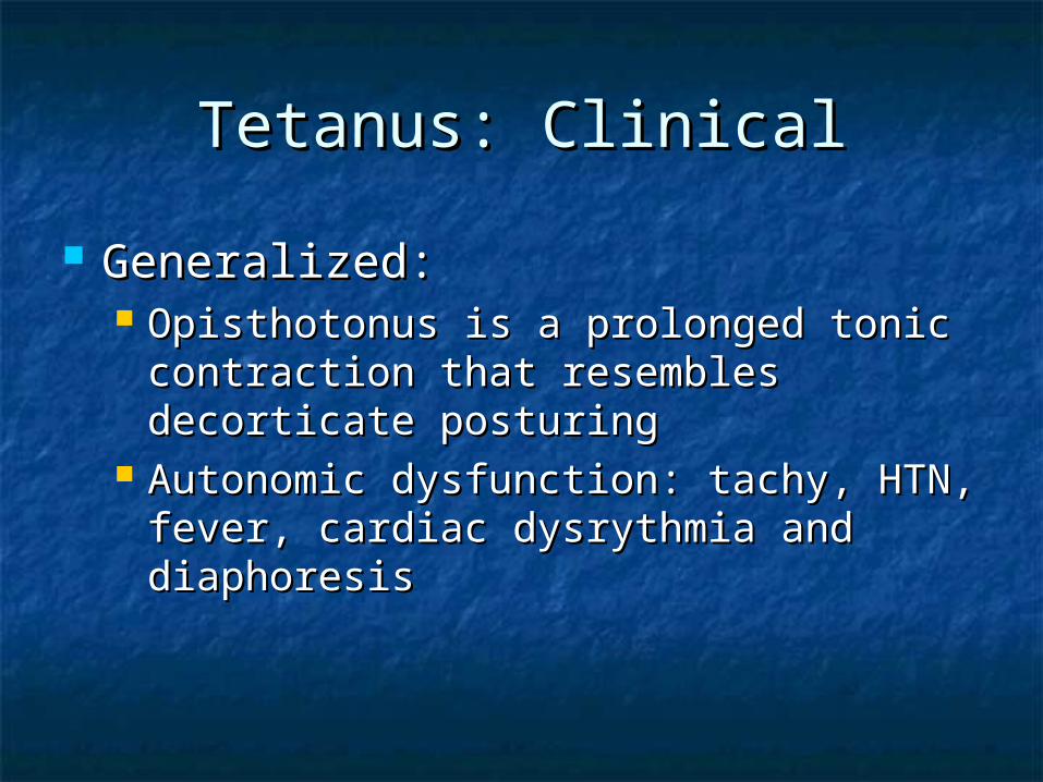
Generalized:Generalized: Opisthotonus is a prolonged tonic Opisthotonus is a prolonged tonic
contraction that resembles decorticate contraction that resembles decorticate posturingposturing
Autonomic dysfunction: tachy, HTN, Autonomic dysfunction: tachy, HTN, fever, cardiac dysrythmia and fever, cardiac dysrythmia and diaphoresis diaphoresis
Tetanus: ClinicalTetanus: Clinical

Tetanus: ClinicalTetanus: Clinical
Localized: persistent muscle spasm Localized: persistent muscle spasm located near site of injurylocated near site of injury
Most cases do not progress to Most cases do not progress to generalized tetanusgeneralized tetanus
Tetanus: ClinicalTetanus: Clinical
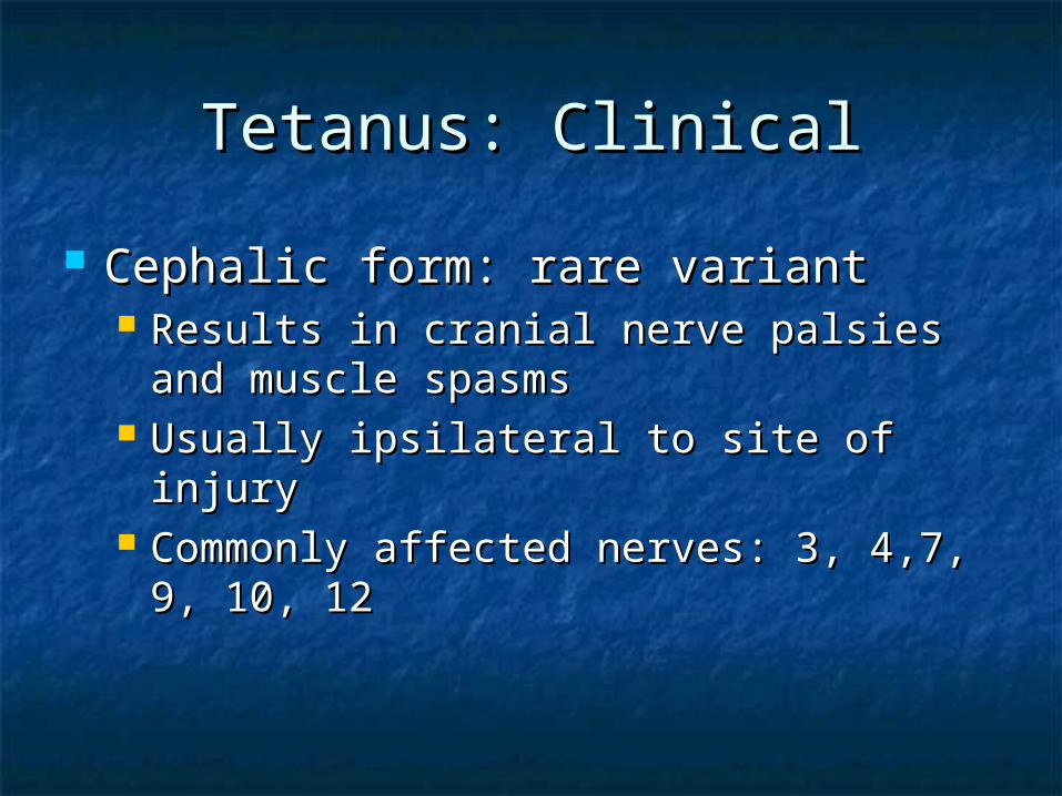
Cephalic form: rare variantCephalic form: rare variant Results in cranial nerve palsies and Results in cranial nerve palsies and
muscle spasmsmuscle spasms Usually ipsilateral to site of injuryUsually ipsilateral to site of injury Commonly affected nerves: 3, 4,7, 9, 10, Commonly affected nerves: 3, 4,7, 9, 10,
1212

Tetanus: ClinicalTetanus: Clinical
Neonatal form: generalized tetanus Neonatal form: generalized tetanus of newbornof newborn
Symptoms begin during 1Symptoms begin during 1stst week of week of lifelife
Irritability and poor feeding seenIrritability and poor feeding seen

Tetanus: DiagnosisTetanus: Diagnosis
Clinical diagnosisClinical diagnosis No lab test that confirm or exclude No lab test that confirm or exclude
diseasedisease CDC clinical case definition:CDC clinical case definition:
““Acute onset of hypertonia or painful Acute onset of hypertonia or painful muscular contractions and generalized muscular contractions and generalized muscle spasms without other apparent muscle spasms without other apparent medical cause.”medical cause.”

Tetanus: D/DxTetanus: D/Dx
Acute abdomenAcute abdomen Black widow spider Black widow spider
bitebite Dental Dental
abscess/peritonisillar abscess/peritonisillar abscessabscess
Dislocated Dislocated mandible/TMJmandible/TMJ
Dystonic rxnDystonic rxn meningoencephalitismeningoencephalitis
Head trauma/SAHHead trauma/SAH HyperventilationHyperventilation HypocalcemiaHypocalcemia RabiesRabies PsychogenicPsychogenic SepsisSepsis Status EpilepticusStatus Epilepticus Strychnine poisoningStrychnine poisoning

Tetanus: ComplicationsTetanus: Complications
Acute respiratory failure: results from Acute respiratory failure: results from respiratory muscle spasms, respiratory muscle spasms, laryngospasm and airway obstructionlaryngospasm and airway obstruction
Dysrythmias, HTN, myocarditis, pulm. Dysrythmias, HTN, myocarditis, pulm. EdemaEdema
Forceful contractions can cause Forceful contractions can cause vertebral subluxations & fx’s, long bone vertebral subluxations & fx’s, long bone fx’s, shoulder & TMJ joint dislocations fx’s, shoulder & TMJ joint dislocations

Tetanus: TreatmentTetanus: Treatment
4 treatment strategies:4 treatment strategies: 1. Aggressive supportive care1. Aggressive supportive care 2. elimination of unbound TS2. elimination of unbound TS 3. Active immunization3. Active immunization 4. Prevention of further toxin production4. Prevention of further toxin production

Tetanus: TreatmentTetanus: Treatment
Benzodiazepines are DOC for Benzodiazepines are DOC for supportive caresupportive care
Mechanical ventilation Mechanical ventilation w/neuromuscular blockadew/neuromuscular blockade
Autonomic instability: use labetalolAutonomic instability: use labetalol Human tetanus immunoglobulin(TIG) Human tetanus immunoglobulin(TIG)
neutralizes unbound toxinneutralizes unbound toxin Administer at a site separate from toxoidAdminister at a site separate from toxoid
Tetanus: TreatmentTetanus: Treatment
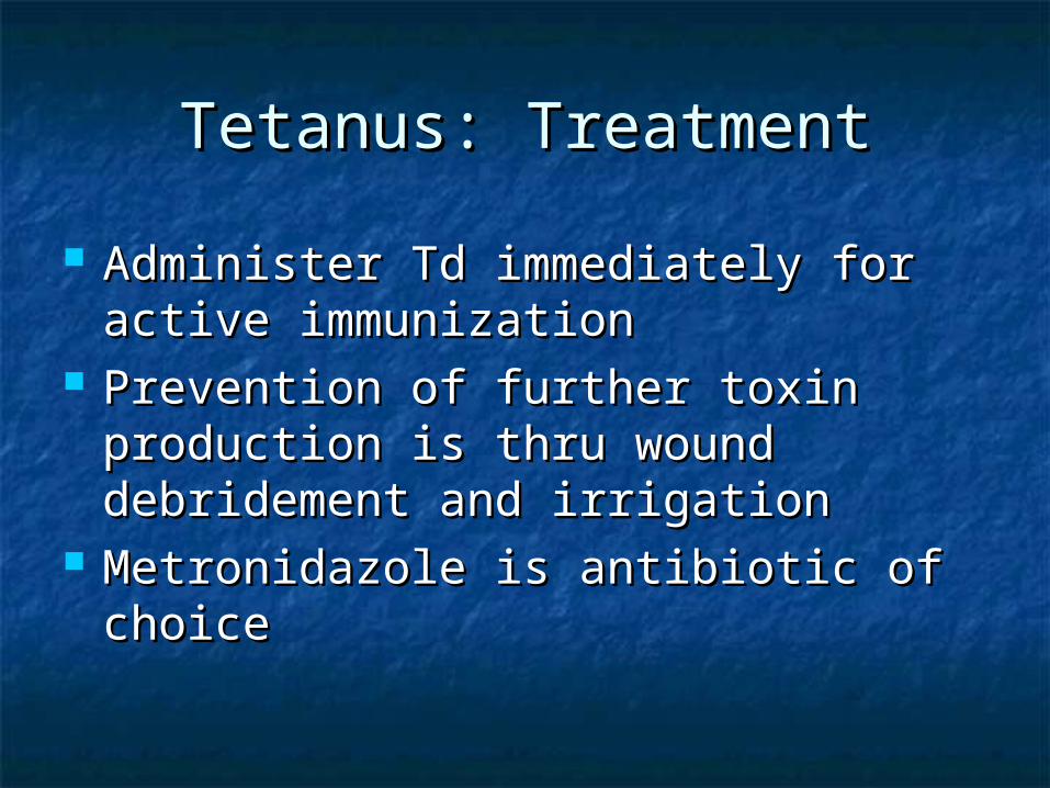
Administer Td immediately for active Administer Td immediately for active immunizationimmunization
Prevention of further toxin Prevention of further toxin production is thru wound production is thru wound debridement and irrigationdebridement and irrigation
Metronidazole is antibiotic of choiceMetronidazole is antibiotic of choice

Tetanus: VaccinationTetanus: Vaccination
Tetanus toxoid is an inactivated form Tetanus toxoid is an inactivated form of TSof TS
Immunity wanes after 5-10 yearsImmunity wanes after 5-10 years Those younger than 7 years should Those younger than 7 years should
receive DPTreceive DPT No evidence that Td is teratogenicNo evidence that Td is teratogenic TIG is not contraindicated in TIG is not contraindicated in
pregnancypregnancy
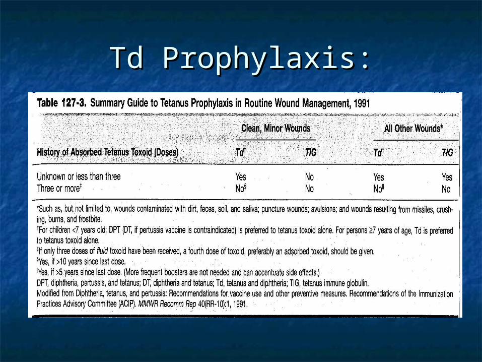
Td Prophylaxis:Td Prophylaxis:

Botulism:Botulism:

Botulism:Botulism:
Caused by neurotoxins produced by Caused by neurotoxins produced by Clostridium botulinumClostridium botulinum
5 forms of the disease:5 forms of the disease: 1. Food born botulism1. Food born botulism 2. Infant botulism2. Infant botulism 3. Wound botulism3. Wound botulism 4. Unclassified botulism4. Unclassified botulism 5. Inadvertent botulism5. Inadvertent botulism

Botulism: EpidemiologyBotulism: Epidemiology
7 types of toxins produced but only 7 types of toxins produced but only types A,B,E,F cause illness in humanstypes A,B,E,F cause illness in humans
110 cases/year reported to CDC110 cases/year reported to CDC Food borne botulism results from the Food borne botulism results from the
ingestion of preformed heat labile ingestion of preformed heat labile toxin which is found from exposure toxin which is found from exposure to home canned foodsto home canned foods

Botulism: EpidemiologyBotulism: Epidemiology
Infant botulism is most common form, caused Infant botulism is most common form, caused by ingestion of spores w/in vivo production of by ingestion of spores w/in vivo production of toxin. Found in honey and corn syruptoxin. Found in honey and corn syrup
Wound botulism is rare, 1 case/year, assoc. Wound botulism is rare, 1 case/year, assoc. w/IV drug abusew/IV drug abuse
Inadvertent botulism is iatrogenic, occurs in Inadvertent botulism is iatrogenic, occurs in people who have been injected w/botulism people who have been injected w/botulism toxin for dystonia, movement disorders and toxin for dystonia, movement disorders and cosmetic purposescosmetic purposes

Botulism: EtiologyBotulism: Etiology
C. botulinumC. botulinum anaerobic, gram positive anaerobic, gram positive rodrod
Bacteria produces a potent exotoxin that Bacteria produces a potent exotoxin that is responsible for the diseaseis responsible for the disease
Botulinum toxin targets peripheral Botulinum toxin targets peripheral neuromuscular junctions and autonomic neuromuscular junctions and autonomic synapses causing flaccid paralysissynapses causing flaccid paralysis BlocksBlocks the release of acetylcholine resulting the release of acetylcholine resulting
in neuromuscular blockadein neuromuscular blockade

Botulism: ClinicalBotulism: Clinical
Botulism manifests by cranial nerve Botulism manifests by cranial nerve palsies, parasympathetic blockade, palsies, parasympathetic blockade, descending flaccid paralysisdescending flaccid paralysis
Food borne botulism is prototype of Food borne botulism is prototype of diseasedisease
Symptoms begin 18-36 hrs. after Symptoms begin 18-36 hrs. after ingestion of toxin containing foodingestion of toxin containing food

Botulism: ClinicalBotulism: Clinical
Early symptoms include weakness, malaise, Early symptoms include weakness, malaise, lightheadedness, N/V, constipationlightheadedness, N/V, constipation
Neurologic symptoms: cranial nerves Neurologic symptoms: cranial nerves affectedaffected Diplopia, blurry vision, dysphonia, dysphagia, Diplopia, blurry vision, dysphonia, dysphagia,
dysarthriadysarthria Symmetric descending muscular weakness Symmetric descending muscular weakness
occurs involving upper and lower extremities occurs involving upper and lower extremities and respiratory musclesand respiratory muscles

Botulism: ClinicalBotulism: Clinical
Ocular signs: ptosis, extraocular Ocular signs: ptosis, extraocular palsies, dilated & fixed pupilspalsies, dilated & fixed pupils
Muscle weakness: upper extremities Muscle weakness: upper extremities more affected than lower, proximal more affected than lower, proximal weakness>distal musclesweakness>distal muscles
Sensory exam is normalSensory exam is normal

Botulism: ClinicalBotulism: Clinical
Infant botulism: Infant botulism: Constipation is common symptom, poor Constipation is common symptom, poor
feeding, weak cry, loss of head control, feeding, weak cry, loss of head control, hypotonia, decreased muscle tone, hypotonia, decreased muscle tone, depressed deep tendon reflexesdepressed deep tendon reflexes
Wound botulism: incubation period is Wound botulism: incubation period is longer 4-14 dayslonger 4-14 days Clinical presentation is similar to food Clinical presentation is similar to food
borne botulismborne botulism

Infant Botulism:Infant Botulism:

Wound Botulism:Wound Botulism:

Botulism: DiagnosisBotulism: Diagnosis
Initial diagnosis is clinical: suspect in Initial diagnosis is clinical: suspect in someone who presents with constellation someone who presents with constellation of GI, Autonomic, cranial nerve of GI, Autonomic, cranial nerve dysfunctiondysfunction
Confirmed by: Confirmed by: 1. botulinum toxin in pts. Blood1. botulinum toxin in pts. Blood 2. Botulinum toxin or C. botulinum in GI 2. Botulinum toxin or C. botulinum in GI
contents, stool, or woundcontents, stool, or wound 3. Toxin in the food source3. Toxin in the food source Notify CDCNotify CDC

Botulism: D/DxBotulism: D/Dx
PharyngitisPharyngitis GastroenteritisGastroenteritis Guillain Barre Guillain Barre
SyndromeSyndrome Tick paralysisTick paralysis Myasthenia GravisMyasthenia Gravis PoliomyelitisPoliomyelitis DiphtheriaDiphtheria Eaton Lambert Eaton Lambert
SyndromeSyndrome
Anticholinergic toxicityAnticholinergic toxicity Organophosphate Organophosphate
toxicitytoxicity Heavy metal poisoningHeavy metal poisoning Mg+2 toxicityMg+2 toxicity

Botulism: ComplicationsBotulism: Complications
Complications are related to Complications are related to respiratory failurerespiratory failure Weakness of respiratory musclesWeakness of respiratory muscles

Botulism: TreatmentBotulism: Treatment
Treatment consists of: supportive Treatment consists of: supportive care, administer antitoxincare, administer antitoxin
ICU, NG tube, foleyICU, NG tube, foley Antitoxin: contains antibodies to Antitoxin: contains antibodies to
toxins types A,B, Etoxins types A,B, E Neutralizes only circulating toxins and Neutralizes only circulating toxins and
has no effect on bound toxinhas no effect on bound toxin One vial is requiredOne vial is required

Botulism: TreatmentBotulism: Treatment
Infant botulism: antitoxin is not Infant botulism: antitoxin is not recommended b/c not efficacy, there is a recommended b/c not efficacy, there is a risk of anaphylaxis to horse serumrisk of anaphylaxis to horse serum Use human botulism immunoglobulin(BIG)Use human botulism immunoglobulin(BIG)
Wound botulism:Wound botulism: Debridement and antibiotics should be given Debridement and antibiotics should be given
only after antitoxin has been givenonly after antitoxin has been given

Pneumococcus:Pneumococcus:

Pneumococcemia:Pneumococcemia:
Strep. Pneumoniae:Strep. Pneumoniae: clinical clinical presentation ranges from mild illness presentation ranges from mild illness to fulminant, life threatening to fulminant, life threatening systemic syndromesystemic syndrome
Also causes localized infections such Also causes localized infections such as: OM, pneumonia, meningitis, as: OM, pneumonia, meningitis, endocarditis, septic arthritis, endocarditis, septic arthritis, peritonitis. peritonitis.

Strep. Pneumoniae: Strep. Pneumoniae: EpidemiologyEpidemiology
Exact incidence is unknown. Spread from Exact incidence is unknown. Spread from person to person by close contactperson to person by close contact
The introduction of the heptavalent The introduction of the heptavalent vaccine has decreased incidence of vaccine has decreased incidence of disease by 69% in children < 2 y/odisease by 69% in children < 2 y/o
Risk Factors for pneumococcemia: chronic Risk Factors for pneumococcemia: chronic respiratory or CV disease, chronic ETOH respiratory or CV disease, chronic ETOH abuse, cirrhosis, DM, impaired abuse, cirrhosis, DM, impaired spleen(sickle cell), CRF, AIDS, cancer, spleen(sickle cell), CRF, AIDS, cancer, organ transplantorgan transplant

Strep. Pneumoniae: EtiologyStrep. Pneumoniae: Etiology
Encapsulated, gram positive Encapsulated, gram positive anaerobic coccus that occurs in pairs anaerobic coccus that occurs in pairs and chainsand chains
Over 90 serotypesOver 90 serotypes Prevnar vaccine account for 7 Prevnar vaccine account for 7
serotypes which cause 80% of serotypes which cause 80% of invasive disease in childreninvasive disease in children

Pneumococcus:Pneumococcus:

Strep. Pneumoniae: EtiologyStrep. Pneumoniae: Etiology
Strep Pneumo enters bloodstream by Strep Pneumo enters bloodstream by one of two routes:one of two routes: 1. Begins as pulmonary infection, thru 1. Begins as pulmonary infection, thru
lymphatics and into bloodstreamlymphatics and into bloodstream 2. Colonizes or cause URI and spreads to 2. Colonizes or cause URI and spreads to
Subarachnoid space, then to arachnoid Subarachnoid space, then to arachnoid villi to venous sinus to bloodvilli to venous sinus to blood
Can cause a clinical picture from a minor Can cause a clinical picture from a minor febrile illness to septic shockfebrile illness to septic shock

Strep. Pneumoniae: ClinicalStrep. Pneumoniae: Clinical
Presents as SIRS syndromePresents as SIRS syndrome Also may present as lethargy, signs Also may present as lethargy, signs
of poor tissue perfusion, cyanosis, of poor tissue perfusion, cyanosis, hypo/hyperventilationhypo/hyperventilation
Findings on physical exam vary with Findings on physical exam vary with site of localized infectionsite of localized infection

Strep. Pneumoniae: Strep. Pneumoniae: DiagnosisDiagnosis
The only specific test is blood cultureThe only specific test is blood culture Check CBC w/diff, blood & urine Check CBC w/diff, blood & urine
cultures, electrolytes, CXR, sputum, cultures, electrolytes, CXR, sputum, ABG prn, LP prn, coags prnABG prn, LP prn, coags prn

Strep. Pneumoniae: Strep. Pneumoniae: ComplicationsComplications
CV collapse, DIC, Septic emboli, CV collapse, DIC, Septic emboli, Respiratory failure, meningitis, Respiratory failure, meningitis, hypothermia, GI Bleeding, hepatic hypothermia, GI Bleeding, hepatic coma, renal failure, MIcoma, renal failure, MI
Pneumococcemia can cause Pneumococcemia can cause hematogenous seeding which results hematogenous seeding which results in: peritonitis, arthritis, endocarditis, in: peritonitis, arthritis, endocarditis, meningitis, cellulitismeningitis, cellulitis

Strep. Pneumoniae: Strep. Pneumoniae: TreatmentTreatment
Prompt initiation of antibiotics: Penicillin Prompt initiation of antibiotics: Penicillin G, ceftriaxone(covers also N. meningitis, G, ceftriaxone(covers also N. meningitis, H. flu)H. flu)
PCN allergic pt: cefotaxime, ceftriaxone, PCN allergic pt: cefotaxime, ceftriaxone, vanco, chloramphenicolvanco, chloramphenicol
For PCN resistant Strep. Pneumo: use For PCN resistant Strep. Pneumo: use ceftriaxone, cefotaxime, vancomycin or ceftriaxone, cefotaxime, vancomycin or imipenemimipenem
For suspected pneumococcal meningitis: For suspected pneumococcal meningitis: use vanco + cefuroxime or cefotaximeuse vanco + cefuroxime or cefotaxime

Pneumococcal Vaccine:Pneumococcal Vaccine:
Effective in preventing disease, accounts Effective in preventing disease, accounts for 85-90% of pneumococcus infectionsfor 85-90% of pneumococcus infections
Recommended for children ages 2-23 Recommended for children ages 2-23 monthsmonths
Recommended for adults with the Recommended for adults with the following: following: Chronic illness: CV/Pulm, DM, ETOHicsChronic illness: CV/Pulm, DM, ETOHics Immunocompromised people including Immunocompromised people including
asplenic pts.,HIVasplenic pts.,HIV

Meningococcemia:Meningococcemia:
Think of this in a patient who appears Think of this in a patient who appears relatively well on initial presentation, relatively well on initial presentation, then becomes morbiund and then becomes morbiund and critically ill w/fulminant infection critically ill w/fulminant infection several hours laterseveral hours later

Meningococcemia: Meningococcemia: EpidemiologyEpidemiology
2400-3000 cases annually2400-3000 cases annually Crowded living conditions increase the Crowded living conditions increase the
risk: military recruits, college freshmanrisk: military recruits, college freshman Risk Factors: close contact w/an Risk Factors: close contact w/an
infected pt., complement def., asplenia, infected pt., complement def., asplenia, chronic ETOH abuse, smoking, chronic chronic ETOH abuse, smoking, chronic steroid use, recent respiratory infectionsteroid use, recent respiratory infection
Overall mortality rate is 10%!Overall mortality rate is 10%!

Meningococcemia: EtiologyMeningococcemia: Etiology
Caused by Caused by Neisseria meningitidisNeisseria meningitidis a gram a gram neg. diplococcus, aerobic, encapsulated neg. diplococcus, aerobic, encapsulated organism w/13 serotypesorganism w/13 serotypes
N. meningitidis attaches to nonciliated N. meningitidis attaches to nonciliated epithelial cells in the nasopharynx and epithelial cells in the nasopharynx and either becomes an asymptomatic carrier either becomes an asymptomatic carrier state or produces a mild URI syndrome. state or produces a mild URI syndrome. If it enters the bloodstream: may see localized If it enters the bloodstream: may see localized
infection, bacteremia, sepsis or fulminant infection, bacteremia, sepsis or fulminant infectioninfection

Meningococcemia: ClinicalMeningococcemia: Clinical
Clinical presentation ranges from mild Clinical presentation ranges from mild febrile illness to fulminant disease and febrile illness to fulminant disease and death w/in hoursdeath w/in hours
May see fever, irritability, lethargy, May see fever, irritability, lethargy, myalgias, emesis, diarrhea, cough, myalgias, emesis, diarrhea, cough, rhinorrhearhinorrhea Only 60% of pts. Have classic signs of Only 60% of pts. Have classic signs of
meningococcemia: fever, petechiae or meningococcemia: fever, petechiae or purpurapurpura

Meningococcemia: ClinicalMeningococcemia: Clinical
Meningococcal meningitis may present as Meningococcal meningitis may present as fever, HA, photophobia, vomiting and fever, HA, photophobia, vomiting and signs of meningeal inflammationsigns of meningeal inflammation
Infants and small children may present as Infants and small children may present as fever, irritability, vomitingfever, irritability, vomiting
Purpura fulminans: occurs in children, Purpura fulminans: occurs in children, usually assoc. w/DIC.usually assoc. w/DIC. characterized by rapidly spreading ecchymosis characterized by rapidly spreading ecchymosis
and gangrene of the extremitiesand gangrene of the extremities

Purpura:Purpura:

Petechiae:Petechiae:

Meningococcemia: ClinicalMeningococcemia: Clinical
Fulminant meningococcemia: Fulminant meningococcemia: Waterhouse Friderichsen SyndromeWaterhouse Friderichsen Syndrome Extreme severity of illness, shock like Extreme severity of illness, shock like
statestate Diffuse petechiae and purpuric rashDiffuse petechiae and purpuric rash Shock, CHF, DIC, Renal failure, coma Shock, CHF, DIC, Renal failure, coma
possiblepossible Bilateral adrenal hemorrhageBilateral adrenal hemorrhage

Meningococcemia: Meningococcemia: DiagnosisDiagnosis
Diagnosis of Diagnosis of N. meningitidisN. meningitidis is is confirmed by isolation from blood confirmed by isolation from blood cultures, CSF, synovial, pleural or cultures, CSF, synovial, pleural or pericardial fluidpericardial fluid
WBC count may be high, low or WBC count may be high, low or normalnormal
Bandemia is typically present Bandemia is typically present

Meningococcemia: D/DxMeningococcemia: D/Dx
Strep. PneumoStrep. Pneumo H. FluH. Flu N. GonorrheaN. Gonorrhea Viral exanthemViral exanthem RMSFRMSF TyphusTyphus EndocarditisEndocarditis Vasculitis(HSP)Vasculitis(HSP)
Toxic ShockToxic Shock Acute Rheumatic Acute Rheumatic
feverfever Drug rxnsDrug rxns ITPITP TTPTTP

Meningococcemia: Meningococcemia: ComplicationsComplications
Most common complication is Most common complication is myocarditis w/CHF or conduction myocarditis w/CHF or conduction abnormalitiesabnormalities
Respiratory failureRespiratory failure Renal failureRenal failure Cranial nerve palsiesCranial nerve palsies VasculitisVasculitis Purulent arthritisPurulent arthritis

Meningococcemia: Meningococcemia: TreatmentTreatment
Immediate antibiotics: lab ID use PCN Immediate antibiotics: lab ID use PCN G or ampicillinG or ampicillin
Alternatives are: cefotaxime, Alternatives are: cefotaxime, ceftriaxone, chloramphenicolceftriaxone, chloramphenicol
Airway mgmt, IVF support and Airway mgmt, IVF support and vasopressor support, glucocorticoid vasopressor support, glucocorticoid therapy for refractory shocktherapy for refractory shock

Meningococcemia: Prophylaxis Meningococcemia: Prophylaxis & Vaccination& Vaccination
Antibiotic prophylaxis for close Antibiotic prophylaxis for close contacts such as household, nursery contacts such as household, nursery school, daycare, intimate contacts, school, daycare, intimate contacts, health care workershealth care workers
Use rifampin for 4 doses or CiproUse rifampin for 4 doses or Cipro Vaccine to children 2 years or older in Vaccine to children 2 years or older in
high risk groups such as functional or high risk groups such as functional or anatomic asplenia or complement def.anatomic asplenia or complement def.


Kawasaki’s Disease: Kawasaki’s Disease:
KD is an acute febrile systemic KD is an acute febrile systemic vasculitis of unknown etiologyvasculitis of unknown etiology
Also called Acute Febrile Also called Acute Febrile Mucocutaneous Lymph Node Mucocutaneous Lymph Node SyndromeSyndrome

Kawasaki’s Disease: Kawasaki’s Disease: EpidemiologyEpidemiology
Peak incidence is in 1-2 year oldsPeak incidence is in 1-2 year olds Most prevalent in Asian descentMost prevalent in Asian descent 3000 cases annually3000 cases annually Overall mortality is 2.5% and is a Overall mortality is 2.5% and is a
result of cardiac complicationsresult of cardiac complications

Kawasaki’s Disease: Kawasaki’s Disease: EtiologyEtiology
KD is a systemic vasculitisKD is a systemic vasculitis There is some link between mechanism There is some link between mechanism
of vascular injury and immune system of vascular injury and immune system activationactivation
Vasculitis affects medium sized vesselsVasculitis affects medium sized vessels Inflammation in the coronary arteries Inflammation in the coronary arteries
can cause myocarditis and aneurysmscan cause myocarditis and aneurysms

Kawasaki’s Disease: ClinicalKawasaki’s Disease: Clinical
Fever for at least 5 days and four of the following Fever for at least 5 days and four of the following 5 signs:5 signs: 1. Bilateral conjunctival injection1. Bilateral conjunctival injection 2. Oral mucosa changes2. Oral mucosa changes
Erythematous dry fissured lipsErythematous dry fissured lips Strawberry tongueStrawberry tongue Erythematous oropharynxErythematous oropharynx
3. Hand and feet changes3. Hand and feet changes Erythema of palms & solesErythema of palms & soles Edema of hand & feetEdema of hand & feet Periungal desquamationPeriungal desquamation
4. Rash4. Rash 5. Cervical lymphadenopathy5. Cervical lymphadenopathy
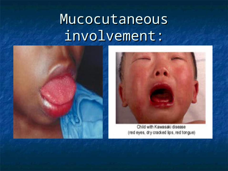
Mucocutaneous Mucocutaneous involvement:involvement:

Edema of hands:Edema of hands:

Kawasaki’s Disease: ClinicalKawasaki’s Disease: Clinical
Cardiac involvement is the hallmark Cardiac involvement is the hallmark of the disease:of the disease: KD is most common cause of acquired KD is most common cause of acquired
pediatric heart disease in U.S.pediatric heart disease in U.S. May see myocarditis, CHF, TachycardiaMay see myocarditis, CHF, Tachycardia Coronary artery aneurysmsCoronary artery aneurysms Mortality is from coronary artery Mortality is from coronary artery
aneurysm rupture or MIaneurysm rupture or MI

Coronary Artery Aneurysm:Coronary Artery Aneurysm:

Kawasaki’s Disease: Kawasaki’s Disease: DiagnosisDiagnosis
Fever > 5 days plus 4/5 criteria Fever > 5 days plus 4/5 criteria No definitive testNo definitive test EKG, EchoEKG, Echo CBC, CRP, ESR, CMP w/LFT’sCBC, CRP, ESR, CMP w/LFT’s May see elevated WBC w/left shift, May see elevated WBC w/left shift,
normochromic normocytic anemia, elevated plt normochromic normocytic anemia, elevated plt ct> 1,000,000ct> 1,000,000
Pts. w/KD should have UA, blood cx, CXR, ASO Pts. w/KD should have UA, blood cx, CXR, ASO titer, GABHS throat cx to exclude other titer, GABHS throat cx to exclude other diagnosisdiagnosis
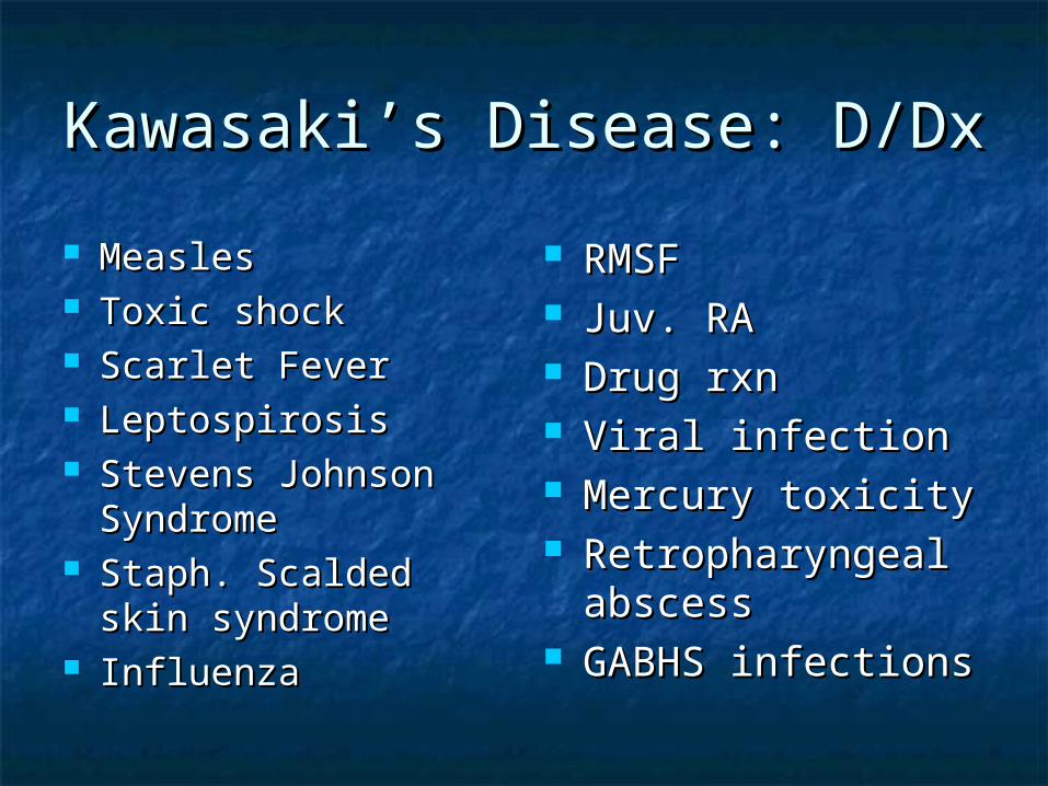
Kawasaki’s Disease: D/DxKawasaki’s Disease: D/Dx
MeaslesMeasles Toxic shockToxic shock Scarlet FeverScarlet Fever LeptospirosisLeptospirosis Stevens Johnson Stevens Johnson
SyndromeSyndrome Staph. Scalded skin Staph. Scalded skin
syndromesyndrome InfluenzaInfluenza
RMSFRMSF Juv. RAJuv. RA Drug rxnDrug rxn Viral infectionViral infection Mercury toxicityMercury toxicity Retropharyngeal Retropharyngeal
abscessabscess GABHS infectionsGABHS infections

Kawaski’s Disease: Kawaski’s Disease: ComplicationsComplications
Coronary artery aneurysm is most Coronary artery aneurysm is most serious complicationserious complication
Occurs in 20-25% of untreated pts., Occurs in 20-25% of untreated pts., occurs in 3-4% of those treated w/IV occurs in 3-4% of those treated w/IV immunoglobulin and ASAimmunoglobulin and ASA
Other complications: MI, CHF, Other complications: MI, CHF, myocarditismyocarditis

Kawasaki’s Treatment:Kawasaki’s Treatment:
Hospitalize pt.Hospitalize pt. Includes administration of ASA and IV Includes administration of ASA and IV
gamma globulingamma globulin ASA 80-100mg/kg/day for 6-8 wksASA 80-100mg/kg/day for 6-8 wks If coronary artery abnormalities If coronary artery abnormalities
exist, can use dipyridamoleexist, can use dipyridamole Coumadin or heparin for severe Coumadin or heparin for severe
coronary diseasecoronary disease


Toxic Shock Syndrome: Toxic Shock Syndrome: EpidemiologyEpidemiology
Toxic Shock Syndrome is a toxin mediated Toxic Shock Syndrome is a toxin mediated systemic inflammatory response syndromesystemic inflammatory response syndrome
Menstruation remains most common Menstruation remains most common settingsetting
200 cases a year200 cases a year Nonmenstrual TSS is assoc. Nonmenstrual TSS is assoc.
w/superinfection of various skin w/superinfection of various skin lesions/soft tissue infections: strep TSSlesions/soft tissue infections: strep TSS
Mortality rate of staph TSS is 3%, strep TSS Mortality rate of staph TSS is 3%, strep TSS is 30-70%is 30-70%

Toxic Shock Syndrome: Toxic Shock Syndrome: EtiologyEtiology
Staphylococcal TSS is caused by Staphylococcal TSS is caused by colonization or infection w/toxigenic colonization or infection w/toxigenic strains of S. aureusstrains of S. aureus
Streptococcal TSS is caused by infection Streptococcal TSS is caused by infection w/toxigenic strains of group A strep(GAS).w/toxigenic strains of group A strep(GAS).
Staph. Aureus produces toxic shock Staph. Aureus produces toxic shock syndrome toxin(TSST-1)syndrome toxin(TSST-1)
GAS produces streptococcal pyrogenic GAS produces streptococcal pyrogenic exotoxinexotoxin

Toxic Shock Syndrome: Risk Toxic Shock Syndrome: Risk FactorsFactors
Use of super Use of super absorbent tamponabsorbent tampon
Post-op wound Post-op wound infectioninfection
Post-partum periodPost-partum period Nasal packingNasal packing Bacterial infectionsBacterial infections
Varicella or Varicella or Influenza A Influenza A infectioninfection
DMDM HIVHIV Chronic cardiac & Chronic cardiac &
pulmonary diseasepulmonary disease

Toxic Shock Syndrome: Case Toxic Shock Syndrome: Case Definition for Staph. Aureus Definition for Staph. Aureus
TSSTSS Fever >38.9CFever >38.9C Diffuse erythroderma rashDiffuse erythroderma rash Palm and sole desquamation rash 1-2 wks laterPalm and sole desquamation rash 1-2 wks later HypotensionHypotension Multisystem involvement: 3 or more of followingMultisystem involvement: 3 or more of following
GI: N/V/DGI: N/V/D Muscular: myalgias or incr. CPKMuscular: myalgias or incr. CPK Mucus Membrane hyperemiaMucus Membrane hyperemia Renal: elevated BUN/Cr.Renal: elevated BUN/Cr. Hepatic: elevated LFT’s, biliHepatic: elevated LFT’s, bili Heme: plts. <100,000Heme: plts. <100,000 CNS: Altered mental statusCNS: Altered mental status

Toxic Shock Syndrome: Case Toxic Shock Syndrome: Case Definition for Strep TSSDefinition for Strep TSS
Isolation of group A strep from body siteIsolation of group A strep from body site Clinical signs: 2 or more Clinical signs: 2 or more
Hypotension andHypotension and Renal impairmentRenal impairment CoagulopathyCoagulopathy Liver abnormalitiesLiver abnormalities ARDSARDS Necrotizing fasciitisNecrotizing fasciitis Erythematous rashErythematous rash

TSS: ClinicalTSS: Clinical
The clinical presentations of strep The clinical presentations of strep TSS and staph TSS are similarTSS and staph TSS are similar
The primary difference is an The primary difference is an identifiable infectious source is identifiable infectious source is always present w/strep TSS and always present w/strep TSS and colonization alone may be the only colonization alone may be the only source in staph TSSsource in staph TSS

TSS: ClinicalTSS: Clinical
Fever, chillsFever, chills N/V/DN/V/D MyalgiasMyalgias PharyngitisPharyngitis HAHA Sepsis w/organ Sepsis w/organ
dysfunctiondysfunction RashRash Altered mental statusAltered mental status
Conjunctival erythemaConjunctival erythema Strawberry tongueStrawberry tongue Peripheral edemaPeripheral edema

Erythematous rash:Erythematous rash:

TSS: DiagnosisTSS: Diagnosis
See case definitionsSee case definitions May see leukocytosis or leukopenia, May see leukocytosis or leukopenia,
bandemiabandemia CXRCXR LP prnLP prn EKGEKG

TSS: D/DxTSS: D/Dx
Kawasaki’s DiseaseKawasaki’s Disease Staph Scaled skin Staph Scaled skin
syndromesyndrome Scarlet feverScarlet fever Drug rxns/Stevens Drug rxns/Stevens
JohnsonJohnson RMSFRMSF
LeptospirosisLeptospirosis MeningococcemiaMeningococcemia Gram neg. sepsisGram neg. sepsis MeaslesMeasles Viral illnessViral illness
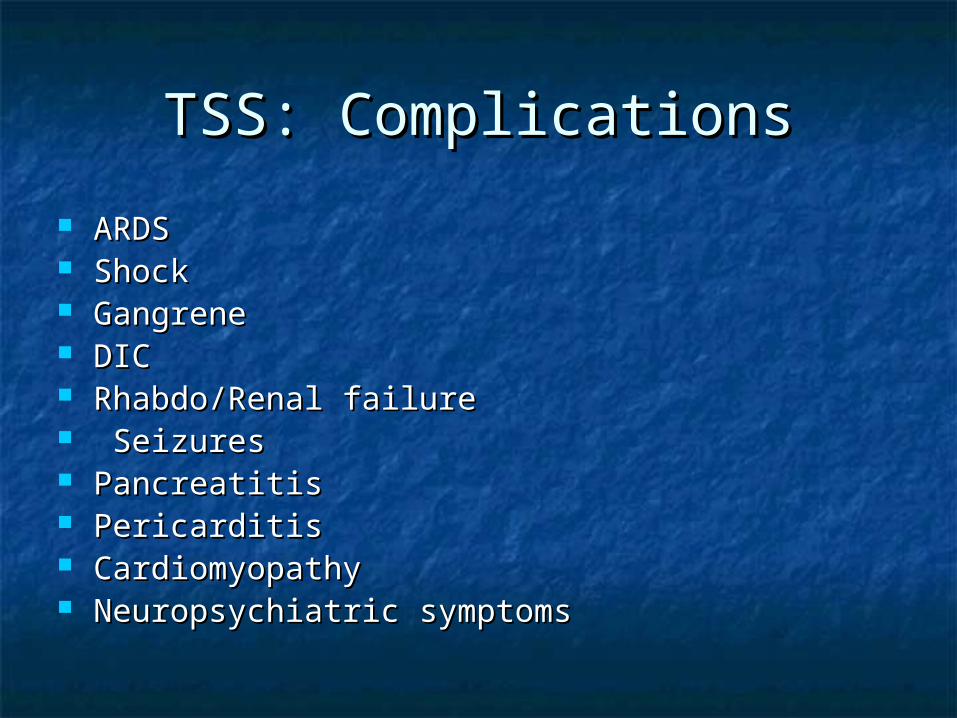
TSS: ComplicationsTSS: Complications
ARDSARDS ShockShock GangreneGangrene DICDIC Rhabdo/Renal failureRhabdo/Renal failure SeizuresSeizures PancreatitisPancreatitis PericarditisPericarditis CardiomyopathyCardiomyopathy Neuropsychiatric symptomsNeuropsychiatric symptoms

TSS: ManagementTSS: Management
Aggressive fluidsAggressive fluids O2, monitorO2, monitor Source of bacteria removed(tampons, Source of bacteria removed(tampons,
nasal packing, wound debridement)nasal packing, wound debridement) Antibiotics: clinda, nafcillin or oxacillinAntibiotics: clinda, nafcillin or oxacillin Refractory case use IV immunoglobulinRefractory case use IV immunoglobulin

Summary:Summary:
All pts. Appearing septic should be treated All pts. Appearing septic should be treated w/broad spectrum antibiotics ASAPw/broad spectrum antibiotics ASAP
Immunity to Diphtheria, pertussis, tetanus wanes Immunity to Diphtheria, pertussis, tetanus wanes in adults. Think of pertusssis as a cause of in adults. Think of pertusssis as a cause of persistent cough in adults. Update Td in trauma persistent cough in adults. Update Td in trauma or infectionor infection
Botulism should be in differential for infant who Botulism should be in differential for infant who presents w/failure to thrive, constipation or presents w/failure to thrive, constipation or decreased muscle tone. Also in IV drug abuser decreased muscle tone. Also in IV drug abuser w/neurologic symptomsw/neurologic symptoms
IV GG should be given as soon as KD is diagnosedIV GG should be given as soon as KD is diagnosed


![Welcome [weillcornellbrainandspine.org] · Maricruz Rivera, MD, PhD PGY-3. Neurological Surgery Residents. Evan Bander, MD PGY-5 Alexander D. Ramos, MD, PhD PGY-5 Joseph Carnevale,](https://static.fdocuments.in/doc/165x107/5f7167444c714e55d46f024a/welcome-weill-maricruz-rivera-md-phd-pgy-3-neurological-surgery-residents.jpg)