
Bac-to-Bac Baculovirus Expression System · The Bac-to-Bac™ Baculovirus Expression System...
74
For Research Use Only. Not for use in diagnostic procedures. Bac-to-Bac ™ Baculovirus Expression System USER GUIDE An efficient site-specific transposition system to generate baculovirus for high-level expression of recombinant proteins Catalog Numbers 10359-016, 10360-014, 10584-027, 10712-024 Publication Number MAN0000414 Revision B.0
Transcript of Bac-to-Bac Baculovirus Expression System · The Bac-to-Bac™ Baculovirus Expression System...

For Research Use Only. Not for use in diagnostic procedures.
Bac-to-Bac™ Baculovirus Expression SystemUSER GUIDE
An efficient site-specific transposition system to generatebaculovirus for high-level expression of recombinant proteins
Catalog Numbers 10359-016, 10360-014, 10584-027, 10712-024Publication Number MAN0000414
Revision B.0

Manufacturer: Life Technologies Corporation | 5781 Van Allen Way | Carlsbad, CA 92008
The information in this guide is subject to change without notice.
DISCLAIMER: TO THE EXTENT ALLOWED BY LAW, THERMO FISHER SCIENTIFIC INC. AND/OR ITS AFFILIATE(S) WILL NOT BE LIABLE FOR SPECIAL,INCIDENTAL, INDIRECT, PUNITIVE, MULTIPLE, OR CONSEQUENTIAL DAMAGES IN CONNECTION WITH OR ARISING FROM THIS DOCUMENT,INCLUDING YOUR USE OF IT.
Revision history: Pub. No. MAN0000414
Revision Date DescriptionB.0 16 July 2018 Rebrand
A.0 17 August 2015 Baseline for revisions
Important Licensing Information: This product may be covered by one or more Limited Use Label Licenses. By use of this product, you accept theterms and conditions of all applicable Limited Use Label Licenses.TRADEMARKS: All trademarks are the property of Thermo Fisher Scientific and its subsidiaries unless otherwise specified.
©2018 Thermo Fisher Scientific Inc. All rights reserved.
Contents
■ Product information . . . . . . . . . . . . . . . . . . . . . . . . . . . . . . . . . . . . . . . . . . . . . . . . . . . . . . . 8
Product description . . . . . . . . . . . . . . . . . . . . . . . . . . . . . . . . . . . . . . . . . . . . . . . . . . . . . . . . . . . . . 8
Kit contents and storage . . . . . . . . . . . . . . . . . . . . . . . . . . . . . . . . . . . . . . . . . . . . . . . . . . . . . . . . . 8Types of products . . . . . . . . . . . . . . . . . . . . . . . . . . . . . . . . . . . . . . . . . . . . . . . . . . . . . . . . . . . 8Shipping and storage . . . . . . . . . . . . . . . . . . . . . . . . . . . . . . . . . . . . . . . . . . . . . . . . . . . . . . . . 8pFastBac™ vectors . . . . . . . . . . . . . . . . . . . . . . . . . . . . . . . . . . . . . . . . . . . . . . . . . . . . . . . . . . 9MAX Efficiency™ DH10Bac™ Competent E. coli . . . . . . . . . . . . . . . . . . . . . . . . . . . . . . . . . . 9ExpiFectamine™ Sf Transfection Reagent . . . . . . . . . . . . . . . . . . . . . . . . . . . . . . . . . . . . . 10
The Bac-to-Bac™ Baculovirus Expression System . . . . . . . . . . . . . . . . . . . . . . . . . . . . . . . . . . 10System components . . . . . . . . . . . . . . . . . . . . . . . . . . . . . . . . . . . . . . . . . . . . . . . . . . . . . . . . 10pFastBac™ vector . . . . . . . . . . . . . . . . . . . . . . . . . . . . . . . . . . . . . . . . . . . . . . . . . . . . . . . . . . 10DH10Bac™ E. coli . . . . . . . . . . . . . . . . . . . . . . . . . . . . . . . . . . . . . . . . . . . . . . . . . . . . . . . . . . 10Baculovirus shuttle vector . . . . . . . . . . . . . . . . . . . . . . . . . . . . . . . . . . . . . . . . . . . . . . . . . . 11Helper plasmid . . . . . . . . . . . . . . . . . . . . . . . . . . . . . . . . . . . . . . . . . . . . . . . . . . . . . . . . . . . . 11ExpiFectamine™ Sf Transfection Reagent . . . . . . . . . . . . . . . . . . . . . . . . . . . . . . . . . . . . . 11Diagram of the Bac-to-Bac™ System . . . . . . . . . . . . . . . . . . . . . . . . . . . . . . . . . . . . . . . . . 12Workflow . . . . . . . . . . . . . . . . . . . . . . . . . . . . . . . . . . . . . . . . . . . . . . . . . . . . . . . . . . . . . . . . . 13
■ Methods . . . . . . . . . . . . . . . . . . . . . . . . . . . . . . . . . . . . . . . . . . . . . . . . . . . . . . . . . . . . . . . . . . . 14
Culture insect cells . . . . . . . . . . . . . . . . . . . . . . . . . . . . . . . . . . . . . . . . . . . . . . . . . . . . . . . . . . . . . 14Introduction . . . . . . . . . . . . . . . . . . . . . . . . . . . . . . . . . . . . . . . . . . . . . . . . . . . . . . . . . . . . . . . 14Serum-free medium . . . . . . . . . . . . . . . . . . . . . . . . . . . . . . . . . . . . . . . . . . . . . . . . . . . . . . . 14Insect cell culture reference guide . . . . . . . . . . . . . . . . . . . . . . . . . . . . . . . . . . . . . . . . . . . 14General cell culture guidelines . . . . . . . . . . . . . . . . . . . . . . . . . . . . . . . . . . . . . . . . . . . . . . 15Cells for transfection . . . . . . . . . . . . . . . . . . . . . . . . . . . . . . . . . . . . . . . . . . . . . . . . . . . . . . . 15
Generate recombinant pFastBac™ donor plasmid . . . . . . . . . . . . . . . . . . . . . . . . . . . . . . . . . . 15Introduction . . . . . . . . . . . . . . . . . . . . . . . . . . . . . . . . . . . . . . . . . . . . . . . . . . . . . . . . . . . . . . . 15Select E. coli host . . . . . . . . . . . . . . . . . . . . . . . . . . . . . . . . . . . . . . . . . . . . . . . . . . . . . . . . . . 15Transformation method . . . . . . . . . . . . . . . . . . . . . . . . . . . . . . . . . . . . . . . . . . . . . . . . . . . . 16Analyze the transformants . . . . . . . . . . . . . . . . . . . . . . . . . . . . . . . . . . . . . . . . . . . . . . . . . . 16Analyze the transformants by PCR . . . . . . . . . . . . . . . . . . . . . . . . . . . . . . . . . . . . . . . . . . . 16
Bac-to-Bac™ Baculovirus Expression System User Guide 3

Sequencing . . . . . . . . . . . . . . . . . . . . . . . . . . . . . . . . . . . . . . . . . . . . . . . . . . . . . . . . . . . . . . . 16Make a glycerol stock for long-term storage . . . . . . . . . . . . . . . . . . . . . . . . . . . . . . . . . . 16
Generate recombinant bacmid DNA: transform DH10Bac™ E. coli . . . . . . . . . . . . . . . . . . . . 17Introduction . . . . . . . . . . . . . . . . . . . . . . . . . . . . . . . . . . . . . . . . . . . . . . . . . . . . . . . . . . . . . . . 17Positive control . . . . . . . . . . . . . . . . . . . . . . . . . . . . . . . . . . . . . . . . . . . . . . . . . . . . . . . . . . . . 17Required materials . . . . . . . . . . . . . . . . . . . . . . . . . . . . . . . . . . . . . . . . . . . . . . . . . . . . . . . . 17Recommendation . . . . . . . . . . . . . . . . . . . . . . . . . . . . . . . . . . . . . . . . . . . . . . . . . . . . . . . . . . 18Prepare for transformation . . . . . . . . . . . . . . . . . . . . . . . . . . . . . . . . . . . . . . . . . . . . . . . . . 18Transform DH10Bac™ E. coli . . . . . . . . . . . . . . . . . . . . . . . . . . . . . . . . . . . . . . . . . . . . . . . . 18Verify the phenotype . . . . . . . . . . . . . . . . . . . . . . . . . . . . . . . . . . . . . . . . . . . . . . . . . . . . . . . 19
Isolate recombinant bacmid DNA . . . . . . . . . . . . . . . . . . . . . . . . . . . . . . . . . . . . . . . . . . . . . . . . . 20Introduction . . . . . . . . . . . . . . . . . . . . . . . . . . . . . . . . . . . . . . . . . . . . . . . . . . . . . . . . . . . . . . . 20Before you begin . . . . . . . . . . . . . . . . . . . . . . . . . . . . . . . . . . . . . . . . . . . . . . . . . . . . . . . . . . . 20Equilibrate the column . . . . . . . . . . . . . . . . . . . . . . . . . . . . . . . . . . . . . . . . . . . . . . . . . . . . . 20Prepare the cell lysate . . . . . . . . . . . . . . . . . . . . . . . . . . . . . . . . . . . . . . . . . . . . . . . . . . . . . . 21Bind and wash the DNA . . . . . . . . . . . . . . . . . . . . . . . . . . . . . . . . . . . . . . . . . . . . . . . . . . . . . 21Elute and precipitate DNA . . . . . . . . . . . . . . . . . . . . . . . . . . . . . . . . . . . . . . . . . . . . . . . . . . 21
Analyze recombinant bacmid DNA by PCR . . . . . . . . . . . . . . . . . . . . . . . . . . . . . . . . . . . . . . . . . 22Introduction . . . . . . . . . . . . . . . . . . . . . . . . . . . . . . . . . . . . . . . . . . . . . . . . . . . . . . . . . . . . . . . 22Analyze by PCR with pUC/M13 primers . . . . . . . . . . . . . . . . . . . . . . . . . . . . . . . . . . . . . . . 23DNA polymerase . . . . . . . . . . . . . . . . . . . . . . . . . . . . . . . . . . . . . . . . . . . . . . . . . . . . . . . . . . . 23Generate the PCR product . . . . . . . . . . . . . . . . . . . . . . . . . . . . . . . . . . . . . . . . . . . . . . . . . . 23What you should see . . . . . . . . . . . . . . . . . . . . . . . . . . . . . . . . . . . . . . . . . . . . . . . . . . . . . . . 24
Produce recombinant baculovirus: transfect insect cells . . . . . . . . . . . . . . . . . . . . . . . . . . . . 25Introduction . . . . . . . . . . . . . . . . . . . . . . . . . . . . . . . . . . . . . . . . . . . . . . . . . . . . . . . . . . . . . . . 25Plasmid preparation . . . . . . . . . . . . . . . . . . . . . . . . . . . . . . . . . . . . . . . . . . . . . . . . . . . . . . . 25ExpiFectamine™ Sf Transfection Reagent . . . . . . . . . . . . . . . . . . . . . . . . . . . . . . . . . . . . . 25Insect cell lines . . . . . . . . . . . . . . . . . . . . . . . . . . . . . . . . . . . . . . . . . . . . . . . . . . . . . . . . . . . . 25Media for transfection . . . . . . . . . . . . . . . . . . . . . . . . . . . . . . . . . . . . . . . . . . . . . . . . . . . . . . 25Positive control . . . . . . . . . . . . . . . . . . . . . . . . . . . . . . . . . . . . . . . . . . . . . . . . . . . . . . . . . . . . 25Required materials . . . . . . . . . . . . . . . . . . . . . . . . . . . . . . . . . . . . . . . . . . . . . . . . . . . . . . . . 25Transfection conditions . . . . . . . . . . . . . . . . . . . . . . . . . . . . . . . . . . . . . . . . . . . . . . . . . . . . . 26Important guidelines for transfection . . . . . . . . . . . . . . . . . . . . . . . . . . . . . . . . . . . . . . . . . 26Transfect cells cultured in supplemented Grace’s Medium . . . . . . . . . . . . . . . . . . . . . . 27Transfect cells cultured in serum-free medium . . . . . . . . . . . . . . . . . . . . . . . . . . . . . . . . 28
Isolate P0 virus stock . . . . . . . . . . . . . . . . . . . . . . . . . . . . . . . . . . . . . . . . . . . . . . . . . . . . . . . . . . . 28Introduction . . . . . . . . . . . . . . . . . . . . . . . . . . . . . . . . . . . . . . . . . . . . . . . . . . . . . . . . . . . . . . . 28Characteristics of infected cells . . . . . . . . . . . . . . . . . . . . . . . . . . . . . . . . . . . . . . . . . . . . . 29Prepare the P0 virus stock . . . . . . . . . . . . . . . . . . . . . . . . . . . . . . . . . . . . . . . . . . . . . . . . . . 29Store the virus stocks . . . . . . . . . . . . . . . . . . . . . . . . . . . . . . . . . . . . . . . . . . . . . . . . . . . . . . 29Virus stock applications . . . . . . . . . . . . . . . . . . . . . . . . . . . . . . . . . . . . . . . . . . . . . . . . . . . . 30
Amplify baculovirus stock . . . . . . . . . . . . . . . . . . . . . . . . . . . . . . . . . . . . . . . . . . . . . . . . . . . . . . . 30Introduction . . . . . . . . . . . . . . . . . . . . . . . . . . . . . . . . . . . . . . . . . . . . . . . . . . . . . . . . . . . . . . . 30Required materials . . . . . . . . . . . . . . . . . . . . . . . . . . . . . . . . . . . . . . . . . . . . . . . . . . . . . . . . 30Multiplicity of infection (MOI) . . . . . . . . . . . . . . . . . . . . . . . . . . . . . . . . . . . . . . . . . . . . . . . . 30
Contents
4 Bac-to-Bac™ Baculovirus Expression System User Guide
Important considerations . . . . . . . . . . . . . . . . . . . . . . . . . . . . . . . . . . . . . . . . . . . . . . . . . . . 31Amplify P0 virus stock in a 1-L flask . . . . . . . . . . . . . . . . . . . . . . . . . . . . . . . . . . . . . . . . . . 31Scale up the amplification procedure . . . . . . . . . . . . . . . . . . . . . . . . . . . . . . . . . . . . . . . . . 31Generate high-titer stocks from frozen master stock . . . . . . . . . . . . . . . . . . . . . . . . . . . 31
Perform a plaque assay . . . . . . . . . . . . . . . . . . . . . . . . . . . . . . . . . . . . . . . . . . . . . . . . . . . . . . . . . 32Introduction . . . . . . . . . . . . . . . . . . . . . . . . . . . . . . . . . . . . . . . . . . . . . . . . . . . . . . . . . . . . . . . 32Experimental outline . . . . . . . . . . . . . . . . . . . . . . . . . . . . . . . . . . . . . . . . . . . . . . . . . . . . . . . 32Factors affecting virus titer . . . . . . . . . . . . . . . . . . . . . . . . . . . . . . . . . . . . . . . . . . . . . . . . . 32Required materials . . . . . . . . . . . . . . . . . . . . . . . . . . . . . . . . . . . . . . . . . . . . . . . . . . . . . . . . 32Prepare the plaquing medium . . . . . . . . . . . . . . . . . . . . . . . . . . . . . . . . . . . . . . . . . . . . . . . 33Perform plaque assay . . . . . . . . . . . . . . . . . . . . . . . . . . . . . . . . . . . . . . . . . . . . . . . . . . . . . . 34Neutral Red staining . . . . . . . . . . . . . . . . . . . . . . . . . . . . . . . . . . . . . . . . . . . . . . . . . . . . . . . 35Prepare Neutral Red agarose overlay (for use on Day 4) . . . . . . . . . . . . . . . . . . . . . . . . 35Prepare Neutral Red stain (for use on Day 7–10 prior to counting plaques) . . . . . . . . 35Calculate the titer . . . . . . . . . . . . . . . . . . . . . . . . . . . . . . . . . . . . . . . . . . . . . . . . . . . . . . . . . 36What you should see . . . . . . . . . . . . . . . . . . . . . . . . . . . . . . . . . . . . . . . . . . . . . . . . . . . . . . . 36Plaque purification . . . . . . . . . . . . . . . . . . . . . . . . . . . . . . . . . . . . . . . . . . . . . . . . . . . . . . . . . 36
Express recombinant protein . . . . . . . . . . . . . . . . . . . . . . . . . . . . . . . . . . . . . . . . . . . . . . . . . . . . 37Introduction . . . . . . . . . . . . . . . . . . . . . . . . . . . . . . . . . . . . . . . . . . . . . . . . . . . . . . . . . . . . . . . 37Positive control . . . . . . . . . . . . . . . . . . . . . . . . . . . . . . . . . . . . . . . . . . . . . . . . . . . . . . . . . . . . 37Guidelines for expression . . . . . . . . . . . . . . . . . . . . . . . . . . . . . . . . . . . . . . . . . . . . . . . . . . . 37Calculate virus volumes . . . . . . . . . . . . . . . . . . . . . . . . . . . . . . . . . . . . . . . . . . . . . . . . . . . . 38Optimize expression . . . . . . . . . . . . . . . . . . . . . . . . . . . . . . . . . . . . . . . . . . . . . . . . . . . . . . . 38Harvest baculovirus infected insect cells . . . . . . . . . . . . . . . . . . . . . . . . . . . . . . . . . . . . . 38
Analyze recombinant protein . . . . . . . . . . . . . . . . . . . . . . . . . . . . . . . . . . . . . . . . . . . . . . . . . . . . 39Introduction . . . . . . . . . . . . . . . . . . . . . . . . . . . . . . . . . . . . . . . . . . . . . . . . . . . . . . . . . . . . . . . 39Protease inhibitors . . . . . . . . . . . . . . . . . . . . . . . . . . . . . . . . . . . . . . . . . . . . . . . . . . . . . . . . . 39Prepare cell lysates . . . . . . . . . . . . . . . . . . . . . . . . . . . . . . . . . . . . . . . . . . . . . . . . . . . . . . . . 40Detect recombinant protein . . . . . . . . . . . . . . . . . . . . . . . . . . . . . . . . . . . . . . . . . . . . . . . . . 40Assay for β-glucuronidase . . . . . . . . . . . . . . . . . . . . . . . . . . . . . . . . . . . . . . . . . . . . . . . . . . 40Assay for CAT . . . . . . . . . . . . . . . . . . . . . . . . . . . . . . . . . . . . . . . . . . . . . . . . . . . . . . . . . . . . . 40Remove the 6x His tag using AcTEV™ Protease . . . . . . . . . . . . . . . . . . . . . . . . . . . . . . . . 40
■ APPENDIX A Troubleshooting . . . . . . . . . . . . . . . . . . . . . . . . . . . . . . . . . . . . . . . . . 41
Cloning into pFastBac™ vectors . . . . . . . . . . . . . . . . . . . . . . . . . . . . . . . . . . . . . . . . . . . . . . . . . . 41
Recombinant bacmid DNA generation . . . . . . . . . . . . . . . . . . . . . . . . . . . . . . . . . . . . . . . . . . . . 42
Bacmid DNA isolation . . . . . . . . . . . . . . . . . . . . . . . . . . . . . . . . . . . . . . . . . . . . . . . . . . . . . . . . . . 43
Insect cells transfection . . . . . . . . . . . . . . . . . . . . . . . . . . . . . . . . . . . . . . . . . . . . . . . . . . . . . . . . 44
Protein expression . . . . . . . . . . . . . . . . . . . . . . . . . . . . . . . . . . . . . . . . . . . . . . . . . . . . . . . . . . . . . 44
Contents
Bac-to-Bac™ Baculovirus Expression System User Guide 5
■ APPENDIX B Vectors . . . . . . . . . . . . . . . . . . . . . . . . . . . . . . . . . . . . . . . . . . . . . . . . . . . 46
pFastBac™ vectors . . . . . . . . . . . . . . . . . . . . . . . . . . . . . . . . . . . . . . . . . . . . . . . . . . . . . . . . . . . . . 46
pFastBac™1 . . . . . . . . . . . . . . . . . . . . . . . . . . . . . . . . . . . . . . . . . . . . . . . . . . . . . . . . . . . . . . . . . . . 46Cloning considerations . . . . . . . . . . . . . . . . . . . . . . . . . . . . . . . . . . . . . . . . . . . . . . . . . . . . . 46Leading sequences . . . . . . . . . . . . . . . . . . . . . . . . . . . . . . . . . . . . . . . . . . . . . . . . . . . . . . . . 47Features of the vector . . . . . . . . . . . . . . . . . . . . . . . . . . . . . . . . . . . . . . . . . . . . . . . . . . . . . . 47Multiple cloning site of pFastBac™1 . . . . . . . . . . . . . . . . . . . . . . . . . . . . . . . . . . . . . . . . . . 48pFastBac™ 1 vector map . . . . . . . . . . . . . . . . . . . . . . . . . . . . . . . . . . . . . . . . . . . . . . . . . . . . 48
pFastBac™HT A, B, and C . . . . . . . . . . . . . . . . . . . . . . . . . . . . . . . . . . . . . . . . . . . . . . . . . . . . . . . . 49Introduction . . . . . . . . . . . . . . . . . . . . . . . . . . . . . . . . . . . . . . . . . . . . . . . . . . . . . . . . . . . . . . . 49Cloning considerations . . . . . . . . . . . . . . . . . . . . . . . . . . . . . . . . . . . . . . . . . . . . . . . . . . . . . 49Leading sequences . . . . . . . . . . . . . . . . . . . . . . . . . . . . . . . . . . . . . . . . . . . . . . . . . . . . . . . . 49Features of the vector . . . . . . . . . . . . . . . . . . . . . . . . . . . . . . . . . . . . . . . . . . . . . . . . . . . . . . 49Multiple cloning site of pFastBac™HT A . . . . . . . . . . . . . . . . . . . . . . . . . . . . . . . . . . . . . . . 50Multiple cloning site of pFastBac™HT B . . . . . . . . . . . . . . . . . . . . . . . . . . . . . . . . . . . . . . . 51Multiple cloning site of pFastBac™HT C . . . . . . . . . . . . . . . . . . . . . . . . . . . . . . . . . . . . . . . 52pFastBac™HT vector map . . . . . . . . . . . . . . . . . . . . . . . . . . . . . . . . . . . . . . . . . . . . . . . . . . . 53
pFastBac™ Dual . . . . . . . . . . . . . . . . . . . . . . . . . . . . . . . . . . . . . . . . . . . . . . . . . . . . . . . . . . . . . . . . 53Introduction . . . . . . . . . . . . . . . . . . . . . . . . . . . . . . . . . . . . . . . . . . . . . . . . . . . . . . . . . . . . . . . 53Cloning considerations . . . . . . . . . . . . . . . . . . . . . . . . . . . . . . . . . . . . . . . . . . . . . . . . . . . . . 54Leading sequences . . . . . . . . . . . . . . . . . . . . . . . . . . . . . . . . . . . . . . . . . . . . . . . . . . . . . . . . 54Features of the vector . . . . . . . . . . . . . . . . . . . . . . . . . . . . . . . . . . . . . . . . . . . . . . . . . . . . . . 54Multiple cloning site downstream of the PH promoter . . . . . . . . . . . . . . . . . . . . . . . . . . 55Multiple cloning site downstream of the p10 promoter . . . . . . . . . . . . . . . . . . . . . . . . . . 55pFastBac™ Dual vector map . . . . . . . . . . . . . . . . . . . . . . . . . . . . . . . . . . . . . . . . . . . . . . . . . 56
pFastBac™ 1-Gus . . . . . . . . . . . . . . . . . . . . . . . . . . . . . . . . . . . . . . . . . . . . . . . . . . . . . . . . . . . . . . 56Description . . . . . . . . . . . . . . . . . . . . . . . . . . . . . . . . . . . . . . . . . . . . . . . . . . . . . . . . . . . . . . . 56pFastBac™ 1-Gus Control Vector map . . . . . . . . . . . . . . . . . . . . . . . . . . . . . . . . . . . . . . . . 57
pFastBac™ HT-CAT . . . . . . . . . . . . . . . . . . . . . . . . . . . . . . . . . . . . . . . . . . . . . . . . . . . . . . . . . . . . . 57Description . . . . . . . . . . . . . . . . . . . . . . . . . . . . . . . . . . . . . . . . . . . . . . . . . . . . . . . . . . . . . . . 57pFastBac™HT-CAT vector map . . . . . . . . . . . . . . . . . . . . . . . . . . . . . . . . . . . . . . . . . . . . . . . 58
pFastBac™ Dual-Gus/CAT . . . . . . . . . . . . . . . . . . . . . . . . . . . . . . . . . . . . . . . . . . . . . . . . . . . . . . . 58Description . . . . . . . . . . . . . . . . . . . . . . . . . . . . . . . . . . . . . . . . . . . . . . . . . . . . . . . . . . . . . . . 58pFastBac™ Dual-Gus/CAT vector map . . . . . . . . . . . . . . . . . . . . . . . . . . . . . . . . . . . . . . . . 59
■ APPENDIX C Supporting protocols . . . . . . . . . . . . . . . . . . . . . . . . . . . . . . . . . . . . 60
Antibiotic stock solutions . . . . . . . . . . . . . . . . . . . . . . . . . . . . . . . . . . . . . . . . . . . . . . . . . . . . . . . 60
IPTG (200 mg/mL) stock solution . . . . . . . . . . . . . . . . . . . . . . . . . . . . . . . . . . . . . . . . . . . . . . . . . 60
Bluo-gal (20 mg/mL) stock solution . . . . . . . . . . . . . . . . . . . . . . . . . . . . . . . . . . . . . . . . . . . . . . 60
LB (Luria-Bertani) Medium . . . . . . . . . . . . . . . . . . . . . . . . . . . . . . . . . . . . . . . . . . . . . . . . . . . . . . 61
LB (Luria-Bertani) Plates . . . . . . . . . . . . . . . . . . . . . . . . . . . . . . . . . . . . . . . . . . . . . . . . . . . . . . . 61
Contents
6 Bac-to-Bac™ Baculovirus Expression System User Guide

■ APPENDIX D Bacmid DNA Isolation Using PureLink™ HiPureMaxiprep Kit . . . . . . . . . . . . . . . . . . . . . . . . . . . . . . . . . . . . . . . . . . . . . . . . . . . . . . . . . . . . . . 62
Introduction . . . . . . . . . . . . . . . . . . . . . . . . . . . . . . . . . . . . . . . . . . . . . . . . . . . . . . . . . . . . . . . . . . . 62
Grow bacmid DNA stock . . . . . . . . . . . . . . . . . . . . . . . . . . . . . . . . . . . . . . . . . . . . . . . . . . . . . . . . 62
Before Starting . . . . . . . . . . . . . . . . . . . . . . . . . . . . . . . . . . . . . . . . . . . . . . . . . . . . . . . . . . . . . . . . 63
Equilibrate the column . . . . . . . . . . . . . . . . . . . . . . . . . . . . . . . . . . . . . . . . . . . . . . . . . . . . . . . . . . 63
Prepare the cell lysate . . . . . . . . . . . . . . . . . . . . . . . . . . . . . . . . . . . . . . . . . . . . . . . . . . . . . . . . . . 63
Bind and wash the DNA . . . . . . . . . . . . . . . . . . . . . . . . . . . . . . . . . . . . . . . . . . . . . . . . . . . . . . . . . 64
Elute and precipitate the DNA . . . . . . . . . . . . . . . . . . . . . . . . . . . . . . . . . . . . . . . . . . . . . . . . . . . 64
■ APPENDIX E Ordering information . . . . . . . . . . . . . . . . . . . . . . . . . . . . . . . . . . . . 66
Additional products . . . . . . . . . . . . . . . . . . . . . . . . . . . . . . . . . . . . . . . . . . . . . . . . . . . . . . . . . . . . 66
Insect cell culture products . . . . . . . . . . . . . . . . . . . . . . . . . . . . . . . . . . . . . . . . . . . . . . . . . . . . . 67
Purifying recombinant fusion proteins . . . . . . . . . . . . . . . . . . . . . . . . . . . . . . . . . . . . . . . . . . . . 68
Nalgene flasks and tissue culture plates . . . . . . . . . . . . . . . . . . . . . . . . . . . . . . . . . . . . . . . . . . 68
■ APPENDIX F Safety . . . . . . . . . . . . . . . . . . . . . . . . . . . . . . . . . . . . . . . . . . . . . . . . . . . . . 69
Chemical safety . . . . . . . . . . . . . . . . . . . . . . . . . . . . . . . . . . . . . . . . . . . . . . . . . . . . . . . . . . . . . . . . 70
Biological hazard safety . . . . . . . . . . . . . . . . . . . . . . . . . . . . . . . . . . . . . . . . . . . . . . . . . . . . . . . . . 71
■ APPENDIX G Documentation and support . . . . . . . . . . . . . . . . . . . . . . . . . . . . 72
Customer and technical support . . . . . . . . . . . . . . . . . . . . . . . . . . . . . . . . . . . . . . . . . . . . . . . . . 72
Limited product warranty . . . . . . . . . . . . . . . . . . . . . . . . . . . . . . . . . . . . . . . . . . . . . . . . . . . . . . . 72
Contents
Bac-to-Bac™ Baculovirus Expression System User Guide 7

Product information
IMPORTANT! Before using this product, read and understand the information in the“Safety” appendix in this document.
Product description
The Bac-to-Bac™ Baculovirus Expression System enables rapid and efficientgeneration of recombinant baculovirus. The system takes advantage of the site-specific transposition properties of the Tn7 transposon to simplify and enhance theprocess of generating recombinant bacmid DNA.
Kit contents and storage
Types of products
Product Amount Cat. No.
Bac-to-Bac™ Baculovirus Expression System 1 kit, 3 boxes 10359-016
Bac-to-Bac™ Vector Kit 1 kit, 1 box 10360014
Bac-to-Bac™ HT Vector Kit 1 kit, 1 box 10584027
pFastBac™ Dual Expression Vector 1 kit, 1 box 10712-024
The pFastBac™ Dual Expression Vector and the Bac-to-Bac™ Vector and HT Vector Kitsare each shipped in one box, whereas the Bac-to-Bac™ Baculovirus Expression Systemis shipped in three boxes. On receipt, store each box as described. All reagents areguaranteed for at least six months when stored properly.
Shipping andstorage
8 Bac-to-Bac™ Baculovirus Expression System User Guide

Each product includes specific pFastBac™ vectors and a corresponding expressioncontrol, according to the following table.
Product pFastBac™ vector Amount Storage
Bac-to-Bac™ BaculovirusExpression System
pFastBac™1 20 µL at 0.5 µg/µL in TE, pH8.0[1] (10 µg total)
2 to 8°C
pFastBac™-Gus 20 µL at 0.2 ng/µL in TE, pH 8.0(4 ng total)
Bac-to-Bac™ Vector Kit pFastBac™1 20 µL at 0.5 µg/µL in TE, pH 8.0(10 µg total)
pFastBac™-Gus 20 µL at 0.2 ng/µL in TE, pH 8.0(4 ng total)
Bac-to-Bac™ HT Vector Kit pFastBac™HT A
pFastBac™HT B
pFastBac™HT C
20 µL each at 0.5 µg/µL in TE,pH 8.0 (10 µg total of each
vector)
pFastBac™HT-CAT 15 µL at 1 ng/µL in TE, pH 8.0(15 ng total)
pFastBac™ Dual pFastBac™ Dual 20 µL at 0.5 µg/µL in TE, pH 8.0(10 µg total)
pFastBac™ Dual-Gus/CAT 20 µL at 0.2 ng/µL in TE, pH 8.0(4 ng total)
[1] TE buffer, pH 8.0: 10 mM Tris-HCl, 1 mM EDTA, pH 8.0.
MAX Efficiency™ DH10Bac™ Competent E. coli
Box 2
Reagent Composition Amount Storage
MAX Efficiency™ DH10Bac™ Competent E. coli—
4 kits(5 × 100 µL
per kit) −80°C
pUC19 Control DNA 0.01 µg/mL in 5 mM Tris-HCl,0.5 mM EDTA, pH 8.0 100 µL
MAX Efficiency™ DH10Bac™ Competent E. coli
Genotype F−mcrA Δ(mrr-hsdRMS-mcrBC) ϕ80lacZΔM15 ΔlacX74 recA1 endA1 araD139Δ(ara,leu)7697 galU galK λ−rpsL nupG/bMON14272/pMON7124
Transformation efficiency 1 × 10⁸ cfu/μg of DNA
pFastBac™ vectors
Product informationKit contents and storage
Bac-to-Bac™ Baculovirus Expression System User Guide 9

ExpiFectamine™ Sf Transfection Reagent
Box 3
Reagent Amount Storage
ExpiFectamine™ Sf Transfection Reagent 1 mL 4°C (do not freeze)
The Bac-to-Bac™ Baculovirus Expression System
The Bac-to-Bac™ Baculovirus Expression System provides a rapid and highly effectivemethod to generate recombinant baculoviruses based on site-specific transposition ofan expression cassette into a baculovirus shuttle vector (bacmid) propagated in E. coli.
The major components of the Bac-to-Bac™ Baculovirus Expression System include:• A choice of pFastBac™ donor plasmid that allows generation of an expression
construct containing the gene of interest under the control of a baculovirus-specific promoter.
• An E. coli host strain, DH10Bac™ , that contains a bacmid and a helper plasmid,and allows generation of a recombinant bacmid following transposition of thepFastBac™ expression construct.
• ExpiFectamine™ Sf Transfection Reagent for fast and efficient transfection ofinsect cells to generate recombinant baculovirus.
• A control expression plasmid containing the Gus and or CAT gene that allowsproduction of a recombinant baculovirus which, when used to infect insect cells,expresses the Arabidopsis thaliana β-glucuronidase and or chloramphenicol acetyl-transferase.
The first major component of the System is a pFastBac™ vector into which your geneof interest will be cloned.
Expression of the gene of interest is controlled by the Autographa californica multiplenuclear polyhedrosis virus (AcMNPV) polyhedrin (PH) or p10 promoter for high-levelexpression in insect cells. This expression cassette is flanked by the left and right armsof Tn7, and contains a gentamicin resistance gene and an SV40 polyadenylation signalto form a mini Tn7.
The second major component of the System is the DH10Bac™ E. coli strain that is usedas the host for your pFastBac™ construct containing your gene of interest. DH10Bac™
cells contain a bacmid with a mini-attTn7 target site and a helper plasmid.
Once the pFastBac™ expression plasmid (the "donor plasmid") is transformed intoDH10Bac™ cells, transposition occurs between the mini-Tn7 element on the pFastBac™
vector and the mini-attTn7 target site on the bacmid to generate a recombinantbacmid. This transposition reaction occurs in the presence of transposition proteinssupplied by the helper plasmid.
Once you have performed the transposition reaction, you will isolate the highmolecular weight recombinant bacmid DNA and transfect the bacmid DNA intoinsect cells using the ExpiFectamine™ Sf Transfection Reagent to generate arecombinant baculovirus that can be used for preliminary expression experiments.
Systemcomponents
pFastBac™ vector
DH10Bac™ E. coli
Product informationThe Bac-to-Bac™ Baculovirus Expression System
10 Bac-to-Bac™ Baculovirus Expression System User Guide

After the baculovirus stock is amplified and titered, the high-titer stock can be used toinfect insect cells for large-scale expression of the recombinant protein of interest.
The baculovirus shuttle vector (bacmid), bMON14272 (136 kb), present in DH10Bac™
E. coli contains:• A low-copy number mini-F replicon.• Kanamycin resistance marker.• A segment of DNA encoding the lacZ alpha peptide gene from a pUC-based
cloning vector into which the attachment site for the bacterial transposon, Tn7(mini-attTn7) has been inserted. Insertion of the mini-attTn7 attachment site doesnot disrupt the reading frame of the lacZ alpha peptide gene.
The bacmid propagates in DH10Bac™ E. coli as a large plasmid that confers resistanceto kanamycin and can complement a lacZ gene deletion present on the chromosome toform colonies that are blue (lac+) in the presence of a chromogenic substrate such asBluo-gal or X-gal and the inducer, IPTG.
Recombinant bacmids are generated by transposing a mini-Tn7 element from apFastBac™ donor plasmid to the mini-attTn7 attachment site on the bacmid. The Tn7transposition functions are provided by a helper plasmid.
DH10Bac™ E. coli also contain the helper plasmid, pMON7124 (13.2 kb), whichencodes the transposase and confers resistance to tetracycline. The helper plasmidprovides the Tn7 transposition function in trans.
ExpiFectamine™ Sf Transfection Reagent is a next-generation proprietary cationic lipidtransfection reagent for efficienct transfection of plasmid DNA and baculovirusproduction in insect cells. ExpiFectamine™ Sf is a component of the ExpiSf™
Expression System (Cat. No. A38841, A39112, or A39111). To learn more about thisreagent and the ExpiSf™ Expression System for superior protein yields, see thermofisher.com/expisf.
Baculovirusshuttle vector
Helper plasmid
ExpiFectamine™ SfTransfectionReagent
Product informationThe Bac-to-Bac™ Baculovirus Expression System
Bac-to-Bac™ Baculovirus Expression System User Guide 11
pFastBacTM TOPOdonor plasmid
DonorTn7R
RecombinantDonor Plasmid
Tn7LPPH
Helper
TetR
Amp R
Donor
GmR
lacZ mini-attTn7
KanR
Bacmid
Gene of interest
Bacmid
Helper
Transposition
Antibiotic selection
Transformation
Mini-prep of high molecular
weight DNA
Competent DH10BacTM E.coli cells
Determine virus titer by plaque assay or FACS
Infection of insect cells
Recombinant baculovirus
particles
Recombinant baculovirus particles
Transfection of insect cellswith
Recombinant bacmid DNA
Recombinant gene expression or
virus amplification
ExpifectamineTM Sf Reagent
E.coli (lacZ-) containing recombinant bacmid
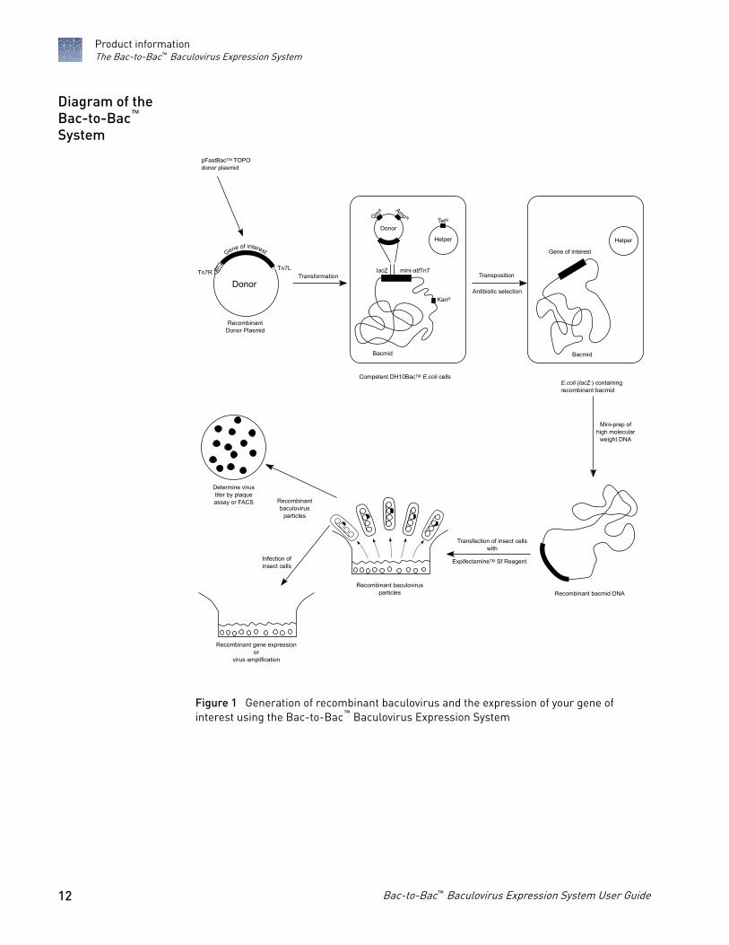
Figure 1 Generation of recombinant baculovirus and the expression of your gene ofinterest using the Bac-to-Bac™ Baculovirus Expression System
Diagram of theBac-to-Bac™
System
Product informationThe Bac-to-Bac™ Baculovirus Expression System
12 Bac-to-Bac™ Baculovirus Expression System User Guide
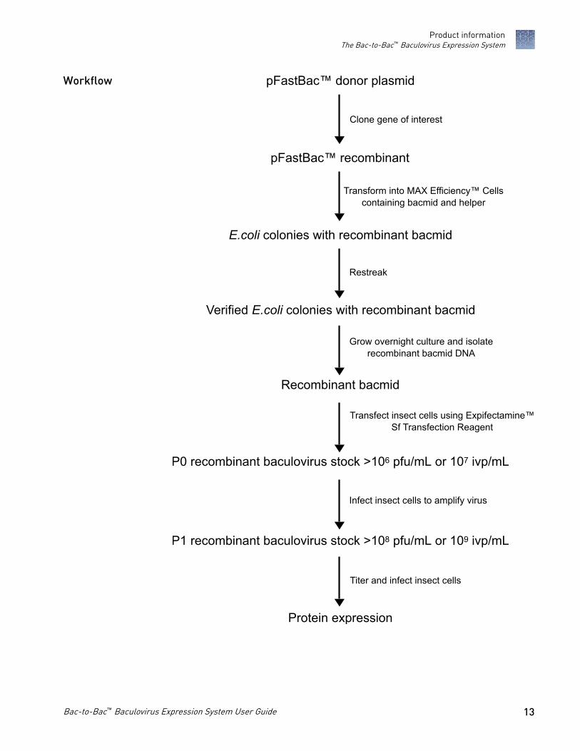
pFastBac™ donor plasmid
pFastBac™ recombinant
E.coli colonies with recombinant bacmid
Verified E.coli colonies with recombinant bacmid
Recombinant bacmid
P0 recombinant baculovirus stock >106 pfu/mL or 107 ivp/mL
P1 recombinant baculovirus stock >108 pfu/mL or 109 ivp/mL
Protein expression
Workflow
Product informationThe Bac-to-Bac™ Baculovirus Expression System
Bac-to-Bac™ Baculovirus Expression System User Guide 13
Methods
Culture insect cells
We recommend using Spodoptera frugiperda Sf9 or Sf21 insect cells as the host forrecombinant baculovirus production. You can also use the high-density, suspensionExpiSf9™ cell line, a component of the ExpiSf™ Expression System, to produce yourrecombinant baculovirus. For additional information on baculovirus generation andprotein expression using the ExpiSf™ Expression System, go to thermofisher.com/expisf. Before you start your transfection and expression experiments, ensure to havecultures of Sf9 or Sf21 cells growing and have frozen master stocks available. Sf9 orSf21 cells and cell culture reagents are available separately.
Note: High Five™ cells and Mimic™ Sf9 insect cells are appropriate for expressiononly.
Insect cells may be cultured under serum-free conditions. We recommend usingSf-900™ II SFM or Sf-900™ III SFM. If you are using the ExpiSf™ Expression System,use ExpiSf™ CD Medium for cell culture.
Both Sf-900™ II SFM and Sf-900™ III SFM are protein-free media optimized for thegrowth and maintenance of Sf9 and Sf21 cells, as well as for the large-scale productionof recombinant proteins expressed using the Bac-to-Bac™ System.
ExpiSf™ CD Medium is an innovative chemically-defined, yeastolate-free, animalorigin-free, serum-free, protein-free formulation that has been optimized for thegrowth and maintenance of high-density suspension ExpiSf9™ cells, and for large-scale production of recombinant proteins expressed using the Bac-to-Bac™ System. Formore information, see thermofisher.com/expisf.
For guidelines and detailed information on insect cell culture, such as in the followinglist, refer to the Guide to Baculovirus Expression Vector System (BEVS) and Insect CellCulture Techniques at thermofisher.com.
• Maintaining and passaging insect cells in adherent and suspension culture• Freezing cells• Using serum-free medium (includes protocols to adapt cells to serum-free
medium)• Scaling up cell culture
Introduction
Serum-freemedium
Insect cell culturereference guide
14 Bac-to-Bac™ Baculovirus Expression System User Guide
Insect cells are sensitive to environmental factors. In addition to chemical andnutritional culture factors, physical factors can also affect insect cell growth. Thereforeoptimization is required to maximize cell growth. Consider the following whenculturing insect cells:
• Temperature: The optimal range to grow and infect cultured insect cells is 27°Cto 28°C.
• pH: A range of 6.1 to 6.4 works well for most culture systems. Sf-900™ II SFM andSf-900™ III SFM maintain a pH in this range under conditions of normal air andopen-capped culture systems.
• Osmolality: The optimal osmolality of medium for use with lepidopteran celllines is 345 to 380 mOsm/kg.
• Aeration: Insect cells require passive oxygen diffusion for optimal growth andrecombinant protein expression. Active or controlled oxygenated systems requiredissolved oxygen at 10% to 50% of air saturation.
• Shear forces: Suspension culture generates mechanical shear forces. Growinginsect cells in serum-containing media (10% to 20% FBS) generally providesadequate protection from cellular shear forces. If you are growing insect cells inserum-free conditions other than Sf-900™ II SFM or Sf-900™ III SFM,supplementation with a shear force protectant such as Pluronic™ F-68 can berequired.
You need log-phase Sf9 or Sf21 cells with >95% viability to perform a successfultransfection. To determine how many cells you need for transfection, see “Transfection conditions“ on page 26.
Generate recombinant pFastBac™ donor plasmid
To generate a recombinant plasmid containing your gene of interest for use in the Bac-to-Bac™ Baculovirus Expression System, use restriction enzyme digestion and ligationto clone your gene into one of the pFastBac™ vectors. For guidelines to help designyour cloning strategy, and for information on vector maps, multiple cloning sites, andfeatures, see Appendix B, “Vectors“.
Transform the cloned-insert ligation reaction into E. coli and select for ampicillin-resistant transformants. Use any recA, endA E. coli strain, such as TOP10, DH10B™, orDH5α™ for transformation. Do not transform the ligation reaction into DH10Bac™
cells.
Table 1 Chemically competent E.Coli cells
Item Quantity Cat. No.
One Shot™ TOP10 Chemically Competent E. coli 20 × 50 µL C404003
One Shot™MAX Efficiency™ DH10B™ T1 Phage-ResistantCells 20 × 50 µL 12331013
One Shot™MAX Efficiency™ DH5α™-T1R Competent Cells 20 × 50 µL 12297016
General cellculture guidelines
Cells fortransfection
Introduction
Select E. coli host
MethodsGenerate recombinant pFastBac™ donor plasmid
Bac-to-Bac™ Baculovirus Expression System User Guide 15

You may use any method of choice to transform E. coli. Chemical transformation is themost convenient method, while electroporation is the most efficient and method ofchoice for large plasmids. To select for transformants, use LB agar plates containing100 µg/mL ampicillin.
1. Pick 10 transformants and culture them overnight in LB or S.O.B. containing100 µg/mL ampicillin.
2. Isolate the plasmid DNA using your method of choice.We recommend using the PureLink™HiPure Plasmid DNA Miniprep Kit topurify high quality plasmid DNA from your E. coli transformants.
3. Analyze the plasmids by restriction analysis to confirm the presence and correctorientation of the insert. Use a restriction enzyme or a combination of enzymesthat cut once in the vector and once in the insert.
Requires the Platinum™ SuperFi™ PCR Master Mix or equivalent.
1. For each sample, aliquot 48 µL of Platinum™ SuperFi™ PCR Master Mix into a0.5-mL microcentrifuge tube, then add 1 µL each of the forward and reverse PCRprimer.
2. Pick up to 10 colonies and resuspend them individually in 50 µL of the PCRMaster Mix containing primers.Make a patch plate to preserve the colonies for further analysis.
3. Incubate the reaction for 10 minutes at 94°C to lyse the cells and inactivatenucleases.
4. Amplify for 20 to 30 cycles.
5. For the final extension, incubate at 72°C for 10 minutes, then store at 4°C.
6. Visualize by agarose gel electrophoresis.
Sequence the construct to confirm the gene of interest is in the correct orientation forexpression. If the gene was cloned into one of the pFastBac™HT vectors, verify that thegene is cloned in frame with the N-terminal tag.
1. Streak the original colony out for single colony on LB plates containing100 µg/mL ampicillin.
2. Isolate a single colony and inoculate into 1–2 mL of LB containing 100 µg/mLampicillin.
3. Grow until the culture reaches the stationary phase.
4. Mix 0.85 mL of culture with 0.15 mL of sterile glycerol, then transfer to acryovial.
5. Store at –80°C.
Transformationmethod
Analyze thetransformants
Analyze thetransformants byPCR
Sequencing
Make a glycerolstock for long-term storage
MethodsGenerate recombinant pFastBac™ donor plasmid
16 Bac-to-Bac™ Baculovirus Expression System User Guide

Generate recombinant bacmid DNA: transform DH10Bac™ E. coli
The pFastBac™ construct containing your gene of interest in the correct orientation isused to transform purified plasmid DNA into MAX Efficiency™ DH10Bac™
Competent Cells for transposition into the bacmid. Then, blue/white selectionidentifies colonies containing the recombinant bacmid.
Guidelines and instructions to transform DH10Bac™ E. coli using the pFastBac™
construct are provided in this section.
Each pFastBac™ plasmid is supplied with a corresponding control plasmid for use as apositive transfection and expression control (see the following table). We recommendincluding the corresponding control plasmid in your DH10Bac™ transformationexperiment. For maps and a description of the features of each control plasmid, seeAppendix B: Vectors.
pFastBac™ Vector Control Plasmid
pFastBac™1 pFastBac™ 1-Gus
pFastBac™HT pFastBac™HT-CAT
pFastBac™ Dual pFastBac™ Dual-Gus/CAT
• Your purified pFastBac™ construct (200 pg/mL in TE, pH 8.0)• Positive expression control plasmid (as a transposition control)• MAX Efficiency™ DH10Bac™ Competent Cells (use 1 tube per transformation)• pUC19 (as a transformation control)• LB agar plates containing kanamycin, gentamicin, tetracycline, Bluo-gal, and
IPTG (3 freshly prepared plates per transformation, see “Recommendation“ onpage 18 and Appendix C, “Supporting protocols“)
• LB agar plate containing 100 mg/mL ampicillin (for pUC19 transformationcontrol)
• SOC Medium• 15-mL round-bottom polypropylene tubes• 42°C water bath• 37°C shaking and non-shaking incubator
Introduction
Positive control
Requiredmaterials
MethodsGenerate recombinant bacmid DNA: transform DH10Bac™ E. coli
Bac-to-Bac™ Baculovirus Expression System User Guide 17
Prepare LB agar plates containing:• 50 µg/mL kanamycin• 7 µg/mL gentamicin• 10 µg/mL tetracycline• 100 µg/mL Bluo-gal• 40 µg/mL IPTG
Note: Use Bluo-gal instead of X-gal for blue/white selection as Bluo-gal generallyproduces a darker blue color than X-gal.
To order antibiotics, Bluo-gal, and IPTG, see Appendix E, “Ordering information“,and for instructions to prepare plates, see “LB (Luria-Bertani) Plates“ on page 61. Werecommend using Luria Broth Base (see “LB (Luria-Bertani) Medium“ on page 61)instead of Lennox L (LB) as the color intensity and number of colonies that areobtained on Lennox L plates is reduced.
Each transformation requires one vial of DH10Bac™ competent cells and threeselective plates.
• Equilibrate a water bath to 42°C.• Warm selective plates at 37°C for 30 minutes.• Warm the SOC Medium to room temperature.• Pre-chill one 15-mL round-bottom polypropylene tube for each transformation.
1. Thaw on ice one vial of MAX Efficiency™ DH10Bac™ Competent Cells for eachtransformation.
2. For each transformation, gently mix, then transfer 100 µL of the DH10Bac™ cellsinto a pre-chilled, 15-mL round-bottom polypropylene tube.
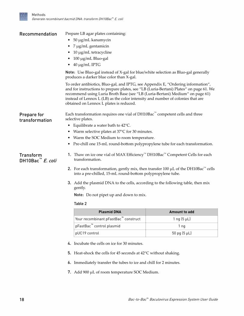
3. Add the plasmid DNA to the cells, according to the following table, then mixgently.
Note: Do not pipet up and down to mix.
Table 2
Plasmid DNA Amount to add
Your recombinant pFastBac™ construct 1 ng (5 µL)
pFastBac™ control plasmid 1 ng
pUC19 control 50 pg (5 µL)
4. Incubate the cells on ice for 30 minutes.
5. Heat-shock the cells for 45 seconds at 42°C without shaking.
6. Immediately transfer the tubes to ice and chill for 2 minutes.
7. Add 900 µL of room temperature SOC Medium.
Recommendation
Prepare fortransformation
TransformDH10Bac™ E. coli
MethodsGenerate recombinant bacmid DNA: transform DH10Bac™ E. coli
18 Bac-to-Bac™ Baculovirus Expression System User Guide

8. Transform cells according to the following table.
To transform Description
pFastBac™ Shake tubes at 37°C at 225 rpm for 4 hours
pUC19 Shake tube at 37°C at 225 rpm for 1 hour
9. Prepare dilutions according to the following table.
For plasmid Description
pFastBac™ Prepare 10-fold serial (10−1 to 10−3) dilutions of the cells with SOCMedium, then plate 100 µL of each dilution on an LB agar platecontaining:
• 50 µg/mL kanamycin
• 7 µg/mL gentamicin
• 10 µg/mL tetracycline
• 100 µg/mL Bluo-gal
• 40 µg/mL IPTG
pUC19 Dilute the cells 1:100 with SOC Medium, then plate 100 µL of thedilution on an LB agar plate containing 100 µg/mL ampicillin.
10. Incubate plates for 24–48 hours at 37°C.
IMPORTANT! Insertion of the mini-Tn7 into the mini-attTn7 attachment site onthe bacmid disrupts the expression of the LacZ peptide. Therefore, coloniescontaining the recombinant bacmid are white. Blue colonies contain unalteredbacmid.
11. Pick white colonies for analysis.
Note: True white colonies tend to be large, therefore avoid false positives byselecting the largest, most isolated white colonies. Avoid picking colonies thatappear gray or that are darker in the center as they can contain a mixture of cellswith empty bacmid and recombinant bacmid.
1. Pick up to 10 white colonies, then restreak them on fresh LB agar platescontaining:
• 50 µg/mL kanamycin• 7 µg/mL gentamicin• 10 µg/mL tetracycline• 100 µg/mL Bluo-gal• 40 µg/mL IPTG
2. Incubate the plates overnight at 37°C.
3. Pick a white colony, then inoculate a liquid culture that contains:• 50 µg/mL kanamycin• 7 µg/mL gentamicin• 10 µg/mL tetracycline
Verify thephenotype
MethodsGenerate recombinant bacmid DNA: transform DH10Bac™ E. coli
Bac-to-Bac™ Baculovirus Expression System User Guide 19
4. Isolate recombinant bacmid DNA, see “Isolate recombinant bacmid DNA“ onpage 20.
Note: For increased recombinant bacmid yield, use the procedure for thePureLink™ HiPure Plasmid Maxiprep Kit, see Appendix D, “Bacmid DNAIsolation Using PureLink™ HiPure Maxiprep Kit“.
Note: Bacmid DNA must be clean and free from phenol and sodium chloride ascontaminants can kill insect cells, and salt interferes with lipid complexing,which decreases the transfection efficiency.
5. Analyze the recombinant bacmid DNA by PCR to verify successful transpositionto the bacmid, see “Analyze recombinant bacmid DNA by PCR“ on page 22.
Isolate recombinant bacmid DNA
The PureLink™HiPure Plasmid DNA Miniprep Kit allows you to purify high-qualitybacmid DNA from DH10Bac™ E. coli (see Appendix E, “Ordering information“). Theisolated bacmid DNA is appropriate for use in insect cell transfections.
Note: We do not recommend the PureLink™ HiPure Precipitator Module or thePureLink™ HiPure Plasmid Filter Mini/Midi/Maxiprep Kits for isolating bacmid DNA.
Note: When using the ExpiSf™ Expression System, we recommend isolating bacmidDNA using the PureLink™ HiPure Plasmid Maxiprep Kit (Cat. No. K2100-06). Thiswill ensure there is enough, high-quality bacmid DNA for baculovirus generationusing suspension-based transfection. Follow instructions in PureLink™ HiPure PlasmidPurification Kits User Guide, (Pub. No. MAN0000486) using the MaxiPrep low-copyplasmid instructions.
• Prepare LB medium containing:– 50 µg/mL kanamycin– 7 µg/mL gentamicin– 10 µg/mL tetracycline
Inoculate a single white bacterial colony into 2 mL of this LB medium, thenincubate the culture at 37°C in a shaking water bath at 250 rpm overnight.
• Verify that RNase A is added to the Resuspension Buffer (R3).• Ensure the Lysis Buffer (L7) contains no precipitates.
1. Place the PureLink™ HiPure Mini column on the PureLink™ Nucleic AcidPurification Rack.
2. Add 2 mL of Equilibration Buffer (EQ1) to the column.
3. Allow the solution in the column to drain by gravity flow.
Introduction
Before you begin
Equilibrate thecolumn
MethodsIsolate recombinant bacmid DNA
20 Bac-to-Bac™ Baculovirus Expression System User Guide

1. Harvest 1.5 mL of bacterial cells by centrifuging at 9,000 × g for 15 minutes, thenremove all the medium.
2. Add 0.4 mL Resuspension Buffer (R3) containing RNase A to the pellet, thenresuspend the cells until homogeneous.
3. Transfer the cell suspension to a centrifuge tube.
4. Add 0.4 mL Lysis Buffer (L7), then mix gently by inverting the capped tube fivetimes.Do not vortex.
5. Incubate at room temperature for 5 minutes.
6. Add 0.4 mL Precipitation Buffer (N3), then mix immediately by inverting thecapped tube until the mixture is homogeneous.Do not vortex.
7. Centrifuge the mixture at >15,000 × g at room temperature for 10 minutes.
Note: If the pellet does not adhere to the bottom of the tube, incubate the tube atroom temperature for 5 minutes to allow the separation of the lysate andgelatinous pellet. Pipette the clear lysate into a sterile tube and centrifuge at>15,000 × g for 5 minutes at room temperature to remove any remaining cellulardebris.
1. Load the supernatant from step 7 onto the equilibrated column.
2. Allow the solution in the column to drain by gravity flow.
3. Wash the column twice with 2.5 mL of Wash Buffer (W8).
4. Allow the solution in the column to drain by gravity flow after each wash.Discard the flow-through.
1. Place a sterile centrifuge tube (elution tube) under the column.
2. Add 0.9 mL of Elution Buffer (E4) to the column to elute DNA.
Note: Allow the solution to drain by gravity flow. Do not force out anyremaining solution.
3. The elution tube contains the purified DNA.Discard the column.
4. Add 0.63 mL of isopropanol to the elution tube.
5. Mix, then place on ice for 10 minutes.
6. Centrifuge the mixture at >15,000 × g at 4°C for 20 minutes.
7. Carefully remove, then discard the supernatant.
Prepare the celllysate
Bind and wash theDNA
Elute andprecipitate DNA
MethodsIsolate recombinant bacmid DNA
Bac-to-Bac™ Baculovirus Expression System User Guide 21

8. Resuspend the DNA pellet in 1 mL of 70% ethanol.
9. Centrifuge at >15,000 × g at 4°C for 5 minutes.
10. Carefully remove, then discard the supernatant.
11. Air-dry the pellet for 10 minutes.
12. Resuspend the DNA pellet in 40 µL TE Buffer.
Note: Allow the pellet to dissolve for at least 10 minutes on ice. To avoidshearing the DNA, pipette only 1–2 times to resuspend.
13. Store the bacmid DNA at 4°C.
IMPORTANT! Aliquot bacmid DNA in TE buffer pH 8.0, into separate tubes andstore at –20°C (not in a frost-free freezer). Avoid multiple freeze/thaw cycles as itdecreases the transfection efficiency. Bacmid DNA can also be stored for up to 2weeks at 4°C in TE Buffer pH 8.0.
Prepare glycerol stocks of DH10Bac™ E. coli containing the bacmid DNA frommid-logarithmic phase culture grown from white colonies picked during theblue-white screening, and store at –80°C for future bacmid DNA isolation.
Note: For increased recombinant bacmid yield, use the PureLink™ HiPurePlasmid Maxiprep Kit, see Appendix D, “Bacmid DNA Isolation UsingPureLink™ HiPure Maxiprep Kit“. The PureLink™ HiPure Plasmid Prep Kitsallow purification of all types and sizes of plasmid DNA and are ideally suitedfor bacmid purification (see Appendix E, “Ordering information“).
Analyze recombinant bacmid DNA by PCR
Recombinant bacmid DNA is greater than 135 kb in size. As restriction analysis isdifficult to perform with DNA of this size, use PCR analysis to verify the presence ofyour gene of interest in the recombinant bacmid. Use the pUC/M13 Forward andReverse primers that hybridize to sites flanking the mini-attTn7 site within the lacZ-complementation region to facilitate PCR analysis.
pUC/M13
Bacmid DNA
Transposed pFastBacsequence
Forward
™ Tn7R Tn7LGene of interest
pUC/M13Reverse
mini-attTn7
139 bp 157 bp
Guidelines and instructions are provided in this section to perform PCR using thepUC/M13 Forward and Reverse primers.
Introduction
MethodsAnalyze recombinant bacmid DNA by PCR
22 Bac-to-Bac™ Baculovirus Expression System User Guide

To verify the presence of the gene of interest in the recombinant bacmid using PCR,either:
• Use the pUC/M13 Forward and Reverse primers.• Use a combination of the pUC/M13 Forward or Reverse primer and a primer that
hybridizes within the insert.
pUC/M13 Forward and Reverse primers, see the following table, are not provided.
Primer Sequence
pUC/M13 Forward 5'-CCCAGTCACGACGTTGTAAAACG-3'
pUC/M13 Reverse 5'-AGCGGATAACAATTTCACACAGG-3'
Use any DNA polymerase for PCR, including Platinum™ Taq DNA Polymerase. If theexpected PCR product is > 4 kb, we recommend using a polymerase mixture such asPlatinum™ Taq DNA Polymerase High Fidelity for best results. See Appendix E,“Ordering information“.
Use the procedure below to amplify your recombinant bacmid DNA using thepUC/M13 Forward and Reverse primers and Platinum™ Taq polymerase. If you areusing a combination of the pUC/M13 Forward or Reverse primers and a primerspecific for your gene, determine the amplification conditions to use. If you are usinganother polymerase, follow the manufacturer's recommendations for the polymeraseyou are using.
Note: Optimize amplification conditions when the insert is > 4 kb.
1. For each sample, set up the following 50-µL PCR reaction in a 0.5-mLmicrocentrifuge tube:
Component Amount
Recombinant bacmid DNA (100 ng) 1 µL
10X PCR Buffer (appropriate for enzyme) 5 µL
10 mM dNTP Mix 1 µL
50 mM MgCl2 1.5 µL
PCR Primers (1.25 µL each 10 µM stock) 2.5 µL
Sterile Water 38.5 µL
Platinum™Taq polymerase (5 units/µL) 0.5 µL
Total 50 µL
2. Overlay with 50 µL (1 drop) of mineral or silicone oil.
Analyze by PCRwith pUC/M13primers
DNA polymerase
Generate the PCRproduct
MethodsAnalyze recombinant bacmid DNA by PCR
Bac-to-Bac™ Baculovirus Expression System User Guide 23

3. Amplify using the following cycling parameters:
Step Time Temperature Cycles
Initial Denaturation 3 minutes 94°C 1X
Denaturation 45 seconds 94°C 25–35X
Annealing 45 seconds 55°C
1XExtension 5 minutes 72°C
Final extension 7 minutes 72°C
4. Remove 5–10 µL from the reaction, then analyze by agarose gel electrophoresis.
Successful transposition using the pUC/M13 Forward and Reverse primers foramplification results in PCR product sizes on agarose gel according to the followingtable.
Bacmid transposed with Size of PCR Product
Bacmid alone ~300 bp
pFastBac™ 1 ~2300 bp + size of your insert
pFastBac™ 1-Gus ~4200 bp
pFastBac™ HT ~2430 bp + size of your insert
pFastBac™ HT-CAT ~3075 bp
pFastBac™ Dual ~2560 bp + size of your insert
pFastBac™ Dual Gus/CAT ~5340 bp
If you have used a combination of the pUC/M13 Forward and Reverse primers and agene-specific primer for amplification, determine the expected size of your PCRproduct.
What you shouldsee
MethodsAnalyze recombinant bacmid DNA by PCR
24 Bac-to-Bac™ Baculovirus Expression System User Guide

Produce recombinant baculovirus: transfect insect cells
After confirmation that the recombinant bacmid contains the gene of interest,transfect insect cells to produce recombinant baculovirus. Guidelines and instructionsto transfect Sf9 and Sf21 insect cells are provided in this section.
Note: For instructions to generate recombinant baculovirus using ExpiSf9™ cells aspart of the ExpiSf™ Expression System workflow, refer to the ExpiSf™ ExpressionSystem User Guide (Pub. No. MAN0017532) at thermofisher.com/expisf.
You may use any method to prepare purified recombinant bacmid DNA fortransfection. Bacmid DNA must be clean and free from phenol and sodium chlorideas contaminants may kill the cells, and salt will interfere with lipid complexing anddecrease transfection efficiency. We recommend isolating bacmid DNA using thePureLink™HiPure Plasmid DNA Miniprep Kit following the procedure that isprovided, see Appendix D, “Bacmid DNA Isolation Using PureLink™ HiPureMaxiprep Kit“.
ExpiFectamine™ Sf Transfection Reagent is a next-generation proprietary cationic lipidformulation that offers high transfection efficiency and protein expression levels inadherent or suspension Sf9 and Sf21 cell cultures. This versatile reagent exhibit lowtoxicity and enables transfection of insect cells using fast and simple optimizedprotocols to streamline the workflow.
We recommend using Sf9 or Sf21 cells for transfection and identification ofrecombinant plaques. High Five™ and Mimic™ Sf9 cells are not recommended becausethey generally transfect less efficiently. However, after you have generated yourbaculovirus stock, you can use High Five™ or Mimic™ Sf9 cells for expression studies.
The ExpiFectamine™ Sf Transfection Reagent:• allows for high transfection efficiency directly into growth medium in both
adherent and suspension cell culture formats• ensures no need to replace the culture medium prior to transfection• has low toxicity that ensures no need to replace the medium after transfection
Include the recombinant bacmid from the pFastBac™ control plasmid as a positivecontrol in your transfection and expression experiments. In these bacmids, the geneencoding Gus and or CAT is expressed under the control of the strong polyhedrin(PH) or p10 promoter. Assay expression of Gus and or CAT after transfection.
• Purified recombinant bacmid DNA generated from the pFastBac™ construct(500 ng/µL in TE Buffer, pH 8.0)
• (Optional) Purified recombinant bacmid DNA generated from the pFastBac™
control construct (500 ng/µL in TE Buffer, pH 8.0)• Sf9 or Sf21 cells cultured in the appropriate medium• ExpiFectamine™ Sf Transfection Reagent (store at 4°C until use)• Opti-MEM™ I Reduced Serum Medium• 6-well tissue culture plates and other tissue culture supplies• 1.5-mL sterile microcentrifuge tubes
Introduction
Plasmidpreparation
ExpiFectamine™ SfTransfectionReagent
Insect cell lines
Media fortransfection
Positive control
Requiredmaterials
MethodsProduce recombinant baculovirus: transfect insect cells
Bac-to-Bac™ Baculovirus Expression System User Guide 25

• Complete growth medium for culturing insect cells, such as:– Sf-900™ II SFM– Sf-900™ III SFM– TNM-FH– Grace's Supplemented Insect Cell Culture Medium
1. Calculate the number of Sf9 or Sf21 cells required for your transfectionexperiment, then expand cells accordingly.Before transfection, ensure cells are healthy, with greater than 95% viability, andare growing in the logarithmic phase with a density of 1.5–2.5 × 10⁶ cells/mL.
2. Produce baculovirus stocks in Sf9 or Sf21 cells according to the following table oftransfection conditions.
Condition Amount per well
Tissue culture plate size 6-well (35 mm) plate (one well/bacmid)
Number of Sf9 or Sf21 cells to transfect 1 x 10⁶ cells
Medium volume 3 mL
Bacmid DNA 1 µg (can vary from 1 to 2 µg)
ExpiFectamine™ Sf Transfection Reagent 10 µL (can vary from 5 to 10 µL)
Note: Optimize transfection efficiency by varying cell density, and DNA andExpiFectamine™ Sf Transfection Reagent concentrations.
• ExpiFectamine™ Sf Transfection Reagent does not require removal of the culturemedium or washing of the cells. It is not necessary to replace the medium aftertransfection.
• The DNA-lipid complex formation time is shorter (~5 minutes) when usingExpiFectamine™ Sf Transfection Reagent as compared to Cellfectin™ II Reagent.
• Do not add antibiotics during transfection as this causes cell death or interfereswith transfection.
Transfectionconditions
Importantguidelines fortransfection
MethodsProduce recombinant baculovirus: transfect insect cells
26 Bac-to-Bac™ Baculovirus Expression System User Guide

Use the following protocol to transfect cells in a 6-well format. All amounts andvolumes are given on a per well basis.
1. Verify that the Sf9 or Sf21 cells are in the log phase (1.5–2.5 × 10⁶ cells/mL) withgreater than 95% viability.
2. Select the appropriate procedure according to the following table.
Culture conditions Procedure
For cells withoutantibiotics and adensity within 1.5–2.5 × 10⁶ cells/mL
1. Detach the cells, then seed 8 × 10⁵ cells per well in 2 mLunsupplemented (without antibiotics and serum) Grace'sInsect medium.
Note: Do not change medium or wash the cells as themedium carried over will enhance the transfectionefficiency.
2. Allow cells to attach for 15 minutes at room temperaturein the hood.
For cells containingantibiotics or thecell density is notwithin 1.5–2.5 × 10⁶
cells/mL
1. Prepare 10 mL Plating Medium by mixing 1.5 mLSupplemented (10% FBS) Grace’s Insect Medium(without antibiotics) and 8.5 mL Grace’s Insect Medium,Unsupplemented (without FBS and antibiotics).
2. Detach the cells, then seed 8 × 10⁵ cells per well.
3. Allow cells to attach for 15 minutes at room temperaturein the hood.
4. Replace the medium with 2.5 mL Plating Medium.
3. For each transfection sample, dilute 10 µL ExpiFectamine™ Sf TransfectionReagent in 250 µL Opti-MEM™ I Reduced Serum Medium.
4. Mix by inverting 5–10 times (do not vortex), then incubate for 5 minutes at roomtemperature.
5. Add 1 µg undiluted bacmid DNA per sample.
Note: It is not necessary to pre-dilute bacmid DNA prior to addition.
6. Mix by inverting 5–10 times (do not vortex), then incubate for 5 minutes at roomtemperature.
Note: Do not incubate the mixture for longer than 20 minutes as this decreasestransfection efficiency.
7. Add the entire DNA-lipid mixture dropwise onto the cells.
8. Incubate the cells at 27°C for 72–96 hours or until visible signs of virus infection.
Transfect cellscultured insupplementedGrace’s Medium
MethodsProduce recombinant baculovirus: transfect insect cells
Bac-to-Bac™ Baculovirus Expression System User Guide 27
Use the following protocol to transfect cells in a 6-well format. All amounts andvolumes are given on a per well basis.
1. Verify that the Sf9 or Sf21 cells are in the log phase (2–4 × 10⁶ cells/mL) withgreater than 95% viability.
2. Detach the cells, then seed 1 × 10⁶ cells per well in 3 mL culture medium.Allow cells to attach for 30–60 minutes at room temperature in the hood or byreturning the plate to the incubator.
3. For each transfection sample, dilute 10 µL ExpiFectamine™ Sf TransfectionReagent in 250 µL Opti-MEM™ I Reduced Serum Medium.
4. Mix by inverting 5–10 times (do not vortex), then incubate for 5 minutes at roomtemperature.
5. Add 1 µg undiluted bacmid DNA per sample.
Note: It is not necessary to pre-dilute bacmid DNA prior to addition.
6. Mix by inverting 5–10 times (do not vortex), then incubate for 5 minutes at roomtemperature.
Note: Do not incubate the mixture for longer than 20 minutes as this decreasestransfection efficiency.
7. Add the entire DNA-lipid mixture dropwise onto the cells.
8. Incubate the cells at 27°C for 72–96 hours or until visible signs of virus infection.
Isolate P0 virus stock
Budded virus is released into the medium ~72–96 hours after transfection. However, iftransfection efficiency is suboptimal, the cells might not show all the signs of virusinfection until 5 days posttransfection. Starting at 72 hours after transfection, visuallyinspect the cells daily for signs of infection. After the cells appear infected (that is,display characteristics typical of late to very late infection), harvest the virus from thecell culture medium, as described in this section.
Transfect cellscultured inserum-freemedium
Introduction
MethodsIsolate P0 virus stock
28 Bac-to-Bac™ Baculovirus Expression System User Guide
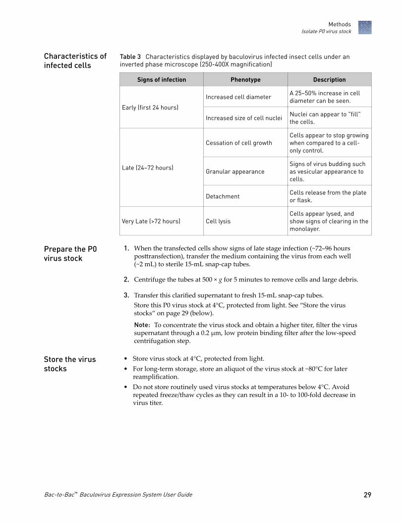
Table 3 Characteristics displayed by baculovirus infected insect cells under aninverted phase microscope (250-400X magnification)
Signs of infection Phenotype Description
Early (first 24 hours)
Increased cell diameter A 25–50% increase in celldiameter can be seen.
Increased size of cell nuclei Nuclei can appear to "fill"the cells.
Late (24–72 hours)
Cessation of cell growthCells appear to stop growingwhen compared to a cell-only control.
Granular appearanceSigns of virus budding suchas vesicular appearance tocells.
Detachment Cells release from the plateor flask.
Very Late (>72 hours) Cell lysisCells appear lysed, andshow signs of clearing in themonolayer.
1. When the transfected cells show signs of late stage infection (~72–96 hoursposttransfection), transfer the medium containing the virus from each well(~2 mL) to sterile 15-mL snap-cap tubes.
2. Centrifuge the tubes at 500 × g for 5 minutes to remove cells and large debris.
3. Transfer this clarified supernatant to fresh 15-mL snap-cap tubes.Store this P0 virus stock at 4°C, protected from light. See “Store the virusstocks“ on page 29 (below).
Note: To concentrate the virus stock and obtain a higher titer, filter the virussupernatant through a 0.2 µm, low protein binding filter after the low-speedcentrifugation step.
• Store virus stock at 4°C, protected from light.• For long-term storage, store an aliquot of the virus stock at −80°C for laterreamplification.
• Do not store routinely used virus stocks at temperatures below 4°C. Avoidrepeated freeze/thaw cycles as they can result in a 10- to 100-fold decrease invirus titer.
Characteristics ofinfected cells
Prepare the P0virus stock
Store the virusstocks
MethodsIsolate P0 virus stock
Bac-to-Bac™ Baculovirus Expression System User Guide 29
Clarified P0 baculoviral stock can be used to:• Amplify the virus stock (see “Amplify baculovirus stock“ on page 30). This
procedure is recommended to obtain the highest virus titers and optimal resultsin expression studies.
• Determine the titer of the virus stock (see “Perform a plaque assay“ on page 32)• Plaque purify recombinant baculovirus (see “Perform a plaque assay“ on
page 32)• Infect Sf9 or Sf21 cells for preliminary expression experiments
Note: The amount of virus stock is limited and expression conditions may not bereproducible (such as, MOI is unknown if the titer has not been determined).
Amplify baculovirus stock
The P0 virus stock is a small-scale, low-titer stock that can be used to infect cells togenerate a high-titer P1 stock. The titer of the initial virus stock obtained fromtransfecting Sf9 or Sf21 cells generally ranges from 1 × 10⁶ to 1 × 10⁷ plaque formingunits (pfu)/mL. Amplification allows production of a P1 virus stock with a titerranging from 1 × 10⁷ to 1 × 10⁸ pfu/mL, and is recommended.
Guidelines and protocols are provided in this section to amplify the recombinantbaculovirus to prepare a P1 virus stock.
• Sf9 or Sf21 cells (in monolayer or suspension) cultured in the appropriate growthmedium
• P0 baculovirus stock• Any appropriate tissue culture container
Note: We recommend amplification in a 10 mL to 200 mL suspension culture at2 × 106 cells/mL.
• Tissue culture reagents• 27°C humidified incubator
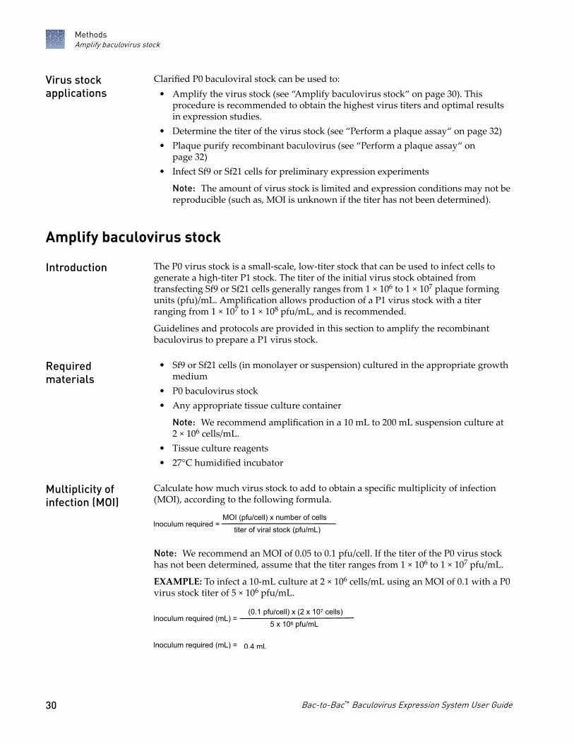
Calculate how much virus stock to add to obtain a specific multiplicity of infection(MOI), according to the following formula.
Inoculum required =MOI (pfu/cell) x number of cells
titer of viral stock (pfu/mL)
Note: We recommend an MOI of 0.05 to 0.1 pfu/cell. If the titer of the P0 virus stockhas not been determined, assume that the titer ranges from 1 × 10⁶ to 1 × 10⁷ pfu/mL.
EXAMPLE: To infect a 10-mL culture at 2 × 10⁶ cells/mL using an MOI of 0.1 with a P0virus stock titer of 5 × 10⁶ pfu/mL.
Common Callouts and Arrows
1. Copy-paste a callout or arrow to use in this SVG.
Note: If you need more advanced callouts or arrows use the TechComm_Inkscape_Callout&Arrow_Libary.
3. Delete this text, this rectangle, and unused callouts, arrows, or other SVG elements before adding this SVG to the repository.
2. Edit number and/or line-length, as needed.
1 1
1
1 Inoculum required (mL) =(0.1 pfu/cell) x (2 x 10⁷ cells)
5 x 10⁶ pfu/mL
Inoculum required (mL) = 0.4 mL
Virus stockapplications
Introduction
Requiredmaterials
Multiplicity ofinfection (MOI)
MethodsAmplify baculovirus stock
30 Bac-to-Bac™ Baculovirus Expression System User Guide

For successful amplification of baculovirus, pay attention to several key points:• Use Sf9 or Sf21 cells that are in excellent health, low passage (5–20), log-phase
growth, and have >95% viability.• Use a low MOI between 0.05–0.1 as higher MOI reduces baculovirus quality.• Harvest the virus when 60–80% of cells are dead.• Do not amplify baculovirus indefinitely, as baculovirus acquire deleterious
mutations with each passage. Usually, P3 is the highest usable passage.
1. On the day of infection, prepare an Sf9 or Sf21 cell suspension by seeding cells at2 × 106 cells/mL in 200 mL of culture medium.
2. Add the appropriate amount of P0 virus stock to the flask.
3. Incubate the cells for 72–96 hours in a humidified incubator at 27°C on an orbitalshaker, according to the following table.
Shaking orbit Speed (rpm)
19-mm 125 ±5
25-mm 125 ±5
50-mm 95 ±5
4. Transfer the medium containing virus from the flask to sterile ≥50-mL tubes, thencentrifuge the tubes at 500 × g for 5 minutes to clarify the baculovirus stock.
Note: Determine optimal harvest times for each baculovirus construct. Cultureviability decreases over time as cells lyse.
5. Transfer the supernatant to fresh collecting tubes.This is the P1 virus stock.
6. Store at 4°C, protected from light. For long-term storage, aliquot the P1 stock andstore at –80°C, protected from light. For storage guidelines see “Store the virusstocks“ on page 29.
After generating a high-titer P1 baculovirus stock, you can scale up the amplificationprocedure to any volume of your choice. To produce this high-titer P2 stock, scale upthe number of cells and volume of virus that is used appropriately, and follow theguidelines and procedure that is outlined in “Amplify baculovirus stock“ on page 30.
We recommend amplifying virus master stock that is stored at −80°C to generateanother high-titer stock for use in expression experiments. Virus titer generallydecreases over time when virus is stored at −80°C. Follow the guidelines andamplification procedure that is detailed in “Amplify baculovirus stock“ on page 30.
Importantconsiderations
Amplify P0 virusstock in a 1-Lflask
Scale up theamplificationprocedure
Generate high-titer stocks fromfrozen masterstock
MethodsAmplify baculovirus stock
Bac-to-Bac™ Baculovirus Expression System User Guide 31

Perform a plaque assay
We recommend that you perform a plaque assay to determine the titer of your virusstock. You can also perform a plaque assay to purify a single virus clone. In thisprocedure, you infect cells with dilutions of virus stock and identify focal points ofinfection (plaques) on an agarose overlay. You can also titer your virus stock by theendpoint dilution method.
To determine the titer of a baculovirus stock.
Plate Sf9 or Sf21 cells in 6-well plates
▼
Prepare 10-fold serial dilutions of baculovirus stock
▼
Add the different dilutions of baculovirus to cells, then incubatefor 1 hour
▼
Remove the virus, then overlay the cell monolayer withPlaquing Medium
▼
Incubate the cells for 7–10 days, then stain and count thenumber of plaques in each dilution
• The size of the gene of interest: Titer generally decreases as the size of the insertincreases.
• Transfection efficiency: For the highest transfection efficiency, we recommendtransfecting Sf9 or Sf21 cells using ExpiFectamine™ Sf Transfection Reagent.Prepare DNA: lipid complexes in Opti-MEM™ I Reduced Serum Medium.
• The age of the baculovirus stock: Virus titer can decrease with long-term storageat 4°C or −80°C. Titer baculovirus stock that has been stored for ≥6 months beforeuse in an expression experiment.
• Freeze/thaw cycles: Virus titer can decrease as much as 10% with eachfreeze/thaw cycle.
• Improper storage of your baculovirus stock: For routine use, aliquot baculovirusstock and store at 4°C, protected from light.
• Clarified baculovirus stock (store at 4°C until use)• Sf9 or Sf21 cells cultured in the appropriate medium (30 mL of log-phase cells at 5
x 105 cells/mL for each baculovirus stock to be titered)• Sf-900™ II SFM, Sf-900™ III SFM or other appropriate complete growth medium• Sf-900™ Medium (1.3X) (100 mL) or other appropriate plaquing medium• 4% agarose gel (specifically formulated for optimal insect cell growth)• Sterile, cell-culture grade distilled water
Introduction
Experimentaloutline
Factors affectingvirus titer
Requiredmaterials
MethodsPerform a plaque assay
32 Bac-to-Bac™ Baculovirus Expression System User Guide

• 100 mL sterile glass bottle• 6-well tissue-culture plates (2 plates for each virus stock to be titered)• Sterile hood• Water baths at 40°C and 70°C• (Optional) Microwave oven• 27°C humidified incubator• Neutral red (Sigma-Aldrich™ Cat. No. N7005)
Note: When culturing Sf9 or Sf21 cells in serum-supplemented media, such ascomplete TNM-FH, have the following reagents on hand:· Grace's Insect Cell Culture Medium, Supplemented· Grace's Insect Cell Culture Medium (2X)· Fetal Bovine Serum (FBS), qualified, heat-inactivated
Plaquing medium consists of a mixture of culture medium and agarose, and is used toimmobilize the infected cells for the plaque assay. Prepare Plaquing Mediumimmediately before use by the following procedure.
Note: If you are culturing Sf9 cells in Sf-900™ II SFM or Sf-900™ III SFM, prepareSf-900 Plaquing Medium. If you are culturing cells in TNM-FH, prepare Grace'sPlaquing Medium.
1. Melt the 4% agarose gel by placing the bottle in a 70°C water bath for 20 to30 minutes or by heating the agarose in a microwave oven.
2. While the 4% agarose gel is melting, place the following in the 40°C water bath:• Empty, sterile 100-mL bottle• Sf-900 Medium (1.3X) or Grace's Insect Cell Culture Medium (2X), as
appropriate
3. When the 4% agarose gel has liquefied, move the agarose gel, medium, andempty 100-mL bottle to a sterile hood.
4. Prepare the Plaquing Medium according to the following table.
Medium Procedure
Sf-900PlaquingMedium
In the empty 100-mL bottle, combine 30 mL of Sf-900 Medium(1.3X) with 10 mL of the melted 4% agarose gel, then mix gently.
Grace'sPlaquingMedium
1. Add 20 mL of heat-inactivated FBS to the 100-mL bottle ofGrace's Insect Medium (2X), then mix.
2. In the empty 100-mL bottle, combine 25 mL of Grace's InsectMedium (2X, with serum added) with 12.5 mL of cell-culturegrade, sterile, distilled water and 12.5 mL of the melted 4%agarose gel, then mix gently.
Note: Work quickly so that the agarose does not harden.
5. Return the bottle of Plaquing Medium to the 40°C water bath until use.
Prepare theplaquing medium
MethodsPerform a plaque assay
Bac-to-Bac™ Baculovirus Expression System User Guide 33

Use this procedure to determine the titer of pFastBac™ baculovirus stock, includingcontrol vector stocks. The amounts provided are to titer one baculovirus stock (two 6-well plates per virus stock).
Note: Include a negative control (no virus) in your experiment.
1. On the day of infection, harvest Sf9 or Sf21 cells, then prepare a 30 mL cellsuspension at 5 × 10⁵ cells/mL in Sf-900™ II SFM (or other complete growthmedium).
2. Aliquot 2 mL of cell suspension into each well of two 6-well plates.
3. Incubate the cells, covered, at room temperature for 1 hour, to allow the cells tosettle to the bottom of the plate.
4. After the 1 hour incubation, ensure, by observation, that the cells are attachedand at 50% confluence.
5. Prepare an 8-log serial dilution (10-1 to 10-8) of the clarified baculovirus stock.
Medium Description
Sf‑900™ II SFM In 12 mL disposable tubes, sequentiallydilute 0.5 mL of the baculovirus stock (orprevious dilution) in 4.5 mL of medium.Grace's Insect Cell Culture Medium,
Supplemented, without FBS
Note: This results in 8 tubes of diluted virus stock (10-1 to 10-8 ). Only dilutions10-4 to 10-8 are to be used.
6. Move the 6-well plates of cells and the tubes of diluted virus to a sterile hood.
7. Label the plates, in columns of 2 (a sample well plus a duplicate well) as follows:no virus (negative control), 10-4, 10-5, 10-6, 10-7, 10-8.
8. Remove the medium from each well, then immediately add 1 mL of theappropriate inoculum (virus dilution). For the negative control, add theappropriate medium without virus.
9. Incubate cells with virus for 1 hour at room temperature.
10. After the 1 hour incubation, move the bottle of plaquing medium from the 40°Cwater bath and the 6-well plates of infected cells to a sterile hood.
11. Sequentially, going from the highest dilution (10-8) to the lowest dilution (10-4),remove the inoculum from the wells, then add 2 mL of plaquing medium.
Note: Work quickly to avoid desiccation of the cell monolayer.
12. Allow the agarose overlay to harden for 1 hour at room temperature beforemoving the plates.
Perform plaqueassay
MethodsPerform a plaque assay
34 Bac-to-Bac™ Baculovirus Expression System User Guide

13. Incubate the cells in a 27°C humidified incubator for 7–10 days until plaques arevisible and ready to count.
• To stain the plaques to facilitate counting, see “Neutral Red staining“ onpage 35
• To calculate the titer, see “Calculate the titer“ on page 36
Staining with Neutral Red improves the visualization of plaques. Crystalline Blue andother plaque staining dyes containing organic solvents are not recommended becausethey kill the host cells. Stain the plaques by either of the following ways:
• Prepare an agarose solution containing Neutral Red and overlay this solution onthe plates 4 days post-infection. Count the plaques 7–10 days post-infection.
• Prepare a Neutral Red solution and add to the plates for 1–2 hours just prior tocounting plaques (7–10 days post-infection).
IMPORTANT! For plaque purification, do not stain with Neutral Red as it is a knownmutagen that can alter the recombinant virus.
1. Prepare a 1 mg/mL Neutral Red solution in Sf-900™ II SFM (or other appropriatecomplete growth medium), then filter-sterilize it.
2. In a 50-mL tube, combine 16.5 mL of Sf-900™ II SFM with 1.5 mL of Neutral Redsolution (1 mg/mL), then place in a 40°C water bath.
3. Microwave the 4% agarose gel until melted, then place in a 40°C water bath for5 minutes.
4. Move the 50 mL tube of Neutral Red solution and the 4% agarose gel to a sterilehood.
5. Prepare the Neutral Red overlay by adding 6 mL of 4% agarose gel to the NeutralRed solution.
6. Add 1 mL of the Neutral Red overlay to each well.
7. When the agarose has hardened, return the plates to a 27°C humidified incubatoruntil the plaques are ready to count.
Note: Plaques will appear as clear spots on a red monolayer.
1. Prepare a 1 mg/mL Neutral Red solution in cell-culture grade, distilled water.
2. Add 0.5 mL of Neutral Red solution to each well, then incubate for 1–2 hours atroom temperature.
3. Gently remove the excess stain with a pipet or blotter, then count the plaques.
Note: Plaques will appear as clear spots on a red monolayer.
Neutral Redstaining
Prepare NeutralRed agaroseoverlay (for use onDay 4)
Prepare NeutralRed stain (for useon Day 7–10 priorto countingplaques)
MethodsPerform a plaque assay
Bac-to-Bac™ Baculovirus Expression System User Guide 35
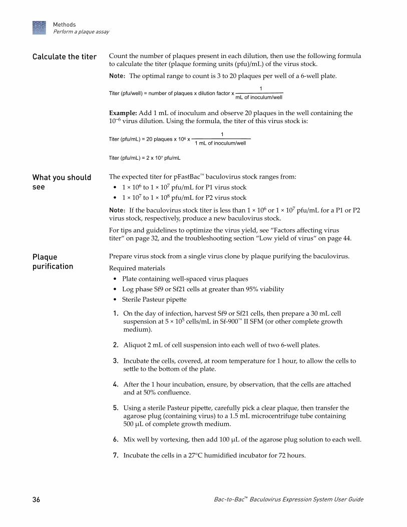
Count the number of plaques present in each dilution, then use the following formulato calculate the titer (plaque forming units (pfu)/mL) of the virus stock.
Note: The optimal range to count is 3 to 20 plaques per well of a 6-well plate.
Titer (pfu/well) = number of plaques x dilution factor x1
mL of inoculum/well
Example: Add 1 mL of inoculum and observe 20 plaques in the well containing the10−6 virus dilution. Using the formula, the titer of this virus stock is:
Common Callouts and Arrows
1. Copy-paste a callout or arrow to use in this SVG.
Note: If you need more advanced callouts or arrows use the TechComm_Inkscape_Callout&Arrow_Libary.
3. Delete this text, this rectangle, and unused callouts, arrows, or other SVG elements before adding this SVG to the repository.
2. Edit number and/or line-length, as needed.
1 1
1
1 Titer (pfu/mL) = 20 plaques x 106 x 1
1 mL of inoculum/well
Titer (pfu/mL) = 2 x 10⁷ pfu/mL
The expected titer for pFastBac™ baculovirus stock ranges from:• 1 × 10⁶ to 1 × 10⁷ pfu/mL for P1 virus stock• 1 × 10⁷ to 1 × 10⁸ pfu/mL for P2 virus stock
Note: If the baculovirus stock titer is less than 1 × 10⁶ or 1 × 10⁷ pfu/mL for a P1 or P2virus stock, respectively, produce a new baculovirus stock.
For tips and guidelines to optimize the virus yield, see “Factors affecting virustiter“ on page 32, and the troubleshooting section “Low yield of virus“ on page 44.
Prepare virus stock from a single virus clone by plaque purifying the baculovirus.
Required materials• Plate containing well-spaced virus plaques• Log phase Sf9 or Sf21 cells at greater than 95% viability• Sterile Pasteur pipette
1. On the day of infection, harvest Sf9 or Sf21 cells, then prepare a 30 mL cellsuspension at 5 × 10⁵ cells/mL in Sf-900™ II SFM (or other complete growthmedium).
2. Aliquot 2 mL of cell suspension into each well of two 6-well plates.
3. Incubate the cells, covered, at room temperature for 1 hour, to allow the cells tosettle to the bottom of the plate.
4. After the 1 hour incubation, ensure, by observation, that the cells are attachedand at 50% confluence.
5. Using a sterile Pasteur pipette, carefully pick a clear plaque, then transfer theagarose plug (containing virus) to a 1.5 mL microcentrifuge tube containing500 µL of complete growth medium.
6. Mix well by vortexing, then add 100 µL of the agarose plug solution to each well.
7. Incubate the cells in a 27°C humidified incubator for 72 hours.
Calculate the titer
What you shouldsee
Plaquepurification
MethodsPerform a plaque assay
36 Bac-to-Bac™ Baculovirus Expression System User Guide
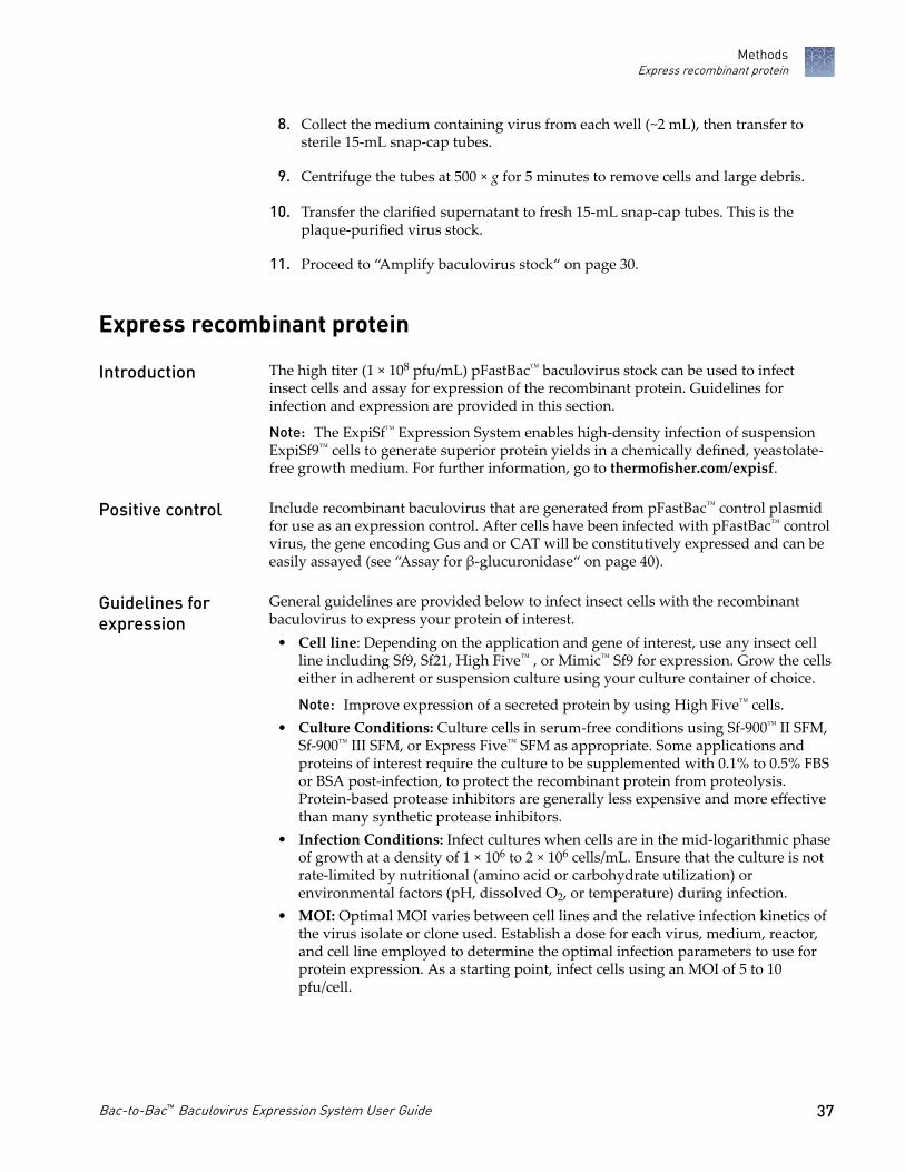
8. Collect the medium containing virus from each well (~2 mL), then transfer tosterile 15-mL snap-cap tubes.
9. Centrifuge the tubes at 500 × g for 5 minutes to remove cells and large debris.
10. Transfer the clarified supernatant to fresh 15-mL snap-cap tubes. This is theplaque-purified virus stock.
11. Proceed to “Amplify baculovirus stock“ on page 30.
Express recombinant protein
The high titer (1 × 10⁸ pfu/mL) pFastBac™ baculovirus stock can be used to infectinsect cells and assay for expression of the recombinant protein. Guidelines forinfection and expression are provided in this section.
Note: The ExpiSf™ Expression System enables high-density infection of suspensionExpiSf9™ cells to generate superior protein yields in a chemically defined, yeastolate-free growth medium. For further information, go to thermofisher.com/expisf.
Include recombinant baculovirus that are generated from pFastBac™ control plasmidfor use as an expression control. After cells have been infected with pFastBac™ controlvirus, the gene encoding Gus and or CAT will be constitutively expressed and can beeasily assayed (see “Assay for β-glucuronidase“ on page 40).
General guidelines are provided below to infect insect cells with the recombinantbaculovirus to express your protein of interest.
• Cell line: Depending on the application and gene of interest, use any insect cellline including Sf9, Sf21, High Five™ , or Mimic™ Sf9 for expression. Grow the cellseither in adherent or suspension culture using your culture container of choice.
Note: Improve expression of a secreted protein by using High Five™ cells.• Culture Conditions: Culture cells in serum-free conditions using Sf-900™ II SFM,
Sf-900™ III SFM, or Express Five™ SFM as appropriate. Some applications andproteins of interest require the culture to be supplemented with 0.1% to 0.5% FBSor BSA post-infection, to protect the recombinant protein from proteolysis.Protein-based protease inhibitors are generally less expensive and more effectivethan many synthetic protease inhibitors.
• Infection Conditions: Infect cultures when cells are in the mid-logarithmic phaseof growth at a density of 1 × 10⁶ to 2 × 10⁶ cells/mL. Ensure that the culture is notrate-limited by nutritional (amino acid or carbohydrate utilization) orenvironmental factors (pH, dissolved O2, or temperature) during infection.
• MOI: Optimal MOI varies between cell lines and the relative infection kinetics ofthe virus isolate or clone used. Establish a dose for each virus, medium, reactor,and cell line employed to determine the optimal infection parameters to use forprotein expression. As a starting point, infect cells using an MOI of 5 to 10pfu/cell.
Introduction
Positive control
Guidelines forexpression
MethodsExpress recombinant protein
Bac-to-Bac™ Baculovirus Expression System User Guide 37
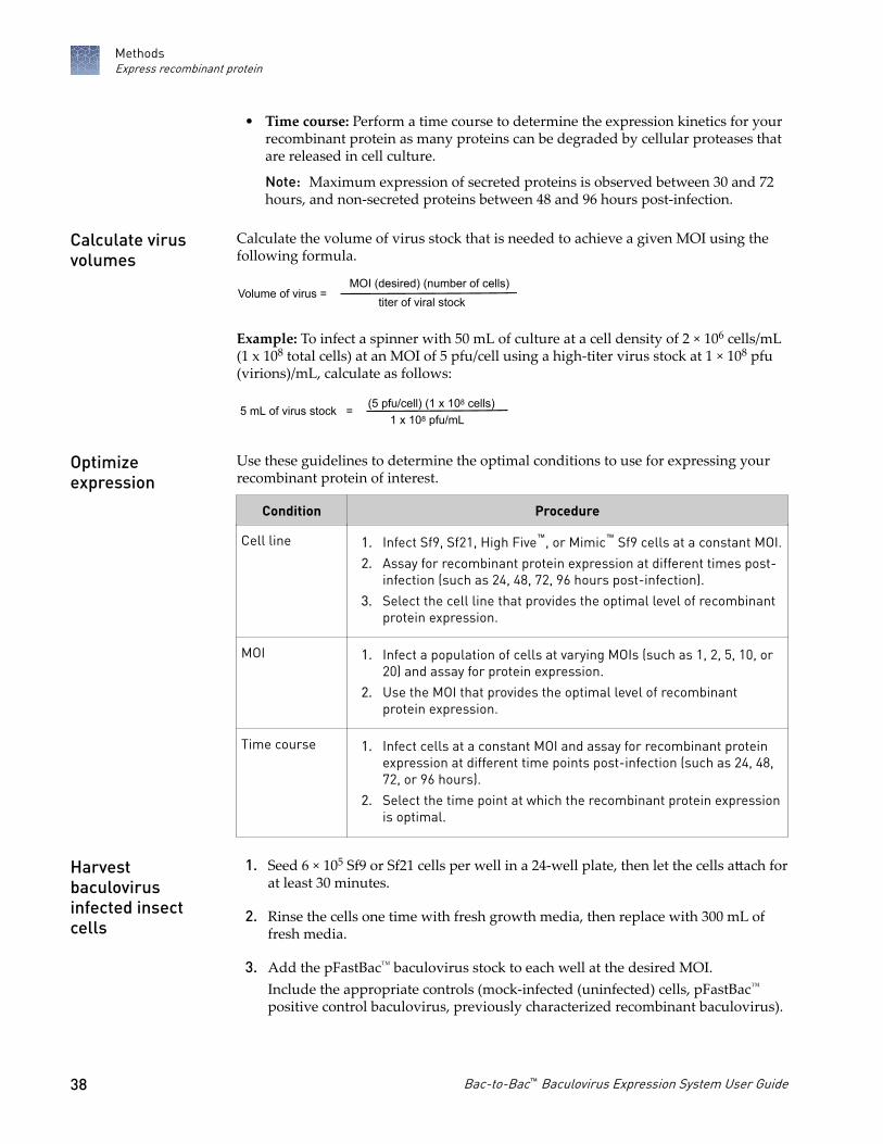
• Time course: Perform a time course to determine the expression kinetics for yourrecombinant protein as many proteins can be degraded by cellular proteases thatare released in cell culture.
Note: Maximum expression of secreted proteins is observed between 30 and 72hours, and non-secreted proteins between 48 and 96 hours post-infection.
Calculate the volume of virus stock that is needed to achieve a given MOI using thefollowing formula.
Volume of virus =MOI (desired) (number of cells)
titer of viral stock
Example: To infect a spinner with 50 mL of culture at a cell density of 2 × 10⁶ cells/mL(1 x 108 total cells) at an MOI of 5 pfu/cell using a high-titer virus stock at 1 × 10⁸ pfu(virions)/mL, calculate as follows:
5 mL of virus stock = (5 pfu/cell) (1 x 10⁸ cells)1 x 10⁸ pfu/mL
Use these guidelines to determine the optimal conditions to use for expressing yourrecombinant protein of interest.
Condition Procedure
Cell line 1. Infect Sf9, Sf21, High Five™, or Mimic™ Sf9 cells at a constant MOI.
2. Assay for recombinant protein expression at different times post-infection (such as 24, 48, 72, 96 hours post-infection).
3. Select the cell line that provides the optimal level of recombinantprotein expression.
MOI 1. Infect a population of cells at varying MOIs (such as 1, 2, 5, 10, or20) and assay for protein expression.
2. Use the MOI that provides the optimal level of recombinantprotein expression.
Time course 1. Infect cells at a constant MOI and assay for recombinant proteinexpression at different time points post-infection (such as 24, 48,72, or 96 hours).
2. Select the time point at which the recombinant protein expressionis optimal.
1. Seed 6 × 10⁵ Sf9 or Sf21 cells per well in a 24-well plate, then let the cells attach forat least 30 minutes.
2. Rinse the cells one time with fresh growth media, then replace with 300 mL offresh media.
3. Add the pFastBac™ baculovirus stock to each well at the desired MOI.Include the appropriate controls (mock-infected (uninfected) cells, pFastBac™
positive control baculovirus, previously characterized recombinant baculovirus).
Calculate virusvolumes
Optimizeexpression
Harvestbaculovirusinfected insectcells
MethodsExpress recombinant protein
38 Bac-to-Bac™ Baculovirus Expression System User Guide

4. Incubate the cells in a 27°C humidified incubator.
5. Harvest cells (or media, if the recombinant protein is secreted) at the appropriatetime (24, 48, 72, or 96 hours post-infection).
Note: If harvesting cells, remove the media, then rinse the cells one time withserum-free medium.
6. Lyse the cells with 400 µL of 1X SDS-PAGE Buffer (62.5 mM Tris-HCl, pH 6.8, 2%SDS).
7. Freeze samples at −20°C or boil samples for at least 3 minutes before separatingproteins by SDS-PAGE.
Analyze recombinant protein
After harvesting baculovirus infected insect cells, analyze the expression of yourprotein by SDS-PAGE or western blot. For secreted proteins, determine that all theprotein is being secreted by analyzing both the supernatant and the cell lysate.Analyzing cell lysates can help with optimizing the MOI and time course forexpression (see “Optimize expression“ on page 38). After determining the optimalexperimental parameters for secreted expression, proceed with large-scale expressionexperiments.
We recommend that you add one or more protease inhibitors to your lysis bufferduring protein isolation.
Proteaseinhibitor
Method ofaction Stock solution Working
concentration Storage
PMSF Serine proteaseinhibitor
10 mg/mL inisopropanol 100 µg/mL 20–25°C[1]
Leupeptin Serine and thiolproteaseinhibitor
50 µg/mL indeionized water 0.5 µg/mL
−20°CAprotinin Serine proteaseinhibitor
50 µg/mL indeionized water 0.5 µg/mL
Pepstatin A Acid proteaseinhibitor
100 µg/mL inmethanol 1 µg/mL
[1] Store in isopropnaol
Note: PMSF is not stable in aqueous solution. Add it to the lysis buffer just beforeuse.
CAUTION! PMSF (phenylmethylsulfonylfluoride) is very harmful wheninhaled, swallowed, or contacted by the skin. Wear protective clothing andgloves when handling.
Introduction
Proteaseinhibitors
MethodsAnalyze recombinant protein
Bac-to-Bac™ Baculovirus Expression System User Guide 39

Use any method to prepare cell lysates for analysis, including:
• Detergent lysis• Sonication lysis• Freeze-thaw lysis
Select any method of choice to detect the recombinant protein of interest, includingfunctional analysis or western blot.
Note: 6xHis tags and the Tobacco Etch Virus (TEV) recognition site increase the sizeof your protein by at least 3 kDa on PAGE gels.
• Identify blue plaques on agarose plates containing the chromogenic indicatorX-glucuronide.
a. Mix 5 mL of a 20 mg/mL X-glucuronide solution (in DMSO ordimethylformamide) with 50 mL of cell-free medium from the infected cells.
b. Monitor for the development of blue color within 2 hours.
• Alternatively, histochemical detection of β-glucuronidase in infected insect cellscan be performed using x-Gluc, see Appendix E, “Ordering information“.
Assay for CAT expression using your method of choice. There are commercial kitsavailable for assaying CAT expression as well as a rapid radioactive assay.
The pFastBac™ HT vector contains a Tobacco Etch Virus (TEV) recognition site thatallows the removal of the 6xHis tag from your recombinant fusion protein usingAcTEV™ Protease (available separately). For more information, go to thermofisher.com.
Prepare celllysates
Detectrecombinantprotein
Assay for β-glucuronidase
Assay for CAT
Remove the 6x Histag using AcTEV™
Protease
MethodsAnalyze recombinant protein
40 Bac-to-Bac™ Baculovirus Expression System User Guide

Troubleshooting
Cloning into pFastBac™ vectors
Observation Possible cause Recommended action
Recombinant pFastBac™
construct lacks insertIncomplete digestion ofpFastBac™ plasmid or insertDNA
Use additional restriction enzyme for digestion.
Purify insert DNA.
Incomplete or excessivephosphatase treatment ofpFastBac™ plasmid
Optimize dephosphorylation conditionsaccording to the manufacturer'srecommendations for the phosphatase you areusing.
Poor recovery of pFastBac™
plasmid or insert DNA fromagarose gel
Use PureLink™ Quick Gel Extraction Kit(Cat. No. K2100-12 ) to purify high qualityplasmid DNA from your agarose gel.
Incomplete ligation reactions Follow ligation conditions according to themanufacturer's recommendations for theligase you are using.
Optimize ligation reaction by varyingvector:insert molar ratios, such as 1:3, 1:1, 3:1.
Insert contains unstable DNAsequences such as LTRsequences and inverted repeats
Grow transformed cells at lower temperatures,such as 30°C.
Use MAX Efficiency™ Stbl2™ Competent Cellsfor transformation. Stbl2™ E. coli arespecifically designed for cloning unstableinserts.
No or few colonies obtainedafter transformation
Low transformation efficiencyof competent E. coli
If stored incorrectly, prepare or obtain newcompetent cells.
Use One Shot™ TOP10 Competent Cells orOne Shot™MAX Efficiency™ DH10B™ T1 Phage-Resistant Cells for transformation.
Impurities in DNA Purify insert DNA. Make sure to remove excessphenol, proteins, detergents, and ethanol fromthe DNA solution.
Too much DNA transformed For chemically competent cells, add 1 to 10 ngof DNA in a volume of 5 μL or less per 100 μLof cells. For electrocompetent cells, add 10 to50 ng of DNA in a volume of 1 μL or less per20 μL of cells.
If you have purchased competent cells, followthe manufacturer's instructions.
Incomplete ligation reaction Optimize the ligation reaction.
A
Bac-to-Bac™ Baculovirus Expression System User Guide 41

Observation Possible cause Recommended action
No or few colonies obtainedafter transformation
Include a ligation control (digested pFastBac™
vector + ligase). Check the ligation reaction ona gel.
Note: Ligated products and linear DNAtransform 10X and 100-1000X less efficiently,respectively, than super-coiled DNA.
Ligation reaction mix inhibitstransformation of competentcells
Reduce the amount of ligation reactiontransformed. Dilute ligation reaction 5X withTE Buffer prior to transformation.
Problem with antibiotic Confirm the use of the correct antibiotic at thecorrect concentration.
Use fresh antibiotic.
Competent cells storedimproperly
Store competent cells at −80°C.
Competent cells handledimproperly
Thaw cells on ice.
Use cells immediately after thawing.
Do not vortex the cells.
Cells not heat-shocked orincubated properly duringtransformation
Follow the recommended transformationprocedure for the cells you are using.
Recombinant bacmid DNA generation
Observation Possible cause Recommended action
No blue colonies
Although you pick whitecolonies, expect to see someblue colonies (contain non-recombinant bacmids).
Insufficient time for colordevelopment
Wait at least 48 hours before identifying colonyphenotypes.
Used X-gal instead of Bluo-galin agar plates
Use Bluo-gal in selective plates to increase thecontrast between blue and white colonies.
Insufficient growth aftertransposition
Grow transformed cells in S.O.C. Medium for atleast 4 hours before plating.
Bluo-gal and IPTG omitted fromplates
Prepare fresh selective plates containing 50mg/mL kanamycin, 7 mg/mL gentamicin, 10mg/mL tetracycline, 100 mg/mL Bluo-gal, and40 mg/mL IPTG.
Too many colonies on the plate Serially dilute the transformation mixture andplate to give well-separated colonies.
Adjust the serial dilutions of cells (10-2 to 10-4)to obtain well-spaced colonies.
Plates too old or stored in light Do not use plates that are more than 4 weeksold.
Store plates protected from light.
Incubation time too short ortemperature too low
Wait at least 48 hours before picking colonies.Incubate plates at 37°C.
Appendix A TroubleshootingRecombinant bacmid DNA generationA
42 Bac-to-Bac™ Baculovirus Expression System User Guide
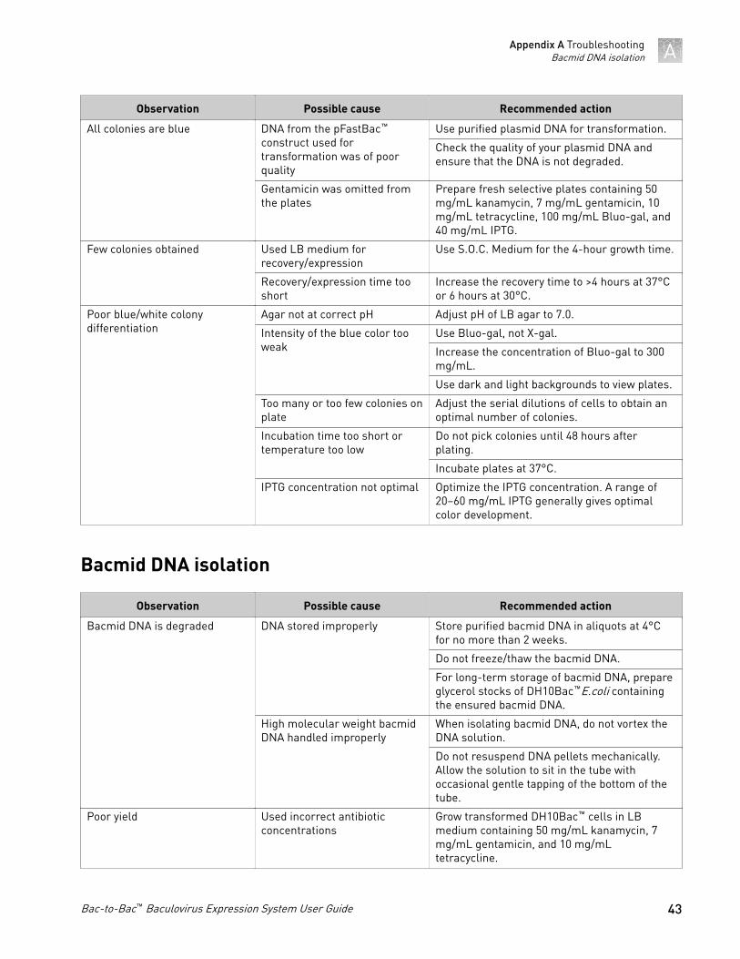
Observation Possible cause Recommended action
All colonies are blue DNA from the pFastBac™
construct used fortransformation was of poorquality
Use purified plasmid DNA for transformation.
Check the quality of your plasmid DNA andensure that the DNA is not degraded.
Gentamicin was omitted fromthe plates
Prepare fresh selective plates containing 50mg/mL kanamycin, 7 mg/mL gentamicin, 10mg/mL tetracycline, 100 mg/mL Bluo-gal, and40 mg/mL IPTG.
Few colonies obtained Used LB medium forrecovery/expression
Use S.O.C. Medium for the 4-hour growth time.
Recovery/expression time tooshort
Increase the recovery time to >4 hours at 37°Cor 6 hours at 30°C.
Poor blue/white colonydifferentiation
Agar not at correct pH Adjust pH of LB agar to 7.0.
Intensity of the blue color tooweak
Use Bluo-gal, not X-gal.
Increase the concentration of Bluo-gal to 300mg/mL.
Use dark and light backgrounds to view plates.
Too many or too few colonies onplate
Adjust the serial dilutions of cells to obtain anoptimal number of colonies.
Incubation time too short ortemperature too low
Do not pick colonies until 48 hours afterplating.
Incubate plates at 37°C.
IPTG concentration not optimal Optimize the IPTG concentration. A range of20–60 mg/mL IPTG generally gives optimalcolor development.
Bacmid DNA isolation
Observation Possible cause Recommended action
Bacmid DNA is degraded DNA stored improperly Store purified bacmid DNA in aliquots at 4°Cfor no more than 2 weeks.
Do not freeze/thaw the bacmid DNA.
For long-term storage of bacmid DNA, prepareglycerol stocks of DH10Bac™E.coli containingthe ensured bacmid DNA.
High molecular weight bacmidDNA handled improperly
When isolating bacmid DNA, do not vortex theDNA solution.
Do not resuspend DNA pellets mechanically.Allow the solution to sit in the tube withoccasional gentle tapping of the bottom of thetube.
Poor yield Used incorrect antibioticconcentrations
Grow transformed DH10Bac™ cells in LBmedium containing 50 mg/mL kanamycin, 7mg/mL gentamicin, and 10 mg/mLtetracycline.
Appendix A TroubleshootingBacmid DNA isolation A
Bac-to-Bac™ Baculovirus Expression System User Guide 43

Observation Possible cause Recommended action
Bacmid DNA contains a mixtureof recombinant bacmid andempty bacmid
Picked a colony that was grayor dark in the center
Analyze more white DH10Bac™ transformantsand select one that contains recombinantbacmid DNA only.
Insect cells transfection
Observation Possible cause Recommended action
Low yield of virus Low transfection efficiency Use ExpiFectamine™ Sf Transfection Reagentfor transfection.
Ensure that no supplements, FBS, orantibiotics are present in the media duringtransfection.
Harvest virus supernatant when signs ofinfection are visible, that is >72 hours post-transfection.
Cells plated too sparsely Plate insect cells at the recommended celldensity.
Used too much or too littleExpiFectamine™ Sf TransfectionReagent or other lipid reagent
Optimize the amount of ExpiFectamine™ SfTransfection Reagent or other lipid reagentused.
Time of incubation withDNA/lipid complexes too shortor too long
Incubate DNA/lipid complexes for only 5minutes before addition to cells. Incubatingthis mixture for longer than 20 minutes canlead to lower transfection efficiencies.
Recombinant bacmid DNA isdegraded
Check the quality of your recombinant DNA byagarose gel electrophoresis beforetransfection.
Prepare bacmid DNA using PureLink™ HiPurePlasmid Prep Kits.
Store purified bacmid at 4°C. Do not freeze at−80°C as it decreases transfection efficiency.
Bacmid DNA is not pure, that isit contains recombinant bacmidand empty bacmid
Screen other DH10Bac™ transformants andselect one that contains only recombinantbacmid.
Perform plaque purification to isolaterecombinant baculovirus.
Protein expression
Observation Possible cause Recommended action
Low protein expression virus stock contains a mixtureof recombinant and non-recombinant baculovirus
Perform plaque purification to isolaterecombinant baculovirus.
Appendix A TroubleshootingInsect cells transfectionA
44 Bac-to-Bac™ Baculovirus Expression System User Guide

Observation Possible cause Recommended action
Low protein expression Baculovirus not recombinant Ensure transposition by PCR analysis ofbacmid DNA using the pUC/M13 Forward andReverse primers.
Re-transfect insect cells with new recombinantbacmid DNA.
Used too low or too high virustiter
Optimize infection conditions by varying theMOI.
Time of cell harvest not optimal Perform a time course of expression todetermine the optimal time to obtain maximalprotein expression.
Cell growth conditions andmedium not optimal
Optimize culture conditions according to thesize of your culture container and expressionconditions.
Culture cells in Sf‑900™ II SFM or Sf‑900™ IIISFM for optimal cell growth and proteinexpression.
Cell line not optimal Try other insect cell lines.
Protein expression is notoptimal.
Optimize protein expression by varying suchparameters as incubation temperature andoxygenation.
Appendix A TroubleshootingProtein expression A
Bac-to-Bac™ Baculovirus Expression System User Guide 45

Vectors
Sequences of all the vectors in this section are available at www.lifetechnologies.comor by contacting Technical Support, see “Customer and technical support“ onpage 72.
pFastBac™ vectors
Vector Features
pFastBac™ 1 • Strong AcMNPV polyhedrin (PH)promoter for high-level proteinexpression
• Large multiple cloning site forsimplified cloning
pFastBac™ HT • Strong polyhedrin (PH) promoter forhigh-level protein expression
• N-terminal 6×His tag for purification ofrecombinant fusion proteins usingmetal-chelating resin and a TEVprotease cleavage site for removal ofthe 6×His tag following proteinpurification
• Vector supplied in 3 reading frames forsimplified cloning
pFastBac™ Dual • Two strong baculovirus promoters (PHand p10) to allow simultaneousexpression of two proteins
• Two large multiple cloning sites forsimplified cloning
pFastBac™1
The pFastBac™1 vector is a non-fusion vector (i.e., no fusion tags are present in thevector). To ensure proper expression of the recombinant protein, the insert mustcontain:
• An ATG start codon for initiation of translation• A stop codon for termination of the gene
Note: Stop codons are included in the multiple cloning site in all three readingframes.
B
Cloningconsiderations
46 Bac-to-Bac™ Baculovirus Expression System User Guide
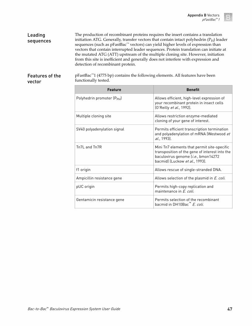
The production of recombinant proteins requires the insert contains a translationinitiation ATG. Generally, transfer vectors that contain intact polyhedrin (PH) leadersequences (such as pFastBac™ vectors) can yield higher levels of expression thanvectors that contain interrupted leader sequences. Protein translation can initiate atthe mutated ATG (ATT) upstream of the multiple cloning site. However, initiationfrom this site is inefficient and generally does not interfere with expression anddetection of recombinant protein.
pFastBac™1 (4775 bp) contains the following elements. All features have beenfunctionally tested.
Feature Benefit
Polyhedrin promoter (PPH) Allows efficient, high-level expression ofyour recombinant protein in insect cells(O’Reilly et al., 1992).
Multiple cloning site Allows restriction enzyme-mediatedcloning of your gene of interest.
SV40 polyadenylation signal Permits efficient transcription terminationand polyadenylation of mRNA (Westwood etal., 1993).
Tn7L and Tn7R Mini Tn7 elements that permit site-specifictransposition of the gene of interest into thebaculovirus genome (i.e., bmon14272bacmid) (Luckow et al., 1993).
f1 origin Allows rescue of single-stranded DNA.
Ampicillin resistance gene Allows selection of the plasmid in E. coli.
pUC origin Permits high-copy replication andmaintenance in E. coli.
Gentamicin resistance gene Permits selection of the recombinantbacmid in DH10Bac™ E. coli.
Leadingsequences
Features of thevector
Appendix B VectorspFastBac™1 B
Bac-to-Bac™ Baculovirus Expression System User Guide 47
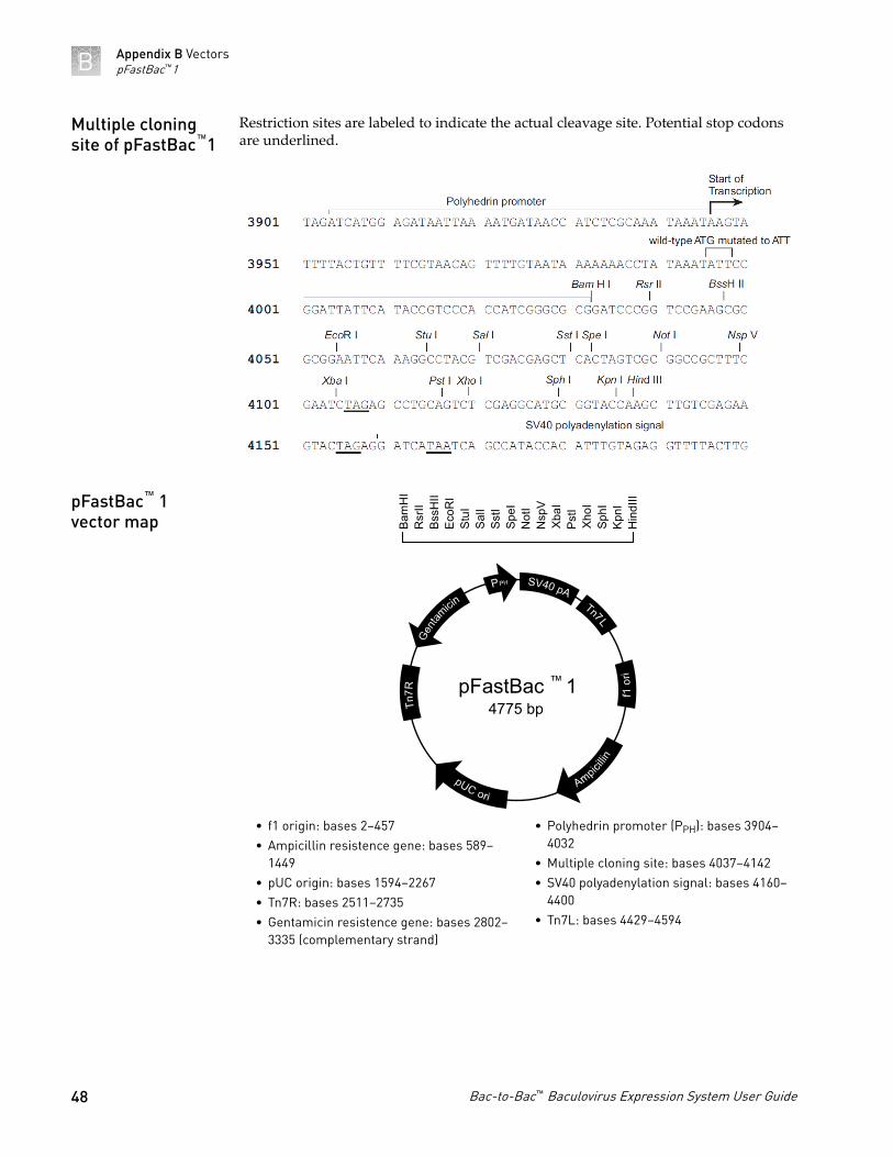
Restriction sites are labeled to indicate the actual cleavage site. Potential stop codonsare underlined.
pFastBac4775 bp
1
Bam
HI
Rsr
IIBs
sHII
EcoR
ISt
uISa
lISs
tISp
eIN
otI
Nsp
VXb
aIPs
tIXh
oISp
hIKp
nIH
indI
II
• f1 origin: bases 2–457• Ampicillin resistence gene: bases 589–
1449• pUC origin: bases 1594–2267• Tn7R: bases 2511–2735• Gentamicin resistence gene: bases 2802–
3335 (complementary strand)
• Polyhedrin promoter (PPH): bases 3904–4032
• Multiple cloning site: bases 4037–4142• SV40 polyadenylation signal: bases 4160–
4400• Tn7L: bases 4429–4594
Multiple cloningsite of pFastBac™1
pFastBac™ 1vector map
Appendix B VectorspFastBac™1B
48 Bac-to-Bac™ Baculovirus Expression System User Guide

pFastBac™HT A, B, and C
The pFastBac™HT vector is supplied with the multiple cloning site in three readingframes (A, B, and C) to facilitate cloning the gene of interest in frame with the N-terminal 6×His tag.
The pFastBac™HT vectors are fusion vectors. To ensure proper expression of therecombinant protein:
• Clone the gene in frame with the initiation ATG at base pairs 4050–4052. This willcreate a fusion with the N-terminal 6×His tag and a cleavage site for the AcTEV™
Protease.• Include a stop codon with your insert.
The production of recombinant proteins requires the insert contains a translationinitiation ATG. Generally, transfer vectors that contain intact polyhedrin (PH) leadersequences (such as pFastBac™ vectors) can yield higher levels of expression thanvectors that contain interrupted leader sequences. Protein translation can initiate atthe mutated ATG (ATT) upstream of the multiple cloning site. However, initiationfrom this site is inefficient and generally does not interfere with expression anddetection of recombinant protein.
The pFastBac™HT A (4856 bp), B (4857 bp), and C (4858 bp) vectors contain thefollowing elements. All features have been functionally tested.
Feature Benefit
Polyhedrin promoter (PPH) Allows efficient, high-level expression ofthe recombinant protein in insect cells.
6×His tag Allows purification of the recombinantprotein using a metal-chelating resin suchas ProBond™ or Ni-NTA.
TEV recognition site Permits removal of the N-terminal tag fromthe recombinant protein using AcTEV™
Protease.
Multiple cloning site Allows restriction enzyme-mediatedcloning of the gene of interest.
SV40 polyadenylation signal Permits efficient transcription terminationand polyadenylation of mRNA.
Tn7L and Tn7R Mini Tn7 elements that permit site-specifictransposition of the gene of interest into thebaculovirus genome (i.e., bmon14272bacmid).
f1 origin Allows rescue of single-stranded DNA.
Ampicillin resistance gene Allows selection of the plasmid in E. coli.
Introduction
Cloningconsiderations
Leadingsequences
Features of thevector
Appendix B VectorspFastBac™HT A, B, and C B
Bac-to-Bac™ Baculovirus Expression System User Guide 49

Feature Benefit
pUC origin Permits high-copy replication andmaintenance in E. coli.
Gentamicin resistance gene Permits selection of the recombinantbacmid in DH10Bac™ E. coli.
The initiation ATG is indicated in bold. Restriction sites are labeled to indicate theactual cleavage site.
Multiple cloningsite ofpFastBac™HT A
Appendix B VectorspFastBac™HT A, B, and CB
50 Bac-to-Bac™ Baculovirus Expression System User Guide
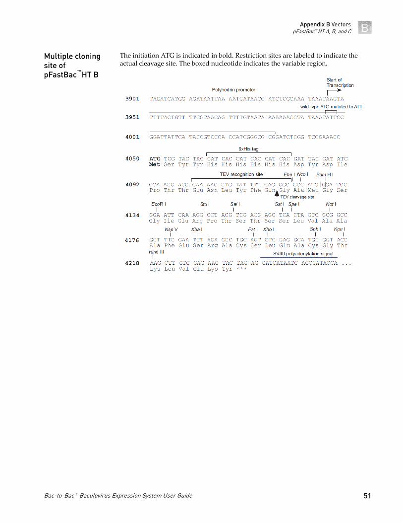
The initiation ATG is indicated in bold. Restriction sites are labeled to indicate theactual cleavage site. The boxed nucleotide indicates the variable region.
Multiple cloningsite ofpFastBac™HT B
Appendix B VectorspFastBac™HT A, B, and C B
Bac-to-Bac™ Baculovirus Expression System User Guide 51

The initiation ATG is indicated in bold. Restriction sites are labeled to indicate theactual cleavage site. The boxed nucleotide indicates the variable region.
Note: In pFastBac™HT C, there is a stop codon within the Xba I site that is in framewith the N-terminal tag. Ensure that the 5’ end of your gene is cloned upstream of theXba I site.
Multiple cloningsite ofpFastBac™HT C
Appendix B VectorspFastBac™HT A, B, and CB
52 Bac-to-Bac™ Baculovirus Expression System User Guide
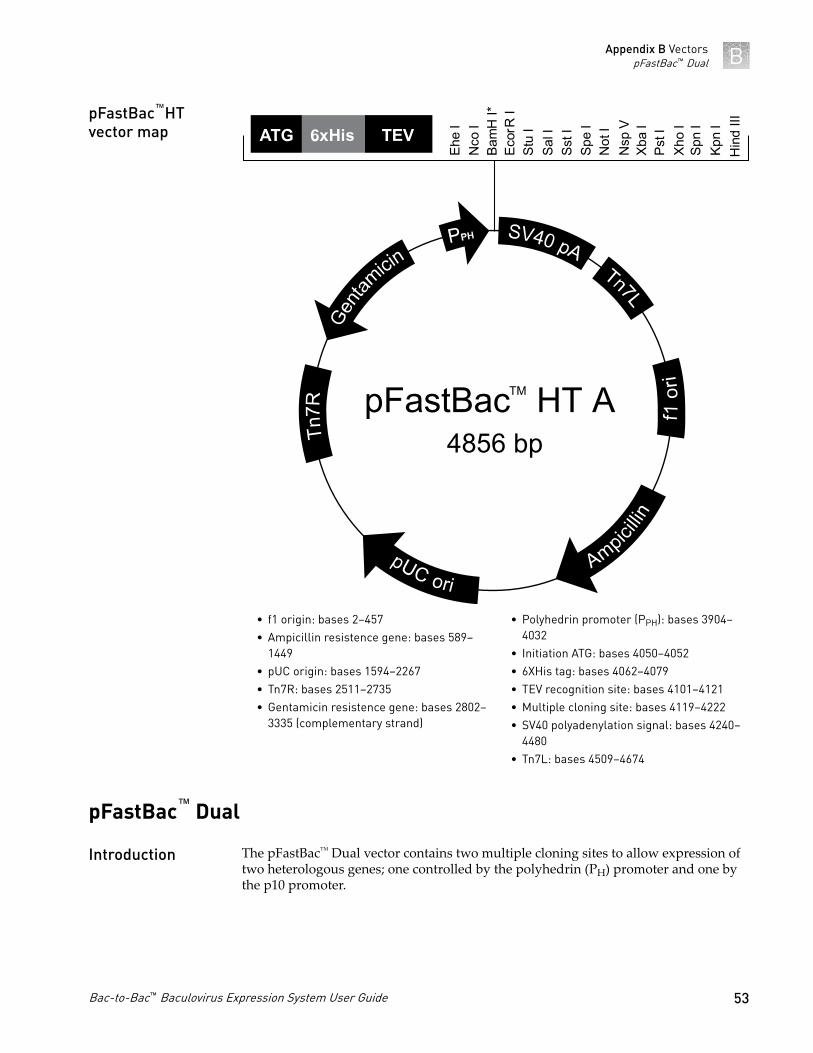
Ecor
R I
Bam
H I*
Nco
IEh
e I
Stu
ISa
l ISs
t ISp
e I
Not
IN
sp V
Xba
IPs
t IXh
o I
Spn
IKp
n I
Hin
d III
pFastBac4856 bp
HT ATM
• f1 origin: bases 2–457• Ampicillin resistence gene: bases 589–
1449• pUC origin: bases 1594–2267• Tn7R: bases 2511–2735• Gentamicin resistence gene: bases 2802–
3335 (complementary strand)
• Polyhedrin promoter (PPH): bases 3904–4032
• Initiation ATG: bases 4050–4052• 6XHis tag: bases 4062–4079• TEV recognition site: bases 4101–4121• Multiple cloning site: bases 4119–4222• SV40 polyadenylation signal: bases 4240–
4480• Tn7L: bases 4509–4674
pFastBac™ Dual
The pFastBac™ Dual vector contains two multiple cloning sites to allow expression oftwo heterologous genes; one controlled by the polyhedrin (PH) promoter and one bythe p10 promoter.
pFastBac™HTvector map
Introduction
Appendix B VectorspFastBac™ Dual B
Bac-to-Bac™ Baculovirus Expression System User Guide 53
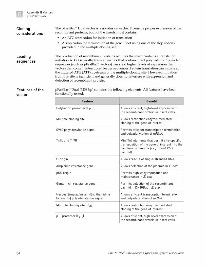
The pFastBac™ Dual vector is a non-fusion vector. To ensure proper expression of therecombinant proteins, both of the inserts must contain:
• An ATG start codon for initiation of translation• A stop codon for termination of the gene if not using one of the stop codons
provided in the multiple cloning site
The production of recombinant proteins requires the insert contains a translationinitiation ATG. Generally, transfer vectors that contain intact polyhedrin (PH) leadersequences (such as pFastBac™ vectors) can yield higher levels of expression thanvectors that contain interrupted leader sequences. Protein translation can initiate atthe mutated ATG (ATT) upstream of the multiple cloning site. However, initiationfrom this site is inefficient and generally does not interfere with expression anddetection of recombinant protein.
pFastBac™ Dual (5238 bp) contains the following elements. All features have beenfunctionally tested.
Feature Benefit
Polyhedrin promoter (PPH) Allows efficient, high-level expression ofthe recombinant protein in insect cells.
Multiple cloning site Allows restriction enzyme-mediatedcloning of the gene of interest.
SV40 polyadenylation signal Permits efficient transcription terminationand polyadenylation of mRNA.
Tn7L and Tn7R Mini Tn7 elements that permit site-specifictransposition of the gene of interest into thebaculovirus genome (i.e., bmon14272bacmid).
f1 origin Allows rescue of single-stranded DNA.
Ampicillin resistance gene Allows selection of the plasmid in E. coli.
pUC origin Permits high-copy replication andmaintenance in E. coli.
Gentamicin resistance gene Permits selection of the recombinantbacmid in DH10Bac™ E. coli.
Herpes Simplex Virus (HSV) thymidinekinase (tk) polyadenylation signal
Allows efficient transcription terminationand polyadenylation of mRNA.
Multiple cloning site (Pp10) Allows restriction enzyme-mediatedcloning of the gene of interest.
p10 promoter (Pp10) Allows efficient, high-level expression ofthe recombinant protein in insect cells.
Cloningconsiderations
Leadingsequences
Features of thevector
Appendix B VectorspFastBac™ DualB
54 Bac-to-Bac™ Baculovirus Expression System User Guide
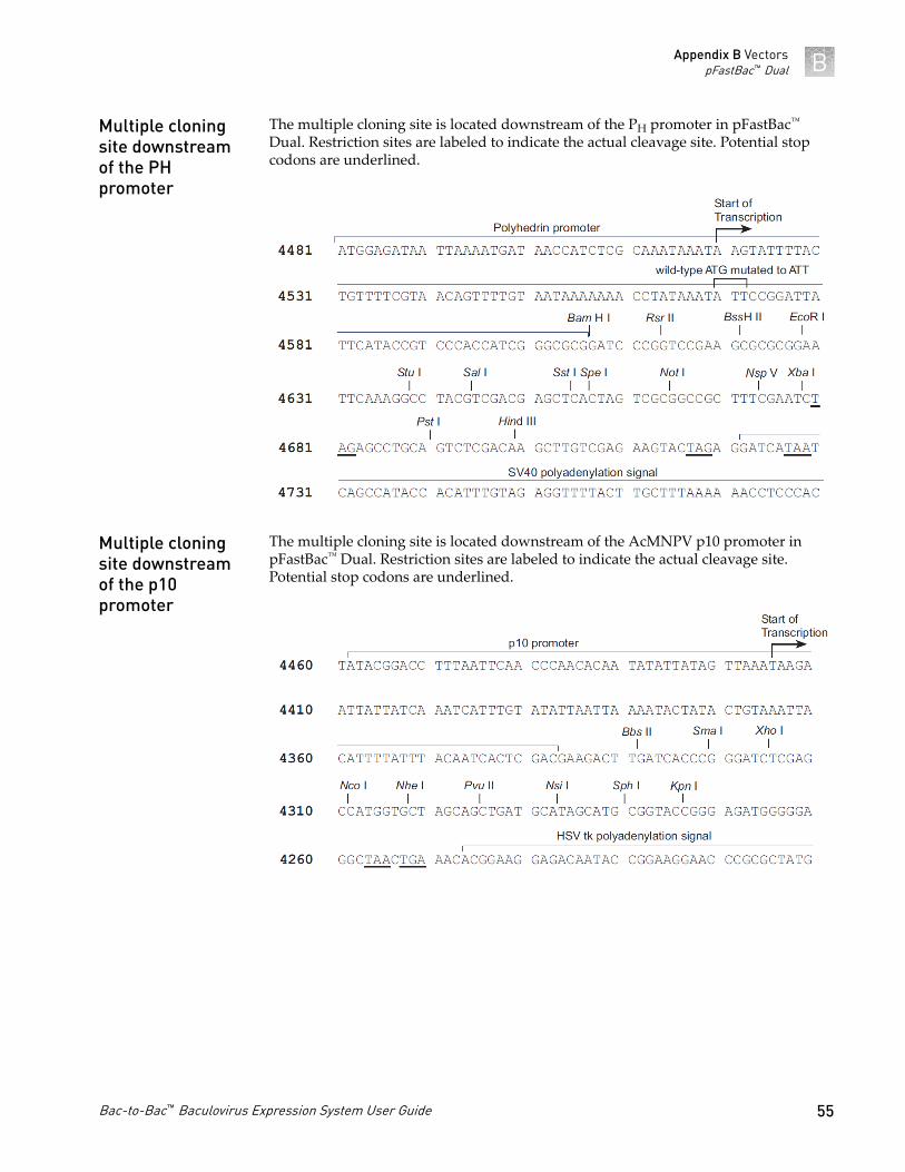
The multiple cloning site is located downstream of the PH promoter in pFastBac™
Dual. Restriction sites are labeled to indicate the actual cleavage site. Potential stopcodons are underlined.
The multiple cloning site is located downstream of the AcMNPV p10 promoter inpFastBac™ Dual. Restriction sites are labeled to indicate the actual cleavage site.Potential stop codons are underlined.
Multiple cloningsite downstreamof the PHpromoter
Multiple cloningsite downstreamof the p10promoter
Appendix B VectorspFastBac™ Dual B
Bac-to-Bac™ Baculovirus Expression System User Guide 55
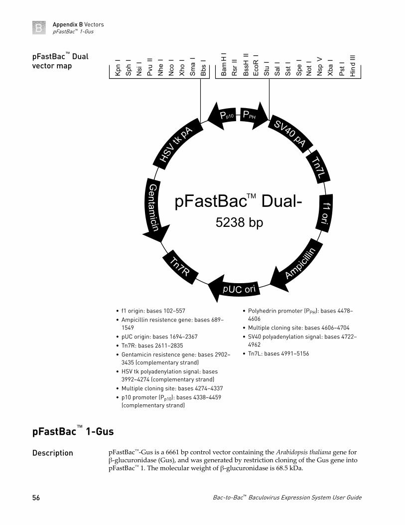
Kpn
ISp
h I
Nsi
IPv
u II
N
he I
Nco
IXh
o I
Sma
IBb
s I
Bam
H I
Rsr
II
BssH
IIEc
oR I
Stu
ISa
l ISs
t ISp
e I
Not
IN
sp V
Xba
I
Pst I
Hin
d III
pFastBac5238 bp
Dual-TM
• f1 origin: bases 102–557• Ampicillin resistence gene: bases 689–
1549• pUC origin: bases 1694–2367• Tn7R: bases 2611–2835• Gentamicin resistence gene: bases 2902–
3435 (complementary strand)• HSV tk polyadenylation signal: bases
3992–4274 (complementary strand)• Multiple cloning site: bases 4274–4337• p10 promoter (Pp10): bases 4338–4459
(complementary strand)
• Polyhedrin promoter (PPH): bases 4478–4606
• Multiple cloning site: bases 4606–4704• SV40 polyadenylation signal: bases 4722–
4962• Tn7L: bases 4991–5156
pFastBac™ 1-Gus
pFastBac™-Gus is a 6661 bp control vector containing the Arabidopsis thaliana gene forβ-glucuronidase (Gus), and was generated by restriction cloning of the Gus gene intopFastBac™ 1. The molecular weight of β-glucuronidase is 68.5 kDa.
pFastBac™ Dualvector map
Description
Appendix B VectorspFastBac™ 1-GusB
56 Bac-to-Bac™ Baculovirus Expression System User Guide

pFastBac6661 bp
1-Gus
Bam
HI
EcoR
ISt
uISp
eIN
otI
XbaI
PstI
XhoI
SphI
KpnI
Hin
dIII
Gus
• f1 origin: bases 2–457• Ampicillin resistence gene: bases 589–
1449• pUC origin: bases 1594–2267• Tn7R: bases 2511–2735• Gentamicin resistence gene: bases 2802–
3335 (complementary strand)
• Polyhedrin promoter (PPH): bases 3904–4032
• GUS ORF: bases 4081–5892• SV40 polyadenylation signal: bases 6047–
6287• Tn7L: bases 6315–6480
pFastBac™ HT-CAT
pFastBac™HT-CAT is a 5500 bp control vector containing the gene forchloramphenicol acetyltransferase (CAT), and was generated by restriction cloning ofthe CAT gene into pFastBac™HT. The CAT gene is expressed as a fusion to the N-terminal 6×His tag. The molecular weight of the fusion protein is 28 kDa.
pFastBac™ 1-GusControl Vectormap
Description
Appendix B VectorspFastBac™ HT-CAT B
Bac-to-Bac™ Baculovirus Expression System User Guide 57

Xba
IPs
t ISp
h I
Hin
d III
pFastBac5500 bp
HT-CATTM
• f1 origin: bases 2–457• Ampicillin resistence gene: bases 589–
1449• pUC origin: bases 1594–2267• Tn7R: bases 2511–2735• Gentamicin resistence gene: bases 2802–
3335 (complementary strand)
• Polyhedrin promoter (PPH): bases 3904–4032
• Initiation ATG: bases 4050–4052• 6XHis tag: bases4062–4079• TEV recognition site: bases 4101–4121• CAT ORF: bases 4131–4790• SV40 polyadenylation signal: bases 4884–
5124• Tn7L: bases 5153–5318
pFastBac™ Dual-Gus/CAT
pFastBac™ Dual-Gus/CAT is a 7843 bp control vector containing the Arabidopsisthaliana gene for β-glucuronidase (Gus) and the chloramphenicol acetyltransferase(CAT) gene. The vector was generated by restriction cloning of the Gus and CATgenes into pFastBac™ Dual. Expression of CAT and Gus are controlled by thepolyhedrin (PH) and p10 promoters, respectively. The molecular weight of β-glucuronidase and CAT are 68.5 kDa and 26 kDa, respectively.
pFastBac™HT-CATvector map
Description
Appendix B VectorspFastBac™ Dual-Gus/CATB
58 Bac-to-Bac™ Baculovirus Expression System User Guide

pFastBacGus/CAT
7843 bp
Dual-TM
Kpn
ISp
h II
Xho
ISm
a I
Bbs
I
Pst I
Bam
H I
Hin
d III
• Polyhedrin promoter (PPH): bases 16–144• CAT ORF: bases 181–840• SV40 polyadenylation signal: bases 964–
1204• Tn7L: bases 4991–5156• f1 origin: bases 1582–2037• Ampicillin resistence gene: bases 2169–
3029• pUC origin: bases 3174–3847
• Tn7R: bases 4091–4315• Gentamicin resistence gene: bases 4382–
4915 (complementary strand)• HSV tk polyadenylation signal: bases
5472–5754 (complementary strand)• GUS ORF: bases 5878–7689
(complementary strand)• p10 promoter (Pp10): bases 7719–7840
(complementary strand)
pFastBac™ Dual-Gus/CAT vectormap
Appendix B VectorspFastBac™ Dual-Gus/CAT B
Bac-to-Bac™ Baculovirus Expression System User Guide 59

Supporting protocols
Antibiotic stock solutions
Antibiotics can be ordered in either dry powdered form or as a stabilized, sterile,premixed solution. Store these solutions according to the manufacturer'srecommendations. Prepare and store stock solutions of the antibiotics according to thefollowing table.
Antibiotic Stock Solution Concentration Storage
Ampicillin 50 mg/mL in water
filter-sterilize−20°C,
protectedfrom light
Kanamycin 10 mg/mL in water
Tetracycline 10 mg/mL in 100% ethanol
Gentamicin 7 mg/mL in water
IPTG (200 mg/mL) stock solution
1. Dissolve 2 g of IPTG in 8 mL of sterile water.
2. Adjust the volume of the solution to 10 mL with sterile water.
3. Filter-sterilize through a 0.22-micron filter.
4. Dispense the stock solution into 1-mL aliquots.
5. Store at −20°C.
Bluo-gal (20 mg/mL) stock solution
1. Dissolve the Bluo-gal in dimethylformamide or dimethylsulfoxide (DMSO) tomake a 20 mg/mL stock solution.
Note: Use a glass or polypropylene tube.
IMPORTANT! Exercise caution when working with dimethylformamide.Dispense solutions in a vented chemical hood only.
C
60 Bac-to-Bac™ Baculovirus Expression System User Guide

2. Do not filter the stock solution.
3. Store at −20°C protected from light.
LB (Luria-Bertani) Medium
Composition:• 1.0% Tryptone (casein peptone)• 0.5% Yeast Extract• 1.0% NaCl• pH 7.0
1. For 1 L, dissolve the following in 950 mL deionized water.• 10 g of tryptone• 5 g of yeast extract• 10 g of NaCl
2. Adjust the solution to pH 7.0 with NaOH, then bring the volume up to 1 L.
3. Autoclave on liquid cycle for 20 minutes.
4. Allow the solution to cool to ~55°C, then add antibiotics if needed.
5. Store at room temperature or at 4°C.
LB (Luria-Bertani) Plates
1. Prepare LB medium as described in “LB (Luria-Bertani) Medium“ on page 61,but add 15 g/L of agar before autoclaving.
2. Autoclave on liquid cycle for 20 minutes.
3. Allow the solution to cool to ~55°C, then add antibiotics before pouring into10 cm plates.
4. Let the solution harden, then invert and store at 4°C, in the dark.
Note: Plates containing antibiotics are stable for up to 4 weeks at 4°C.
Appendix C Supporting protocolsLB (Luria-Bertani) Medium C
Bac-to-Bac™ Baculovirus Expression System User Guide 61

Bacmid DNA Isolation UsingPureLink™ HiPure Maxiprep Kit
Introduction
After you have transformed your pFastBac™ TOPO™ construct containing your geneof interest into DH10Bac™ E. coli and performed the transposition reaction, use thePureLink™ HiPure Plasmid Maxiprep Kit to purify recombinant bacmid DNA fromDH10Bac™ transformant.
Bacmid DNA purified by this method is suitable for use in PCR analysis or insect celltransfections.
Note: We do not recommend the PureLink™ HiPure Precipitator Module or thePureLink™ HiPure Plasmid Filter Mini/Midi/Maxiprep Kits for isolating bacmid DNA.
For more information on PureLink™ HiPure purification products, see thermofisher.com .
Grow bacmid DNA stock
Growing bacmid DNA stock from DH10Bac™ transformants in LB medium requiresthree days.
1. Day 1: Pick a single white bacterial colony from among the DH10Bac™
transformants (see “Transform DH10Bac™ E. coli“ on page 18) and inoculate 4mL of LB medium containing 50 mg/mL kanamycin, 7 mg/mL gentamicin and 10mg/mL tetracycline. Alternatively, you can thaw glycerol stocks of DH10Bac ™cells harboring your verified recombinant bacmid and use 100 mL to inoculation.
2. Incubate the culture at 37°C in a shaking water bath at 250 rpm overnight.
3. Day 2: Transfer the entire 4 mL of overnight culture into 50 mL of fresh LBmedium with antibiotics (as above) and incubate at 37°C in a shaking water bathat 250 rpm overnight.
4. Day 3: Transfer the entire 50 mL of overnight culture into 500 mL of fresh LBmedium with antibiotics (as above) and incubate at 37°C in a shaking water bathat 250 rpm overnight.
5. Day 4: proceed with the PureLink™ HiPure bacmid DNA isolation procedure.
D
62 Bac-to-Bac™ Baculovirus Expression System User Guide
Before Starting
Before beginning, verify that RNase A has been added to the Resuspension Buffer(R3) and that no precipitate has formed in the Lysis Buffer (L7).
Equilibrate the column
Place the PureLink™ HiPure Maxi column on the PureLink™ Nucleic Acid PurificationRack (see the manual supplied with the rack for more details). Apply 30 mLEquilibration Buffer (EQ1) to the column. Allow the solution in the column to drainby gravity flow.
Prepare the cell lysate
1. Harvest 250–500 mL of the overnight culture by centrifuging at 4,000 × g for 10minutes in a bucket.Remove all medium.
2. Add 20 mL Resuspension Buffer (R3) with RNase A to the pellet and resuspendthe cells until homogeneous.
3. Transfer cell suspension to a 50-mL centrifuge tube.
4. Add 20 mL Lysis Buffer (L7). Mix gently by inverting the capped tube five times.Do not vortex.
5. Incubate at room temperature for 5 minutes.
Note: Do not allow lysis to proceed for more than 5 minutes.
6. Add 20 mL Precipitation Buffer (N3) and mix immediately by inverting thecapped tube until the mixture is homogeneous.Do not vortex.
7. Centrifuge the mixture at >12,000 × g at room temperature for 10 minutes.
Note: If the pellet does not adhere to the bottom of the tube, incubate the tube atroom temperature for 5 minutes to allow the separation of the lysate andgelatinous pellet.
8. Pipette the clear lysate into another tube and centrifuge at >15,000 × g for 5minutes at room temperature to remove any remaining cellular debris.
Appendix D Bacmid DNA Isolation Using PureLink™ HiPure Maxiprep KitBefore Starting D
Bac-to-Bac™ Baculovirus Expression System User Guide 63

Bind and wash the DNA
1. Load the supernatant onto the equilibrated column. Allow the solution in thecolumn to drain by gravity flow.
2. Wash the column with 60 mL Wash Buffer (W8).
3. Allow the solution in the column to drain by gravity flow after each wash.Discard the flow-through.
Elute and precipitate the DNA
1. Place a sterile 30 mL centrifuge tube (elution tube) under the column.
2. Add 15 mL Elution Buffer (E4) to the column to elute DNA. Allow the solution todrain by gravity flow. Do not force out any remaining solution. The elution tubecontains the purified DNA. Discard the column.
3. Add 10.5 mL isopropanol to the elution tube. Mix well.
4. Centrifuge the mixture at >15,000 × g at 4°C for 30 minutes. Carefully remove anddiscard the supernatant.
5. Add 1 mL 70% ethanol to the pellet in the 30 mL elution tube, displace the pelletfrom the side of the tube, and transfer all the pellet fragments into a 1.5 mLmicrocentrifuge tube.
6. Centrifuge at >15,000 × g at 4°C for 10 minutes. Carefully remove and discard thesupernatant.
7. Add another 1 mL fresh 70% ethanol to the pellet in the microcentrifuge tube,and centrifuge at >15,000 × g at 4°C for another 10 minutes (second wash).Carefully remove and discard the supernatant.
8. Air-dry the pellet at room temperature until the appearance of the pellet changesfrom white-opaque to translucent and crystalline.
9. Resuspend the DNA pellet in 200–500 µL TE Buffer, pH 8.0 by vortexing.
10. Measure the concentration of the purified bacmid DNA. The concentrationshould be in range of 150–300 ng/mL
Appendix D Bacmid DNA Isolation Using PureLink™ HiPure Maxiprep KitBind and wash the DNAD
64 Bac-to-Bac™ Baculovirus Expression System User Guide

11. Store the tube at 4°C.
IMPORTANT!
You may store your bacmid DNA at −20°C if you avoid frequent freeze/thawcycles as it decreases the transfection efficiency.To store your purified bacmid DNA at −20°C, aliquot into separate tubes in TEBuffer, pH 8.0 to avoid more than one freeze/thaw cycle and do not store in afrost-free freezer. You may also store the purified bacmid DNA for up to 2 weeksat 4°C in TE Buffer, pH 8.0.You may prepare glycerol stocks of DH10Bac ™E. coli containing the bacmidDNA from mid-logarithmic phase culture grown from white colonies pickedduring the blue-white screening, and store at −80°C for future bacmid DNAisolation.
Appendix D Bacmid DNA Isolation Using PureLink™ HiPure Maxiprep KitElute and precipitate the DNA D
Bac-to-Bac™ Baculovirus Expression System User Guide 65
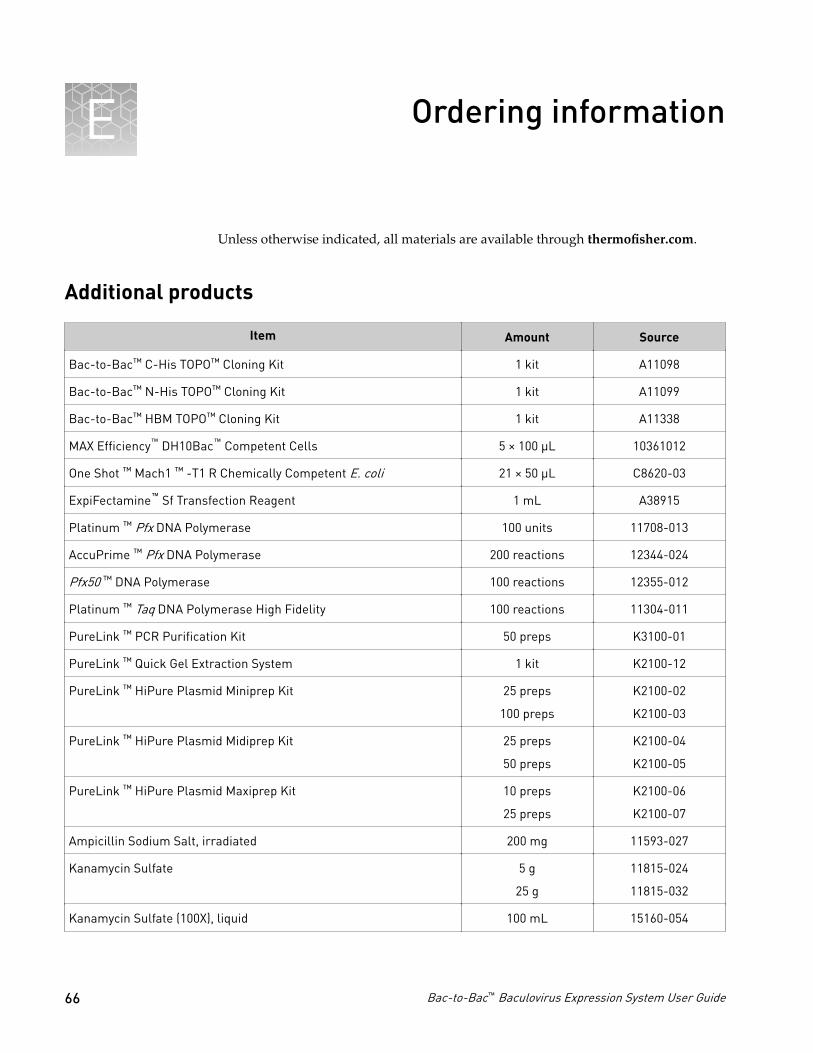
Ordering information
Unless otherwise indicated, all materials are available through thermofisher.com.
Additional products
Item Amount Source
Bac-to-Bac™ C-His TOPO™ Cloning Kit 1 kit A11098
Bac-to-Bac™ N-His TOPO™ Cloning Kit 1 kit A11099
Bac-to-Bac™ HBM TOPO™ Cloning Kit 1 kit A11338
MAX Efficiency™ DH10Bac™ Competent Cells 5 × 100 µL 10361012
One Shot ™ Mach1 ™ -T1 R Chemically Competent E. coli 21 × 50 µL C8620-03
ExpiFectamine™ Sf Transfection Reagent 1 mL A38915
Platinum ™ Pfx DNA Polymerase 100 units 11708-013
AccuPrime ™ Pfx DNA Polymerase 200 reactions 12344-024
Pfx50 ™ DNA Polymerase 100 reactions 12355-012
Platinum ™ Taq DNA Polymerase High Fidelity 100 reactions 11304-011
PureLink ™ PCR Purification Kit 50 preps K3100-01
PureLink ™ Quick Gel Extraction System 1 kit K2100-12
PureLink ™ HiPure Plasmid Miniprep Kit 25 preps
100 preps
K2100-02
K2100-03
PureLink ™ HiPure Plasmid Midiprep Kit 25 preps
50 preps
K2100-04
K2100-05
PureLink ™ HiPure Plasmid Maxiprep Kit 10 preps
25 preps
K2100-06
K2100-07
Ampicillin Sodium Salt, irradiated 200 mg 11593-027
Kanamycin Sulfate 5 g
25 g
11815-024
11815-032
Kanamycin Sulfate (100X), liquid 100 mL 15160-054
E
66 Bac-to-Bac™ Baculovirus Expression System User Guide

Item Amount Source
Gentamicin Reagent Solution, liquid (50 mg/mL) 10 mL
10 × 10 mL
15750-060
15750-078
Bluo-gal 1 g 15519-028
Isopropylthio- b -galactoside (IPTG) 1 g 15529-019
S.O.C. Medium 10 × 10 mL 15544-034
(Miller's LB Broth Base) ® Luria Broth Base, powder 500 g 12795-027
Water, distilled (cell-culture grade) 500 mL 15230-162
4% Agarose gel (optimal for insect cell growth) 40 mL 18300-012
Fetal Bovine Serum (FBS), Qualified, Heat Activated 100 mL 16140-063
X-Gluc 0.1 g R0851
Platinum™ SuperFi™ PCR Master Mix 100 reactions 12358010
Insect cell culture products
A variety of insect cell lines and Gibco™ cell culture products are available from LifeTechnologies™ to facilitate baculovirus-mediated expression of your recombinantprotein in insect cells. For more information about the insect cell lines and Gibco™ cellculture products, refer to www.lifetechnologies.com or contact Technical Support see “Customer and technical support“ on page 72.
Note: Reagents are also available in other sizes.
Item Quantity Cat. no.
Sf9 Cells, SFM™ Adapted 1.5 × 10⁷ cells 11496-015
Sf21 Cells, SFM™ Adapted 1.5 × 10⁷ cells 11497-013
High Five™ Cells 3 × 10⁶ cells B855-02
Mimic™ Sf9 Insect Cells 1 × 10⁷ cells 12552-014
Sf-900™ II SFM™ 500 mL 10902-096
Sf-900™ III SFM™ 500 mL 12658-019
Sf-900™ Medium (1.3X) 100 mL 10967-032
Express Five™ SFM™ 1000 mL 10486-025
Grace’s Insect Cell Culture Medium,Unsupplemented
500 mL 11595-030
Grace’s Insect Cell Culture Medium,Supplemented
500 mL 11605-094
Appendix E Ordering informationInsect cell culture products E
Bac-to-Bac™ Baculovirus Expression System User Guide 67
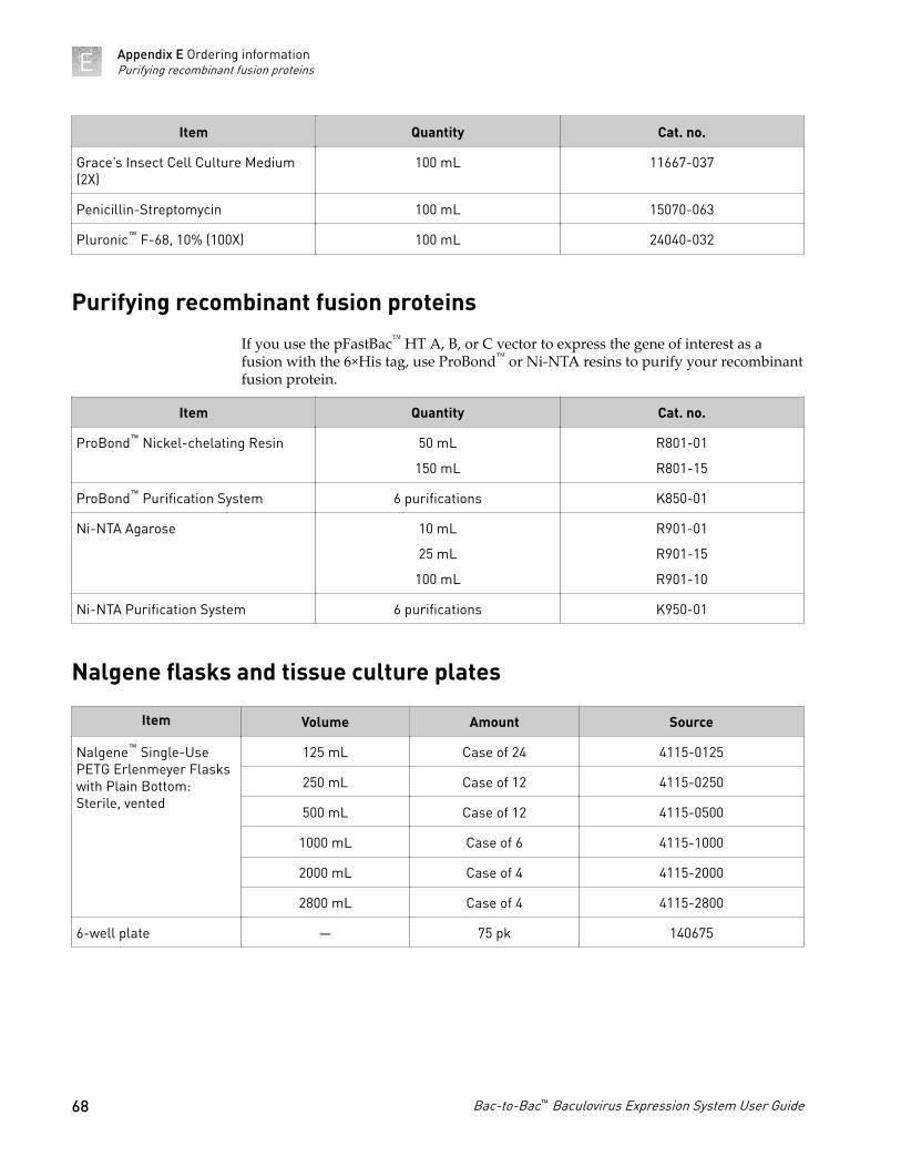
Item Quantity Cat. no.
Grace’s Insect Cell Culture Medium(2X)
100 mL 11667-037
Penicillin-Streptomycin 100 mL 15070-063
Pluronic™ F-68, 10% (100X) 100 mL 24040-032
Purifying recombinant fusion proteins
If you use the pFastBac™ HT A, B, or C vector to express the gene of interest as afusion with the 6×His tag, use ProBond™ or Ni-NTA resins to purify your recombinantfusion protein.
Item Quantity Cat. no.
ProBond™ Nickel-chelating Resin 50 mL
150 mL
R801-01
R801-15
ProBond™ Purification System 6 purifications K850-01
Ni-NTA Agarose 10 mL
25 mL
100 mL
R901-01
R901-15
R901-10
Ni-NTA Purification System 6 purifications K950-01
Nalgene flasks and tissue culture plates
Item Volume Amount Source
Nalgene™ Single-UsePETG Erlenmeyer Flaskswith Plain Bottom:Sterile, vented
125 mL Case of 24 4115-0125
250 mL Case of 12 4115-0250
500 mL Case of 12 4115-0500
1000 mL Case of 6 4115-1000
2000 mL Case of 4 4115-2000
2800 mL Case of 4 4115-2800
6-well plate — 75 pk 140675
Appendix E Ordering informationPurifying recombinant fusion proteinsE
68 Bac-to-Bac™ Baculovirus Expression System User Guide

Safety
WARNING! GENERAL SAFETY. Using this product in a manner not specifiedin the user documentation may result in personal injury or damage to theinstrument or device. Ensure that anyone using this product has receivedinstructions in general safety practices for laboratories and the safetyinformation provided in this document.
· Before using an instrument or device, read and understand the safetyinformation provided in the user documentation provided by themanufacturer of the instrument or device.
· Before handling chemicals, read and understand all applicable Safety DataSheets (SDSs) and use appropriate personal protective equipment (gloves,gowns, eye protection, etc). To obtain SDSs, see the “Documentation andSupport” section in this document.
F
Bac-to-Bac™ Baculovirus Expression System User Guide 69
Chemical safety
WARNING! GENERAL CHEMICAL HANDLING. To minimize hazards,ensure laboratory personnel read and practice the general safety guidelines forchemical usage, storage, and waste provided below. Consult the relevant SDSfor specific precautions and instructions:
· Read and understand the Safety Data Sheets (SDSs) provided by thechemical manufacturer before you store, handle, or work with any chemicalsor hazardous materials. To obtain SDSs, see the “Documentation andSupport” section in this document.
· Minimize contact with chemicals. Wear appropriate personal protectiveequipment when handling chemicals (for example, safety glasses, gloves, orprotective clothing).
· Minimize the inhalation of chemicals. Do not leave chemical containers open.Use only with adequate ventilation (for example, fume hood).
· Check regularly for chemical leaks or spills. If a leak or spill occurs, followthe manufacturer's cleanup procedures as recommended in the SDS.
· Handle chemical wastes in a fume hood.· Ensure use of primary and secondary waste containers. (A primary waste
container holds the immediate waste. A secondary container contains spillsor leaks from the primary container. Both containers must be compatiblewith the waste material and meet federal, state, and local requirements forcontainer storage.)
· After emptying a waste container, seal it with the cap provided.· Characterize (by analysis if necessary) the waste generated by the particular
applications, reagents, and substrates used in your laboratory.· Ensure that the waste is stored, transferred, transported, and disposed of
according to all local, state/provincial, and/or national regulations.· IMPORTANT! Radioactive or biohazardous materials may require special
handling, and disposal limitations may apply.
Appendix F SafetyChemical safetyF
70 Bac-to-Bac™ Baculovirus Expression System User Guide

Biological hazard safety
WARNING! BIOHAZARD. Biological samples such as tissues, body fluids,infectious agents, and blood of humans and other animals have the potential totransmit infectious diseases. Conduct all work in properly equipped facilitieswith the appropriate safety equipment (for example, physical containmentdevices). Safety equipment can also include items for personal protection, suchas gloves, coats, gowns, shoe covers, boots, respirators, face shields, safetyglasses, or goggles. Individuals should be trained according to applicableregulatory and company/ institution requirements before working withpotentially biohazardous materials. Follow all applicable local, state/provincial,and/or national regulations. The following references provide generalguidelines when handling biological samples in laboratory environment.
· U.S. Department of Health and Human Services, Biosafety in Microbiologicaland Biomedical Laboratories (BMBL), 5th Edition, HHS Publication No. (CDC)21-1112, Revised December 2009; found at:www.cdc.gov/biosafety/publications/bmbl5/BMBL.pdf
· World Health Organization, Laboratory Biosafety Manual, 3rd Edition,WHO/CDS/CSR/LYO/2004.11; found at:www.who.int/csr/resources/publications/biosafety/Biosafety7.pdf
Appendix F SafetyBiological hazard safety F
Bac-to-Bac™ Baculovirus Expression System User Guide 71

Documentation and support
Customer and technical support
Visit thermofisher.com/support for the latest in services and support, including:• Worldwide contact telephone numbers• Product support, including:
– Product FAQs– Software, patches, and updates– Training for many applications and instruments
• Order and web support• Product documentation, including:
– User guides, manuals, and protocols– Certificates of Analysis– Safety Data Sheets (SDSs; also known as MSDSs)
Note: For SDSs for reagents and chemicals from other manufacturers,contact the manufacturer.
Limited product warranty
Life Technologies Corporation and/or its affiliate(s) warrant their products as set forthin the Life Technologies' General Terms and Conditions of Sale found on LifeTechnologies' website at www.thermofisher.com/us/en/home/global/terms-and-conditions.html. If you have any questions, please contact LifeTechnologies at www.thermofisher.com/support.
G
72 Bac-to-Bac™ Baculovirus Expression System User Guide


thermofisher.com/support | thermofisher.com/askaquestion
thermofisher.com
16 July 2018