B-ENT 12 Facial trauma
18
B-ENT, 2016, 12, Suppl. 26/2, 1-18 Introduction The face is fundamental to human appearance and function. Facial injuries can have adverse impact on a patient’s ability to eat, speak, interact with others and perform other important functions. The treatment of facial trauma must first focus on threats to life, but important secondary considerations are function and long-term cosmesis. 1 Facial trauma, with or without life- and/ or sight-threatening complications, may arise following isolated injury or may be associated with significant injuries elsewhere. The most common causes of facial trauma are assault and motor vehicle accidents. 2 Maxillofacial trauma occurs in a significant number of severely injured patients and may be a sign of concomitant serious or life- threatening injuries, which may not be immediately evident on arrival in the emergency setting. For this reason, assessment of patients with facial trauma needs to be systematic and must be repeated, establishing clearly stated priorities in the overall care of the patient. Of course, the priority of treating maxillofacial trauma is usually subordinated to more critical, life-threatening injuries. Depending on the extent of the facial injuries, maxillofacial surgeons, ear, nose and throat (ENT) surgeons and/ or ophthalmologists will manage these patients after they have received acute trauma care. Facial injuries can be classified into four groups according to the urgency of the treatment necessary 1,3 : I. Immediate or emergent treatment required: facial injuries that are life- or sight-threatening and that require immediate intervention aimed at securing the airway, stopping profuse haemorrhage or relieving intraocular pressure via lateral canthotomy and cantholysis. II. Treatment required within a few hours: facial injuries that are heavily contaminated, with some open fractures, in a hemodynamically stable patient. III. Treatment required within 24 hours: some facial fractures and clean lacerations. IV. Treatment can wait for more than 24 hours if necessary: most other facial fractures. The aim of this manuscript is to consider the correct emergency care approach to patients with Facial trauma N. Peeters 1 , P. Lemkens 1 , R. Leach 2 , B. Gemels 3 , S. Schepers 3 and W. Lemmens 1 1 Department of Ear, Nose and Throat Diseases, Head and Neck Surgery, Ziekenhuis Oost-Limburg, Genk, Belgium; 2 Department of Emergency Medicine, Kliniek Sint Jan, 1000 Brussel; 3 Department of Oral and Maxillofacial Surgery, Ziekenhuis Oost-Limburg, Genk, Belgium Key-words. Facial trauma; facial injury; fractures; nose; orbit; midface; mandible; assessment; treatment Abstract. Facial trauma. Patients with facial trauma must be assessed in a systematic way so as to avoid missing any injury. Severe and disfiguring facial injuries can be distracting. However, clinicians must first focus on the basics of trauma care, following the Advanced Trauma Life Support (ATLS) system of care. Maxillofacial trauma occurs in a sig- nificant number of severely injured patients. Life- and sight-threatening injuries must be excluded during the primary and secondary surveys. Special attention must be paid to sight-threatening injuries in stabilized patients through early referral to an appropriate specialist or the early initiation of emergency care treatment. The gold standard for the radiographic evaluation of facial injuries is computed tomography (CT) imaging. Nasal fractures are the most frequent isolated facial fractures. Isolated nasal fractures are principally diagnosed through history and clinical examination. Closed reduction is the most frequently performed treatment for isolated nasal fractures, with a fractured nasal septum as a predictor of fai- lure. Ear, nose and throat surgeons, maxillofacial surgeons and ophthalmologists must all develop an adequate treatment plan for patients with complex maxillofacial trauma. 01- peeters-.indd 1 7/11/16 14:48
Transcript of B-ENT 12 Facial trauma

B-ENT, 2016, 12, Suppl. 26/2, 1-18
Introduction
The face is fundamental to human appearance and function. Facial injuries can have adverse impact on a patient’s ability to eat, speak, interact with others and perform other important functions. The treatment of facial trauma must first focus on threats to life, but important secondary considerations are function and long-term cosmesis.1
Facial trauma, with or without life- and/or sight-threatening complications, may arise following isolated injury or may be associated with significant injuries elsewhere. The most common causes of facial trauma are assault and motor vehicle accidents.2 Maxillofacial trauma occurs in a significant number of severely injured patients and may be a sign of concomitant serious or life-threatening injuries, which may not be immediately evident on arrival in the emergency setting. For this reason, assessment of patients with facial trauma needs to be systematic and must be repeated, establishing clearly stated priorities in the overall care of the patient. Of course, the priority of treating maxillofacial trauma is usually subordinated to
more critical, life-threatening injuries. Depending on the extent of the facial injuries, maxillofacial surgeons, ear, nose and throat (ENT) surgeons and/or ophthalmologists will manage these patients after they have received acute trauma care.
Facial injuries can be classified into four groups according to the urgency of the treatment necessary1,3:
I. Immediate or emergent treatment required: facial injuries that are life- or sight-threatening and that require immediate intervention aimed at securing the airway, stopping profuse haemorrhage or relieving intraocular pressure via lateral canthotomy and cantholysis.
II. Treatment required within a few hours: facial injuries that are heavily contaminated, with some open fractures, in a hemodynamically stable patient.
III. Treatment required within 24 hours: some facial fractures and clean lacerations.
IV. Treatment can wait for more than 24 hours if necessary: most other facial fractures.
The aim of this manuscript is to consider the correct emergency care approach to patients with
Facial trauma
N. Peeters1, P. Lemkens1, R. Leach 2, B. Gemels3, S. Schepers 3 and W. Lemmens1
1Department of Ear, Nose and Throat Diseases, Head and Neck Surgery, Ziekenhuis Oost-Limburg, Genk, Belgium; 2Department of Emergency Medicine, Kliniek Sint Jan, 1000 Brussel; 3Department of Oral and Maxillofacial Surgery, Ziekenhuis Oost-Limburg, Genk, Belgium
Key-words. Facial trauma; facial injury; fractures; nose; orbit; midface; mandible; assessment; treatment
Abstract. Facial trauma. Patients with facial trauma must be assessed in a systematic way so as to avoid missing any injury. Severe and disfiguring facial injuries can be distracting. However, clinicians must first focus on the basics of trauma care, following the Advanced Trauma Life Support (ATLS) system of care. Maxillofacial trauma occurs in a sig-nificant number of severely injured patients. Life- and sight-threatening injuries must be excluded during the primary and secondary surveys. Special attention must be paid to sight-threatening injuries in stabilized patients through early referral to an appropriate specialist or the early initiation of emergency care treatment. The gold standard for the radiographic evaluation of facial injuries is computed tomography (CT) imaging. Nasal fractures are the most frequent isolated facial fractures. Isolated nasal fractures are principally diagnosed through history and clinical examination. Closed reduction is the most frequently performed treatment for isolated nasal fractures, with a fractured nasal septum as a predictor of fai-lure. Ear, nose and throat surgeons, maxillofacial surgeons and ophthalmologists must all develop an adequate treatment plan for patients with complex maxillofacial trauma.
01- peeters-.indd 1 7/11/16 14:48

2 N. Peeters
2. Assessment and management at the hospital
2.1. Initial general assessment
severe and disfiguring facial injuries can be distracting for both patient and clinician. However, clinicians must focus on the basics of trauma care. The Advanced Trauma Life Support (ATLS) system of care has generally come to be accepted as the gold standard in the initial management of multiple trauma patients.1,3 It consists of a systematic initial assessment of each trauma patient. The main goal is to recognize patients with severe life-threatening injuries, establish treatment priorities and manage them efficiently. All life-threatening injuries must be addressed before performing a complete facial examination.
The assessment of trauma patients as outlined by ATLS protocols places high value on the ABCDEs (Airway maintenance with cervical spine protection, Breathing with ventilation, Circulation with haemorrhage control, Disability and Exposure/Environment) in the primary survey. Although the aim of the primary survey is to identify and treat life-threatening problems, the early identification of a sight-threatening condition may be possible during ‘D’ (Disability). The primary reason for examining the pupils is to assess for neurological disability; however, associated ocular findings must also be noted to identify visual disability. In the alert patient, it only takes a few seconds to ask the patient if he/she can see clearly with each eye (in turn) during assessment of the Glasgow Coma Scale and to carefully palpate the globes through closed eyelids. Early identification of a potential sight-threatening injury enables early referral to an appropriate specialist and the initiation of treatment where necessary (e.g., retrobulbar haemorrhage). After problems identified during the primary survey are adequately addressed, a secondary survey, including careful examination of facial injuries, must be performed. The secondary survey is of great importance for detecting evolving injuries, and should include a systematic approach to and examination of all major facial structures and functions.
Airway obstruction, bleeding and sight-threatening conditions may at first be subtle and may not become apparent until the secondary survey is underway. This serves as a reminder of the two well-established principles in trauma care:
facial trauma. We will start with some general considerations in facial trauma care, followed by a description of the systematic clinical examination of patients with facial injuries. The focus of this manuscript lies in recognizing typical clinical signs of facial trauma patients, which helps the clinician to make the correct diagnosis. We will provide a more extensive overview of nasal trauma, followed by a discussion of orbital, midface and mandibular fractures.
1. Prehospital management
Prehospital management of trauma patients is discussed in detail separately (see the first chapters of this report). Special considerations for patients with facial trauma are described below.
Facial trauma may occur in isolation or may be associated with significant injuries elsewhere in a multiple trauma patient. In the prehospital setting, it is crucial to pay attention to clinically apparent threats to airway, breathing and circulation (ABC).
In a multiple trauma patient, the initial evaluation of facial trauma should be succinct – the minimum necessary to secure the airway prior to further resuscitation measures. In the case of significant facial trauma, associated head and cervical spine injuries are common. Airway management remains unchanged for patients with facial trauma. Facial fractures can influence airway management in several ways. Facial fractures can increase the difficulty of ventilating with a bag valve mask and may possibly impede the performance of the jaw-thrust manoeuvre. If intubation must be performed in the field for patients with facial trauma, orotracheal intubation is strongly preferred to blind nasotracheal intubation.3
Bleeding from severe facial wounds can complicate endotracheal intubation. Suction devices may be required and external compression can help in the acute phase. Patients with severe oropharyngeal bleeding who do not require spinal immobilization should be placed in a comfortable position, which often consists of sitting and leaning forward with the head. If spinal immobilization is required and bleeding cannot be controlled by compression and suctioning, intubation may be necessary to protect the airway5. After intubation, gauze can be placed in the oro- and nasopharynx to obtain control by direct pressure.
01- peeters-.indd 2 7/11/16 14:48

Facial trauma 3
so that any vision-threatening injuries can be ruled out. The timing of the examination is problematic in comatose or sedated patients. Having said this, it is very important to identify injuries of the globe and optic nerve early, so that emergent interventions can be planned for vision preservation. An examination of the eyes should include an inspection of the position and alignment of the eyes and an evaluation of visual acuity, pupillary response, reactivity to light tests and the range of motion of the extraocular muscles of the eyes. Subtle differences can be important. An orbital fracture, for example, can be diagnosed by subtle discrepancies in the positions of the eyeballs. Enophthalmos can be caused by an orbital fracture with the sinking of the contents of the orbit. An increase in orbital volume can cause exophthalmos, which is suggestive of intraorbital haematoma. However, these changes in eye position may not be obvious in acute settings due to oedema in the periorbital area. Lacerations of the ethmoidal arteries may result in intraorbital (retrobulbar) haemorrhage and a progressive increase in intraorbital pressure. This can cause compression of the optic nerve and ophthalmic artery and blindness. Signs of retrobulbar haemorrhage include pain, reduced light perception, a fixed dilated pupil, loss of the direct pupillary reflex, exophthalmos and ophthalmoplegia. Retrobulbar haemorrhage may be decompressed by lateral canthotomy, which saves the vision. When an intraocular injury is suspected, an ophthalmologist should conduct an emergent evaluation of the patient. However, if there is no time and a retrobulbar haemorrhage is suspected, the emergency physician must perform a lateral canthotomy. Eyelid lacerations involving the medial canthus can be signs of damage to the canalicular drainage system. These injuries mostly require a specialist to repair them up to 48 hours after the injury.1
2.3.3. Naso-orbital-ethmoid complexIn the case of a high-energy trauma with oedema of the periorbital region, the naso-orbital-ethmoid (NOE) complex must be evaluated to rule out a NOE fracture. NOE fractures refer to injuries involving the area of confluence of the nose, orbit and ethmoids, the base of the frontal sinus and the floor of the anterior skull base. A missed NOE fracture can lead to serious aesthetic consequences that cannot be easily rectified by revisionary surgical procedures.1 The NOE complex is rarely fractured in isolation and the patient often has other
I. The need for a high index of suspicion (often based on the mechanism of injury and known patterns of injury)
II. The need for frequent patient reassessment For further information on the general principles
of acute medicine, see chapters one to 10.
2.2. History
The priority and extent of history taking should be dictated by the acuity of the patient’s presentation and associated conditions. In addition to obtaining a basic history of the injury and past medical problems, the clinician should ask for:Vision problems
- Nasal obstruction- Facial numbness- Hearing loss- Previous facial injuries or surgeries- Changes in dental occlusion, painful or loose
teeth- Bleeding from the ears, nose or mouth- Vertigo
2.3. Clinical examination
Facial trauma patients should be examined in a systematic way in order to minimize missed injuries. First of all, the patient’s face should be generally observed for any structural asymmetry. It helps to observe the face from different perspectives.
Tissue avulsions, lacerations, contusions and skin wounds are mostly obvious. Tetanus vaccination status should be checked when open skin wounds are present.
The clinician should subsequently palpate the bony structures of the face, feeling for crepitus, step deformities, abnormal motion or local tenderness. Facial nerve motor function and skin sensation (trigeminal nerve) should be carefully assessed. After a general examination of the face, an examination of specific facial parts can be performed.1,6
2.3.1. ForeheadInjuries to the forehead can be easily recognized and may indicate frontal bone fractures. Special attention should be paid to closing lacerations involving the hairline or eyebrows.
2.3.2. OrbitExamining the eyes and orbit is crucial. Orbital and periorbital injuries should be carefully evaluated
01- peeters-.indd 3 7/11/16 14:48

4 N. Peeters
2.3.4. NoseThe nasal skeleton is the next area of the face to be examined. This requires good lighting, suction and a nasal speculum. Look and feel for deformities and lacerations on and inside the nose. Nasal deformities must be examined facing the patient, from both worm’s and bird’s eye views. Palpate the nasal bones for abnormal mobility, crepitus and tenderness. Intranasal examination should focus on lacerations and perforations of the mucosa, the appearance of the nasal septum and, especially, the presence of a septal haematoma, because must be recognized and dealt with early.
The presence of clear fluid in the nasal cavity may indicate CSF resulting from an associated basal skull fracture. For an adequate diagnosis, the fluid should be tested for beta-2 transferrin, although this test takes time. The clinician could ask for a computed tomography (CT) scan with thin coronal cuts and special attention to the cribriform plate. The application of fluorescent intrathecal dyes with a direct visualization of the leak via transnasal endoscopy can be performed. Bedside tests that have been used to identify CSF, such as the detection of glucose in the fluid, the ‘reservoir sign’ (i.e., a patient leaking CSF when flexing the head) or the ‘halo sign’, are rather unreliable. The ‘halo sign’ (Figure 3) refers to CSF’s ability to migrate further on filter paper than red blood cells, creating a halo or bull’s eye with blood in the centre (from bloody rhinorrhoea).
facial fractures. This makes examining this area complex. Significant orbital swelling and increased intercanthal distance (telecanthus) is suggestive of the presence of a NOE fracture. Intercanthal distance varies according to race and gender, but as a rough estimate, the distance should equal 50% of the interpupillary distance.1 Periorbital ecchymosis and circumorbital oedema, which gives the patient the appearance of raccoon eyes, is a hallmark sign of NOE fractures. Other signs that may be present are diplopia, epistaxis, vision abnormalities, anosmia and dizziness. NOE fractures can also occur in combination with basal skull fractures, so it is of paramount importance to look for any signs of cerebrospinal fluid (CSF) leakage.
If a NOE fracture has occurred, palpation of the complex will show mobility and crepitus. An instrument can be placed in the nose, pushing its tip deep into the attachment of the medial canthal tendon (MCT). Meanwhile, you palpate with the index finger and thumb of the other hand, placed on either side of the NOE complex. Pushing laterally with the instrument in the medial canthal area tests for instability and crepitation, which are both signs of an unstable NOE fracture (Figure 1).
Another test is the bowstring test (Figure 2). The surgeon takes the eyelid near the medial canthal tendon with his/her index finger and thumb and pulls the lateral canthal tendon laterally. In the case of a fracture, the physician will be able to detect movement of the MCT and underlying bone fragments.
Figure 1Testing for NOE fracture (from www.aofoundation.org)
Figure 2Bowstring test (from www.aofoundation.org)
01- peeters-.indd 4 7/11/16 14:48

Facial trauma 5
upper alveolar ridge suggests a midface fracture, whereas such injuries to the lower alveolar ridge suggest a mandible fracture. Check the maximum incisal opening if possible. Any deviations while opening may be suggestive of the dislocation of the temporomandibular joint or fractures of the mandible. Difficulty in opening the mouth can also be caused by a zygomatic complex fracture.
Dental occlusion must be evaluated. Ask the alert patient to close his mouth and describe whether the motion and positioning feels normal. Malocclusion is suggestive of a maxillary, dentoalveolar or mandibular fracture. Ecchymosis or oedema around the chin or in the preauricular area is associated with mandibular fractures. Palpation of the mandible both within the oropharynx and externally may reveal step deformities, tenderness or crepitus, any of which suggest a mandibular fracture.
Skin wounds in the parotid area may be indicative of disruption to the Stenson’s duct. During inspection of the mouth, the duct opening, which lies in the inner wall of the cheek opposite the second upper molar, should be examined for bleeding while the gland is compressed.
2.3.6. EarInspect the external ear for deformities or lacerations. Look out for postauricular ecchymosis (Battle’s sign), as this may be suggestive of temporal or basal skull fractures. The clinician can also perform an otoscopy to evaluate the integrity of the external canal and to look for hemotympanum and otorrhoea. Clear fluid draining from the ear following trauma may be CSF.
2.4. Diagnostic imaging
the gold standard for the radiographic evaluation of facial injuries has come to be the helical CT scan with multiplanar reconstructions.1,7 The added ability to obtain a three-dimensional reformatted image helps in planning treatment. Three-dimensional printing can be used for virtual surgical planning. Correlating the findings of the physical examination with the radiographic assessment would lead to an accurate diagnosis and provide assistance with treatment planning.
2.5. Antibiotics
the use of antibiotics in maxillofacial trauma remains controversial. When considering antibiotic therapy, the risk factors for infection need to be
2.3.5. Midface, mouth and mandible Oral examination and examination of the midface, mandible and dentoalveolar structures should be performed at the end of the systematic evaluation to avoid other facial wounds from being contaminated with saliva. Palpation for step deformities and obvious tenderness and mobility can reveal fractures. Midface fractures may manifest as a large contusion over the cheekbone and can be associated with malocclusion of the upper teeth and enophthalmos. Changes in sensitivity of the area supplied by the infraorbital nerve can be a subtle sign of an orbital floor fracture, which is seen in the Le Fort II fracture, see below. Anaesthesia of the maxillary teeth can be caused by a fracture through the anterior wall of the maxillary sinus due to injury of the dentoalveolar nerves.
High-energy trauma can cause midface instability, which can be examined by grasping the superior anterior teeth between thumb and index finger and attempting to move the maxilla anteriorly and posteriorly, while stabilizing the patient’s forehead with the opposite hand. Mobility of the maxillary complex indicates a disjunction of the maxilla from the remaining facial skeleton, which is seen in the presence of a Le Fort fracture.
Look for lacerations in the perioral region and within the oral cavity and examine for debris, contaminants and loose teeth. Lacerations or step deformities in the palate suggest a maxillary fracture. Step-off, ecchymosis and lacerations to the
Figure 3The halo sign in CSF rhinorrhoea (from www.aofoundation.org)
01- peeters-.indd 5 7/11/16 14:48
6 N. Peeters
patients between the ages of 15 and 30 and in the elderly, among whom a small increase can be put down to falls. In young adults, most nasal fractures are the result of assaults, sporting injuries and motor vehicle accidents.14 Nasal trauma can cause subtle or obvious changes in nasal appearance and can lead to functional nasal airway obstruction. It has been reported that more than 90% of nasal fractures may also have a degree of septal injury.15
The overall goal of treatment is to restore the nose to its premorbid shape and function and to minimize the need for subsequent surgery.
3.1. Clinical assessment
Isolated nasal fractures are principally diagnosed through history taking and through a careful clinical examination in particular, marking a difference from many other facial fractures that rely on computed tomography imaging for an accurate diagnosis.
3.1.1. HistoryThe most pertinent understanding of a given nasal fracture emerges from each patient’s history, which will provide clues as to the severity and location of the fractures. History taking will help to distinguish isolated nasal and septal fractures from those likely to be associated with other injuries.
Factors to consider in a patient with an injury of the nasal pyramid are as follows12,16,17:
- mechanism and timing of injury- loss of consciousness- in the case of a blow: direction and energy of
the blow- any epistaxis associated with the injury- any change in nasal appearance- nasal airway obstruction- facial numbness, changes in vision, changes
in dental occlusion - persistent rhinorrhoea, changes in sense of
smell- prior nasal and/or septal trauma and surgery- prior nasal deformity or nasal obstruction- tetanus statusRecent photographs taken prior to the trauma
may facilitate the clinician’s appreciation of the extent of nasal deformities. Concussion and brain injury symptoms such as headache, nausea or vomiting, dizziness, disorientation or lethargy must be elicited and appropriately managed by the trauma and neurology teams. Headaches can also be caused by trauma to the neck.
assessed. Antibiotics are not required for simple facial wounds, which rarely become infected. Exceptions include the following:
- bite wounds- wounds with any evidence of devascularization- wounds that penetrate the buccal mucosa and
the lips- wounds involving exposed cartilage of the
nose or ear- grossly contaminated wounds that require
closure- wounds associated with open fracturesIf prophylactic antibiotic therapy is needed, the
antibiotic should be selected based on the normal bacterial flora found in the affected site.8 Mostly, antibiotics such as amoxicillin or clindamycin in combination with metronidazole are preferred, which are active against oral and pharyngeal organisms.9 For most midfacial fractures, there is limited evidence to suggest that antibiotics offer any benefits. The midface, as well as the condylar head of the mandible, are at low risk of postoperative infection in comparison with the angle, body and symphysis of the mandible (tooth-bearing segments). If CSF leakage is suspected, the patient may be at risk of contracting meningitis. However, the current literature does not support antibiotic prophylaxis based on incomplete and potentially biased evidence.10,11
2.6. Treatment planning
After a thorough assessment of a facial trauma patient who does not present with life- and/or sight-threatening injuries, the facial trauma care team (often consisting of the maxillofacial surgeon, an ENT surgeon and/or a plastic surgeon) must develop a treatment plan in the case of multiple facial injuries, which can be complex.
3. Nasal fractures
The central position of the nose and its anterior projection from the face renders it susceptible to injury. It should not be surprising that nasal fractures are the most frequent of all isolated facial fractures and are reported to be the third most common fracture of the human skeleton.12 The incidence of posttraumatic nasal deformity, if left untreated, varies from 14% to 50%.13 The male-to-female ratio for nasal fractures is greater than 2:1, as with other facial fractures. The incidence peaks bi-modally in
01- peeters-.indd 6 7/11/16 14:48

Facial trauma 7
dislocation or fracture or a septal haematoma. A cotton-tipped applicator can be used to palpate a septum that appears to be deviated. If the septum is compressible, think of a septal haematoma. If the septum is slightly mobile, and even more so when there is a mucosal tear, it could arguably be a new septal deviation and/or fracture. A fractured septum unfavourably affects the alignment of the nasal bone during the healing process and is a predictor of failure of closed nasal fracture reduction.12,16
In the presence of a septal haematoma, incision and drainage must be performed in order to prevent the formation of a septal abscess, which can lead to cartilage destruction and saddle deformity of the nose.
If any septal deformity caused by the trauma is suspected, your management should be adjusted. In the case of clear nasal discharge, cerebrospinal fluid leakage should be suspected.
All the other parts of the facial skeleton must also be examined to rule out associated fractures. This should include visual acuity, extraocular muscle movement, pupil size and reactivity, intercanthal distance, medial canthal tendon position, mandibular range of motion and occlusion. The clinician should palpate the orbital rims, zygomatic arches, maxilla and mandible. In many cases, a force that is great enough to fracture the nasal and septal bones is also substantial enough to injure adjacent structures, such as the globe and orbit floor. When the signs and symptoms of a concussion become evident during the examination, such as headaches, irritability or confusion, a neurological examination should be performed and, eventually, the patient will need to be referred to a neurologist.
3.2. Imaging
Many isolated nasal injuries without loss of consciousness and a clear history of the mechanism can be diagnosed and treated without the need for exposing the patient to radiation. If the patient history and/or clinical examination suggests fractures of the other facial bones, a CT scan must be performed.
Conventional radiography offers little clinical benefit in isolated nasal trauma. Nigam et al.18, as confirmed by other studies19,20, found that there was a poor correlation between the radiological findings on conventional radiography and the presence of external deformities. They therefore
3.1.2. Clinical examinationAn extensive physical examination must be carried out. The goal is to assess the severity and extent of the injury to ensure the selection of the most appropriate management. Before starting the physical examination, cervical spine precautions must be taken until a cervical spine injury is ruled out.
First, start with the external nasal examination. Observing the nasal dorsum from frontal, worm’s eye and bird’s eye views will help the clinician to observe any external nasal deformities. If the examination is performed more than several hours after the injury, overlying oedema and ecchymosis is frequently present. This may mask the severity of the underlying skeletal deformity and is often the reason why a nasal reassessment must be performed after several days. The clinician should examine for the presence of nasal or septal deviation, step deformities and crepitus on palpation. Fractures of the nasal bone most commonly occur distally, where the nasal bone is at its broadest and thinnest. Look and feel for any deviation of the bony pyramid, the degree of displacement and the extent of the depression or lateralization of the nasal bones. Palpation of the nasal tip should assess the potential loss of caudal septal support. A saddle deformity may indicate significant dislocation, or fracture, of the septal cartilage. Palpation of the junction between the nasal bone and upper lateral cartilage can reveal some separation of these structures, which can result in depression and collapse at this point, even when the bony pyramid is not displaced. Many of these patients will benefit from a secondary septorhinoplasty.12
The intranasal examination includes an anterior rhinoscopy and, where necessary, a nasal endoscopy. By using appropriate light, a nasal speculum, suction and eventually a vasoconstrictor, the clinician must assess the status of the cartilaginous and bony nasal septum, rule out the presence of a septal haematoma and determine the origin of epistaxis and/or cerebrospinal fluid rhinorrhoea. In some cases, it may be helpful to perform a nasal endoscopy to evaluate the posterior nose.
The assessment of the nasal septum is of paramount importance, paying attention to tears in the septal mucosa and the degree of deviation. An apparent septal deviation may be explained by a previous septal deviation, a new septal
01- peeters-.indd 7 7/11/16 14:48

8 N. Peeters
3.3. Treatment
The functional and cosmetic problems that arise may sometimes necessitate surgical procedures to restore the anatomy. Not all nasal fractures require treatment. If the fractured nasal bones remain well aligned and the nasal airway is not significantly changed, observation alone is adequate and surgical treatment will not be necessary.12 If the patient is seen shortly after the injury and the oedema is still significant, the patient should be re-evaluated as soon as the swelling has receded, but soon enough to reduce the fractures prior to the consolidation of the fracture site.
For most routine nasal fractures, operative management requires the consideration of three major aspects to ensure the best treatment: the timing of treatment, the choice of an appropriate anaesthetic (local or general) and the surgical technique (open or closed reduction).
3.3.1. Timing of treatmentIn the literature, opinions differ as to the most appropriate timing of treatment. Some nasal trauma requires immediate treatment, whereas others are better treated after a delay. For example, treatment of open nasal fractures and injuries with gross external deformities should be performed within several hours. Septal haematomas require immediate incision and drainage.
Mondin et al.16 state that if the patient is seen before significant distorting oedema sets in, i.e., within the first three to six hours after injury, reduction of the fractured nose could be performed immediately. If there is already soft tissue oedema, the patient should be reassessed three to four days later, because oedema usually masks mild-to-moderate nasal fractures and it would be difficult to determine whether the nasal bones were properly reduced.
It is generally believed that nasal fractures should be treated within two to three weeks before the nasal bones stabilize. Staffel21 noticed the importance of treating nasal bones within two weeks after injury. Other authors22,23,24 argued that the reduction should be performed within 10 days of the trauma for adults and within seven days for children (due to faster bone healing in the paediatric population). On the other hand, Basheeth et al.13 offered manipulations up to five weeks post-trauma and found no change in the degree of satisfaction post-manipulation when comparing patient groups based on the length
deem conventional radiography to be of little use in the diagnosis of nasal bone fractures due to its inaccuracy. The value of ultrasound (US) as a diagnostic tool for detecting fractures has been demonstrated, with several studies showing that the accuracy of US in the diagnosis of nasal fractures is greater than that of conventional radiography.20
However, because ultrasound is a dynamic procedure, it is difficult to reproduce the fracture after the examination. CT scanning is also superior to plain radiography for diagnostic purposes, but because the risks of radiation exposure and the costs of CT scans are too high in relation to the additional information they provide, they should not be performed on every patient with a nasal trauma.
Bremke et al.19 demonstrated the value of Digital Volume Tomography (DVT) in the diagnosis of nasal bone fractures. DVT is a kind of cone beam computed tomography and provides high quality images using low radiation exposure, with the option of three-dimensional reconstruction. DVT has a limited capacity to indicate extensive midfacial trauma because the extent of the DVT image itself is limited. It also has limited scope to be used for incompliant patients who are not able to hold their head fixed during the time it takes to examine them. In these two cases, CT scanning is always a better option. However, if one suspects an injury extensive enough to cause multiple fractures at multiple sites, cone beam CT may not be recommended because imaging of the brain tissues may be needed to evaluate the possibility of an intracranial bleed.12
An isolated nasal fracture is mostly a clinical diagnosis, but imaging can also be helpful in some cases. If physical examination cannot determine the complexity of the fracture and if associated facial fractures are suspected, a CT scan can give a good visualization of the fracture lines and of concomitant fractures. When you suspect a nasal fracture at the proximal part of the nasal bone where the bone is thicker, think about associated facial fractures because it indicates the possibility of a high-energy trauma.
Due to possible legal implications, the accurate imaging of nasal fractures is necessary in some circumstances. Because of the low sensitivity and specificity of conventional radiography, the emergency department should not perform routine radiography due to these legal implications.
01- peeters-.indd 8 7/11/16 14:48

Facial trauma 9
and, eventually, an anaesthetic agent, are used for nasal mucosa anaesthesia and haemostasis. This is supplemented by infiltration with a solution containing 1% lidocaine or 0,5% bupivacaine to block the infraorbital, infratrochlear, supratrochlear and external nasal nerves. Investigators1 have studied the difference in postoperative pain between two LA techniques. One group received intranasal infiltration, the other group received infiltration via an external route. Their study suggested that the external method is preferable.
3.3.3. Surgical techniqueThe operative approach to isolated nasal bone fractures typically consists of closed reduction. However, nasal fractures associated with significant nasal obstruction, septal deviation that cannot be reduced using closed manoeuvres or nasal fractures with a loss of nasal septal support may require open repair and reconstruction.
For most injuries, closed reduction is successful in achieving appropriate form and function. Before reduction is performed, the extent of the nasal deformity and deviation should be noted from frontal, worm’s eye and bird’s eye views. It is important to re-examine the nose under these more ideal conditions, including the nasal septal form, the integrity of the nasal floor, the size of the nasal turbinates and the integrity of the cartilaginous structures. A local vasoconstrictor such as oxymetazoline nasal spray or cottonoids soaked in adrenaline or xylocaine can be applied to the nose for optimal mucosal decongestion prior to the manipulation. If more extensive intervention is anticipated, such as septal repair or osteotomy, then regional infiltration with a local anaesthetic is also performed. Perioperative antibiotics are used at the surgeon’s discretion. However, antibiotics are advised in all cases of septal haematoma.12
Digital pressure may be all that is needed to reduce deformities in simple nasal fractures, using the maxillary and frontal bones for anchorage and guidance for reduction. Additionally, instrumentation can be used. A Boies elevator can be used to elevate the depressed bone, using a bimanual technique to stabilize the bones and to feel when they have been accurately reduced. The Walsham forceps is designed to deal with impacted nasal bones. The Ash forceps can be used to reduce the nasal septum without resorting to septoplasty, but they are also useful in restoring the alignment
of time since their injury. Al-Moraissi et al.14 state that one reason for delaying surgery can be the surgeon’s philosophy, i.e., obtaining a result that might exceed what the patient had prior to the injury, especially in patients with pre-existing nasal and septal deformities. The surgeon will allow the nasal bones heal in a malunited position, only performing a septorhinoplasty after at least six months.
3.3.2. Choice of anaestheticMost nasal fractures are treated with closed reduction, which is typically performed as an outpatient procedure. The management of nasal fractures can be performed under local anaesthesia (LA) or general anaesthesia (GA), depending on the preference of the surgeon and patient, hospital protocol, surgical specialty and practical reasons.14
In cases whereby the nasal septum is involved in the fracture and in patients with nasal obstruction and compromised nasal patency, it is better to perform the reduction under general anaesthesia. The surgeon can manipulate the nasal septum more extensively under general anaesthesia and can even perform a limited septoplasty if necessary. A meta-analysis of Al-Moraissi and Ellis14 compares the outcomes of closed reduction under local and general anaesthesia. The results showed that although there was a trend toward better outcomes with GA, there was not a statistically meaningful difference between LA and GA in the closed reduction of nasal bone fractures in terms of patient satisfaction with the anaesthesia, nasal function, subsequent treatments or preference of anaesthesia in cases where the nose had to be re-fractured. Manipulation under local anaesthesia is appropriate for cooperative adults with simple nasal bone fractures that do not require open reduction of the nasal septum.12,14 General anaesthesia can also be used in these cases, but is definitely the best choice for uncooperative or young patients, patients with severely displaced fractures and those who require extensive septal work.14 Local anaesthesia, when the surgeon can manage the reduction at accident and emergency, is a more economical and well-tolerated alternative. In summary, contributing factors may include patient cooperation, financial constraints, operator experience or comfort, among other reasons. Local anaesthesia can be performed with the addition of sedation. In closed reduction under LA, a topical intranasal solution using pledgets soaked with a vasoconstrictive
01- peeters-.indd 9 7/11/16 14:48

10 N. Peeters
limited osteotomies and septoplasty, soft tissue repair and perhaps bone or cartilage grafting to restore dorsal support.
Post-reduction deformities requiring septorhinoplasty could be prevented by early diagnosis and appropriate intervention and follow-up. Pre-existing nasal deformities, traumatic oedema and occult septal injury can cause acute reduction failures.
3.4. Paediatric nasal fractures
Children’s noses are comparatively more protected from fractures, due to their having a less pronounced nasal dorsum than adults. The more prominent forehead and supraorbital rim in children provide protection for the paediatric nose.
The paediatric nasal skeleton is more cartilaginous and thus more flexible. This renders the nasal septum in children more vulnerable to dislocation or distortion, rather than nasal bone fractures.
One of the most common clinical findings in paediatric nasal fractures is bluish discoloration over the nasal bridge.13 Periorbital ecchymosis, where signs of an orbital fracture are absent, is suggestive of a nasal bone fracture. Clinical examination includes palpation of the nasal bones, looking out for crepitus, mobility, tenderness, step-off or deformities. The intranasal cavity should be evaluated in all children with nasal trauma because septal injury, and especially a septal haematoma, can occur without other signs of a nasal fracture.
As with adults, conventional radiography is unhelpful in the diagnosis of nasal fractures, even less so than with adults because of the large amount of cartilage in the paediatric nose. A CT scan should be performed when other facial trauma is suspected.
The nasal septum plays a crucial role as a growth centre for the nose and midface in children prior to adolescence. Therefore, the surgical management of nasal bone fractures should be rather conservative.12
A child with a septal haematoma must be urgently treated using incision and drainage, as with adults, and followed by antibiotic therapy.
Children who have no symptoms, minimal oedema and no septal deviation or haematoma after the trauma, do not necessary need specific follow-up.
Children with a possible nasal bone fracture, but no septal haematoma, should be referred to the ENT department within five days. After this short
of impacted nasal bones. Often an audible and palpable ‘pop’ will occur during reduction, which may signify that the bony spicules of the fracture sites have become interdigitated.
In open reduction, the choice of a given operative technique is dictated by the patient’s particular fracture type or dislocation. Septoplasty with resection of deviated cartilage or bone is performed in the case of severe septal fractures. Rarely, a full septorhinoplasty approach is necessary in the initial treatment.
External splints are commonly used postoperatively to stabilize and protect the reduction. Nasal packing can be placed in the nose to provide additional support for the previously depressed nasal bone.
Some authors12 have proposed a classification scheme for nasal fractures. They have attempted to categorize nasal trauma based on the degree of injury to the nasal bone and septum, using this to create a treatment algorithm based on the severity of the injury. These algorithms can be used to improve treatment outcomes and to reduce the need for secondary surgery. In all these classifications, the nasal septum plays a crucial role, whereby the correction of significant septal deformities caused by the trauma is often cited as key in optimizing nasal fracture management.
We refer to Hoffman’s treatment algorithm (Figure 4)12. He created four groups. The first group includes those who have displaced but uncomminuted fractures, with no septal deviation and only minor displacement of the midline. The second group includes those with limited comminution and mild, but not obstructive, septal deviation and moderate displacement of the nasal dorsum greater than half of the width of the dorsum. These two groups can be managed through the closed reduction of the nasal bones with external splint stabilization.
A third group includes those with severe bony comminution and obstructive septal deviation or dislocation with displacement of the midline dorsum. These patients will also need either closed reduction of the nasal septum or perhaps a limited septoplasty, in addition to closed reduction of the nasal bones. The final group includes those with severe comminution and impaction of the nasal bones, as well as the collapse of the septal support and damage to, or loss of, external soft tissues. These cases may require open reduction with
01- peeters-.indd 10 7/11/16 14:48

Facial trauma 11
4. Midface fractures
The midface refers to the bones in the middle third of the facial skeleton. The maxilla, zygomatic, lacrimal, nasal, palatine and vomer bones are referred to as the middle third of the facial skeleton. The sphenoid, frontal and ethmoid bones are not classically facial skeleton bones, but they are frequently traumatized in midface fractures and thus should be considered to be part of the midfacial skeleton.
delay, the nasal bones can be better evaluated due to the resolution of the oedema.
Most nasal bone fractures are treated by closed reduction. Early intervention (within seven days) is recommended, because the active growth centres in the nasal bones of children stimulate rapid healing. Septal dislocation can be treated by closed reduction.
Hoffmann12 states that it is prudent to warn parents that nasal trauma at an early age may result in problems with airways or appearance later in life.
Figure 4Treatment algorithm for nasal fractures 12
01- peeters-.indd 11 7/11/16 14:48

12 N. Peeters
4.1.1. Le Fort I fractureIn Le Fort I fractures, also known as Guerin fractures, a transversal fracture line goes through the maxilla, separating the inferior portion of the maxilla, the horizontal plates of the palatal bones and the inferior third of the sphenoid pterygoid processes from the superior two thirds of the face, which remain associated with the skull. The patient may complain of malocclusion. The clinician will detect motion in the maxilla when the upper teeth are grasped and rocked, while the forehead is held stationary with the other hand. On initial examination, these fractures are not always clinically evident. Haematomas in the upper vestibule (Guerin’s sign), palatal ecchymosis and epistaxis may occur.
4.1.2. Le Fort II fracture In Le Fort II fractures, the pyramidal midface is separated from the rest of the facial skeleton and skull base. The fracture line begins inferior to the nasofrontal suture and extends across the nasal bridge, the lacrimal bone, the medial part of the orbital floor, the infraorbital rim (often through
During the initial physical examination, complex midface fractures involving the zygoma and adjacent structures may manifest as a large contusion over the cheekbone, enophthalmos or malocclusion. Oedema and facial asymmetry are the most common clinical signs of midface fractures. Patients may have epistaxis, meaning that an intranasal tamponade may be necessary.
Below, we discuss Le Fort and zygomatic fractures.
4.1. Le fort fractures
Complex fractures of the midface are classified using the Le Fort system. In 1901, French surgeon René Le Fort published a classification of midface fractures (Figure 5).9 They are characterized by typical lines through the midface, involving the maxillary dentoalveolar segment. Most Le Fort fractures are not ‘pure’ and can involve comminution or additional fracture lines. The mechanism and location of impact usually determines the type of fracture. Most Le Fort fractures are associated with other facial fractures and orthopaedic and neurological injuries.
Figure 5Le Fort I-II-III fractures (Figure adapted from www.fprmed.com)
01- peeters-.indd 12 7/11/16 14:48

Facial trauma 13
to 10 days after trauma in order to perform a good aesthetic evaluation after the oedema has decreased. Open fractures must be treated within 24 hours of the trauma.
The use of antibiotics in midface fractures remains controversial and most studies supporting it involve small series.10 A recent study showed that a three day course of antibiotics was not superior to nasal saline solution in preventing the signs and symptoms of acute sinusitis following maxillary sinus fractures from blunt trauma.27 Having said that, antibiotics are recommended for open fractures or in the presence of tissue emphysema from sinus wall disruption.9 Sinus precautions, such avoiding blowing one’s nose or sneezing with one’s mouth open, are the most important.
4.2. Zygomatic fractures
The zygomatic bone is the second most common midfacial injury after nasal fracture, due to its prominence and superficial location.
There are two fracture patterns. The zygomatic arch can be fractured alone, or the zygomatic complex can be separated through a fracture from its four articulations (frontal, sphenoidal, temporal and maxillary). This last fracture is also called a ‘tripod’ fracture. It involves the zygoma, lateral orbit and the maxilla.
In a non-displaced zygomatic injury, the earliest clinical signs are pain, ecchymosis and periorbital oedema. Displaced fractures cause malar depression immediately post-trauma, which can later be masked by oedema or haematoma. Crepitus can be palpated. A limited ability to open the mouth can be a symptom. In the case of tripod fractures, patients may complain of paraesthesia or anaesthesia due to damage to the zygomaticotemporal and infraorbital nerves. Limitation of eye motion, monocle haematoma, enophthalmos or exophthalmos should be noted, as these can be signs of a fracture of the orbital floor or medial or lateral orbital walls. Lateral subconjunctival haemorrhage is indicative of disruption to the lateral orbital wall. Ophthalmologic consultation should then be considered prior to surgical intervention.
The isolated zygomatic arch fracture is the most common fracture pattern. The fracture line has a typical M-shaped pattern, with two fragments collapsed medially and often impinging on the masseter muscle. Trismus can occur due to the
the infraorbital foramen, as this is a weak point) and along the maxilla to the zygomaticomaxillary suture. When examined, the nasal complex moves as a unit with the maxilla when the teeth are grasped and rocked, while the forehead is held stationary. Periorbital haematoma, subconjunctival ecchymosis, crepitus in the inferior orbital rim and anaesthesia of the area supplied by the infraorbital nerve (ipsilateral lower eyelid, lateral nose, upper lip, anterior maxilla) are typical signs and symptoms of this fracture.
4.1.3. Le Fort III fractureIn Le Fort III fractures, the face is dissociated from the skull due to force directed at the level of the orbit (craniofacial dissociation). These fractures are rare. The fracture line runs from the nasofrontal region along the medial orbit, extends posteriorly along the superior and inferior orbital fissures and then along the lateral orbital wall through the frontozygomatic suture. It then extends through the zygomaticotemporal suture and through the sphenoid and the pterygomaxillary suture.
Le Fort III fractures are frequently accompanied by a CSF leak. Only Le Fort III injuries involve the lateral orbital wall and zygomatic arch.
4.1.4. Treatment of Le Fort fracturesThe basic principle of the treatment of Le Fort fractures is the fixation of the maxilla to the next most stable structure, which differs according to the Le Fort level. The restoration of proper occlusion is a main goal of treatment.4
Le Fort II and III fractures are often associated with fractures of the skull or basal skull and can be accompanied by a CSF leak. In the case of CSF leaks, a neurosurgeon should be consulted, although many such leaks will spontaneously resolve themselves after the reduction and fixation of the midface.25 Most bilateral Le Fort fractures need to be treated by open reduction and internal fixation (ORIF), regardless of their category. Unilateral fractures or fractures with minimal or no displacement can heal spontaneously and be treated conservatively.26 In midface fractures, a short delay in reconstruction may be advantageous because in the acute phase, oedema of the overlying soft tissue may minimize the appearance of the defect. If there is no orbital fracture with muscle entrapment, no airway compromise or haemorrhage and no open fracture, reconstruction can be delayed up to seven
01- peeters-.indd 13 7/11/16 14:48
14 N. Peeters
is made behind the hairline and the subcutaneous and superficial temporal fascia is dissected to the level of the temporalis muscle to reach the zygomatic bone. The arch fracture is then reduced using a zygomatic elevator. Displaced zygomatic complex fractures require open reduction and internal fixation.9
4.3. Isolated maxillary fracture
Isolated fractures of the maxillary sinus walls are common and rarely require treatment. These fractures, even if they are displaced, are conservatively managed (through a soft food diet and analgesia) as long as the orbital rim and floor is not involved.1 If the fracture involves the sinus wall, there will be a mucosal tear causing a haematosinus and possibly epistaxis. Acute rhinosinusitis can develop after a few days. Antibiotics in the first days following the trauma have no influence on the development of acute rhinosinusitis.27 To prevent subcutaneous emphysema, patients are not permitted to blow their nose and must sneeze with an open mouth.
5. Orbital fractures
Orbital fractures frequent result from significant trauma and are often associated with other serious injuries, such as intracranial and intraocular injuries. Orbital fractures can occur alongside multiple facial fractures or in isolation. Isolated orbital fractures are uncommon. However, the incidence of midface fractures involving the orbit is high because Le Fort II and III fractures, NOE fractures and fractures of the zygomaticomaxillary complexes all involve orbital injuries.
Orbital fractures may involve one or more walls of the orbit, the orbital rim, or both. Orbital fractures can be divided into four groups1,9,26:
- Orbital zygomatic fractures – the orbital zygomatic region is the most common location of orbital rim fractures. This injury is often caused by a high-impact blow to the lateral orbit. There is often an associated orbital floor fracture.
- Nasoethmoid fracture – fractures in the nasoethmoidal region of the medial orbital rim are also common. Fractures in this portion of the orbital rim can result in the disruption of the medial canthal ligament and the lacrimal duct system. The medial rectus muscle may become trapped in these fractures.
medial displacement of the zygomatic arch.9,18
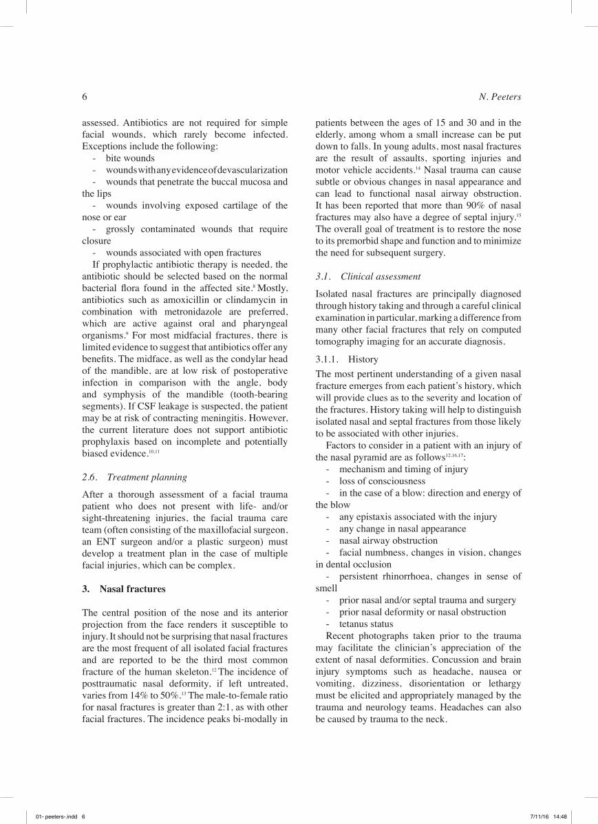
Tripod or zygomaticomaxillary complex fractures are more severe and sustained through direct blunt trauma to the zygomatic buttress of the face. For a complete fracture of the complex, the fracture line must go through its four stability points: a) the frontal bone at the frontozygomatic suture, b) the maxilla at the medial inferior orbital rim, c) at the zygomaticomaxillary buttress and d) the temporal bone at the zygomatic arch (Figure 6).9,18
The treatment of isolated zygomatic arch and zygomatic complex fractures with minimal or no displacements is not surgically managed. A soft food diet can help to avoid secondary fracture displacement. In the case of displacement and minimal comminution of the zygomatic arch fracture, the Gillies technique is the standard reduction treatment used for isolated zygomatic arch fractures. In this approach, a temporal incision
Figure 6Tripod or zygomaticomaxillary complex fracture (Picture from: Buchanan E et al. Zygomaticomaxillary complex fractures and their association with naso-orbito-ethmoid fractures: a 5-year review. Plast Reconstr Surg. 2012: 130; 1296-304)
01- peeters-.indd 14 7/11/16 14:48

Facial trauma 15
Nausea and vomiting are common following eye injuries. Entrapment and ischaemia of the herniated contents of the eye to the sinus can lead to the stimulation of the oculocardic reflex, producing bradycardia, nausea and vomiting.1
5.2. Clinical examination
Common signs of orbital fractures include periocular ecchymosis, diplopia, decreased sensation in the cheek region and orbital emphysema. Physical examination should note crepitus or step deformities on palpation of the facial bones. Examination of the eyelids and surrounding soft tissues may display crepitus, revealing orbital emphysema resulting from a sinus fracture.
When inspecting the globe, the following clinical features indicate an orbital fracture with significant associated injury:
- Exophthalmos/proptosis – orbital haematoma- Telecanthus – disruption of the medial canthal
ligament- Orbital dystopia and/or enophthalmos –
orbital floor fracture- Limitation of extraocular motility – entrapped
muscle- Decreased visual acuity – intraocular injuryPupillary reactivity, size and shape, extraocular
movements and visual acuity should all be evaluated.
5.3. Management
An ophthalmological consultation is necessary during the initial evaluation in the following situations 28:
- Clear or suspected globe injuries. Patients with exophthalmos may have an orbital haematoma, which may require an emergent lateral canthotomy to decompress the orbit.
- Severe vagal symptoms, which are associated with extraocular muscle entrapment. These symptoms, in the absence of intracranial injury, may be caused by the oculocardiac reflex. These symptoms can be relieved by releasing the muscle.
An ophthalmologic consultation is necessary within 24 hours with these injuries:28
- muscle entrapment- enophthalmos or orbital dystopia- naso-orbital-ethmoid fractures with injury
to the medial canthal ligament and/or lacrimal apparatus
- Orbital floor fracture, also known as ‘orbital blowout fracture’ – this fracture typically occurs when a small, round object strikes the eye, causing a sudden increase in intraorbital pressure and in turn creating an outward force that causes the weakest bony structures in the internal orbital walls to fracture. Entrapment of the inferior rectus muscle and/or orbital fat may be observed in this fracture, resulting in diplopia. When the globe is displaced posteriorly in association with an orbital floor fracture and the prolapse of tissue into the maxillary sinus, enophthalmos may develop. Orbital dystopia (where the eye on the affected side is positioned lower on the horizontal plane than the other) can occur because entrapped muscle and orbital fat pull the eye downward. Injury to the infraorbital nerve is possible.
- Orbital roof fracture – this fracture is more common in younger patients, probably because of the high cranium-to-midface ratio of young children.
In paediatric patients, elastic recoil of the orbital floor bone can cause the entrapment and ischaemia of the herniated contents, creating a ‘white-eyed’ blowout fracture that leads to the stimulation of the oculocardiac reflex and results in bradycardia, nausea and vomiting, as well as necrosis of the extra-ocular muscles, stricture and permanent diplopia.
In patients with a facial trauma, sight-threatening pathologies must be identified after the patient’s condition has been stabilized. In patients with an orbital trauma, injuries that threaten the globe must be urgently identified and treated (e.g., ruptured globe, orbital haematoma, optic nerve sheath haematoma).
5.1. History
Key questions for patients who are alert and verbal include:
- Where does it hurt? - Do you have blurry, double or decreased
vision?- Are you having difficulty with eye movement?- Does any part of your face feel numb?Diffuse pain indicates an orbital haematoma,
whereas pain with eye movement suggests injury involving extraocular muscles. Diplopia, particularly with upward gaze, and numbness below the eye, may occur in an orbital blowout fracture.
01- peeters-.indd 15 7/11/16 14:48

16 N. Peeters
inability to bite, limited jaw movement, bleeding, pain, swelling or step deformity at the fracture site. Paraesthesia or anaesthesia of the lower lip can result from damage to the inferior alveolar nerve (V3) within the mandible. Sublingual haematoma is pathognomonic for a mandibular fracture. Dentoalveolar injuries are often observed when the fracture line involves the tooth-bearing regions of the mandible. Almost all mandible fractures involve dentoalveolar segments and are thus open fractures requiring antibiotics.
The ultimate goal of treatment is to restore occlusion (bite), mandibular anatomy and jaw function. With multiple facial fractures, mandibular fractures should be treated first. Mandibular fractures are most commonly treated by (a) conservative observation with modifications to diet/activity; (b) closed reduction with immobilization using mandibulomaxillary fixation (MMF) and (c) more invasive surgical open reduction with an internal fixation approach.9,26,30
Conclusions and take-home messages for ent specialists
The aim of this manuscript was to describe some crucial points concerning facial trauma for both ENT surgeons and clinicians in an emergency care setting. We wanted to emphasize the importance of a systematic clinical assessment of patients with facial trauma. Its conclusions can be summarized in a few take-home messages as follows:
1. Maxillofacial trauma occurs in a significant number of severely injured patients and may be a sign of concomitant life-threatening injuries, which may not be immediately evident upon arrival in the emergency care setting.
2. After the primary survey, which focuses on ruling out life-threatening injuries, attention must be paid to sight-threatening injuries. If necessary, early referral to an appropriate specialist or the initiation of treatment can take place.
3. Clinical examination of the facial trauma patient should be conducted in a systematic way in order to minimize missed injuries.
4. The gold standard for the radiographic evaluation of facial injuries is computed tomography. The additional ability of obtaining three-dimensional reformatted images helps in treatment planning.
The general principles for managing patients with orbital fractures include prophylactic antibiotics for fractures involving a sinus (crepitus in soft tissue) and corticosteroids for patients with limited extraocular movement to decrease swelling. Patients should sleep with the head of the bed elevated and avoid blowing their nose and sniffing.
In the treatment of isolated orbital blowout fractures, the orbital floor is approached via subciliary or transconjunctival incisions.
Linear injuries of the orbital floor without soft tissue entrapment of the orbital structures require no intervention.
The optimal time to repair isolated orbital blowout fractures depends on the signs and symptoms of the patient. Immediate repair should be undertaken for orbital blowout fractures where there is bony impingement on the optic nerve leading to vision loss, where the bony defect is so great as to cause significant immediate asymmetry or enophthalmos, where orbital soft tissue entrapment is causing nausea, vomiting and bradycardia through the oculocardiac reflex, or in the case of ‘white-eyed blowout’ fractures in young patients, where there is little ecchymosis or oedema in a young patient with significantly restricted extraocular movements.29
6. Mandibular fractures
Mandibular fractures are a common form of facial injury in adults and occur most frequently in males. Fractures resulting from violence are most commonly associated with the angle region, while those related to road traffic accidents usually involve the condyle, body and parasymphysis.30
The majority of mandibular fractures occur at more than one site, typically bilaterally, because of the ring structure formed by its articulation at the temporomandibular joint. In addition to the traditional fracture classifications (open, closed, simple, complex or comminuted), mandibular fractures are also described as favourable or unfavourable, depending on whether the mastication muscles tend to reduce or distract the fracture, respectively.
Therefore, when one fracture is identified, a second must be excluded. For example, fractures of the mandibular angle are typically associated with a fracture of the opposite mandibular symphysis or body.
Mandibular fractures may present with a variety of signs and symptoms such as malocclusion, an
01- peeters-.indd 16 7/11/16 14:48

Facial trauma 17
5. Perry M, Dancey A, Mireskandara K, Oakley P, Davies S, Cameron M. Emergency care in facial trauma – a maxillofacial and ophthalmic perspective. Int J Care Injured. 2005;36(8):875-896.
6. Krishnan DG. Systematic assessment of the patient with facial trauma. Oral Maxillofacial Surg Cl N Am. 2013;25(4):537-544.
7. Sun JK, LeMay DR. Imaging of facial trauma. Neuroimaging Clin N Am. 2002;12(2):295-309.
8. Moran GJ, Talan DA, Abrahamian FM. Antimicrobial prophylaxis for wounds and procedures in the emergency department. Infect Dis Clin North Am. 2008;22(1):117-143.
9. Deangelis AF, Barrowman RA, Harrod R, Nastri AL. Review article: Maxillofacial emergencies: maxillofacial trauma. Emerg Med Australas. 2014;26(6):530-537.
10. Andreasen JO, Jensen SS, Schwartz O, Hillerup Y. A systematic review of prophylactic antibiotics in the surgical treatment of maxillofacial fractures. J Oral Maxillofac Surg. 2006;64(11):1664-1668.
11. Ratilal BO, Costa J, Sampaio C, Pappimikail L. Antibiotic prophylaxis for preventing meningitis in patients with basilar skull fractures. Cochrane Database Syst Rev. 2015;(4):CD004884. doi: 10.1002/14651858.CD004884.pub4.
12. Hoffman JF. An algorithm for the initial management of nasal trauma. Facial Plast Surg. 2015;31(3):183-193.
13. Basheeth N, Donnelly M, Smyth D., Shandilya M. Acute nasal fracture management: a prospective study and literature review. Laryngoscope. 2015;125(12):2677-2684.
14. Al-Moraissi EA, Ellis E. Local versus general anesthesia for the management of nasal bone fractures: a systematic review and meta-analysis. J Oral Maxillofac Surg. 2015;73(4):606-615.
15. Rhee SC, Kim YK, Cha JH, Kang SR, Park HS. Septal fracture in simple nasal bone fracture. Plast Reconstr Surg. 2004;113(1):45-52.
16. Mondin V, Rinaldo A, Ferlito A. Management of nasal bone fractures. American journal of otolaryngology. 2005;26(3):181-185.
17. Razavi A, Farboud A, Skinner R. Acute nasal injury. BMJ. 2014;(349):g6537. doi: 10.1136/bmj.g6537.
18. Nigam A, Goni A, Benjamin A, Dasgupta AR. The value of radiographs in the management of fractured nose. Arch Emerg. Med. 1993;10(4):293-297.
19. Bremke M, Wiegand S, Sesterhenn AM, Eken M, Bien S, Werner JA. Digital Volume tomography in the diagnosis of nasal bone fractures. Rhinology. 2009;47(2):126-131.
20. Lee IS, Lee JH, Woo CK, Kim HJ, Sol YL, Song JW, Cho KS. Ultrasonography in the diagnosis of nasal bone fractures: a comparison with conventional radiography and computed tomography. Eur Arch Otorhinolaryngol. 2015;273(2):413-418. doi:10. 1007/s00405-015-3595-8
21. Staffel JG. Optimizing treatment of nasal fractures. Laryngoscope. 2002;112(10):1709-1719.
22. Rubinstein B, Strong EB. Management of nasal fractures. Arch Fam Med. 2000;9(8):738-742.
23. Ridder GJ, Boedeker CC, Fradis M, Schipper J. Technique and timing for closed reduction of isolated nasal fractures: a retrospective study. Ear Nose Throat J. 2002;81(1):49-54.
5. Isolated nasal fractures are principally diagnosed through patient history and clinical examination. Conventional radiography offers only a little clinical benefit. If the history and/or clinical examination is suggestive of fractures of the other facial bones, a CT scan must be performed.
In the literature, there are different opinions about the most appropriate timing of treatment. Most authors argue for the importance of treatment within 10 to 14 days for optimal results. However, manipulation up to five weeks post-trauma is described without alteration to patient satisfaction rates.
Closed reduction is the most commonly performed treatment in isolated nasal fractures, but caution must be exercised in the case of a fractured nasal septum, as this is a predictor of failure of closed reduction.
6. Midface fractures (Le Fort and zygomatic fractures) are often associated with orbital fractures. When globe injuries are suspected, an ophthalmological consultation must be urgently conducted.
Abbreviations
ENT : ear, nose and throat ABC : airway, breathing and circulationATLS : advanced trauma life supportMCT : medial canthal tendonNOE : naso-orbital-ethmoidCT : computed tomography CSF : cerebrospinal fluidUS : ultrasoundDVT : digital volume tomographyLA : local anaesthesiaGA : general anaesthesiaMMF : mandibulomaxillary fixation
References
1. Fonseca RJ, Walker RV, Baxter HD, Powers MP, Frost DE. Oral and maxillofacial trauma. Elsevier, Philadelphia; 2013.
2. Alvi A, Doherty T, Lewen G. Facial fractures and concomitant injuries in trauma patients. Laryngoscope. 2003;113(1):102-106.
3. Perry M. Advanced trauma life support (ATLS) and facial trauma: can one size fit all? Int J Oral Maxillofac Surg. 2008;37(3):209-214.
4. Sertac A, Onur G, Tulin S, Hasan G and Kamil G. Management of midfacial fractures. In: Motamedi M, Ed. A textbook of advanced oral and maxillofacial surgery. Intech, Rijeka - Croatia, 2013:415-445.
01- peeters-.indd 17 7/11/16 14:48

18 N. Peeters
29. Winters R, Chastant R, Graham III HD. When is immediate surgical intervention required for isolated orbital blowout fractures? Laryngoscope. 2014;124(3):585-586.
30. Nasser M, Pandis N, Fleming PS, Fedorowicz Z, Ellis E, Ali K. Interventions for the management of mandibular fractures. Cochrane Database Syst Rev. 2013;(7):CD006087. doi: 10.1002/14651858.CD006087.pub3.
Winde LemmensSchiepse Bos 6 3600 GenkBelgium032/89.60.62.Fax: 089/57.98.79E-mail: [email protected]
24. Rohrich RJ, Adams Jr WP. Nasal fracture management: minimizing secondary nasal deformities. Plast Reconstr Surg. 2000;106(2):266-273.
25. Bell RB, Dierks EJ, Homer L, Potter BE. Management of cerebrospinal fluid leak associated with craniomaxillofacial trauma. J Oral Maxillofac Surg. 2004;62(6):676-684.
26. Stegenga B, Vissink A, De Bont LGM. Mondziekten en kaakchirurgie. Van Gorcum, Assen Nederland; 2000.
27. Schmidt RS, Dodson KM, Goldman RA. Prophylactic antibiotic therapy for fractures of the maxillary sinus. Ear Nose Throat J. 2015;94(4-5):170-177.
28. Richards NQ, Brown NH, Kidwell ED Jr. Visual acuity in orbital floor fractures: does surgical subspecialty management matter? J Craniofac Surg. 2015;26(5):1688-1672.
01- peeters-.indd 18 7/11/16 14:48