axiom - anthogyr.de · 2 237 avenue André-Lasquin - 74700 Sallanches - FRANCE Phone +33 (0)4 50 58...
4
Replacement of a single anterior tooth. Extraction, placement and immediate loading of one axiom ® implant axiom ® clinical case Dr. Christophe FoREsti > Teaching Fellowship at Strasbourg Faculty of Dentistry (France) > Graduated in Oral Implantology > University Degree on Mechanical Engineering in Oral Implantology > University Degree on pre and peri-implant surgery > University Degree on New Dental Technologies > Certificate in Anatomics and Implant Surgery and Advanced Techniques > Certificate in implant-supported denture
Transcript of axiom - anthogyr.de · 2 237 avenue André-Lasquin - 74700 Sallanches - FRANCE Phone +33 (0)4 50 58...

Replacement of a single anterior tooth. Extraction, placement and immediate loading of one axiom® implant
axiom® clinical case
Dr. Christophe FoREsti> Teaching Fellowship at Strasbourg Faculty of Dentistry (France)> Graduated in Oral Implantology> University Degree on Mechanical Engineering in Oral
Implantology> University Degree on pre and peri-implant surgery> University Degree on New Dental Technologies> Certificate in Anatomics and Implant Surgery and
Advanced Techniques> Certificate in implant-supported denture

2
Clinical case presentation
Fig.1: Clinical view of the condemned tooth.
Fig.4: Dental alveolus after extraction.
Fig.7: Drilling.
Fig.2: Extraction of the fractured crown.
Fig.5: Examination of the integrity of bony walls (a vestibular wall is necessary).
Fig.8: Examination of the drilling depth.
Fig.3: Atraumatic extraction of the apical part (periotomes).
Fig.10: Use of the reamer Ø 3.4mm. Fig.11: The axiom® implant is transported to the patient’s mouth. The first 1/3 part of the implant must easily go into the socket.
Fig.6: The pointer drill is used to get palatal anchorage.
Fig.9: Examination of the drilling axis. A palatal axis from the buccal cortical bone can be observed.
The patient is a woman, aged 44. Her maxillary left lateral incisor tooth was fractured.She is a non-smoker and has no contraindications for dental implants.She presents a gingival smile and she has high level aesthetic requirements.
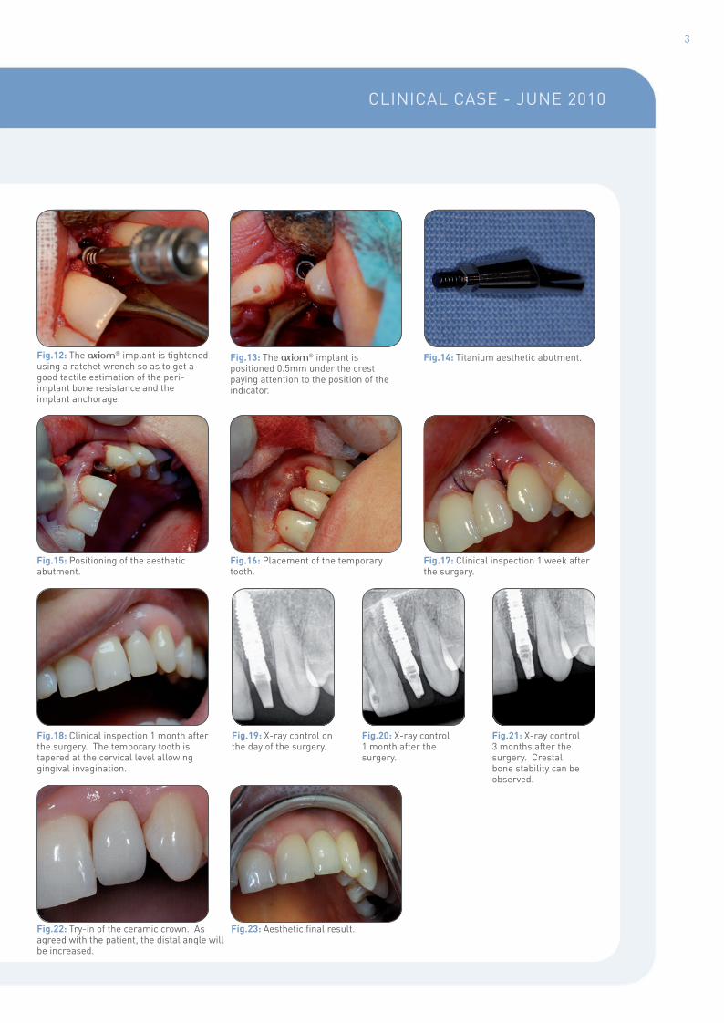
ClINICAl CASE - jUNE 2010
3
Fig.13: The axiom® implant is positioned 0.5mm under the crest paying attention to the position of the indicator.
Fig.16: Placement of the temporary tooth.
Fig.19: X-ray control on the day of the surgery.
Fig.14: Titanium aesthetic abutment.
Fig.17: Clinical inspection 1 week after the surgery.
Fig.20: X-ray control 1 month after the surgery.
Fig.12: The axiom® implant is tightened using a ratchet wrench so as to get a good tactile estimation of the peri-implant bone resistance and the implant anchorage.
Fig.15: Positioning of the aesthetic abutment.
Fig.18: Clinical inspection 1 month after the surgery. The temporary tooth is tapered at the cervical level allowing gingival invagination.
Fig.22: Try-in of the ceramic crown. As agreed with the patient, the distal angle will be increased.
Fig.23: Aesthetic final result.
Fig.21: X-ray control 3 months after the surgery. Crestal bone stability can be observed.

2 237 avenue André-Lasquin - 74700 Sallanches - FRANCEPhone +33 (0)4 50 58 02 37 - Fax +33 (0)4 50 93 78 60
www.anthogyr.com
Tissue preservation Minimally invasiveProsthetics easy to use Comprehensive
axiom®
The new dimension
w One single connection.w Intuitive prosthetic interlocking.w User-friendly protocol.w Implant-holder free.w Convenient and compact kit.w Compatible with the ANTHOGYR Guiding System.
Pho
tos
cred
it : A
ntho
gyr
- D
r. C
hris
toph
e Fo
rest
i - A
ll ri
ght r
eser
ved
© Anthogyr - CC5AGB-0910