Axilla & brachial plexus
22
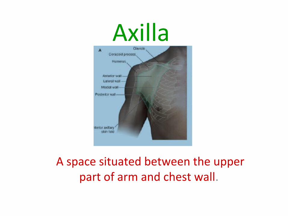
Axilla A space situated between the upper part of arm and chest wall .
-
Upload
shaifaly-rustagi -
Category
Education
-
view
463 -
download
0
Transcript of Axilla & brachial plexus
Axilla
A space situated between the upper part of arm and chest wall.
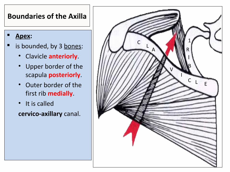
Boundaries of the Axilla
Apex: is bounded, by 3 bones:
• Clavicle anteriorly.• Upper border of the
scapula posteriorly.• Outer border of the
first rib medially.• It is called
cervico-axillary canal.
CL
A
VI
C L E
1
RIB
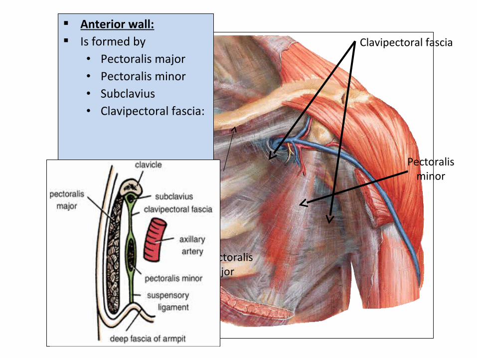
Anterior wall: Is formed by
• Pectoralis major• Pectoralis minor• Subclavius• Clavipectoral fascia:
Pectoralis major
Pectoralis minor
Clavipectoral fascia
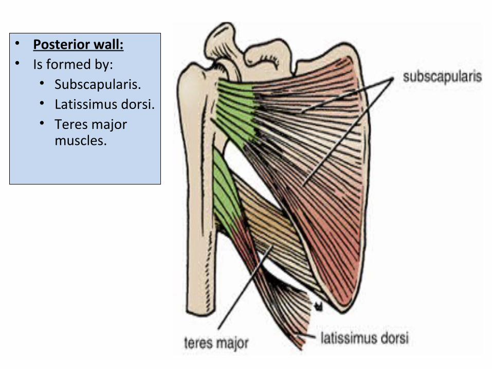
• Posterior wall:• Is formed by:
• Subscapularis.• Latissimus dorsi.• Teres major
muscles.
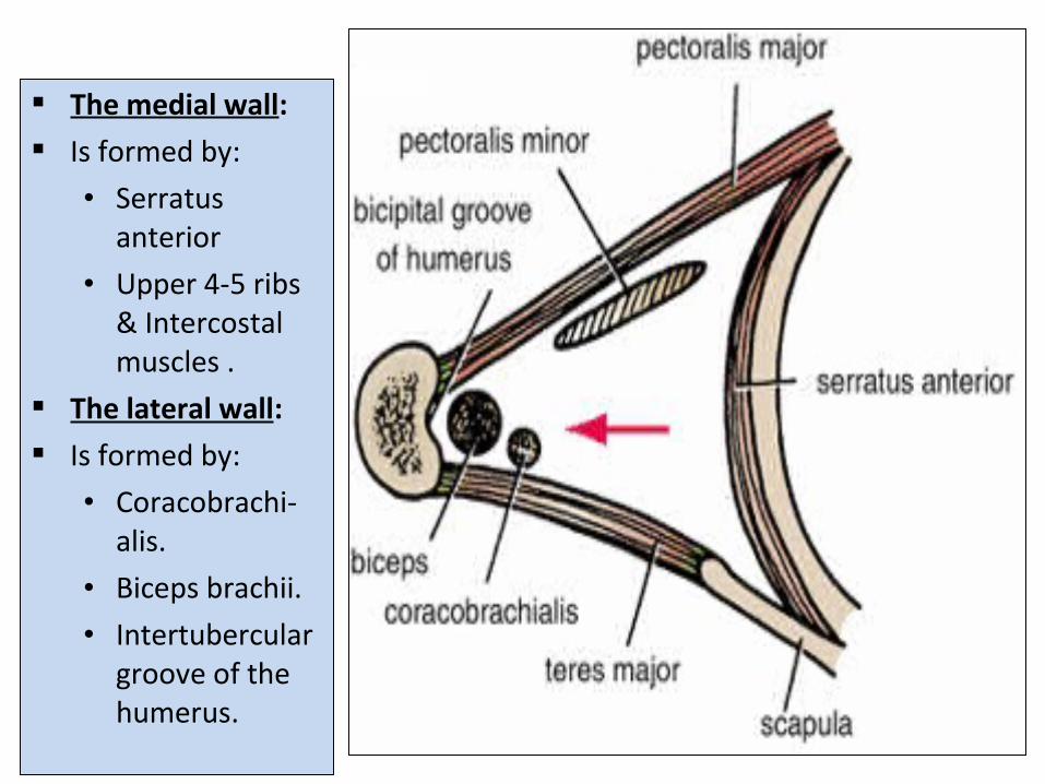
The medial wall: Is formed by:
• Serratus anterior
• Upper 4-5 ribs & Intercostal muscles .
The lateral wall: Is formed by:
• Coracobrachi-alis.
• Biceps brachii.
• Intertubercular groove of the humerus.
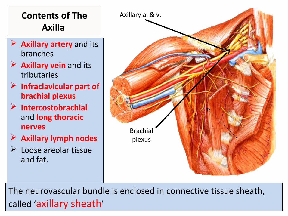
Contents of The Axilla
Axillary artery and its branches
Axillary vein and its tributaries
Infraclavicular part of brachial plexus
Intercostobrachial and long thoracic nerves
Axillary lymph nodes Loose areolar tissue
and fat.
The neurovascular bundle is enclosed in connective tissue sheath, called ‘axillary sheath’
Axillary a. & v.
Brachial plexus
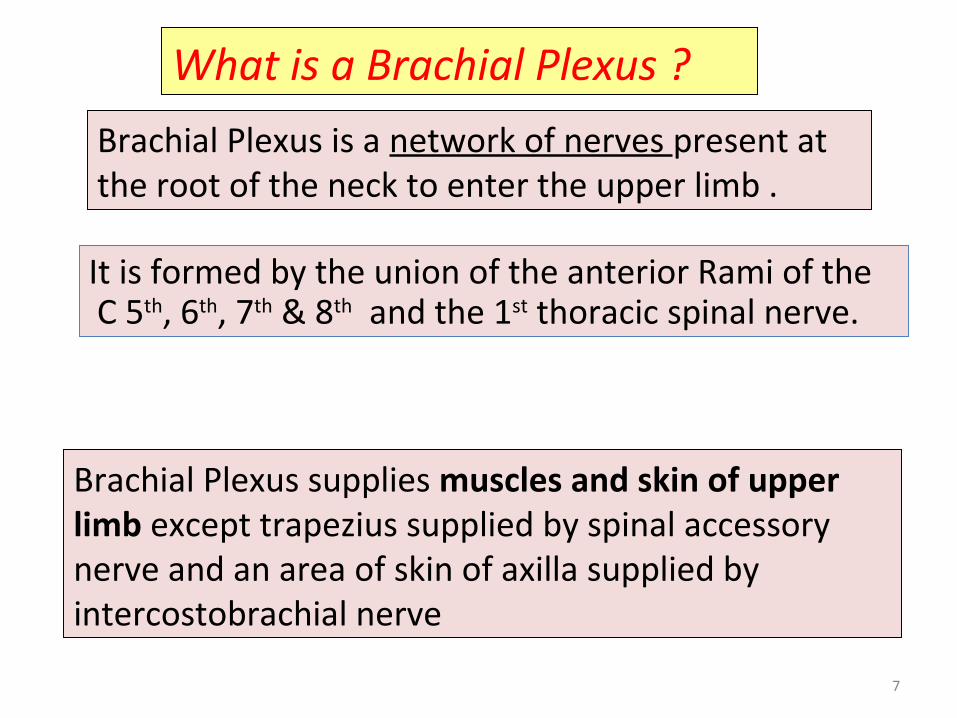
What is a Brachial Plexus ?
Brachial Plexus is a network of nerves present at the root of the neck to enter the upper limb .
Brachial Plexus supplies muscles and skin of upper limb except trapezius supplied by spinal accessory nerve and an area of skin of axilla supplied by intercostobrachial nerve
7
It is formed by the union of the anterior Rami of the C 5th, 6th, 7th & 8th and the 1st thoracic spinal nerve.
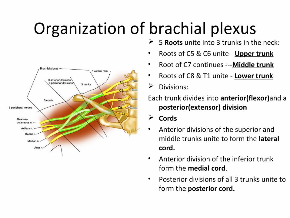
Organization of brachial plexus 5 Roots unite into 3 trunks in the neck: • Roots of C5 & C6 unite - Upper trunk• Root of C7 continues ---Middle trunk• Roots of C8 & T1 unite - Lower trunk Divisions:
Each trunk divides into anterior(flexor)and a posterior(extensor) division
Cords• Anterior divisions of the superior and
middle trunks unite to form the lateral cord.
• Anterior division of the inferior trunk form the medial cord.
• Posterior divisions of all 3 trunks unite to form the posterior cord.
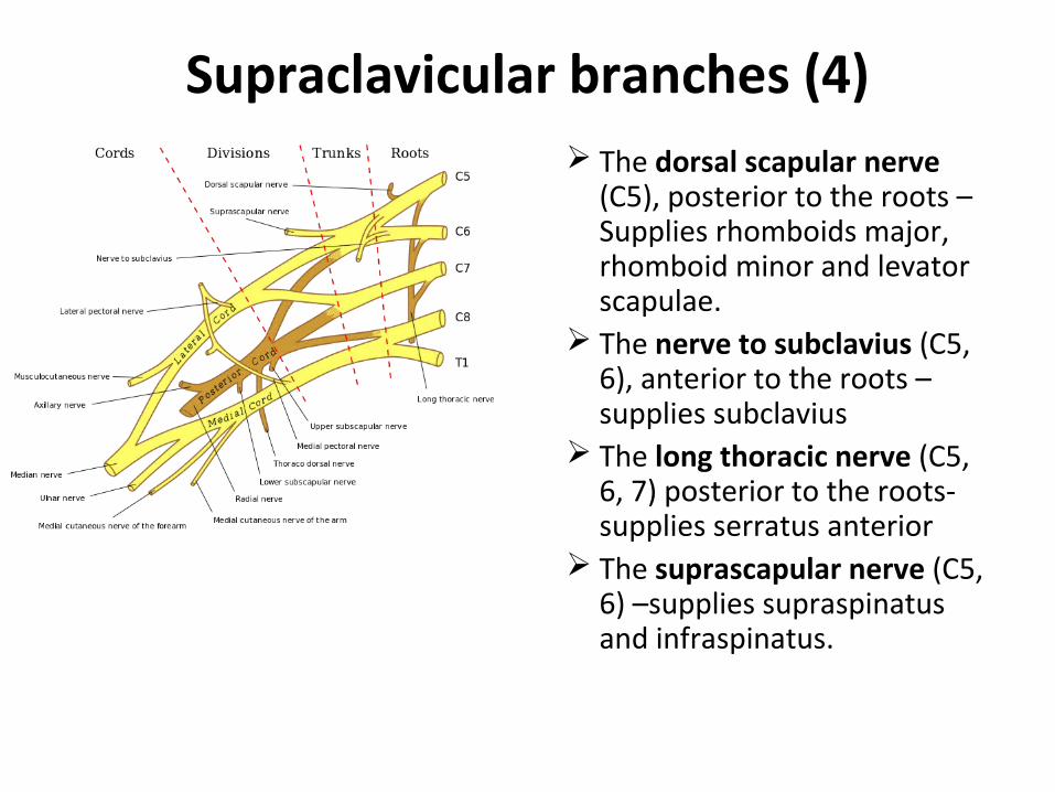
Supraclavicular branches (4) The dorsal scapular nerve
(C5), posterior to the roots –Supplies rhomboids major, rhomboid minor and levator scapulae.
The nerve to subclavius (C5, 6), anterior to the roots –supplies subclavius
The long thoracic nerve (C5, 6, 7) posterior to the roots-supplies serratus anterior
The suprascapular nerve (C5, 6) –supplies supraspinatus and infraspinatus.
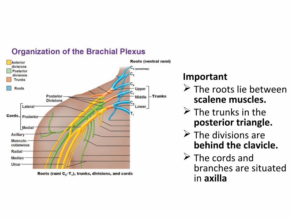
Important The roots lie between
scalene muscles. The trunks in the
posterior triangle. The divisions are
behind the clavicle. The cords and
branches are situated in axilla
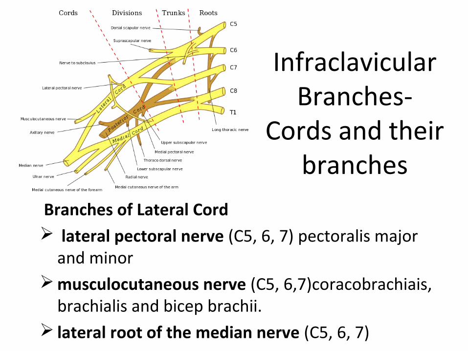
Infraclavicular Branches-
Cords and their branches
Branches of Lateral Cord lateral pectoral nerve (C5, 6, 7) pectoralis major
and minor musculocutaneous nerve (C5, 6,7)coracobrachiais,
brachialis and bicep brachii. lateral root of the median nerve (C5, 6, 7)
.
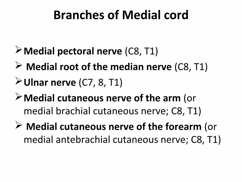
Branches of Medial cord
Medial pectoral nerve (C8, T1) Medial root of the median nerve (C8, T1) Ulnar nerve (C7, 8, T1)Medial cutaneous nerve of the arm (or
medial brachial cutaneous nerve; C8, T1) Medial cutaneous nerve of the forearm (or
medial antebrachial cutaneous nerve; C8, T1)
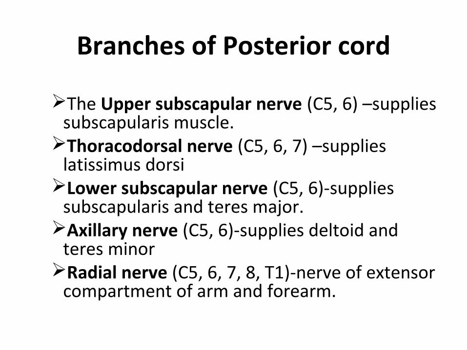
Branches of Posterior cord
The Upper subscapular nerve (C5, 6) –supplies subscapularis muscle.
Thoracodorsal nerve (C5, 6, 7) –supplies latissimus dorsi
Lower subscapular nerve (C5, 6)-supplies subscapularis and teres major.
Axillary nerve (C5, 6)-supplies deltoid and teres minor
Radial nerve (C5, 6, 7, 8, T1)-nerve of extensor compartment of arm and forearm.
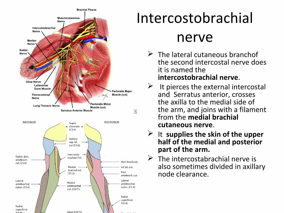
Intercostobrachial nerve
The lateral cutaneous branchof the second intercostal nerve does it is named the intercostobrachial nerve.
It pierces the external intercostal and Serratus anterior, crosses the axilla to the medial side of the arm, and joins with a filament from the medial brachial cutaneous nerve.
It supplies the skin of the upper half of the medial and posterior part of the arm.
The intercostabrachial nerve is also sometimes divided in axillary node clearance.
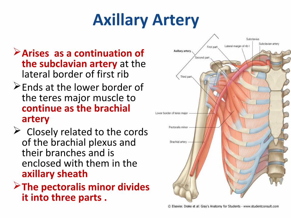
Axillary Artery
Arises as a continuation of the subclavian artery at the lateral border of first rib
Ends at the lower border of the teres major muscle to continue as the brachial artery
Closely related to the cords of the brachial plexus and their branches and is enclosed with them in the axillary sheath
The pectoralis minor divides it into three parts .
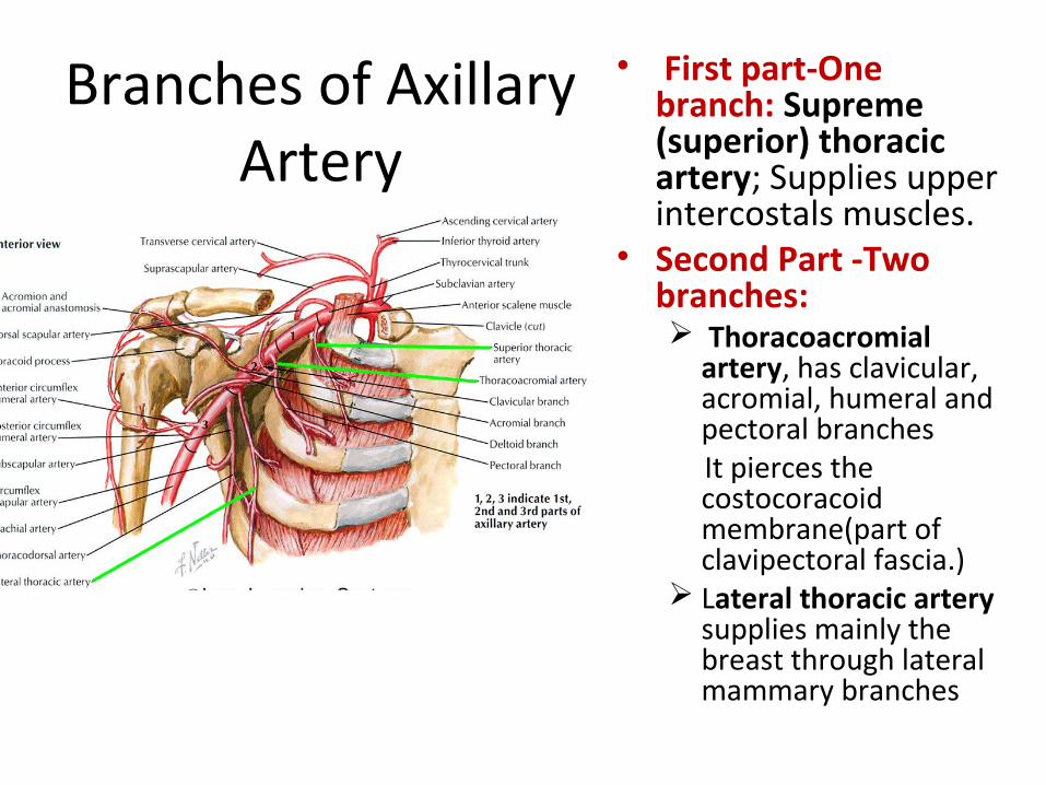
Branches of Axillary Artery
• First part-One branch: Supreme (superior) thoracic artery; Supplies upper intercostals muscles.
• Second Part -Two branches: Thoracoacromial
artery, has clavicular, acromial, humeral and pectoral branches
It pierces the costocoracoid membrane(part of clavipectoral fascia.)
Lateral thoracic artery supplies mainly the breast through lateral mammary branches
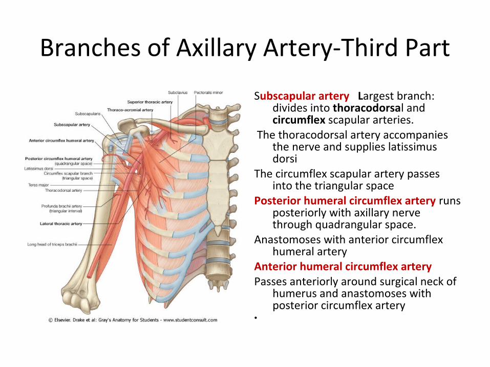
Branches of Axillary Artery-Third Part
Subscapular artery Largest branch: divides into thoracodorsal and circumflex scapular arteries.
The thoracodorsal artery accompanies the nerve and supplies latissimus dorsi
The circumflex scapular artery passes into the triangular space
Posterior humeral circumflex artery runs posteriorly with axillary nerve through quadrangular space.
Anastomoses with anterior circumflex humeral artery
Anterior humeral circumflex artery Passes anteriorly around surgical neck of
humerus and anastomoses with posterior circumflex artery
•
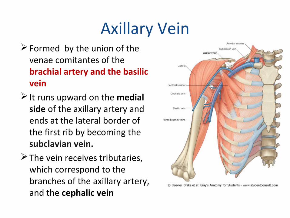
Axillary VeinFormed by the union of the
venae comitantes of the brachial artery and the basilic vein
It runs upward on the medial side of the axillary artery and ends at the lateral border of the first rib by becoming the subclavian vein.
The vein receives tributaries, which correspond to the branches of the axillary artery, and the cephalic vein
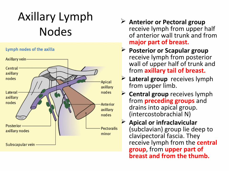
Axillary Lymph Nodes
Anterior or Pectoral group receive lymph from upper half of anterior wall trunk and from major part of breast.
Posterior or Scapular group receive lymph from posterior wall of upper half of trunk and from axillary tail of breast.
Lateral group receives lymph from upper limb.
Central group receives lymph from preceding groups and drains into apical group. (intercostobrachial N)
Apical or infraclavicular (subclavian) group lie deep to clavipectoral fascia. They receive lymph from the central group, from upper part of breast and from the thumb.
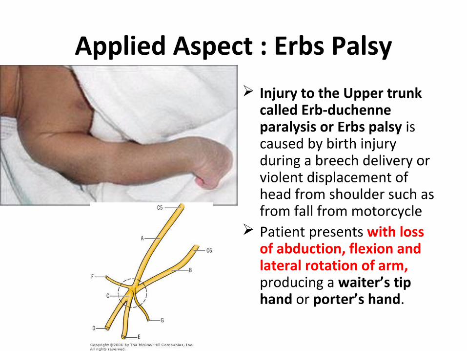
Applied Aspect : Erbs Palsy
Injury to the Upper trunk called Erb-duchenne paralysis or Erbs palsy is caused by birth injury during a breech delivery or violent displacement of head from shoulder such as from fall from motorcycle
Patient presents with loss of abduction, flexion and lateral rotation of arm, producing a waiter’s tip hand or porter’s hand.
Applied aspect: The Cervical rib
A cervical rib represents a persistent ossification of the C7 lateral costal element.
The presence of a cervical rib can cause compression of the lower trunk of the brachial plexus or subclavian artery.
Symptoms are weakness of the muscles around the muscles in the hand, near the base of the thumb.
Compression of the subclavian artery is often diagnosed by finding a positive Adson's sign on examination, where the radial pulse in the arm is lost during abduction and external rotation of the shoulder.
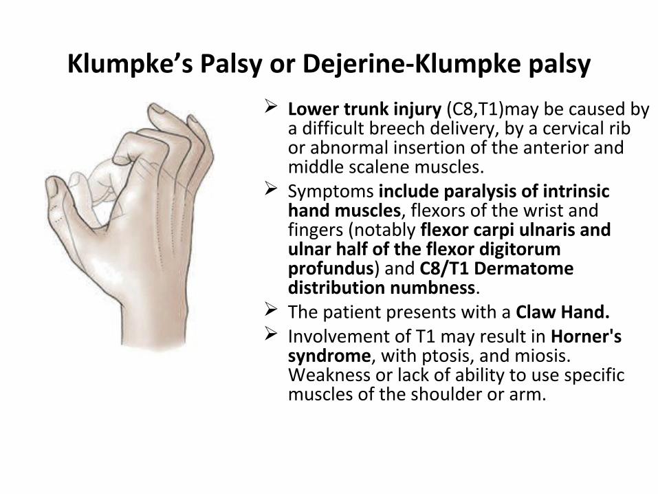
Klumpke’s Palsy or Dejerine-Klumpke palsy Lower trunk injury (C8,T1)may be caused by
a difficult breech delivery, by a cervical rib or abnormal insertion of the anterior and middle scalene muscles.
Symptoms include paralysis of intrinsic hand muscles, flexors of the wrist and fingers (notably flexor carpi ulnaris and ulnar half of the flexor digitorum profundus) and C8/T1 Dermatome distribution numbness.
The patient presents with a Claw Hand. Involvement of T1 may result in Horner's
syndrome, with ptosis, and miosis. Weakness or lack of ability to use specific muscles of the shoulder or arm.