Skeletal Bones and Features Axial Skeleton Skull, Spine, Thoracic Cage.
description
Skull F i l bCranium
Thoracic cage(ribs and
Facial bonesClavicleScapulaSternum(ribs and
sternum)
Vertebralcolumn
SternumRibHumerusVertebraRadiusUlSacrum
column UlnaCarpals
PhalangesPhalangesMetacarpalsFemurPatellaTibiaFibula
Tarsals
Copyright © 2010 Pearson Education, Inc. Figure 7.1a
(a) Anterior viewMetatarsalsPhalanges
AXIAL SKELETON- consists of 80 bones
Consists of 3 MAJOR REGIONS
SKULL (29)8 cranial bones + 14 facial bones + 6 auditory ossicles + 1 hyoid
VERTEBRAL COLUMN (26)7 cervical + 12 thoracic + 5 lumbar vertebrae + 1 sacrum + 1 coccyx
THORACIC CAGE (25)1 sternum + 24 ribs
Upper Limb
Lower Limb
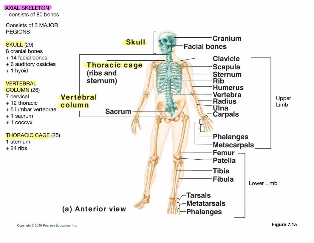
Bones of cranium (cranial vault)
Coronal
Squamoust
Coronalsuture
suture
Lambdoid Facialsuture bones
Copyright © 2010 Pearson Education, Inc.
Cranial and facial divisions of the skull
- framework of the face- CAVITIES for special sense organs for sight, taste, and smell- OPENINGS for air and food passage- SITES of ATTACHMENT for teeth and muscles of facial expression
FACIAL BONES
CRANIAL BONES
- Enclose BRAIN in CRANIAL CAVITY(Cranial vault = calvaria)(Cranial base = anterior,middle, and posteriorcranial fossae)
- Provides SITES ofATTACHMENT forhead and neck muscles
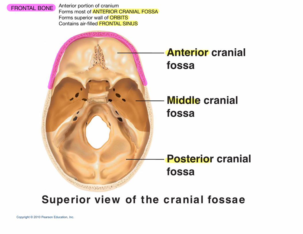
Anterior cranialfossa
Middle cranialMiddle cranialfossa
Posterior cranialPosterior cranialfossa
Copyright © 2010 Pearson Education, Inc.
Superior view of the cranial fossae
FRONTAL BONE Anterior portion of craniumForms most of ANTERIOR CRANIAL FOSSAForms superior wall of ORBITSContains air-filled FRONTAL SINUS
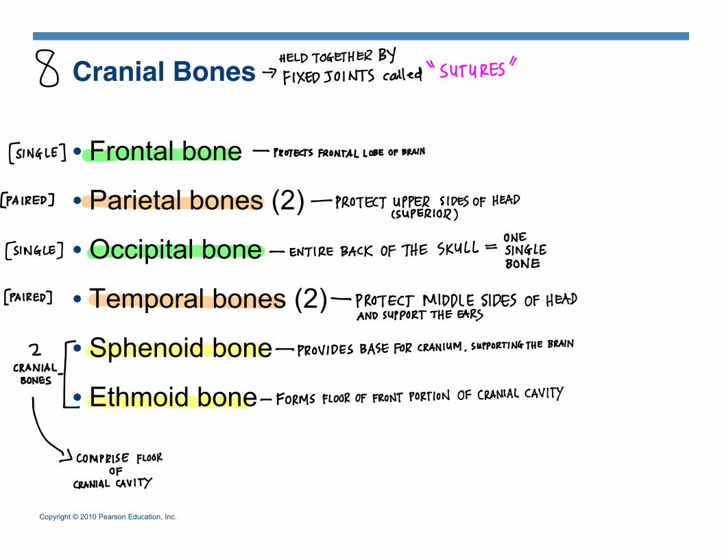
Cranial Bones
• Frontal bone
• Parietal bones (2)
O i it l b• Occipital bone
• Temporal bones (2)p ( )
• Sphenoid bone
• Ethmoid bone
Copyright © 2010 Pearson Education, Inc.
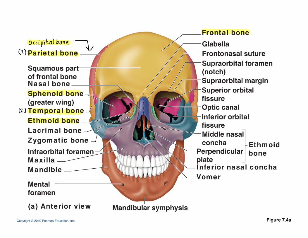
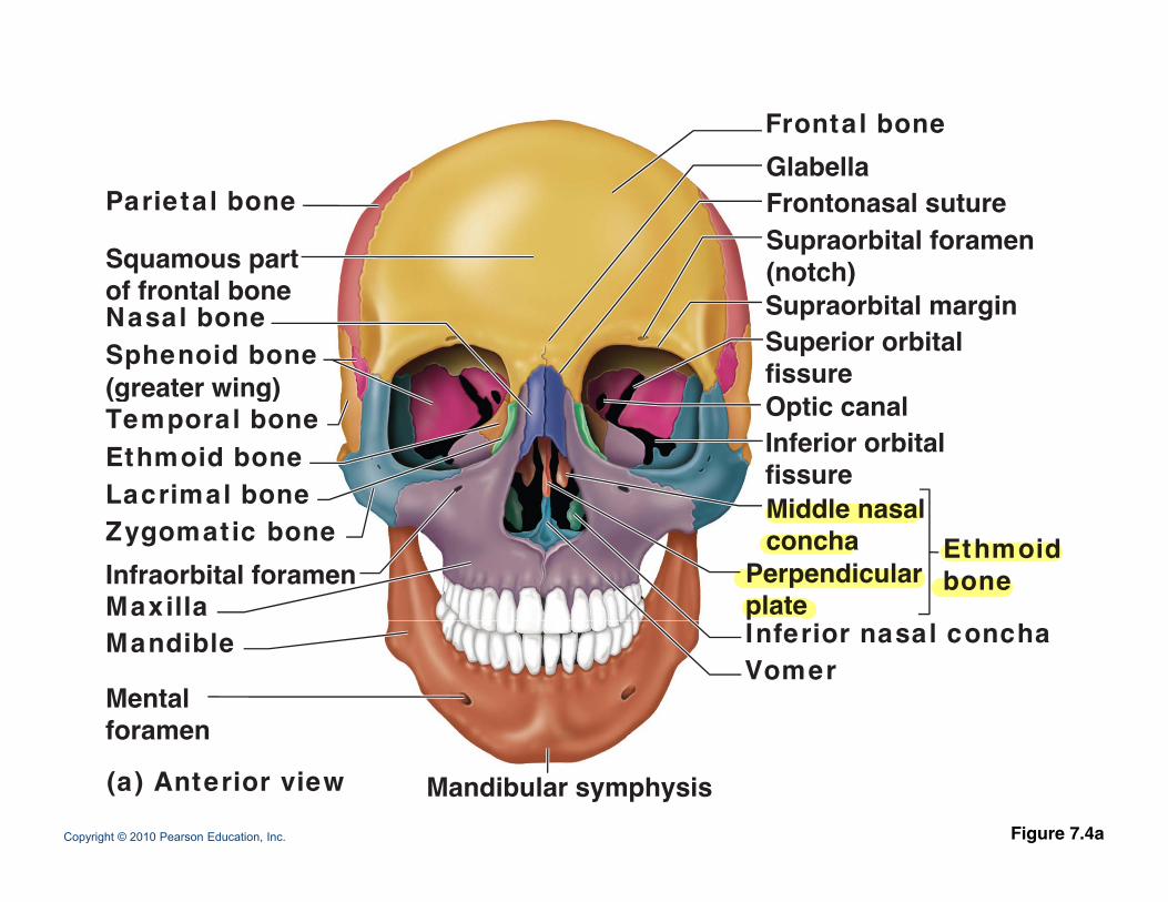
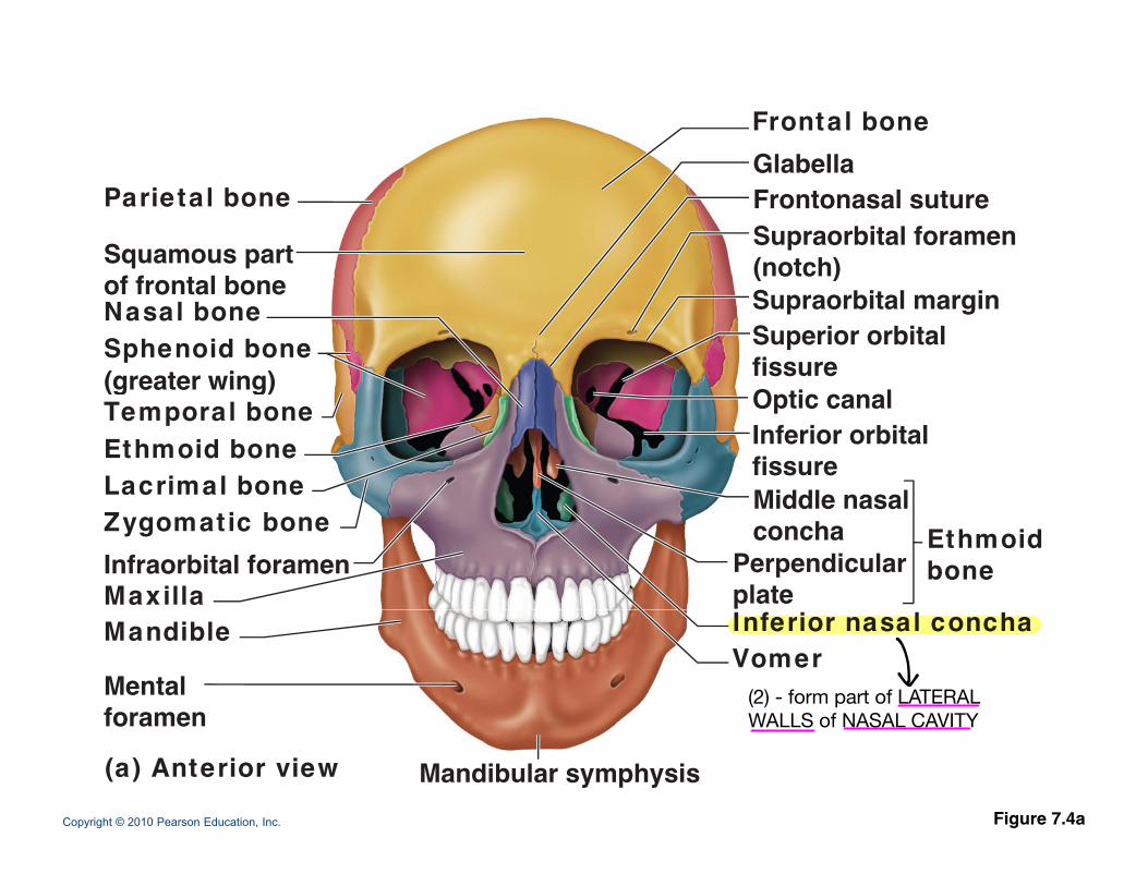
Frontal boneGlabella
Parietal bone
Squamous part
GlabellaFrontonasal sutureSupraorbital foramen(notch)of frontal bone
Nasal boneSphenoid bone(greater wing)
(notch)Supraorbital marginSuperior orbitalfissure(greater wing)
Temporal boneEthmoid boneLacrimal bone
Inferior orbitalfissureMiddle nasal
Optic canal
Zygomatic bone
MaxillaInfraorbital foramen
Middle nasalconcha
Perpendicularplate
Ethmoidbone
Mandible
Mentalforamen
Inferior nasal conchaVomer
Copyright © 2010 Pearson Education, Inc. Figure 7.4a
foramen
(a) Anterior view Mandibular symphysis
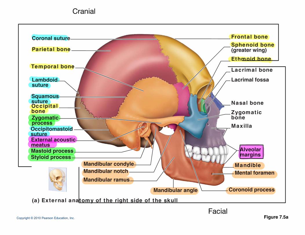
Coronal suture Frontal boneS h id bSphenoid bone(greater wing)Ethmoid bone
Lacrimal bone
Parietal bone
Temporal bone
Lacrimal fossa
Nasal bone
Lambdoidsuture
Squamoussuture Nasal bone
Zygomaticbone Maxilla
sutureOccipitalbone
Occipitomastoid
Zygomaticprocess
Alveolarmargins
sutureExternal acousticmeatusMastoid processStyloid process
MandibleMental foramen
Mandibular condyleMandibular notchMandibular ramus
Mandibular angle Coronoid process
Copyright © 2010 Pearson Education, Inc.
(a) External anatomy of the right side of the skull
Mandibular angle Coronoid process
Figure 7.5a
Cranial
Facial
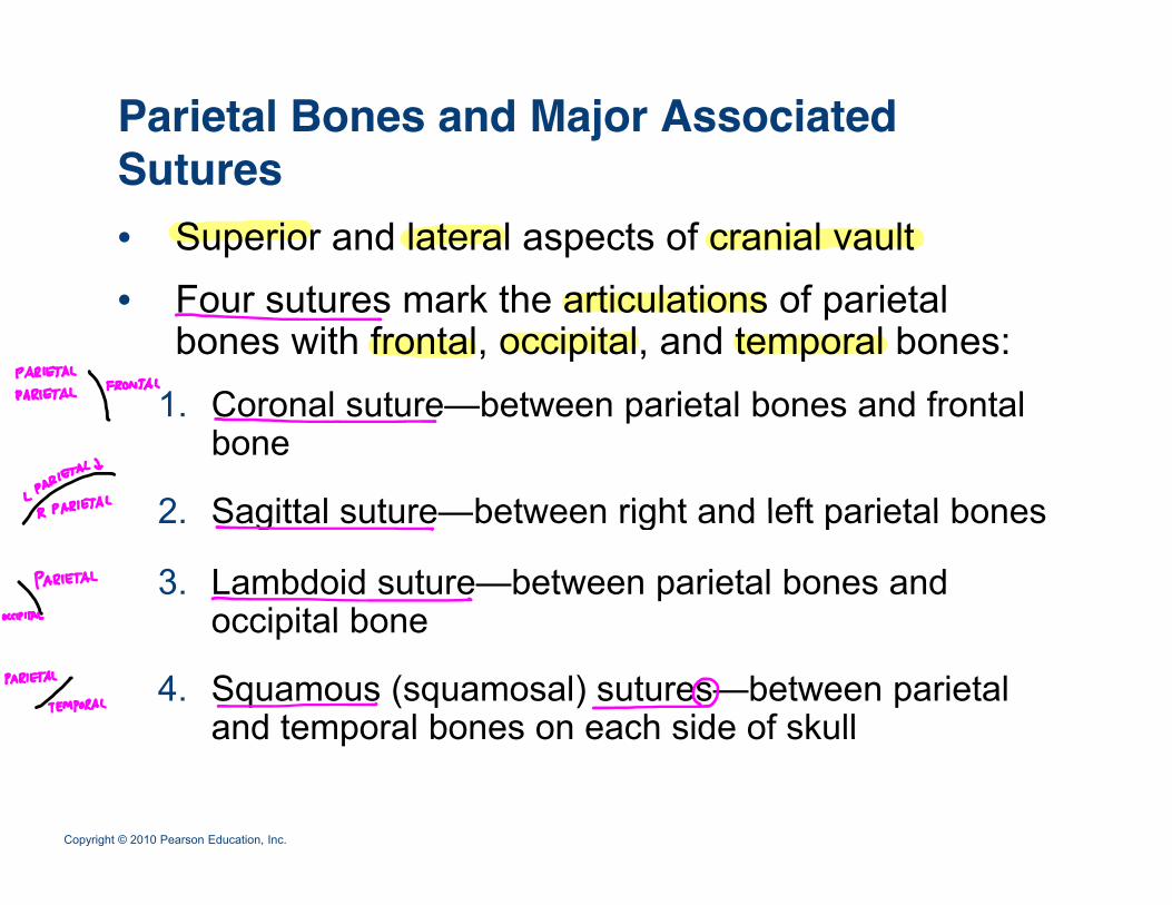
Parietal Bones and Major Associated SuturesSutures• Superior and lateral aspects of cranial vault• Four sutures mark the articulations of parietal
bones with frontal, occipital, and temporal bones:1 C l t b t i t l b d f t l1. Coronal suture—between parietal bones and frontal
bone
2 Sagittal suture between right and left parietal bones2. Sagittal suture—between right and left parietal bones
3. Lambdoid suture—between parietal bones and occipital boneoccipital bone
4. Squamous (squamosal) sutures—between parietal and temporal bones on each side of skull
Copyright © 2010 Pearson Education, Inc.
and temporal bones on each side of skull
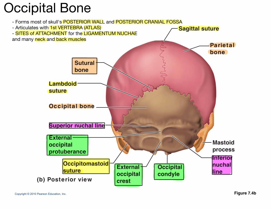
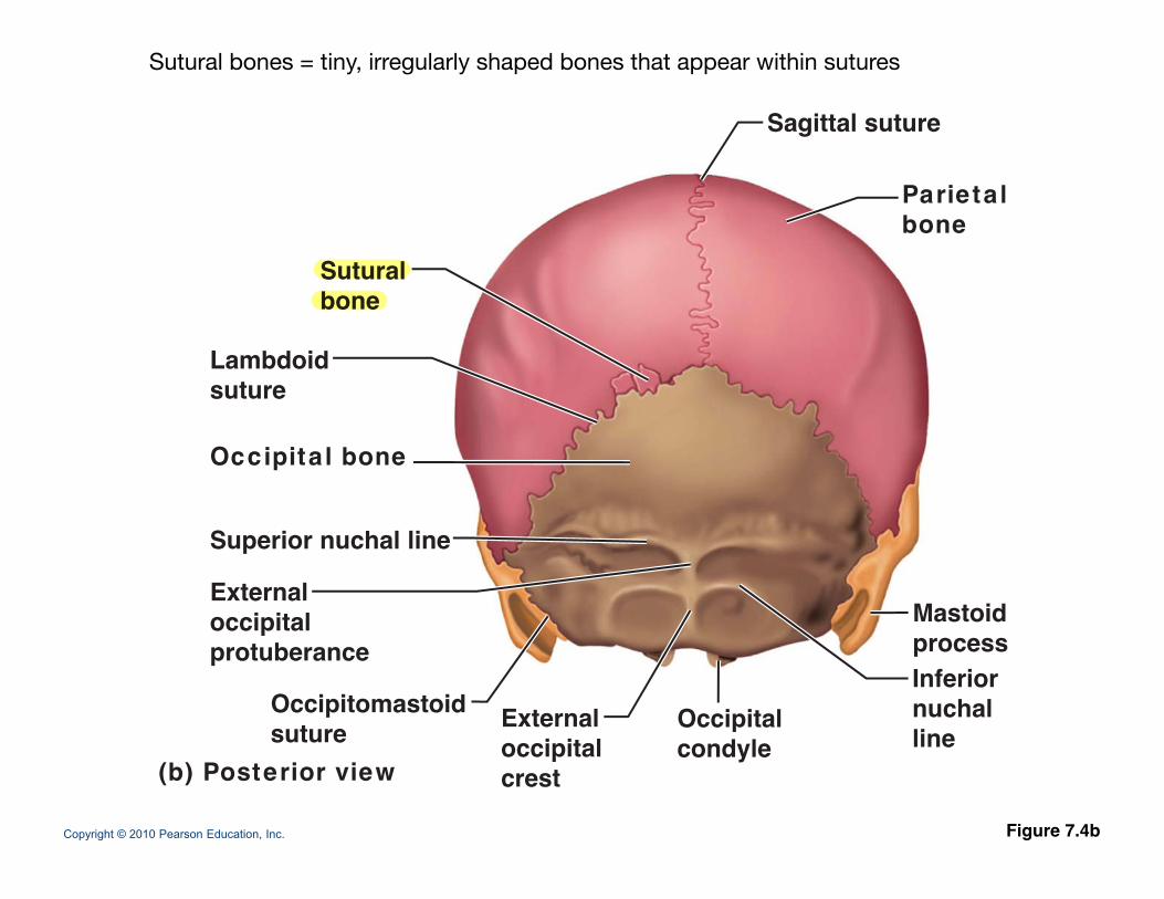
Sagittal suture
Sutural
Parietalbone
Lambdoidsuture
Suturalbone
suture
Occipita l bone
Superior nuchal line
Externaloccipital Mastoidoccipitalprotuberance
Occipitomastoidsuture OccipitalExternal
Inferiornuchalli
Mastoidprocess
Copyright © 2010 Pearson Education, Inc. Figure 7.4b
suture(b) Posterior view
condyleoccipitalcrest
line
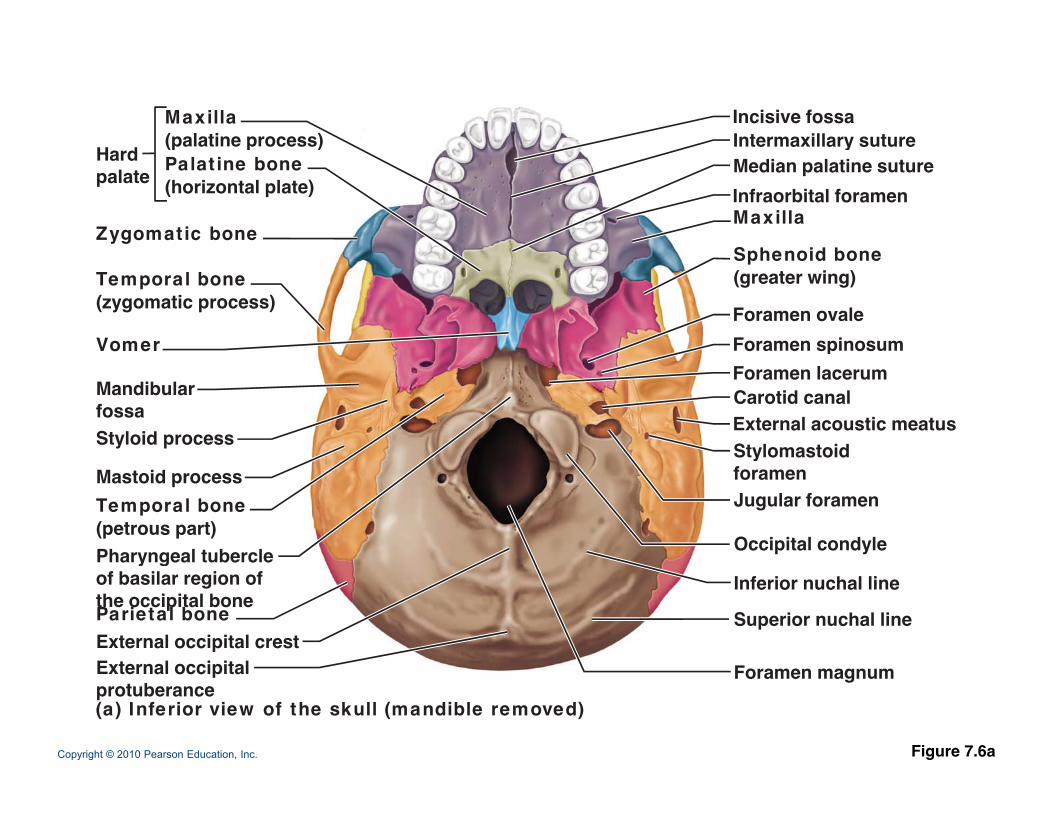
Occipital Bone- Forms most of skull's POSTERIOR WALL and POSTERIOR CRANIAL FOSSA- Articulates with 1st VERTEBRA (ATLAS)- SITES of ATTACHMENT for the LIGAMENTUM NUCHAE and many neck and back muscles
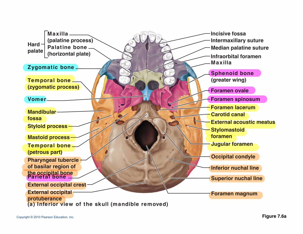
Incisive fossaIntermaxillary suture
Maxilla(palatine process)
H d Median palatine suturey
Infraorbital foramenMaxilla
Sphenoid bone
(p p )Hardpalate
Zygomatic bone
Palat ine bone(horizontal plate)
Sphenoid bone(greater wing)
Foramen ovaleForamen spinosum
Temporal bone(zygomatic process)
VomerForamen lacerumCarotid canalExternal acoustic meatusStylomastoid
MandibularfossaStyloid process Stylomastoid
foramenJugular foramen
Occipital condyle
Mastoid processTemporal bone(petrous part)Pharyngeal tubercle
Inferior nuchal line
Superior nuchal lineExternal occipital crestE t l i it l
Pharyngeal tubercleof basilar region ofthe occipital boneParietal bone
Copyright © 2010 Pearson Education, Inc. Figure 7.6a
Foramen magnumExternal occipitalprotuberance(a) Inferior view of the skull (mandible removed)
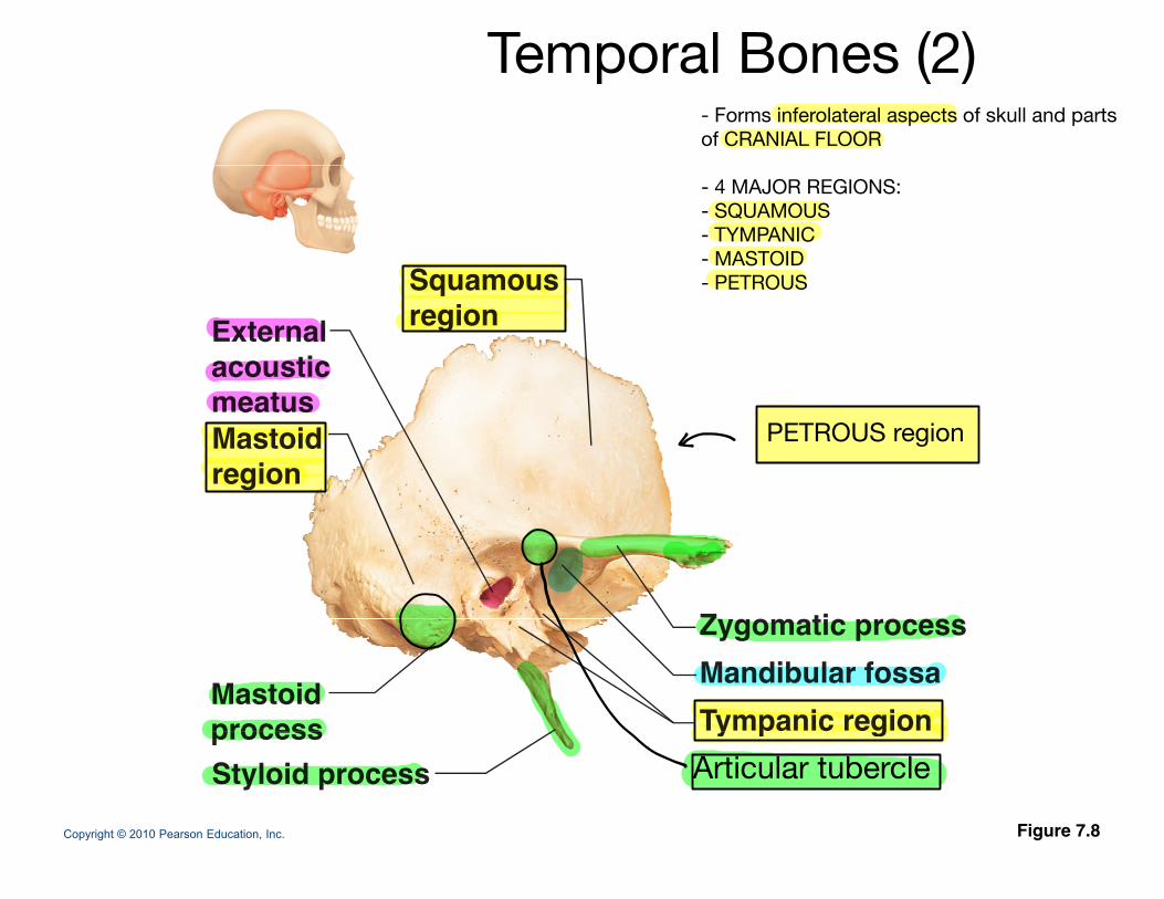
SquamousExternalacoustic
t
Squamousregion
Mastoidregion
meatus
Z ti
Mastoidprocess Tympanic region
Mandibular fossaZygomatic process
Copyright © 2010 Pearson Education, Inc. Figure 7.8
process Styloid process
y p g
Temporal Bones (2)
Articular tubercle
- Forms inferolateral aspects of skull and parts of CRANIAL FLOOR
- 4 MAJOR REGIONS:- SQUAMOUS- TYMPANIC- MASTOID- PETROUS
PETROUS region
Incisive fossaIntermaxillary suture
Maxilla(palatine process)
H d Median palatine suturey
Infraorbital foramenMaxilla
Sphenoid bone
(p p )Hardpalate
Zygomatic bone
Palat ine bone(horizontal plate)
Sphenoid bone(greater wing)
Foramen ovaleForamen spinosum
Temporal bone(zygomatic process)
VomerForamen lacerumCarotid canalExternal acoustic meatusStylomastoid
MandibularfossaStyloid process Stylomastoid
foramenJugular foramen
Occipital condyle
Mastoid processTemporal bone(petrous part)Pharyngeal tubercle
Inferior nuchal line
Superior nuchal lineExternal occipital crestE t l i it l
Pharyngeal tubercleof basilar region ofthe occipital boneParietal bone
Copyright © 2010 Pearson Education, Inc. Figure 7.6a
Foramen magnumExternal occipitalprotuberance(a) Inferior view of the skull (mandible removed)
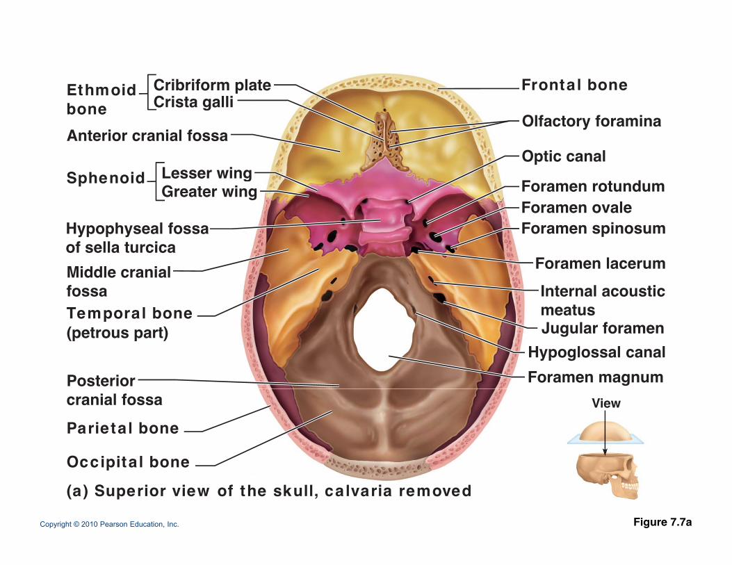
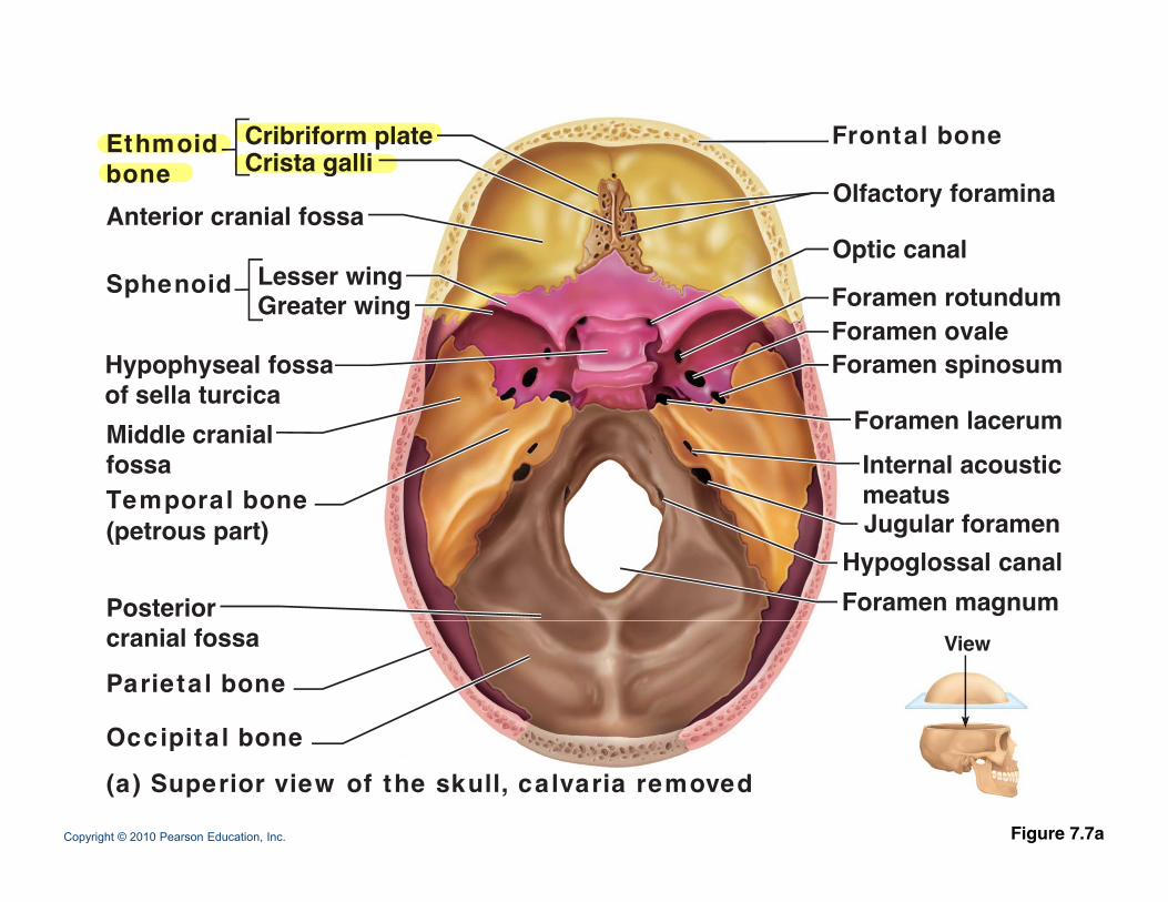
Frontal boneCribriform plateEthmoidb Crista galli
Olfactory foramina
Optic canal
bone Crista galli
Sphenoid
Anterior cranial fossa
Lesser wing
Hypophyseal fossaof sella turcica
Foramen rotundumForamen ovaleForamen spinosum
Sphenoid Lesser wingGreater wing
of sella turcicaMiddle cranialfossaTemporal bone
Foramen lacerumInternal acousticmeatus Temporal bone
(petrous part)
Posterior Foramen magnum
Jugular foramenHypoglossal canal
cranial fossa
Parietal bone
Occipita l bone
View
Copyright © 2010 Pearson Education, Inc. Figure 7.7a
Occipita l bone
(a) Superior view of the skull, calvaria removed
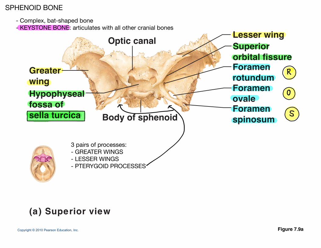
SuperiorOptic canal Lesser wing
Greater Foramenrotundum
Superiororbital fissure
p
wingHypophysealfossa of
rotundumForamenovaleForamen
sella turcicaForamenspinosumBody of sphenoid
Copyright © 2010 Pearson Education, Inc. Figure 7.9a
(a) Superior view
SPHENOID BONE- Complex, bat-shaped bone- KEYSTONE BONE: articulates with all other cranial bones
3 pairs of processes:- GREATER WINGS- LESSER WINGS- PTERYGOID PROCESSES
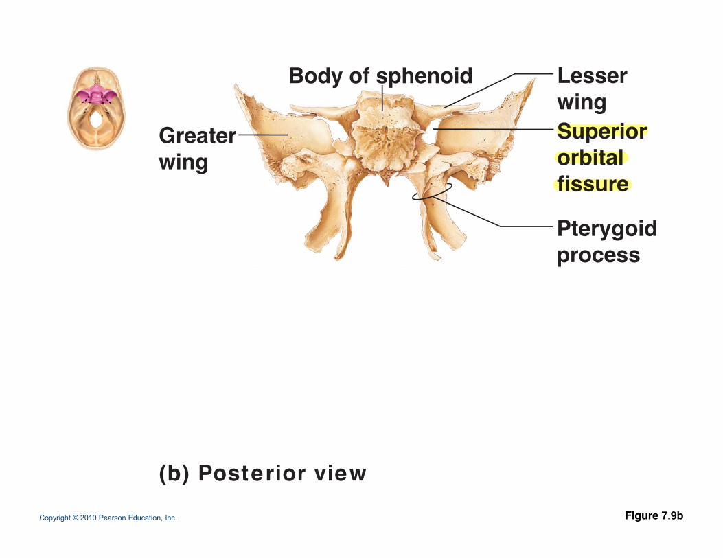
Body of sphenoid Lesserwing
Greaterwing
Superiororbital
wing
gfissure
Pterygoidprocess
Copyright © 2010 Pearson Education, Inc. Figure 7.9b
(b) Posterior view
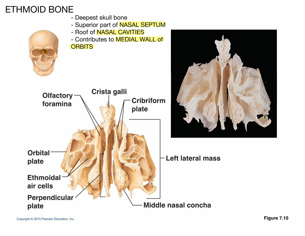
CribriformOlfactoryforamina
Crista galliCribriformplate
foramina
Orbitalplate Left lateral massplate
Ethmoidalair cells
Copyright © 2010 Pearson Education, Inc. Figure 7.10
Perpendicularplate Middle nasal concha
ETHMOID BONE- Deepest skull bone- Superior part of NASAL SEPTUM- Roof of NASAL CAVITIES- Contributes to MEDIAL WALL of ORBITS
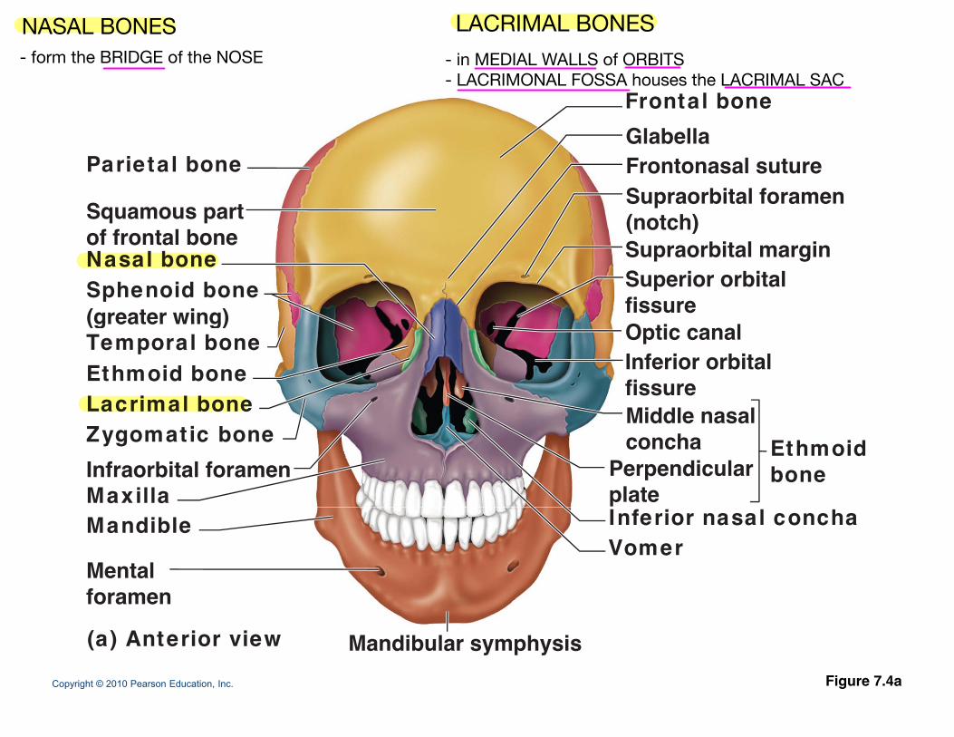
Frontal boneGlabella
Parietal bone
Squamous part
GlabellaFrontonasal sutureSupraorbital foramen(notch)of frontal bone
Nasal boneSphenoid bone(greater wing)
(notch)Supraorbital marginSuperior orbitalfissure(greater wing)
Temporal boneEthmoid boneLacrimal bone
Inferior orbitalfissureMiddle nasal
Optic canal
Zygomatic bone
MaxillaInfraorbital foramen
Middle nasalconcha
Perpendicularplate
Ethmoidbone
Mandible
Mentalforamen
Inferior nasal conchaVomer
Copyright © 2010 Pearson Education, Inc. Figure 7.4a
foramen
(a) Anterior view Mandibular symphysis
Frontal boneCribriform plateEthmoidb Crista galli
Olfactory foramina
Optic canal
bone Crista galli
Sphenoid
Anterior cranial fossa
Lesser wing
Hypophyseal fossaof sella turcica
Foramen rotundumForamen ovaleForamen spinosum
Sphenoid Lesser wingGreater wing
of sella turcicaMiddle cranialfossaTemporal bone
Foramen lacerumInternal acousticmeatus Temporal bone
(petrous part)
Posterior Foramen magnum
Jugular foramenHypoglossal canal
cranial fossa
Parietal bone
Occipita l bone
View
Copyright © 2010 Pearson Education, Inc.
Occipita l bone
(a) Superior view of the skull, calvaria removed
Figure 7.7a
Sagittal suture
Sutural
Parietalbone
Lambdoidsuture
Suturalbone
suture
Occipita l bone
Superior nuchal line
Externaloccipital Mastoidoccipitalprotuberance
Occipitomastoidsuture OccipitalExternal
Inferiornuchalli
Mastoidprocess
Copyright © 2010 Pearson Education, Inc. Figure 7.4b
suture(b) Posterior view
condyleoccipitalcrest
line
Sutural bones = tiny, irregularly shaped bones that appear within sutures
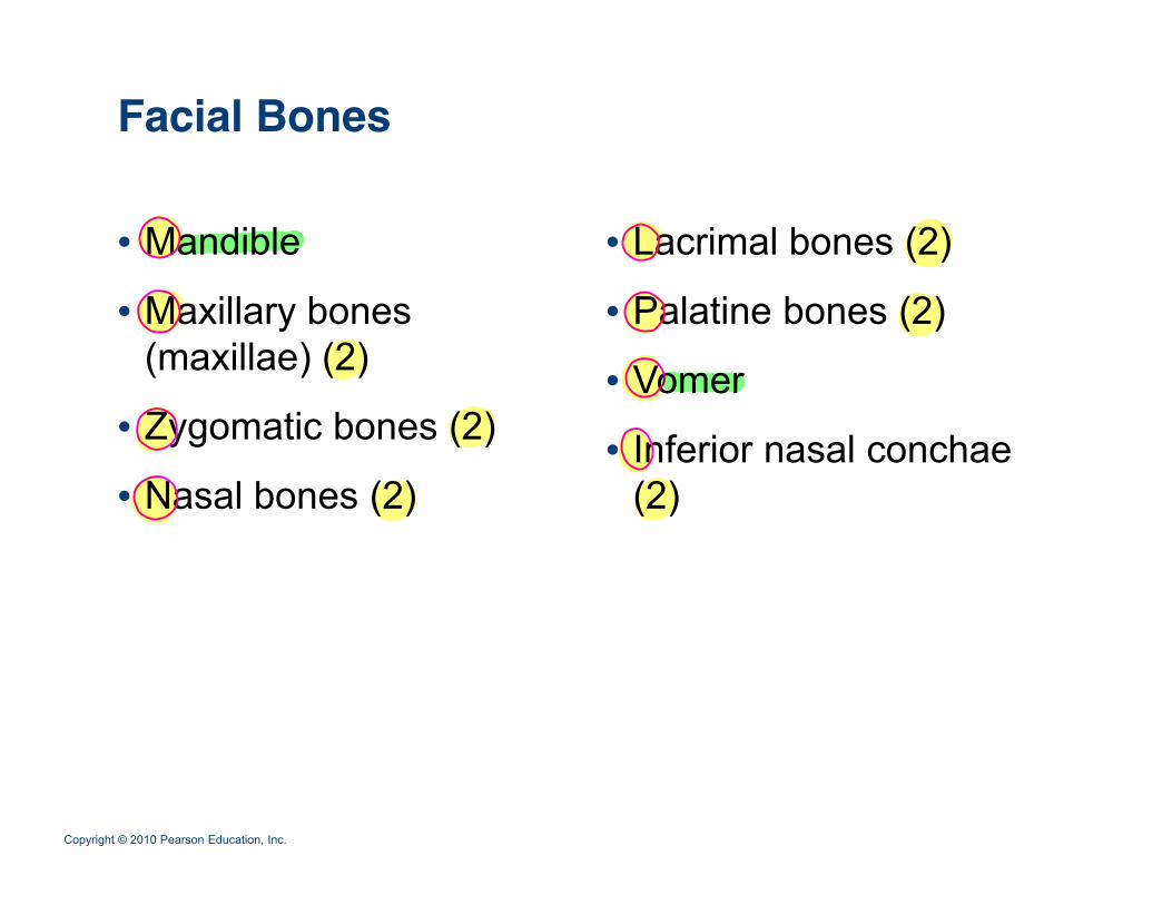
Facial Bones
• Mandible • Lacrimal bones (2)
• Maxillary bones (maxillae) (2)
• Palatine bones (2)
• Vomer• Zygomatic bones (2)
• Nasal bones (2)• Inferior nasal conchae
(2)asa bo es ( ) ( )
Copyright © 2010 Pearson Education, Inc.
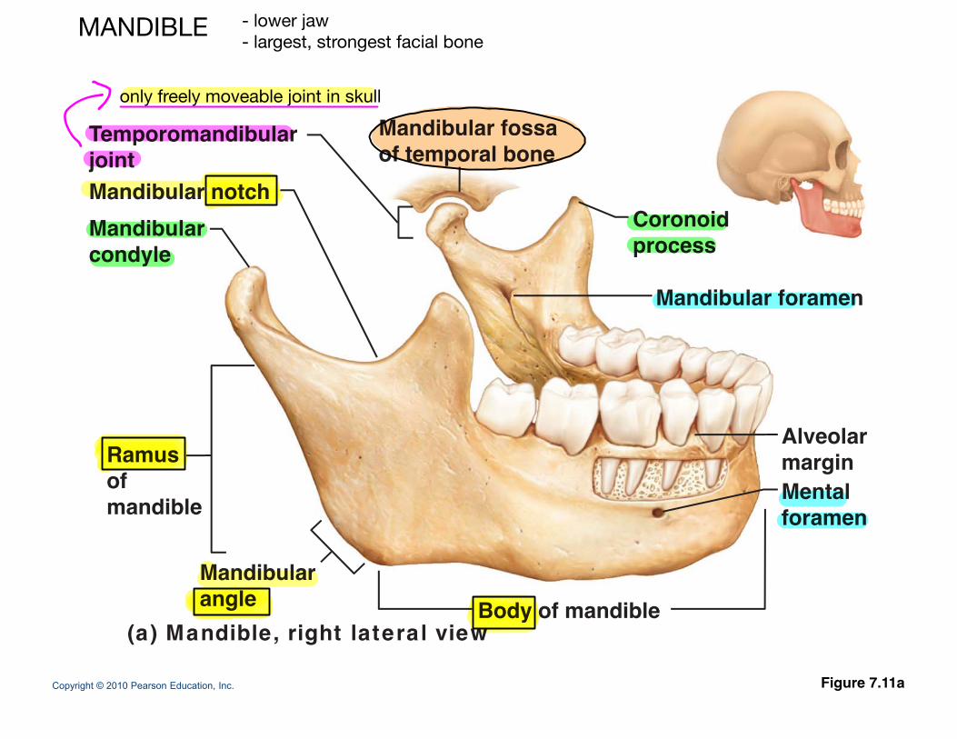
Mandibular fossaTemporomandibular
CoronoidMandibularMandibular notch
of temporal boneTemporomandibularjoint
process
Mandibular foramen
Mandibularcondyle
MentalRamusofmandible
Alveolarmargin
foramen
Mandibularangle
mandible
Copyright © 2010 Pearson Education, Inc. Figure 7.11a
angle Body of mandible(a) Mandible, right lateral view
MANDIBLE - lower jaw- largest, strongest facial bone
only freely moveable joint in skull
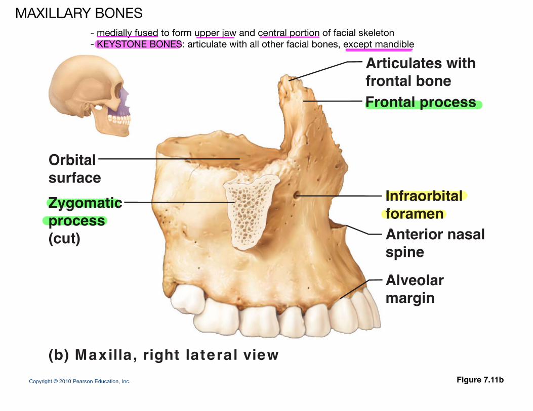
Articulates withf t l bFrontal processfrontal bone
Orbitalsurface
A t i l
Infraorbitalforamen
surface Zygomaticprocess
Anterior nasalspine
Alveolar
(cut)
Alveolarmargin
Copyright © 2010 Pearson Education, Inc. Figure 7.11b
(b) Maxilla , right lateral view
MAXILLARY BONES- medially fused to form upper jaw and central portion of facial skeleton- KEYSTONE BONES: articulate with all other facial bones, except mandible
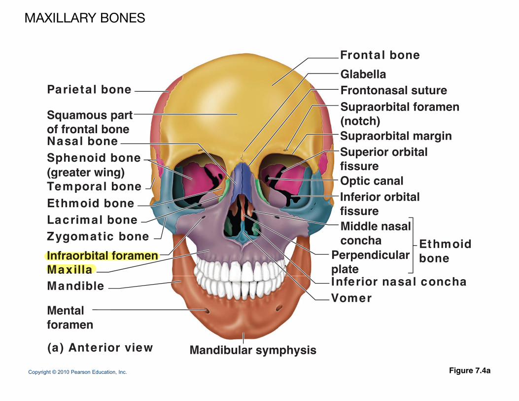
Frontal boneGlabella
Parietal bone
Squamous part
GlabellaFrontonasal sutureSupraorbital foramen(notch)of frontal bone
Nasal boneSphenoid bone(greater wing)
(notch)Supraorbital marginSuperior orbitalfissure(greater wing)
Temporal boneEthmoid boneLacrimal bone
Inferior orbitalfissureMiddle nasal
Optic canal
Zygomatic bone
MaxillaInfraorbital foramen
Middle nasalconcha
Perpendicularplate
Ethmoidbone
Mandible
Mentalforamen
Inferior nasal conchaVomer
Copyright © 2010 Pearson Education, Inc. Figure 7.4a
foramen
(a) Anterior view Mandibular symphysis
MAXILLARY BONES
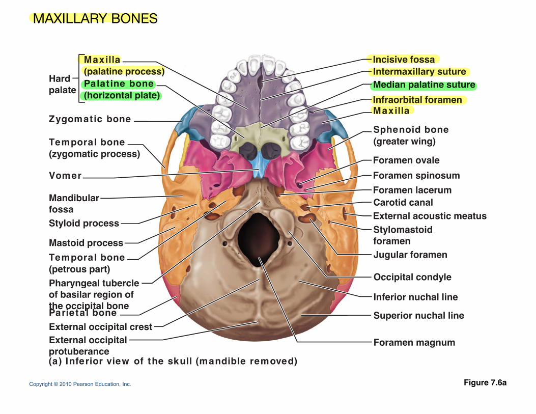
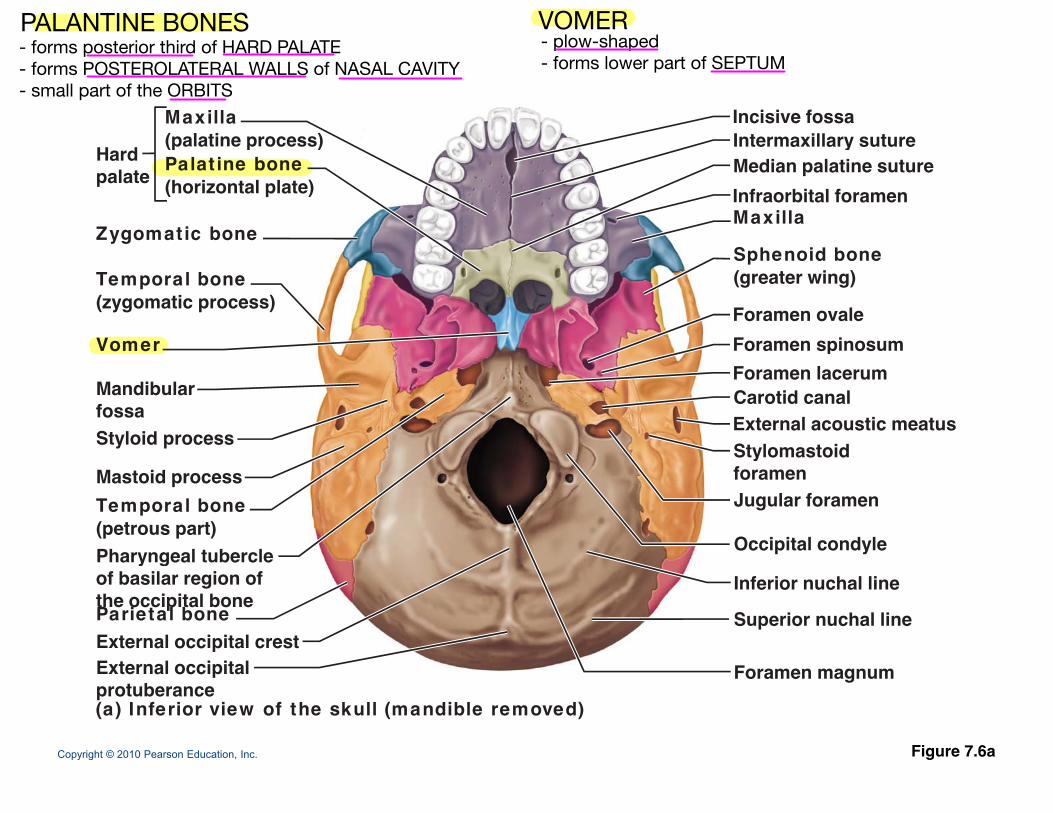
Incisive fossaIntermaxillary suture
Maxilla(palatine process)
H d Median palatine suturey
Infraorbital foramenMaxilla
Sphenoid bone
(p p )Hardpalate
Zygomatic bone
Palat ine bone(horizontal plate)
Sphenoid bone(greater wing)
Foramen ovaleForamen spinosum
Temporal bone(zygomatic process)
VomerForamen lacerumCarotid canalExternal acoustic meatusStylomastoid
MandibularfossaStyloid process Stylomastoid
foramenJugular foramen
Occipital condyle
Mastoid processTemporal bone(petrous part)Pharyngeal tubercle
Inferior nuchal line
Superior nuchal lineExternal occipital crestE t l i it l
Pharyngeal tubercleof basilar region ofthe occipital boneParietal bone
Copyright © 2010 Pearson Education, Inc. Figure 7.6a
Foramen magnumExternal occipitalprotuberance(a) Inferior view of the skull (mandible removed)
MAXILLARY BONES
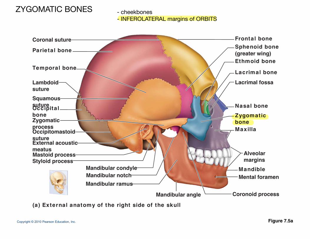
Coronal suture Frontal boneSphenoid bone(greater wing)Ethmoid bone
Lacrimal bone
Parietal bone
Temporal boneLacrimal bone
Lacrimal fossa
N l b
LambdoidsutureSquamoussuture Nasal bone
Zygomaticbone Maxilla
sutureOccipitalbone
Occipitomastoid
Zygomaticprocess
Alveolarmargins
psutureExternal acousticmeatusMastoid processStyloid process
MandibleMental foramen
Mandibular condyleMandibular notchMandibular ramus
Mandibular angle Coronoid process
Copyright © 2010 Pearson Education, Inc. Figure 7.5a
(a) External anatomy of the right side of the skull
Mandibular angle Coronoid process
ZYGOMATIC BONES - cheekbones- INFEROLATERAL margins of ORBITS
Frontal boneGlabella
Parietal bone
Squamous part
GlabellaFrontonasal sutureSupraorbital foramen(notch)of frontal bone
Nasal boneSphenoid bone(greater wing)
(notch)Supraorbital marginSuperior orbitalfissure(greater wing)
Temporal boneEthmoid boneLacrimal bone
Inferior orbitalfissureMiddle nasal
Optic canal
Zygomatic bone
MaxillaInfraorbital foramen
Middle nasalconcha
Perpendicularplate
Ethmoidbone
Mandible
Mentalforamen
Inferior nasal conchaVomer
Copyright © 2010 Pearson Education, Inc. Figure 7.4a
foramen
(a) Anterior view Mandibular symphysis
NASAL BONES - form the BRIDGE of the NOSE - in MEDIAL WALLS of ORBITS
- LACRIMONAL FOSSA houses the LACRIMAL SAC
LACRIMAL BONES
Incisive fossaIntermaxillary suture
Maxilla(palatine process)
H d Median palatine suturey
Infraorbital foramenMaxilla
Sphenoid bone
(p p )Hardpalate
Zygomatic bone
Palat ine bone(horizontal plate)
Sphenoid bone(greater wing)
Foramen ovaleForamen spinosum
Temporal bone(zygomatic process)
VomerForamen lacerumCarotid canalExternal acoustic meatusStylomastoid
MandibularfossaStyloid process Stylomastoid
foramenJugular foramen
Occipital condyle
Mastoid processTemporal bone(petrous part)Pharyngeal tubercle
Inferior nuchal line
Superior nuchal lineExternal occipital crestE t l i it l
Pharyngeal tubercleof basilar region ofthe occipital boneParietal bone
Copyright © 2010 Pearson Education, Inc. Figure 7.6a
Foramen magnumExternal occipitalprotuberance(a) Inferior view of the skull (mandible removed)
PALANTINE BONES VOMER- forms posterior third of HARD PALATE- forms POSTEROLATERAL WALLS of NASAL CAVITY- small part of the ORBITS
- plow-shaped- forms lower part of SEPTUM
Frontal boneGlabella
Parietal bone
Squamous part
GlabellaFrontonasal sutureSupraorbital foramen(notch)of frontal bone
Nasal boneSphenoid bone(greater wing)
(notch)Supraorbital marginSuperior orbitalfissure(greater wing)
Temporal boneEthmoid boneLacrimal bone
Inferior orbitalfissureMiddle nasal
Optic canal
Zygomatic bone
MaxillaInfraorbital foramen
Middle nasalconcha
Perpendicularplate
Ethmoidbone
Mandible
Mentalforamen
Inferior nasal conchaVomer
Copyright © 2010 Pearson Education, Inc. Figure 7.4a
foramen
(a) Anterior view Mandibular symphysis
(2) - form part of LATERAL WALLS of NASAL CAVITY
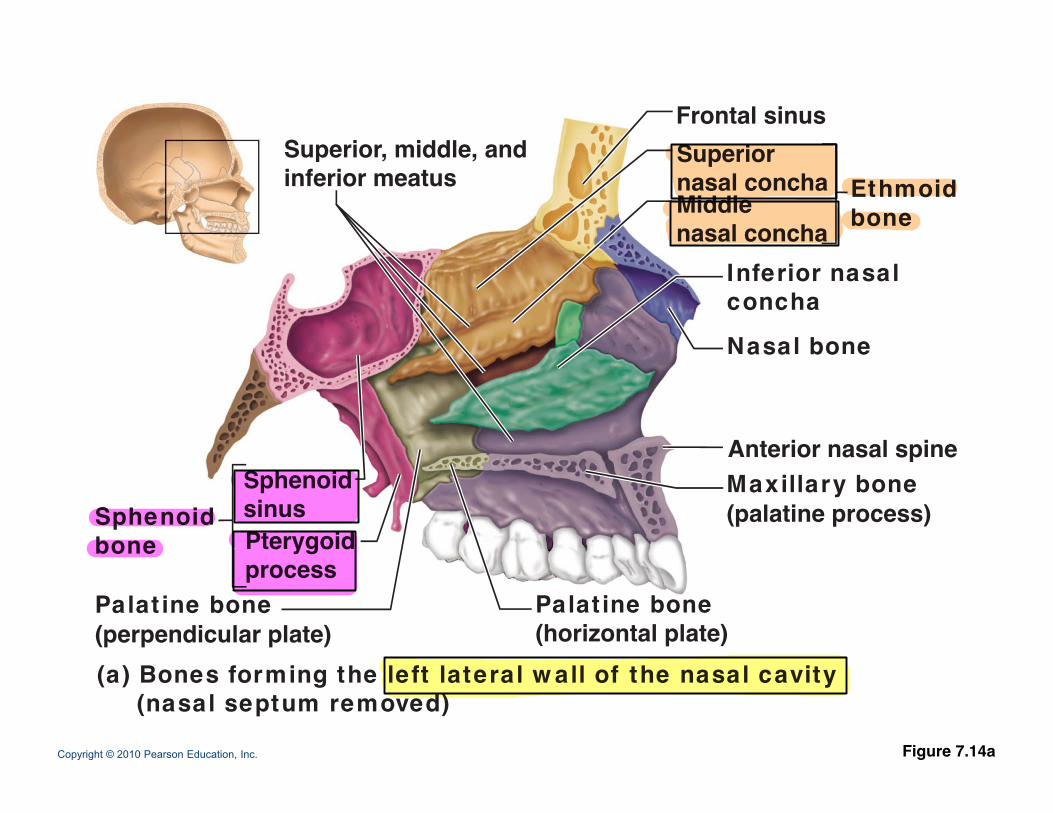
Frontal sinusSuperiorSuperior middle and Superiornasal conchaMiddlenasal concha
Ethmoidbone
Superior, middle, andinferior meatus
Inferior nasalconcha
Nasal bone
Anterior nasal spine
asa bo e
Maxillary bone(palatine process)
Pterygoid
SphenoidsinusSphenoid
bone
te o asa sp e
Palat ine bone(perpendicular plate)
Palat ine bone(horizontal plate)
process
Copyright © 2010 Pearson Education, Inc. Figure 7.14a
(a) Bones forming the left lateral w all of the nasal cavity(nasal septum removed)
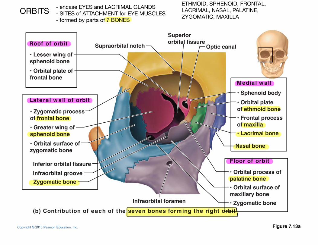
Roof of orbit Supraorbital notch O ti l
Superiororbital fissureRoof of orbit Supraorbital notch Optic canal
• Lesser wing ofsphenoid bone• Orbital plate off t l b
Medial w all
Lateral w all of orbit
frontal bone
• Sphenoid body• Orbital plate
• Zygomatic processof frontal bone• Greater wing ofsphenoid bone
of ethmoid bone• Frontal processof maxilla• Lacrimal bone
Floor of orbit
Nasal bone
Inferior orbital fissure
• Orbital surface ofzygomatic bone
Zygomatic boneInfraorbital groove
I f bit l f
• Orbital process ofpalatine bone• Orbital surface ofmaxillary bone
Copyright © 2010 Pearson Education, Inc.
Infraorbital foramen(b) Contribut ion of each of the seven bones forming the right orbit
• Zygomatic bone
Figure 7.13a
- encase EYES and LACRIMAL GLANDS- SITES of ATTACHMENT for EYE MUSCLES- formed by parts of 7 BONES
ORBITSETHMOID, SPHENOID, FRONTAL,LACRIMAL, NASAL, PALATINE,ZYGOMATIC, MAXILLA
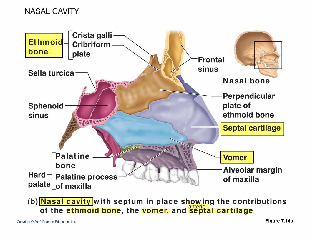
Crista galliCribriformEthmoid Cribriformplate
Ethmoidbone
Frontalsinus Sella turcica
Nasal bone
Perpendicularplate of
Sella turcica
S h id
Septal cartilage
plate of ethmoid bone
Sphenoid sinus
VomerPalat inebone Alveolar margin
of maxillabone Palatine processof maxilla
Hardpalate
Copyright © 2010 Pearson Education, Inc. Figure 7.14b
(b) Nasal cavity w ith septum in place show ing the contribut ionsof the ethmoid bone, the vomer, and septal cart ilageanterior
NASAL CAVITY
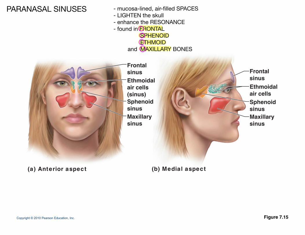
Frontalsinus Frontalsinus Ethmoidalair cells(sinus)Sphenoid
FrontalsinusEthmoidalair cells Sphenoid
Maxillarysinus
Sphenoidsinus
Maxillarysinus
Sphenoidsinus
(a) Anterior aspect (b) Medial aspect
Copyright © 2010 Pearson Education, Inc. Figure 7.15
PARANASAL SINUSES - mucosa-lined, air-filled SPACES- LIGHTEN the skull- enhance the RESONANCE- found in FRONTAL SPHENOID ETHMOID and MAXILLARY BONES
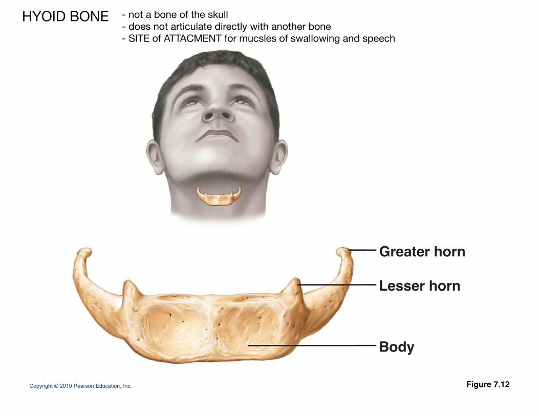
Greater horn
Lesser hornLesser horn
Copyright © 2010 Pearson Education, Inc. Figure 7.12
Body
- not a bone of the skull- does not articulate directly with another bone- SITE of ATTACMENT for mucsles of swallowing and speech
HYOID BONE
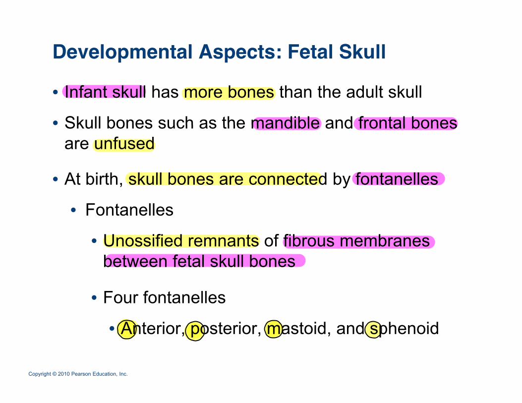
Developmental Aspects: Fetal Skull
• Infant skull has more bones than the adult skull
• Skull bones such as the mandible and frontal bones• Skull bones such as the mandible and frontal bones are unfused
At birth sk ll bones are connected b fontanelles• At birth, skull bones are connected by fontanelles
• Fontanelles
• Unossified remnants of fibrous membranes between fetal skull bones
• Four fontanelles
• Anterior posterior mastoid and sphenoid
Copyright © 2010 Pearson Education, Inc.
• Anterior, posterior, mastoid, and sphenoid
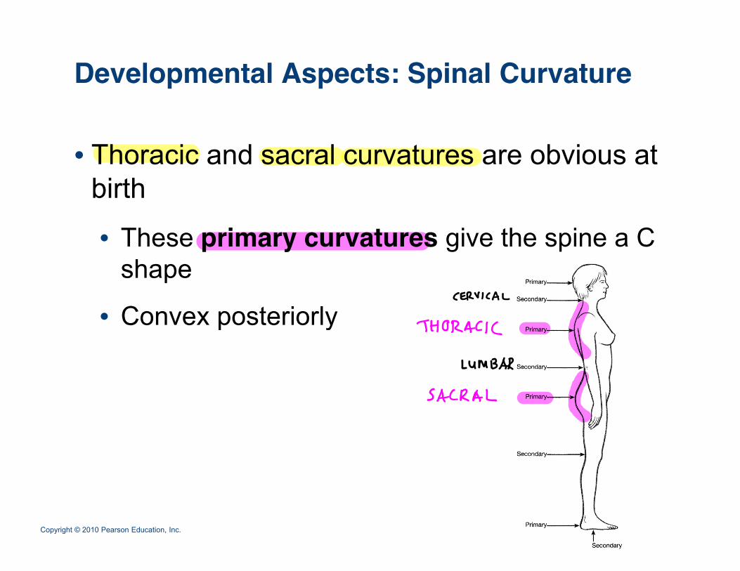
Developmental Aspects: Spinal Curvature
• Thoracic and sacral curvatures are obvious at birth
• These primary curvatures give the spine a C p y g pshape
• Convex posteriorlyConvex posteriorly
Copyright © 2010 Pearson Education, Inc.
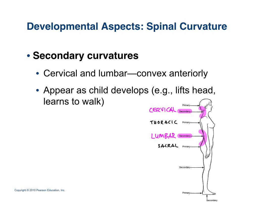
Developmental Aspects: Spinal Curvature
• Secondary curvatures• Cervical and lumbar—convex anteriorly
• Appear as child develops (e g lifts head• Appear as child develops (e.g., lifts head, learns to walk)
Copyright © 2010 Pearson Education, Inc.
Developmental Aspects: Old Age
• Intervertebral discs become thin, less hydrated, and less elasticand less elastic
• Risk of disc herniation increases
• Loss of stature by several centimeters is common by age 55
• Costal cartilages ossify, causing the thorax to become rigid
• All bones lose mass
Copyright © 2010 Pearson Education, Inc.