Award Number: W81XWH-06-1-0740 ERR Gamma: … INVESTIGATOR: Rebecca B. Riggins, ... Ph.D. 5d....
39
AD_________________ Award Number: W81XWH-06-1-0740 TITLE: ERR Gamma: Does an Orphan Nuclear Receptor Link Steroid Hormone Biogenesis to Endocrine Resistance? PRINCIPAL INVESTIGATOR: Rebecca B. Riggins, Ph.D. CONTRACTING ORGANIZATION: Georgetown University Washington, DC 20057 REPORT DATE: September 2007 TYPE OF REPORT: Final PREPARED FOR: U.S. Army Medical Research and Materiel Command Fort Detrick, Maryland 21702-5012 DISTRIBUTION STATEMENT: Approved for Public Release; Distribution Unlimited The views, opinions and/or findings contained in this report are those of the author(s) and should not be construed as an official Department of the Army position, policy or decision unless so designated by other documentation.
Transcript of Award Number: W81XWH-06-1-0740 ERR Gamma: … INVESTIGATOR: Rebecca B. Riggins, ... Ph.D. 5d....

AD_________________ Award Number: W81XWH-06-1-0740 TITLE: ERR Gamma: Does an Orphan Nuclear Receptor Link Steroid Hormone Biogenesis to Endocrine Resistance? PRINCIPAL INVESTIGATOR: Rebecca B. Riggins, Ph.D. CONTRACTING ORGANIZATION: Georgetown University
Washington, DC 20057 REPORT DATE: September 2007 TYPE OF REPORT: Final PREPARED FOR: U.S. Army Medical Research and Materiel Command Fort Detrick, Maryland 21702-5012 DISTRIBUTION STATEMENT: Approved for Public Release; Distribution Unlimited The views, opinions and/or findings contained in this report are those of the author(s) and should not be construed as an official Department of the Army position, policy or decision unless so designated by other documentation.

REPORT DOCUMENTATION PAGE Form Approved
OMB No. 0704-0188 Public reporting burden for this collection of information is estimated to average 1 hour per response, including the time for reviewing instructions, searching existing data sources, gathering and maintaining the data needed, and completing and reviewing this collection of information. Send comments regarding this burden estimate or any other aspect of this collection of information, including suggestions for reducing this burden to Department of Defense, Washington Headquarters Services, Directorate for Information Operations and Reports (0704-0188), 1215 Jefferson Davis Highway, Suite 1204, Arlington, VA 22202-4302. Respondents should be aware that notwithstanding any other provision of law, no person shall be subject to any penalty for failing to comply with a collection of information if it does not display a currently valid OMB control number. PLEASE DO NOT RETURN YOUR FORM TO THE ABOVE ADDRESS. 1. REPORT DATE (DD-MM-YYYY) 01-09-2007
2. REPORT TYPEFinal
3. DATES COVERED (From - To)1 SEP 2006 - 31 AUG 2007
4. TITLE AND SUBTITLE
5a. CONTRACT NUMBER
ERR Gamma: Does an Orphan Nuclear Receptor Link Steroid Hormone Biogenesis to Endocrine Resistance?
5b. GRANT NUMBER W81XWH-06-1-0740
5c. PROGRAM ELEMENT NUMBER
6. AUTHOR(S) Rebecca B. Riggins, Ph.D.
5d. PROJECT NUMBER
5e. TASK NUMBER
E-Mail: [email protected] 5f. WORK UNIT NUMBER
7. PERFORMING ORGANIZATION NAME(S) AND ADDRESS(ES)
8. PERFORMING ORGANIZATION REPORT NUMBER
Georgetown University Washington, DC 20057
9. SPONSORING / MONITORING AGENCY NAME(S) AND ADDRESS(ES) 10. SPONSOR/MONITOR’S ACRONYM(S) U.S. Army Medical Research and Materiel Command
Fort Detrick, Maryland 21702-5012 11. SPONSOR/MONITOR’S REPORT NUMBER(S) 12. DISTRIBUTION / AVAILABILITY STATEMENT Approved for Public Release; Distribution Unlimited
13. SUPPLEMENTARY NOTES
14. ABSTRACT Estrogen-related receptor gamma (ERRγ) is an orphan nuclear receptor with structural similarities to ERα and ERβ (1). In addition to its ability to transactivate classical and imperfect estrogen response elements (EREs), ERRγ is a potent activator of transcription from steroidogenic factor-1 (SF-1) response elements (SF-1REs). Many genes regulated by SF-1REs control key aspects of cholesterol and fatty acid synthesis, important not only for generation of the plasma membrane but also for the synthesis of steroid hormones (2). In this study, we have investigated whether ERRγ expression and/or activity regulates the level of cholesterol in a pair of breast cancer cell lines – one sensitive to endocrine therapy (SUM44) and the other resistant to endocrine therapy (LCCTam, TAM). We found that endocrine-resistant LCCTam (TAM) cells, which overexpress the orphan nuclear receptor ERRγ, contain significantly greater levels of cholesterol than endocrine-sensitive parental SUM44 breast cancer cells, and that siRNA-mediated knockdown of ERRγ in the resistant TAM cell line significantly reduces cholesterol content. SF-1RE activity is 3-fold higher in TAM cells as compared to SUM44 cells, while expression of an endogenous gene (HMGCS2) that contains a consensus SF-1RE is also significantly overexpressed in TAM cells relative to SUM44 cells. Together, these findings represent an important enhancement in our knowledge of breast cancer biology and potential therapeutic response.
15. SUBJECT TERMS Estrogen Receptor Alpha, Estrogen Related Receptor Gamma, SF-1RE, Endocrine Resistance, Lobular Breast Cancer
16. SECURITY CLASSIFICATION OF:
17. LIMITATION OF ABSTRACT
18. NUMBER OF PAGES
19a. NAME OF RESPONSIBLE PERSON USAMRMC
a. REPORT U
b. ABSTRACT U
c. THIS PAGE U
UU
39
19b. TELEPHONE NUMBER (include area code)
Standard Form 298 (Rev. 8-98) Prescribed by ANSI Std. Z39.18

4
Table of Contents
Page Introduction…………………………………………………………….………..….. 5 Body………………………………………………………………………………….. 5 Key Research Accomplishments………………………………………….…….. 9 Reportable Outcomes……………………………………………………………… 9 Conclusion…………………………………………………………………………… 10 References……………………………………………………………………………. 11 Appendices…………………………………………………………………………… 13

Introduction Estrogen receptors alpha and beta (ERα, ERβ) play a central role in the etiology of breast cancer and drive our approach to therapeutic intervention. While these and many other nuclear receptors (NRs) are responsive to specific ligands, there are orphan NRs for which no ligand has yet been identified. The function of these receptors in breast cancer biology is often poorly understood. Estrogen-related receptor gamma (ERRγ) is one orphan NR with structural similarities to ERα and ERβ (1). In addition to its ability to transactivate classical and imperfect estrogen response elements (EREs), ERRγ is a potent activator of transcription from steroidogenic factor-1 (SF-1) response elements (SF-1REs). Many genes regulated by SF-1REs control key aspects of cholesterol and fatty acid synthesis, important not only for generation of the plasma membrane but also for the synthesis of steroid hormones (2). Consequently, upregulation of ERRγ may increase the cholesterol production capacity of breast cancer cells poised to proliferate in response to estrogen. This has the potential to promote the development of hormone-refractory breast cancer, and is also likely to attenuate the therapeutic effectiveness of endocrine agents designed to inhibit ER function. In this study, we have investigated whether ERRγ expression and/or activity regulates the level of cholesterol in a pair of breast cancer cell lines – one sensitive to endocrine therapy (SUM44) and the other resistant (LCCTam, TAM) to endocrine therapy. Body The original text of the Statement of Work (SOW), all progress associated with this SOW, and any additional data contributing to our overall conclusions are described below. Months 1-3: A. Task I – optimize the detection of cholesterol production by SUM44 and TAM cell lines. Findings: We used a commercially-available cholesterol detection assay that detects both cholesterol and cholesteryl esters. Briefly, SUM44 and TAM cells were seeded in 6-well plastic tissue culture dishes for two to three days before being collected. 5 x 10⁵ to 1 x 10⁶ cells were subjected to lipid extraction using a modified version of the Bligh-Dyer method (3). Dried lipids were resuspended in isopropanol containing 10% Triton-X100 detergent and stored at -20oC until use in the assay. The cholesterol detection assay was colorimetric, allowing comparison of our cell line samples to a standard curve containing known amounts (μg) of cholesterol. Blank (zero control), standard curve, and test reactions containing 1 μl of extracted lipids were performed in triplicate and incubated at 37oC for approximately 45 minutes prior to being read on a spectrophotometer at a wavelength of 550 nm. Results are expressed as mean μg cholesterol per 106 cells ± standard error of the mean (SEM) for a representative experiment taken from at least three independent assays (Figure 1). We found that as hypothesized, endocrine-resistant TAM cells do contain modestly, but statistically significantly, more cholesterol than endocrine-sensitive SUM44 cells (2.0 μg/106 cells vs. 1.5 μg/106 cells, p=0.003).
Figure 1: LCCTam (TAM) cells contain significantly higher levels of cholesterol than SUM44 cells (p=0.003).
B. Task II – Transfect SUM44 cells with a plasmid encoding the ERR gamma cDNA or the empty vector control, and select both individual clones and pooled populations. Findings: SUM44 cells were seeded in T-25 plastic tissue culture flasks and co-transfected with 2 μg of a plasmid encoding the murine ERR gamma homologue (ERR3) cDNA tagged at the amino terminus with hemagglutin antigen (generously provided by Dr. Michael Stallcup, USC, and referred to hereafter as HA-ERRγ) (4) or an empty pSG5 vector, and 0.5 μg of a plasmid encoding the puromycin resistance cassette
5
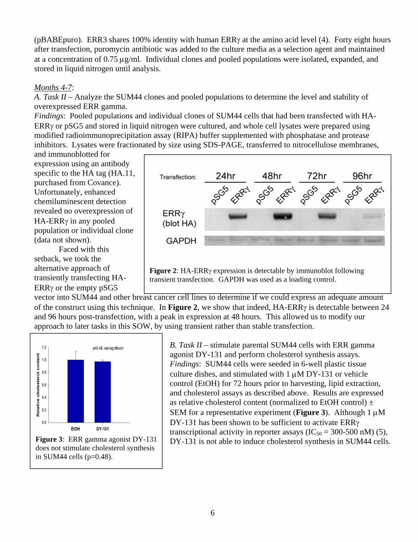
(pBABEpuro). ERR3 shares 100% identity with human ERRγ at the amino acid level (4). Forty eight hours after transfection, puromycin antibiotic was added to the culture media as a selection agent and maintained at a concentration of 0.75 μg/ml. Individual clones and pooled populations were isolated, expanded, and stored in liquid nitrogen until analysis. Months 4-7: A. Task II – Analyze the SUM44 clones and pooled populations to determine the level and stability of overexpressed ERR gamma. Findings: Pooled populations and individual clones of SUM44 cells that had been transfected with HA-ERRγ or pSG5 and stored in liquid nitrogen were cultured, and whole cell lysates were prepared using modified radioimmunoprecipitation assay (RIPA) buffer supplemented with phosphatase and protease inhibitors. Lysates were fractionated by size using SDS-PAGE, transferred to nitrocellulose membranes, and immunoblotted for expression using an antibody specific to the HA tag (HA.11, purchased from Covance). Unfortunately, enhanced chemiluminescent detection revealed no overexpression of HA-ERRγ in any pooled population or individual clone (data not shown). Faced with this setback, we took the alternative approach of transiently transfecting HA-ERRγ or the empty pSG5 vector into SUM44 and other breast cancer cell lines to determine if we could express an adequate amount of the construct using this technique. In Figure 2, we show that indeed, HA-ERRγ is detectable between 24 and 96 hours post-transfection, with a peak in expression at 48 hours. This allowed us to modify our approach to later tasks in this SOW, by using transient rather than stable transfection.
Figure 2: HA-ERRγ expression is detectable by immunoblot following transient transfection. GAPDH was used as a loading control.
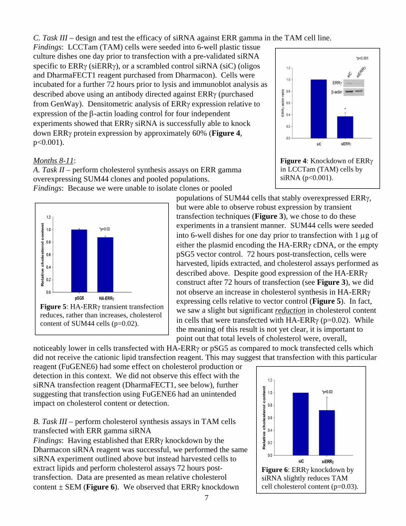
Figure 3: ERR gamma agonist DY-131 does not stimulate cholesterol synthesis in SUM44 cells (p=0.48).
B. Task II – stimulate parental SUM44 cells with ERR gamma agonist DY-131 and perform cholesterol synthesis assays. Findings: SUM44 cells were seeded in 6-well plastic tissue culture dishes, and stimulated with 1 μM DY-131 or vehicle control (EtOH) for 72 hours prior to harvesting, lipid extraction, and cholesterol assays as described above. Results are expressed as relative cholesterol content (normalized to EtOH control) ± SEM for a representative experiment (Figure 3). Although 1 μM DY-131 has been shown to be sufficient to activate ERRγ transcriptional activity in reporter assays (IC50 = 300-500 nM) (5), DY-131 is not able to induce cholesterol synthesis in SUM44 cells.
6
C. Task III – design and test the efficacy of siRNA against ERR gamma in the TAM cell line. Findings: LCCTam (TAM) cells were seeded into 6-well plastic tissue culture dishes one day prior to transfection with a pre-validated siRNA specific to ERRγ (siERRγ), or a scrambled control siRNA (siC) (oligos and DharmaFECT1 reagent purchased from Dharmacon). Cells were incubated for a further 72 hours prior to lysis and immunoblot analysis as described above using an antibody directed against ERRγ (purchased from GenWay). Densitometric analysis of ERRγ expression relative to expression of the β-actin loading control for four independent experiments showed that ERRγ siRNA is successfully able to knock down ERRγ protein expression by approximately 60% (Figure 4, p<0.001).
7
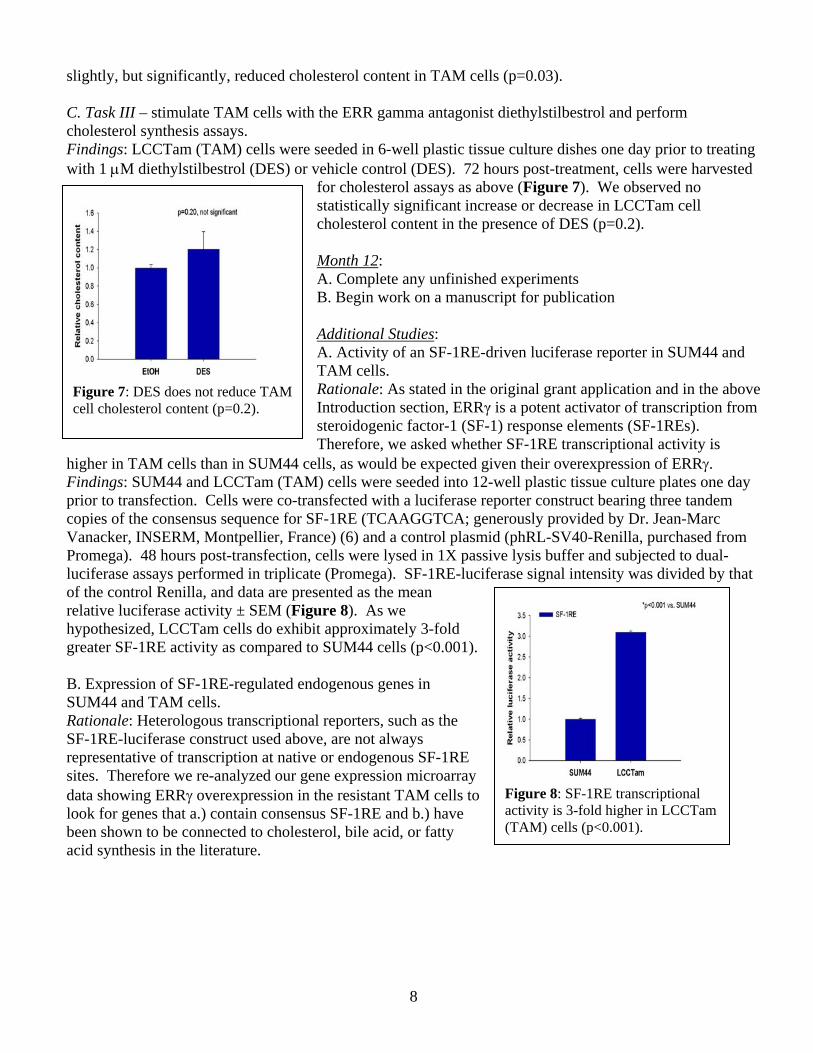
Months 8-11: A. Task II – perform cholesterol synthesis assays on ERR gamma overexpressing SUM44 clones and pooled populations. Findings: Because we were unable to isolate clones or pooled
populations of SUM44 cells that stably overexpressed ERRγ, but were able to observe robust expression by transient transfection techniques (Figure 3), we chose to do these experiments in a transient manner. SUM44 cells were seeded into 6-well dishes for one day prior to transfection with 1 μg of either the plasmid encoding the HA-ERRγ cDNA, or the empty pSG5 vector control. 72 hours post-transfection, cells were harvested, lipids extracted, and cholesterol assays performed as described above. Despite good expression of the HA-ERRγ construct after 72 hours of transfection (see Figure 3), we did not observe an increase in cholesterol synthesis in HA-ERRγ expressing cells relative to vector control (Figure 5). In fact, we saw a slight but significant reduction in cholesterol content in cells that were transfected with HA-ERRγ (p=0.02). While the meaning of this result is not yet clear, it is important to point out that total levels of cholesterol were, overall,
noticeably lower in cells transfected with HA-ERRγ or pSG5 as compared to mock transfected cells which did not receive the cationic lipid transfection reagent. This may suggest that transfection with this particular reagent (FuGENE6) had some effect on cholesterol production or detection in this context. We did not observe this effect with the siRNA transfection reagent (DharmaFECT1, see below), further suggesting that transfection using FuGENE6 had an unintended impact on cholesterol content or detection.
Figure 4: Knockdown of ERRγ in LCCTam (TAM) cells by siRNA (p<0.001).
Figure 5: HA-ERRγ transient transfection reduces, rather than increases, cholesterol content of SUM44 cells (p=0.02).
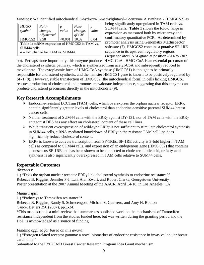
Figure 6: ERRγ knockdown by siRNA slightly reduces TAM cell cholesterol content (p=0.03).
B. Task III – perform cholesterol synthesis assays in TAM cells transfected with ERR gamma siRNA Findings: Having established that ERRγ knockdown by the Dharmacon siRNA reagent was successful, we performed the same siRNA experiment outlined above but instead harvested cells to extract lipids and perform cholesterol assays 72 hours post-transfection. Data are presented as mean relative cholesterol content ± SEM (Figure 6). We observed that ERRγ knockdown
slightly, but significantly, reduced cholesterol content in TAM cells (p=0.03). C. Task III – stimulate TAM cells with the ERR gamma antagonist diethylstilbestrol and perform cholesterol synthesis assays. Findings: LCCTam (TAM) cells were seeded in 6-well plastic tissue culture dishes one day prior to treating with 1 μM diethylstilbestrol (DES) or vehicle control (DES). 72 hours post-treatment, cells were harvested
for cholesterol assays as above (Figure 7). We observed no statistically significant increase or decrease in LCCTam cell cholesterol content in the presence of DES (p=0.2).
8
Month 12: A. Complete any unfinished experiments B. Begin work on a manuscript for publication Additional Studies: A. Activity of an SF-1RE-driven luciferase reporter in SUM44 and TAM cells. Rationale: As stated in the original grant application and in the above Introduction section, ERRγ is a potent activator of transcription from steroidogenic factor-1 (SF-1) response elements (SF-1REs). Therefore, we asked whether SF-1RE transcriptional activity is
higher in TAM cells than in SUM44 cells, as would be expected given their overexpression of ERRγ.
Figure 7: DES does not reduce TAM cell cholesterol content (p=0.2).
Findings: SUM44 and LCCTam (TAM) cells were seeded into 12-well plastic tissue culture plates one day prior to transfection. Cells were co-transfected with a luciferase reporter construct bearing three tandem copies of the consensus sequence for SF-1RE (TCAAGGTCA; generously provided by Dr. Jean-Marc Vanacker, INSERM, Montpellier, France) (6) and a control plasmid (phRL-SV40-Renilla, purchased from Promega). 48 hours post-transfection, cells were lysed in 1X passive lysis buffer and subjected to dual-luciferase assays performed in triplicate (Promega). SF-1RE-luciferase signal intensity was divided by that of the control Renilla, and data are presented as the mean relative luciferase activity ± SEM (Figure 8). As we hypothesized, LCCTam cells do exhibit approximately 3-fold greater SF-1RE activity as compared to SUM44 cells (p<0.001).
Figure 8: SF-1RE transcriptional activity is 3-fold higher in LCCTam (TAM) cells (p<0.001).
B. Expression of SF-1RE-regulated endogenous genes in SUM44 and TAM cells. Rationale: Heterologous transcriptional reporters, such as the SF-1RE-luciferase construct used above, are not always representative of transcription at native or endogenous SF-1RE sites. Therefore we re-analyzed our gene expression microarray data showing ERRγ overexpression in the resistant TAM cells to look for genes that a.) contain consensus SF-1RE and b.) have been shown to be connected to cholesterol, bile acid, or fatty acid synthesis in the literature.
Findings: We identified mitochondrial 3-hydroxy-3-methylglutaryl-Coenzyme A synthase 2 (HMGCS2) as being significantly upregulated in TAM cells vs. SUM44 cells. Table 1 shows the fold-change in expression as measured both by microarray and confirmatory quantitative PCR. As determined by promoter analysis using Genomatix MatInspector software (7), HMGCS2 contains a putative SF-1RE sequence in its upstream regulatory regions (sequence atccCAAGgtaac at position -314 to -302
bp). Perhaps more importantly, this enzyme produces HMG-CoA. HMG-CoA is an essential precursor of the cholesterol synthetic pathway, which is synthesized from acetyl-CoA and subsequently reduced to mevalonate. The cytoplasmic form of HMG-CoA synthase (HMGCS1) is thought to be primarily responsible for cholesterol synthesis, and the hamster HMGCS1 gene is known to be positively regulated by SF-1 (8). However, stable transfection of HMGCS2 (the mitochondrial form) in cells lacking HMGCS1 rescues production of cholesterol and promotes mevalonate independence, suggesting that this enzyme can produce cholesterol precursors directly in the mitochondria (9).
HUGO symbol
Fold-change, Affymetrixa
p value
Fold-change, qPCRa
p value
HMGCS2 9.58 <0.001 10.32 0.04 Table 1: mRNA expression of HMGCS2 in TAM vs. SUM44 cells. a – fold change for TAM vs. SUM44.
Key Research Accomplishments
Endocrine-resistant LCCTam (TAM) cells, which overexpress the orphan nuclear receptor ERRγ, contain significantly greater levels of cholesterol than endocrine-sensitive parental SUM44 breast cancer cells.
Neither treatment of SUM44 cells with the ERRγ agonist DY-131, nor of TAM cells with the ERRγ antagonist DES has any effect on cholesterol content of these cell lines.
While transient overexpression of wild-type ERRγ is not sufficient to stimulate cholesterol synthesis in SUM44 cells, siRNA-mediated knockdown of ERRγ in the resistant TAM cell line does significantly reduce cholesterol content.
ERRγ is known to activate transcription from SF-1REs, SF-1RE activity is 3-fold higher in TAM cells as compared to SUM44 cells, and expression of an endogenous gene (HMGCS2) that contains a consensus SF-1RE and has been shown to be connected to cholesterol, bile acid, or fatty acid synthesis is also significantly overexpressed in TAM cells relative to SUM44 cells.
Reportable Outcomes Abstracts: 1.) “Does the orphan nuclear receptor ERRγ link cholesterol synthesis to endocrine resistance?” Rebecca B. Riggins, Jennifer P-J. Lan, Alan Zwart, and Robert Clarke, Georgetown University Poster presentation at the 2007 Annual Meeting of the AACR, April 14-18, in Los Angeles, CA Manuscripts: 1.) “Pathways to Tamoxifen resistance”* Rebecca B. Riggins, Randy S. Schrecengost, Michael S. Guerrero, and Amy H. Bouton Cancer Letters 256 (2007), pp.1-24. *This manuscript is a mini-review that summarizes published work on the mechanisms of Tamoxifen resistance independent from the studies funded here, but was written during the granting period and the DoD is acknowledged as a source of funding. Funding applied for based on this award: 1.) “Estrogen related receptor gamma: a novel biomarker of endocrine resistance in invasive lobular breast carcinoma.” Submitted to the FY07 DoD Breast Cancer Research Program Idea Grant mechanism.
9

10
2.) “Regulation of ERRγ action in endocrine resistant breast cancer by activation of the Erk/MAPK signaling pathway.” Will be submitted to the Susan G. Komen for the Cure Foundation’s 2007 Career Catalyst Research Grant mechanism, November 2007. Employment: Following my receipt of this award from the Dod BCRP, I applied for and was granted a Research-track faculty position in the Department of Oncology and Lombardi Comprehensive Cancer Center at Georgetown University. I currently hold the position of Research Assistant Professor. Conclusion
There are three major, positive findings from this one-year DoD BCRP Concept award. First, endocrine-resistant LCCTam (TAM) cells, which overexpress the orphan nuclear receptor ERRγ, contain significantly greater levels of cholesterol than endocrine-sensitive parental SUM44 breast cancer cells. Two, siRNA-mediated knockdown of ERRγ in the resistant TAM cell line does significantly reduce cholesterol content. And three, SF-1RE activity is 3-fold higher in TAM cells as compared to SUM44 cells, while expression of an endogenous gene (HMGCS2) that contains a consensus SF-1RE and has been shown to be connected to cholesterol, bile acid, or fatty acid synthesis is also significantly overexpressed in TAM cells relative to SUM44 cells.
Despite the fact that funding for this award is complete, we will continue to actively pursue this research and make several modifications or additions that will complement our current findings. We experienced difficulty producing stable transfectants of HA-ERRγ in SUM44 breast cancer cells. Therefore our next step will be to attempt to produce this cell line again, but instead we will use an adenoviral or retroviral expression vector that expresses this cDNA. Infection rather than transfection is a more rapid and efficient method of gene expression, so we are hopeful that this will be more successful.
The SUM44/LCCTam cell culture model was derived from an invasive lobular breast carcinoma, a distinct histologic subtype of breast cancer that is under-studied despite its rising incidence in Western Europe and the United States (10). Therefore, our data showing that cholesterol content is elevated in an endocrine-resistant variant of this type of breast cancer provides a solid foundation on which to build future studies of resistance that will specifically address risk factors in lobular breast cancer. The function of the orphan NR ERRγ is also poorly understood in breast cancer, although it has been shown to be overexpressed in this context (11), and its family member ERRα1 has received greater attention (12). Therefore, future studies on the role of ERRγ in breast cancer initiation, progression, and therapeutic response are clearly warranted. Finally, the relevant targets of SF-1RE-mediated gene transcription in breast cancer, and the importance of HMGCS2 in this context, are also not known. However it is interesting to note that in a search of the ONCOMINE database of 42 published gene expression microarray datasets of breast cancer tissue (13), SF-1 (HUGO symbol NR5A1) and HMGCS2 expression are both significantly associated with estrogen receptor alpha-positive status (original microarray data from 14-17), and HMGCS2 is also significantly overexpressed in lobular vs. ductal breast carcinomas (http://www.intgen.org/expo.cfm ). Together, the knowledge gained during this research represents an important enhancement in our knowledge of breast cancer biology and potential therapeutic response.
11
References
(1) Ariazi EA, Jordan VC. Estrogen-related receptors as emerging targets in cancer and metabolic
disorders. Curr Top Med Chem 2006;6(3):203-15.
(2) Val P, Lefrancois-Martinez AM, Veyssiere G, Martinez A. SF-1 a key player in the development and differentiation of steroidogenic tissues. Nucl Recept 2003 Sep 18;1(1):8.
(3) BLIGH EG, DYER WJ. A rapid method of total lipid extraction and purification. Can J Biochem Physiol 1959 Aug;37(8):911-7.
(4) Hong H, Yang L, Stallcup MR. Hormone-independent transcriptional activation and coactivator binding by novel orphan nuclear receptor ERR3. J Biol Chem 1999 Aug 6;274(32):22618-26.
(5) Yu DD, Forman BM. Identification of an agonist ligand for estrogen-related receptors ERRbeta/gamma. Bioorg Med Chem Lett 2005 Mar 1;15(5):1311-3.
(6) Vanacker JM, Bonnelye E, Chopin-Delannoy S, Delmarre C, Cavailles V, Laudet V. Transcriptional activities of the orphan nuclear receptor ERR alpha (estrogen receptor-related receptor-alpha). Mol Endocrinol 1999 May;13(5):764-73.
(7) Cartharius K, Frech K, Grote K, Klocke B, Haltmeier M, Klingenhoff A, et al. MatInspector and beyond: promoter analysis based on transcription factor binding sites. Bioinformatics 2005 Jul 1;21(13):2933-42.
(8) Mascaro C, Nadal A, Hegardt FG, Marrero PF, Haro D. Contribution of steroidogenic factor 1 to the regulation of cholesterol synthesis. Biochem J 2000 Sep 15;350 Pt 3:785-90.
(9) Ortiz JA, Gil-Gomez G, Casaroli-Marano RP, Vilaro S, Hegardt FG, Haro D. Transfection of the ketogenic mitochondrial 3-hydroxy-3-methylglutaryl-coenzyme A synthase cDNA into Mev-1 cells corrects their auxotrophy for mevalonate. J Biol Chem 1994 Nov 18;269(46):28523-6.
(10) Li CI, Anderson BO, Daling JR, Moe RE. Trends in incidence rates of invasive lobular and ductal breast carcinoma. J Am Med Assoc 2003 Mar 19;289(11):1421-4.
(11) Ariazi EA, Clark GM, Mertz JE. Estrogen-related receptor alpha and estrogen-related receptor gamma associate with unfavorable and favorable biomarkers, respectively, in human breast cancer. Cancer Res 2002 Nov 15;62(22):6510-8.
(12) Ariazi EA, Kraus RJ, Farrell ML, Jordan VC, Mertz JE. Estrogen-Related Receptor {alpha}1 Transcriptional Activities Are Regulated in Part via the ErbB2/HER2 Signaling Pathway. Mol Cancer Res 2007 Jan;5(1):71-85.
(13) Rhodes DR, Yu J, Shanker K, Deshpande N, Varambally R, Ghosh D, et al. ONCOMINE: a cancer microarray database and integrated data-mining platform. Neoplasia 2004 Jan;6(1):1-6.
(14) Van't Veer LJ, Dai H, van de Vijver MJ, He YD, Hart AA, Mao M, et al. Gene expression profiling predicts clinical outcome of breast cancer. Nature 2002 Jan 31;415(6871):530-6.

12
(15) van de Vijver MJ, He YD, Van't Veer LJ, Dai H, Hart AA, Voskuil DW, et al. A gene-expression signature as a predictor of survival in breast cancer. N Engl J Med 2002 Dec 19;347(25):1999-2009.
(16) Chin K, DeVries S, Fridlyand J, Spellman PT, Roydasgupta R, Kuo WL, et al. Genomic and transcriptional aberrations linked to breast cancer pathophysiologies. Cancer Cell 2006 Dec;10(6):529-41.
(17) Wang Y, Klijn JGM, Zhang Y, Sieuwerts AM, Look MP, Yang F, et al. Gene-expression profiles to predict distant metastasis of lymph-node-negative primary breast cancer. Lancet 2005 Feb 19;365(9460):671-9.

13
Appendices I. Personnel receiving pay from BC051851 in connection with this research: Rebecca B. Riggins II. Abstract presented at the 2007 Annual Meeting of the AACR (1 page). III. Cancer Letters mini-review, written during the granting period and in which DoD is acknowledged as a source of funding (24 pages). IV. Current curriculum vitae (2 pages).

Oasis, Online Abstract Submission and Invitation System - Program Planner
2007 AACR Annual Meeting
April 14-18, 2007 Los Angeles, CA
Print this Page for Your Records Close Window
Abstract Number: 990
Presentation Title: Does the orphan nuclear receptor ERRγ link cholesterol synthesis to endocrine therapy resistance?
Presentation Start/End Time: Sunday, Apr 15, 2007, 1:00 PM - 5:00 PM
Location: Exhibit Hall, Los Angeles Convention Center
Poster Section: 9
Poster Board Number: 11
Author Block: Rebecca B. Riggins, Jennifer P-J Lan, Alan Zwart, Robert Clarke. Georgetown Univ., Lombardi Comp. Cancer Ctr., Washington, DC
Estrogen receptors alpha and beta (ERα, ERβ) play a central role in the etiology of breast cancer and drive our approach to therapeutic intervention. While these and many other nuclear receptors (NRs) are responsive to specific ligands, there are orphan NRs for which no ligand has yet been identified. The function of these receptors in breast cancer biology is often poorly understood. Estrogen-related receptor gamma (ERRγ) is one orphan NR with structural similarities to ERα and ERβ. In addition to its ability to transactivate classical and imperfect estrogen response elements (EREs), ERRγ is a potent activator of transcription from steroidogenic factor-1 (SF-1) response elements (SF-1REs). Many genes regulated by SF-1REs control key aspects of cholesterol and fatty acid synthesis, important not only for generation of the plasma membrane but also for the synthesis of steroid hormones. Consequently, upregulation of ERRγ may increase the cholesterol production capacity of breast cancer cells poised to proliferate in response to estrogen. This has the potential to promote the development of hormone-refractory breast cancer, and is also likely to attenuate the therapeutic effectiveness of antiestrogens designed to inhibit ER function. We have found that ERRγ is upregulated 4-fold (p=0.015) in LCCTam cells, a multi-antiestrogen resistant variant of the ER-positive SUM44 breast cancer cell line, a model of invasive lobular carcinoma. ERRγ overexpression is accompanied by a significant increase in basal and estrogen-stimulated ERE transcriptional activity, and siRNA-mediated knockdown of ERRγ in LCCTam cells completely restores sensitivity to both 4-hydroxytamoxifen and Fulvestrant (p=0.03). Furthermore, LCCTam cells produce greater levels of cholesterol than SUM44, and we have found that the expression of three genes with established connections to cholesterol and steroid hormone synthesis is significantly increased in LCCTam: HMG-CoA synthase 2 (HMGCS2, 9.6-fold), GATA binding protein 4 (GATA4, 4.4-fold), and sterol regulatory element binding transcription factor 1 (SREBF1, 1.8-fold). Together these data suggest that ERRγ may affect endocrine resistance in the LCCTam breast cancer model by regulating cholesterol production.
http://www.abstractsonline.com/viewer/viewAbstrac...D935}&AKey={728BCE9C-121B-46B9-A8EE-DC51FDFC6C15} (1 of 2)9/28/2007 10:09:31 AM

Cancer Letters 256 (2007) 1–24
www.elsevier.com/locate/canlet
Mini-review
Pathways to tamoxifen resistance
Rebecca B. Riggins a, Randy S. Schrecengost b, Michael S. Guerrero b,Amy H. Bouton b,*
a Department of Oncology, Lombardi Comprehensive Cancer Center, Georgetown University, Washington, DC 20057, USAb Department of Microbiology and Cancer Center, University of Virginia Health System, Box 800734, Charlottesville, VA 22908-0734, USA
Received 16 January 2007; received in revised form 15 March 2007; accepted 15 March 2007
Abstract
Therapies that target the synthesis of estrogen or the function of estrogen receptor(s) have been developed to treatbreast cancer. While these approaches have proven to be beneficial to a large number of patients, both de novo andacquired resistance to these drugs is a significant problem. Recent advances in our understanding of the molecular mech-anisms that contribute to resistance have provided a means to begin to predict patient responses to these drugs and developrational approaches for combining therapeutic agents to circumvent or desensitize the resistant phenotype. Here, we reviewcommon mechanisms of antiestrogen resistance and discuss the implications for prediction of response and design of effec-tive combinatorial treatments.� 2007 Elsevier Ireland Ltd. All rights reserved.
Keywords: Tamoxifen; Antiestrogen; Aromatase inhibitor; Fulvestrant; Estrogen receptor
1. Endocrine therapies for breast cancer
Estrogen and the steroid estrogen receptors (ERs)are critical regulators of breast epithelial cell prolifer-ation, differentiation, and apoptosis. Mammalsexpress two ERs, ERa and ERb, which show distincttissue distributions and functions (for review, see [1]).Mice with targeted deletions of one or both ER geneshave established that ERa is the key regulator ofmammary gland development. ERa is expressed in15–30% of the luminal epithelial cells present in nor-mal breast tissue; estrogen stimulation of these cellresults in transcription of various genes, including
0304-3835/$ - see front matter � 2007 Elsevier Ireland Ltd. All rightsdoi:10.1016/j.canlet.2007.03.016
* Corresponding author. Tel.: +1 434 924 2513; fax: +1 434 9821071.
E-mail address: [email protected] (A.H. Bouton).
many that are involved in cell cycle regulation (forreview, see [2]). However, estrogen-dependent prolif-eration of breast epithelial cells is thought to occur ina paracrine fashion, such that ERa-containing cellsproduce growth factors that induce proliferation inadjacent ER-negative cells. In malignant breast tis-sue, the action of estrogen is deregulated, resultingin a shift to proliferation without differentiation orapoptosis. The percentage of epithelial cells express-ing ERa is significantly increased under these condi-tions. Moreover, these cells now proliferate,marking a shift from paracrine to autocrine growth.
ERb is more ubiquitously expressed throughoutmammary tissue than is ERa [3]. ERb is often foundco-expressed with ERa in the luminal epithelial cellsbut has also been detected in myoepithelial cell andsurrounding stromal cell nuclei. While its specific
reserved.
2 R.B. Riggins et al. / Cancer Letters 256 (2007) 1–24
function(s) in normal and neoplastic tissues remainunknown, ERb is essential for the fully differenti-ated phenotype of the normal mammary gland inmice [4]. In ERb�/� mice, the mammary glanddevelops normally until puberty. However, severalabnormalities are apparent during pregnancy andlactation, including incomplete penetration of thefat pad by glandular tissue, an abnormally largeincrease in the size of the lumen of ducts and alveoli,and a decrease in the total number of alveoli. Also,in luminal epithelial cells of these mice, the loss ofERb disrupts the formation of tight junctions andalters the expression of b-catenin, features oftenassociated with a malignant phenotype. It has beensuggested that the function of ERb in these pro-cesses may contribute to the protection conferredby pregnancy and lactation against breast cancer.
Several reports describe a loss of ERb during car-cinogenesis (for review see [5,6]), providing supportfor its possible role as a tumor suppressor. Indeed,ERb has been reported to suppress the function ofERa as a transcriptional activator when both recep-tors are co-expressed [5,7] and to promote anti-prolif-erative and pro-apoptotic effects [8]. Several geneshave been identified that are regulated by ERb butnot ERa; studies are underway to determine whetherthese genes may have anti-proliferative or pro-apop-totic properties that could account for the putativetumor suppressor function of ERb [5,9,10].
A majority of invasive breast tumors express ERa,and as discussed above, ERb is often downregulated.These findings provide a strong rationale for thera-pies that disrupt the actions of estrogen and ERa.Current approaches include inhibiting the functionof ERs with the use of antiestrogens such as tamoxi-fen, raloxifene, and ICI 182,780 (faslodex, fulve-strant), or blocking the conversion of androgensinto biologically active estrogens through the actionof aromatase inhibitors such as letrozole, anastroz-ole, and exemestane (see Section 1.1). Recent studieson ERb have also highlighted its growing value as adiagnostic and prognostic marker in breast cancerand its potential for use as a new strategy for delayingand retarding breast tumorigenesis (see Section1.1.1).
1.1. Classes of therapeutic agents
1.1.1. Selective estrogen receptor modulators
(SERMs)
SERMs have been developed as agents that func-tion as ER antagonists in some tissues, including
breast, and agonists in other tissues such as theheart and bone (for review, see [11]). The antagonisteffects of tamoxifen in breast tissue are thought toresult from its ability to bind to the ligand-bindingdomain (LBD) of the ER, effectively blocking thepotential for estrogen stimulation. Tamoxifen bind-ing further prevents critical ER conformationalchanges that are required for the association ofcoactivators. However, tamoxifen treatment is asso-ciated with an increased risk of endometrial cancer[12]. Other SERMs, such as raloxifene, have beendeveloped with the goal of reducing some of the del-eterious effects of tamoxifen.
New classes of SERMs are being developed thatexhibit mixed agonist/antagonist activities that aresubtype specific (‘‘dual SERMs’’) or preferentiallyaffect one ER over the other (‘‘subtype-selectiveSERMs’’) (reviewed in [13]). However, it is impor-tant to note that selectivity is often assessed in thecontext of binding affinity relative to 17b-estradiol.Therefore, a ‘‘selective’’ compound may actuallyinteract with both receptors but have a higher affin-ity for one versus the other. The relative bindingaffinities of several agents have been determined inHeLa cells engineered to express ERa or ERb [14].Propyl pyrazole triol (PPT) was shown to be aselective agonist for ERa in this system, while thesynthetic ligands diarylpropionitrile (DPN) and8b-VE2, and the natural products genistein andbiochanin A, were preferential agonists of ERb. Incontrast, the antiprogestin RU486 was shown tobe a strong ERb antagonist with only modest agonistactivity toward ERa. DPN has been shown to inhibitproliferation and induce apoptosis in the HC11mouse mammary cell line but this compound hasnot been widely studied in human breast cancer celllines [8]. It has also been reported that a modifiedandrostenediol is a potent activator of ERb byligand-binding assay, but its biological activity inbreast cancer cells requires further investigation [15].
Dual SERM compounds that inhibit ERa whileactivating ERb are particularly attractive, as ERboften performs growth-inhibitory functions inbreast tissues and cell lines (see above, Section 1).An example of this type of SERM is TAS-108, anovel, steroidal antiestrogen that inhibits ERa whileacting as a partial agonist of ERb ([16] and refer-ences therein). In vitro, TAS-108 effectively inhibitsthe growth of tamoxifen-resistant breast cancercells, and Phase I studies have established that thisagent is not only well-tolerated but appears toinduce stable disease in women with advanced
R.B. Riggins et al. / Cancer Letters 256 (2007) 1–24 3
breast cancer. Where and how this particularSERM will be used in the clinic, particularly insequence with the other classes of therapeutic agentsdescribed below, has yet to be determined. How-ever, this and future compounds that have similarprofiles will be important additions to the currentrepertoire of endocrine therapies.
1.1.2. Selective estrogen receptor downregulators
(SERDs)
The pure ER antagonist ICI 182,780 (hereaftercalled fulvestrant) binds to ERa with 100-fold greateraffinity than does tamoxifen and in so doing, inhibitsreceptor dimerization and abrogates estrogen signal-ing [17]. Both laboratory and clinical studies haveshown a decrease in overall ER protein levels inresponse to fulvestrant treatment (discussed in [18]).Importantly, a majority of ERa-expressing tamoxi-fen-resistant breast cancers remain sensitive to fulve-strant treatment. This has led to its approval in theUnited States for use in treating ER-positive patientsfollowing failure to respond to first-line antiestrogentherapy.
1.1.3. Aromatase inhibitors (AIs)
Aromatase inhibitors (AIs) represent a new classof endocrine therapy drugs that inhibit the action ofaromatase, an enzyme necessary for the conversionof androgens to estrogens (for review, see [19]).Clinically, AIs have had success as a second lineof therapy for post-menopausal patients who haveprogressed after tamoxifen treatment. They alsoshow promise as first-line adjuvant treatment forERa-positive breast cancers.
1.2. Resistance to endocrine therapies
While considerable progress has been made indeveloping strategies to target estrogen and ERs inbreast cancer, approximately 30% of ERa-positivebreast cancers do not respond to tamoxifen treat-ment (de novo resistance). In addition, the majorityof tumors that initially respond to treatmentdevelop resistance over time (acquired resistance),despite continued expression of ERa (reviewed in[11,20]). Interestingly, many of these resistanttumors still respond to fulvestrant and AIs, indicat-ing that estrogen remains an important regulator oftumor growth under these circumstances [11]. Thesedata provide support for the idea that endocrine-targeted therapies can lead to the activation of novelsignaling pathways that circumvent the effects of
antiestrogens [21]. De novo and acquired resistanceto AIs have also been reported. The appearanceand development of resistant tumors are thereforemajor obstacles in the struggle to control the growthand metastasis of breast carcinomas.
1.3. Biological effects of endocrine therapies
Homeostasis of non-cancerous cells requires abalance between proliferation, cell cycle arrest,and apoptosis; this equilibrium is disrupted duringcancer progression. Endocrine therapies induce cellcycle arrest and apoptosis through a reduction inexpression of cell cycle regulators such as c-Mycand cyclin D1, accumulation of hypophosphorylat-ed Rb, induction of the cell cycle inhibitorsp21WAF1/CIP1 and p27Kip1, induction of the putativetumor suppressor interferon regulatory factor-1(IRF-1), and inhibition of pro-survival pathwaysthrough decreased Bcl-2 expression and increasedBax expression ([22–25]; for review, see [20,26]).Breast tumor cells that are resistant to the growth-inhibitory and apoptotic effects of antiestrogenssuch as tamoxifen overcome these influencesthrough a variety of mechanisms that are the focusof this review.
2. Molecular mechanisms of antiestrogen resistance
Emerging data from breast tumor biopsies indi-cate that altered expression and/or modification ofseveral growth factor receptors and downstream sig-naling molecules correlate with tamoxifen resistance(Fig. 1). Epidermal growth factor receptor (EGFR),human epidermal growth factor receptor type 2(HER2), and insulin-like growth factor-1 receptor(IGF-1R) signaling pathways are often elevated innon-responsive tumors that exhibit either de novo oracquired resistance [27–30], as is the activity ofkinases that function downstream of these receptorssuch as extracellular-regulated kinase (ERK) 1/2,p38, AKT, and p21-activated kinase-1 (Pak1) [30–32]. Expression and/or phosphorylation of substratesof these receptor and non-receptor kinases are alsoelevated in tumors resistant to tamoxifen; such mole-cules include the cytoplasmic adapter molecule‘‘breast cancer antiestrogen resistance 1’’ (BCAR1;also known as p130Cas; hereafter called Cas), thecoactivator ‘‘amplified in breast cancer 1’’ (AIB1),and ERa itself [28,33–35]. Finally, dysregulatedexpression of molecules involved in cell survival, suchas c-Myc, Bad, and Bcl-2, can also be predictive of a

Fig. 1. Molecular mechanisms of tamoxifen resistance. Model depicting molecules implicated in antiestrogen resistance and discussed inthis review.
4 R.B. Riggins et al. / Cancer Letters 256 (2007) 1–24
poor response to endocrine therapies such as tamox-ifen [36–39]. Clinical data addressing the correlationbetween some of these markers and antiestrogenresistance are presented in greater detain in Section4.1. While these studies are limited in both scopeand number, however, the association betweenexpression/activation of these molecules and resis-tance to endocrine therapies has been corroborated(and in fact extended) in tissue culture and animalmodels. These data are reviewed below.
2.1. ERa, ERb, and receptor co-regulatory proteins
Expression of ERa has long been considered theprimary determinant of a clinical response to endo-
crine or antiestrogen therapy. However in 1996, theconcept of estrogen signaling and ER function inbreast cancer was significantly altered by the discov-ery of ERb [40]. The DNA binding domain of full-length ERb1 shares 96% identity with that of ERa,and although the affinity of this receptor for estro-gen is similar to that of ERa, the response to antag-onists like tamoxifen and fulvestrant is oftendifferent. Both receptors can modulate transcrip-tion, either directly from estrogen response elements(EREs), or indirectly through tethering to AP-1 andSp1 binding sites (reviewed in [41]). However,although ERa perceives tamoxifen and fulvestrantas antagonists, it has been shown that ERb can uti-lize these compounds as agonists when associated

R.B. Riggins et al. / Cancer Letters 256 (2007) 1–24 5
with AP-1 and Sp1 sites [42,43]. Additionally, twonaturally occurring variants, ERb2/ERbcx andERb5, contain carboxy-terminal deletions that pre-vent their binding to ligand, and it has been sug-gested that these receptors may function asdominant-inhibitory mutants of full-length ERb1.Murphy et al. have recently reviewed the evidencefor a role for ERb1 and its variants in breast cancerdiagnosis and prediction of endocrine therapyresponse [6].
The transcriptional activities of ERa, ERb, andother steroid hormone receptors are modulated bycoregulatory proteins in either a positive or negativefashion. The association of coactivators and core-pressors with estrogen receptors is regulated by con-formational changes induced by ligand binding,with estrogen inducing coactivator recruitmentand antiestrogens leading to association with core-pressors. Several excellent reviews have recentlydetailed the expression, function, and clinical rele-vance of these coregulators in breast cancer andendocrine resistance [44–47].
2.1.1. ERa expression and downregulation
In human breast tumors, the vast majority ofERa-negative tumors exhibit de novo or intrinsicresistance to tamoxifen and other antiestrogens;however, small numbers of ERa-negative but pro-gesterone receptor-positive (PR-positive) tumorsrespond to antiestrogen treatment (reviewed in[48]). One possible explanation for this finding isthat PR-positive tumors retain a functional ERasignaling pathway, particularly since expression ofPR is an estrogen-regulated event, and that ERa ispresent in these tumors at levels that are below thelimit of detection for ligand-binding or immunohis-tochemical assays. The importance of PR as a mar-ker of antiestrogen response is substantiated bystudies showing that 70% of ERa-positive/PR-posi-tive tumors effectively respond to tamoxifen, whileonly 34% of ERa-positive/PR-negative patientsrespond to tamoxifen therapy [49].
In the context of de novo antiestrogen resistancedisplayed by ERa-negative breast cancer cells, sev-eral studies suggest that it may be possible to restoretamoxifen sensitivity through re-expression of ERa.Epigenetic silencing of the ER due to hypermethyla-tion of the promoter region has been reported bothin vitro and in vivo [50,51]. For example, ERaexpression can be restored in the ERa-negativeMDA-MB-231 breast cancer cell line by either treat-ment with the demethylating agent 5-aza-2 0-deoxy-
cytidine or the histone deacetylase inhibitortrichostatin A [52]. Importantly, when endogenousERa expression was restored in this way, MDA-MB-231 cells became sensitive to growth inhibitionby tamoxifen. Based on data such as these, it hasbeen suggested that restoration of ERa expressionmay be a viable option for the treatment of someER-negative breast cancers (discussed in [53]). How-ever, a separate study showed that ectopic expres-sion of ERa in MDA-MB-231 cells was notsufficient to render the cells sensitive to tamoxifen[54]. This suggests that the level and cellular contextof ERa expression will be important factors to con-sider when attempting to modulate ERa levels in‘‘ER-negative’’ tumors.
Although downregulation of ERa expression cancontribute to de novo antiestrogen resistance, loss ofthe receptor during the acquisition of tamoxifenresistance is not commonly observed in vivo
([55,56]; reviewed in [57]). However, loss of ERahas been reported in a few models of acquired anti-estrogen resistance. For example, the ZR-75-derived9a1 cell line becomes ERa-negative in the presenceof tamoxifen but reverts to being ERa-positivewhen tamoxifen is removed and estrogen is reintro-duced [58].
Like tamoxifen, fulvestrant inhibits ERa tran-scriptional activity. In addition, fulvestrant has alsobeen shown to accelerate ERa protein degradationvia the ubiquitin–proteasome pathway, leading toits reclassification as a SERD (see above). Loss ordownregulation of ERa has the potential be a viablemechanism for antiestrogen resistance in the contextof acquired resistance to fulvestrant or otherSERDs, but current evidence to this effect is contra-dictory. ERa protein is completely lacking and onlyminimal mRNA is detectable in several fulvestrant-resistant cell lines, including MCF-7-r, T47D-r, andZR-75-r cells [59]. MCF-7-F resistant cells show a90% decrease in both ERa mRNA and protein,accompanied by significant changes in estrogen-induced expression of ER target genes [60]. In con-trast, the MCF-7/182R-1, -6, and -7 models of fulve-strant resistance maintain ERa expression andfunction, although its expression at the protein levelis reduced by one-third as compared to the parentalcell line [61,62]. MCF-7/LCC9 breast cancer cells,which display acquired resistance to fulvestrantand cross-resistance to tamoxifen, show no down-regulation of ERa [63]. This cell line was originallyderived from the estrogen-independent MCF-7/LCC1 cell line [64]; interestingly both MCF-7/
6 R.B. Riggins et al. / Cancer Letters 256 (2007) 1–24
LCC1 and MCF-7/LCC9 cells express higher levelsof ERa mRNA and protein than the parental MCF-7 cell line [65]. MCF-7/LCC9 cells have also beenshown to contain increased basal ERa transcrip-tional activity that is not inhibited by fulvestrant[66]. Factors that drive the differential expressionand activity of ERa among these cell culture modelsof fulvestrant resistance are not known, but onelikely explanation for these disparities may be thedistinct schemes of drug selection that have beenused to generate resistant cells in vitro. Clinical stud-ies confirm that fulvestrant can reduce ERa levelsfollowing either short- (21 days) or long- (6 months)term treatments, but that expression is not com-pletely eliminated (reviewed in [67]). Interestingly,patients who do not show a clinical response to ful-vestrant treatment also do not show a decrease inERa expression.
2.1.2. ERa mutationMutation of the ER may also impact endocrine
therapy responsiveness. For example, one tamoxi-fen-resistant variant of the ERa-positive T47D cellline, T47DCO, has acquired three ERa deletionmutations that affect the hormone- and DNA-binding domains of the receptor [68]. This cell linealso expresses different levels of PR [69], suggest-ing that these variants are functionally distinctfrom the wild type receptor. These and manyother deletions, exon duplications or insertions,point mutations, and alternative splicing eventshave the potential to regulate ERa function inbreast and other tissues. Herynk and Fuqua haverecently published a review of known ER muta-tions and their connection to human disease, sev-eral of which are linked to tamoxifen responseand resistance [70]. For example, duplication ofexons 6 and 7 in an estrogen-independent variantof the MCF-7 cell line leads to the production ofan ER that can no longer bind to estrogen ortamoxifen [71]. Short insertions in exon 6 havealso been identified in tamoxifen-resistant tumors[72]. A tyrosine-to-asparagine substitution at resi-due 537 (Y537N), originally identified in meta-static breast cancer, confers constitutivetranscriptional activity in vitro and renders theER insensitive to both estrogen and tamoxifen,while mutation of this tyrosine to either serineor alanine results in a constitutively active recep-tor whose activity is still inhibited by tamoxifenand fulvestrant [73]. In contrast, a lysine-to-argi-nine substitution at residue 303 (K303R) that
was first identified in breast hyperplasia and anumber of breast tumors results in a receptor thatis hypersensitive to substantially lower concentra-tions of estrogen than normal (10�12 vs. 10�9 M)[74]. The prevalence of this mutation in patientpopulations is unclear, as more recent studies havenot found ERa K303R in other patient groups[75]. The difficulty in identifying functional ERamutations in vivo that affect tamoxifen therapyresponse may stem from the fact that such muta-tions are relatively rare, estimated to be present inonly 1% of breast tumors [51].
2.2. Growth factor receptors
As discussed above, tumors resistant to tamoxi-fen often show high expression and/or activationof EGFR, its close relative HER2, IGF-R1, andproteins that function downstream of these recep-tors such as ERK1/2, and AKT (for review, see[28,76–78]). In vitro data corroborate a role for thesegrowth factor receptors in mediating antiestrogenresistance, both through crosstalk with the ERand through independent pathways.
2.2.1. EGFR and HER2
Several studies have established that overexpres-sion of EGFR or HER2 in ER-positive, antiestro-gen-sensitive, breast cancer cells can conferresistance to these drugs [79–81]. Both ERK1/2and AKT activity appear to contribute to the resis-tant phenotype under these conditions. Othergroups have isolated antiestrogen-resistant cell linesfollowing long-term treatment of ER-positive breastcancer cells with either tamoxifen or fulvestrant. Inthese studies, acquisition of resistance often coin-cides with overexpression of the EGFR and activa-tion of downstream molecules such as ERK1/2,PI3K, and AKT [59,63,82,83]. Expression of oneof the ligands for EGFR, transforming growth fac-tor a (TGFa), is elevated in one of these cell models,resulting in an autocrine loop that is required formaintenance of the resistant phenotype [84]. Unlikethe parental cells, which are relatively impervious toEGFR signaling pathways, the resistant clones arehighly dependent on these pathways for growthand survival.
While the mechanism(s) by which these receptorspromote antiestrogen resistance are not clear, sev-eral lines of evidence suggest that they may functionto provide survival signals that override apoptoticprograms in the cell. First, when EGFR functions
R.B. Riggins et al. / Cancer Letters 256 (2007) 1–24 7
are blocked by either the tyrosine kinase inhibitorIressa or blocking antibodies, apoptosis is induced[85–88]. Second, the expression and/or activity ofseveral anti-apoptotic molecules such as AKT,Bcl-2 and Bcl-xL are upregulated under conditionsof HER2 overexpression in MCF-7 cells [89,90].However, events considered in other contexts to begenerally pro-apoptotic are also associated withoverexpression of EGFR or HER2 in humantumors. For example, elevated levels of phospho-p38 have been reported in HER2-expressing tumorsas well as in tamoxifen-resistant xenografts [29,30].While its role in regulating apoptosis under theseconditions is not clear, activated p38 may promoteresistance to tamoxifen through its ability toenhance the nuclear functions of ER [91]. Finally,recent data from our group indicate that the EGFRmay work together with the non-receptor proteintyrosine kinase c-Src and the adapter moleculeCas to promote survival [92].
In addition to the direct effect of activatedgrowth factor receptors on cell proliferation andsurvival, crosstalk between these receptors and ERplays a significant role in regulating the cellularresponse to antiestrogens. Signaling through EGFRand HER2 can result in downregulation of ERaexpression in cultured cells [93]. This inverse rela-tionship has been corroborated in a recent clinicalstudy by the Southwest Oncology Group [27]. Notonly does growth factor signaling affect ER levels,but it can also strongly impact ER nuclear func-tions, at least in part through promoting phosphor-ylation of the ER, its coactivators, and/or itscorepressors (for review, see [94]).
Conversely, ERa appears to regulate growthfactor receptor activities through rapid, non-geno-mic functions. This may be augmented by anaccumulation of cytoplasmic ERa at the expenseof nuclear ERa under conditions of high HER2expression [95]. Interestingly, it appears thattamoxifen may be able to function as an agonistfor these non-genomic ER activities under somecircumstances, perhaps explaining how cellsexpressing high levels of HER2 and the coactiva-tor AIB1 show increased proliferation in the pres-ence of both estrogen and tamoxifen [90].Additional support for this crosstalk comes frompreclinical studies showing enhanced efficacyand/or delays in the appearance of resistance inthe presence of combined therapies that targetboth tyrosine kinase receptors and estrogen signal-ing ([87,96]; and see below, Section 4.3).
2.2.2. IGF-1R signaling
The involvement of IGF signaling in antiestrogenresistance is less clear than is the role for EGFR andHER2. While data are mixed about whether IGF-1R levels are elevated or decreased in resistanttumors and cell lines [28,97,98], evidence for a roleof IGF-1R signaling in acquired resistance is emerg-ing. This may be mediated at least in part throughcrosstalk with EGFR, HER2, and/or ERa andlikely involves the scaffolding molecule insulinreceptor substrate 1 (IRS-1) [77,99,100].
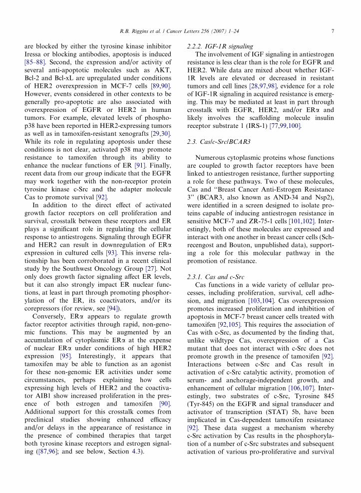
2.3. Cas/c-Src/BCAR3
Numerous cytoplasmic proteins whose functionsare coupled to growth factor receptors have beenlinked to antiestrogen resistance, further supportinga role for these pathways. Two of these molecules,Cas and ‘‘Breast Cancer Anti-Estrogen Resistance3’’ (BCAR3, also known as AND-34 and Nsp2),were identified in a screen designed to isolate pro-teins capable of inducing antiestrogen resistance insensitive MCF-7 and ZR-75-1 cells [101,102]. Inter-estingly, both of these molecules are expressed andinteract with one another in breast cancer cells (Sch-recengost and Bouton, unpublished data), support-ing a role for this molecular pathway in thepromotion of resistance.
2.3.1. Cas and c-Src
Cas functions in a wide variety of cellular pro-cesses, including proliferation, survival, cell adhe-sion, and migration [103,104]. Cas overexpressionpromotes increased proliferation and inhibition ofapoptosis in MCF-7 breast cancer cells treated withtamoxifen [92,105]. This requires the association ofCas with c-Src, as documented by the finding that,unlike wildtype Cas, overexpression of a Casmutant that does not interact with c-Src does notpromote growth in the presence of tamoxifen [92].Interactions between c-Src and Cas result inactivation of c-Src catalytic activity, promotion ofserum- and anchorage-independent growth, andenhancement of cellular migration [106,107]. Inter-estingly, two substrates of c-Src, Tyrosine 845(Tyr-845) on the EGFR and signal transducer andactivator of transcription (STAT) 5b, have beenimplicated in Cas-dependent tamoxifen resistance[92]. These data suggest a mechanism wherebyc-Src activation by Cas results in the phosphoryla-tion of a number of c-Src substrates and subsequentactivation of various pro-proliferative and survival

8 R.B. Riggins et al. / Cancer Letters 256 (2007) 1–24
pathways that allow the cells to grow in the presenceof tamoxifen. Interestingly, ER nuclear functionsremain unaffected by this pathway [92], suggestingthat therapies that target the Src-Cas-EGFR-STAT5b signaling axis may be able to synergize withtamoxifen for the treatment of ‘‘resistant’’ tumors.
Several other studies provide additional insightinto how c-Src and Cas may contribute to tamoxi-fen resistance. First, ERa and phosphatidylinositol3-kinase (PI3K) have been found in the c-Src/Cascomplex in T47D breast cancer cells following rapidestrogen stimulation [108]. While it is clear thatthese complexes help regulate estrogen-dependentcell proliferation and survival, however, it is notyet known whether they are important for promot-ing resistance to antiestrogens. Second, pharmaco-logical inhibition of c-Src in MCF-7 cells enhancesthe growth-inhibitory and apoptotic-inducingeffects of tamoxifen, further strengthening a rolefor c-Src in this process [109]. Finally, cells treatedover a period of two weeks with tamoxifen or fulve-strant show a c-Src-dependent increase in Cas phos-phorylation that correlates with activation of asurvival pathway involving AKT [110].
Together, these studies point to a key role for Casand c-Src in tamoxifen resistance in cell culture.Whether these pathways also play a role in clinicalresistance remains to be determined. c-Src/Casinteractions are favored under conditions in whichboth proteins are expressed at high levels [106].Thus it is intriguing that a large percentage ofhuman breast tumors contain high levels of bothc-Src and Cas, and elevated c-Src kinase activity[33,111,112]. Moreover, c-Src/Cas complexes canbe isolated from a majority of breast cancer celllines (Schrecengost and Bouton, personal communi-cation). This suggests that one of the mechanisms bywhich c-Src activity may be elevated in breast cancercells is through its association with Cas. Cas overex-pression in human tumors correlates with pooroverall and relapse-free survival and a poor intrinsicresponse to tamoxifen treatment [33]. The majorityof the Cas-overexpressing tumors were shown tobe ER-positive, suggesting that Cas promotes denovo resistance to tamoxifen through mechanismsthat do not involve downregulation of the ER. Incontrast, Cas expression levels do not appear tocorrelate with acquired tamoxifen resistance [113].c-Src and the EGFR are also frequently (�30%)co-overexpressed in breast tumors, while 20–30%co-overexpress HER2 and c-Src [114,115]. Basedon these data, it is reasonable to postulate that, in
at least a subset of tumors, tamoxifen resistancemay be mediated through a coordinated mechanisminvolving these receptor tyrosine kinases, c-Src, andCas.
2.3.2. BCAR3/AND-34
BCAR3/AND-34 (hereafter called BCAR3) is aCas binding partner that was identified in a geneticscreen along with Cas as a molecule whose overex-pression conferred antiestrogen resistance [102].Since its discovery, BCAR3 has been implicated inaltering the activity, expression, and intracellularlocation of many proteins, including Cas, Ras andRho family GTPases, and Pak1 ([107,116–118]; forreview, see [119]). BCAR3 overexpression inducesantiestrogen resistance through a process thatinvolves PI3K and Rac1 [118]. Estrogen-indepen-dent growth can also be induced by BCAR3 expres-sion, a process that requires R-Ras activation ofAKT [120]. While these data suggest that BCAR3overexpression may promote antiestrogen resistancein cultured cells, however, it is yet to be determinedwhether this molecule contributes to resistance inthe clinical setting.
2.4. PI3K/AKT
Downstream of growth factor receptor and adap-ter protein activation, signaling intermediates suchas AKT play an important role in cell proliferation,survival, and endocrine resistance. AKT, alsoknown as protein kinase B, is activated by PI3K,leading to substrate phosphorylation and cell sur-vival. AKT activity is subsequently counter-bal-anced by the tumor suppressor PTEN.Historically, all three isoforms of AKT have beenconsidered oncogenic, although recent studies havereported that different isoforms may play distinctroles in invasion and cell migration (discussed in[121]). AKT is most often considered to be a sur-vival factor, preventing apoptosis under conditionsof cell stress. However, it has become increasinglyclear that AKT also participates in cell cycle regula-tion (reviewed in [122]). Cell cycle proteins withestablished connections to antiestrogen resistance,including those that are AKT targets, will bedetailed in Section 2.6.
In ER-positive breast cancer cell lines,PI3K-mediated AKT activation is linked to rapidphosphorylation of ERa and ligand-independentreceptor activation [123]. AKT can also stimulateERb transcriptional activity and enhance

R.B. Riggins et al. / Cancer Letters 256 (2007) 1–24 9
coactivator recruitment to this receptor [124];whether this has an impact on antiestrogen resis-tance has yet to be determined. Overexpression ofconstitutively active AKT in breast cancer cell linescan induce estrogen independence and resistance totamoxifen and fulvestrant [125], while inhibition ofPI3K or AKT restores tamoxifen sensitivity [126].In addition to endocrine agents, activated AKTcan also confer resistance to conventional chemo-therapeutics such as doxorubicin [127]. The linkbetween AKT and tamoxifen resistance is not solelyobserved in cell lines engineered to overexpress thiskinase; MCF-7 cells selected for resistance totamoxifen exhibit upregulated endogenous AKTactivity that is required for antiestrogen resistance[128,129]. However, a separate study showed thatan aromatase-expressing MCF-7 cell line (MCF-7/Ca), when grown as xenografts in nude mice, didnot upregulate the PI3K/AKT pathway in the pro-cess of acquiring resistance to the aromatase inhib-itor letrozole [130].
The mechanisms by which AKT may conferantiestrogen resistance are varied. Phosphorylationof the ER on serine 167 is discussed below (Section2.5). AKT can also activate the mammalian targetof rapamycin (mTOR), which controls cap-depen-dent translation, cell growth, and survival (recentlyreviewed in [131]). This can occur in response toactivation of AKT by growth factor receptors suchas EGFR and IGF-1R; AKT then phosphorylatesand inactivates the tuberous sclerosis complex,which normally keeps mTOR kinase activity incheck. Rapamycin and its analog CCI-779 are ableto restore tamoxifen sensitivity in MCF-7 cellsoverexpressing constitutively active AKT [132].Another mTOR inhibitor (RAD-001) has also beenshown to inhibit the growth of wild type MCF-7cells, as well as MCF-7/Aro cells that stably
Fig. 2. ERa phosphorylation. Domain structure of ERa showingphosphorylations.
over-express aromatase [133]. Moreover, a combi-nation of the aromatase inhibitor letrozole withRAD-001 was shown to more effectively inhibitMCF-7/Aro proliferation than either drug alone.However, given that MCF-7/Ca cells with acquiredresistance to letrozole do not contain elevated lev-els of activated AKT [130], it would seem that adefinitive role for the PI3K/AKT/mTOR pathwayin aromatase inhibitor resistance remains to bedetermined. Nonetheless, mTOR and rapamycin-like drugs that target mTOR are considered to beattractive molecular targets and therapeutic agents,respectively, for treating breast cancer (reviewed by[134,135]; see Section 4.3).
2.5. ER phosphorylation
The estrogen receptor is a target of serine/threo-nine as well as tyrosine phosphorylation (reviewedin [136]). While ER phosphorylation is a key ele-ment of non-genomic or rapid estrogen actions, sev-eral studies have shown that it can also affectconventional, transcription-mediated events.Reported ER phosphorylation sites include tyrosine537, serines 104, 106, 118, 167, 236, and 305, andthreonine 311 (Fig. 2, and discussed in [137]).
Tyr-537, which is located in the activation func-tion-2 (AF-2) domain, is phosphorylated by Srcfamily kinases [138]. Mutation of Tyr-537 to phen-ylalanine increases basal ER transcriptional activitywithout changing the DNA binding capacity of thereceptor [139]. Mutation of Tyr-537 to either serineor alanine also results in estrogen-independent tran-scription, but both 4-hydroxytamoxifen and fulve-strant inhibit this activity [140,141]. Although thefull function(s) of Tyr-537 phosphorylation are stillunder investigation, the evidence that ‘‘constitu-tively active’’ Tyr-537 mutants are still inhibited
phosphorylation sites and the kinases that mediate these
10 R.B. Riggins et al. / Cancer Letters 256 (2007) 1–24
by antiestrogens argues against a critical role forthis residue in endocrine therapy resistance. It hasalso yet to be determined whether Tyr-537 phos-phorylation is observed in breast tumors.
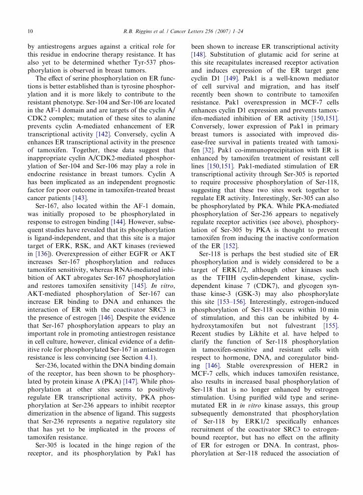
The effect of serine phosphorylation on ER func-tions is better established than is tyrosine phosphor-ylation and it is more likely to contribute to theresistant phenotype. Ser-104 and Ser-106 are locatedin the AF-1 domain and are targets of the cyclin A/CDK2 complex; mutation of these sites to alanineprevents cyclin A-mediated enhancement of ERtranscriptional activity [142]. Conversely, cyclin Aenhances ER transcriptional activity in the presenceof tamoxifen. Together, these data suggest thatinappropriate cyclin A/CDK2-mediated phosphor-ylation of Ser-104 and Ser-106 may play a role inendocrine resistance in breast tumors. Cyclin Ahas been implicated as an independent prognosticfactor for poor outcome in tamoxifen-treated breastcancer patients [143].
Ser-167, also located within the AF-1 domain,was initially proposed to be phosphorylated inresponse to estrogen binding [144]. However, subse-quent studies have revealed that its phosphorylationis ligand-independent, and that this site is a majortarget of ERK, RSK, and AKT kinases (reviewedin [136]). Overexpression of either EGFR or AKTincreases Ser-167 phosphorylation and reducestamoxifen sensitivity, whereas RNAi-mediated inhi-bition of AKT abrogates Ser-167 phosphorylationand restores tamoxifen sensitivity [145]. In vitro,AKT-mediated phosphorylation of Ser-167 canincrease ER binding to DNA and enhances theinteraction of ER with the coactivator SRC3 inthe presence of estrogen [146]. Despite the evidencethat Ser-167 phosphorylation appears to play animportant role in promoting antiestrogen resistancein cell culture, however, clinical evidence of a defin-itive role for phosphorylated Ser-167 in antiestrogenresistance is less convincing (see Section 4.1).
Ser-236, located within the DNA binding domainof the receptor, has been shown to be phosphory-lated by protein kinase A (PKA) [147]. While phos-phorylation at other sites seems to positivelyregulate ER transcriptional activity, PKA phos-phorylation at Ser-236 appears to inhibit receptordimerization in the absence of ligand. This suggeststhat Ser-236 represents a negative regulatory sitethat has yet to be implicated in the process oftamoxifen resistance.
Ser-305 is located in the hinge region of thereceptor, and its phosphorylation by Pak1 has
been shown to increase ER transcriptional activity[148]. Substitution of glutamic acid for serine atthis site recapitulates increased receptor activationand induces expression of the ER target genecyclin D1 [149]. Pak1 is a well-known mediatorof cell survival and migration, and has itselfrecently been shown to contribute to tamoxifenresistance. Pak1 overexpression in MCF-7 cellsenhances cyclin D1 expression and prevents tamox-ifen-mediated inhibition of ER activity [150,151].Conversely, lower expression of Pak1 in primarybreast tumors is associated with improved dis-ease-free survival in patients treated with tamoxi-fen [32]. Pak1 co-immunoprecipitation with ER isenhanced by tamoxifen treatment of resistant celllines [150,151]. Pak1-mediated stimulation of ERtranscriptional activity through Ser-305 is reportedto require processive phosphorylation of Ser-118,suggesting that these two sites work together toregulate ER activity. Interestingly, Ser-305 can alsobe phosphorylated by PKA. While PKA-mediatedphosphorylation of Ser-236 appears to negativelyregulate receptor activities (see above), phosphory-lation of Ser-305 by PKA is thought to preventtamoxifen from inducing the inactive conformationof the ER [152].
Ser-118 is perhaps the best studied site of ERphosphorylation and is widely considered to be atarget of ERK1/2, although other kinases suchas the TFIIH cyclin-dependent kinase, cyclin-dependent kinase 7 (CDK7), and glycogen syn-thase kinse-3 (GSK-3) may also phosphorylatethis site [153–156]. Interestingly, estrogen-inducedphosphorylation of Ser-118 occurs within 10 minof stimulation, and this can be inhibited by 4-hydroxytamoxifen but not fulvestrant [155].Recent studies by Likhite et al. have helped toclarify the function of Ser-118 phosphorylationin tamoxifen-sensitive and resistant cells withrespect to hormone, DNA, and coregulator bind-ing [146]. Stable overexpression of HER2 inMCF-7 cells, which induces tamoxifen resistance,also results in increased basal phosphorylation ofSer-118 that is no longer enhanced by estrogenstimulation. Using purified wild type and serine-mutated ER in in vitro kinase assays, this groupsubsequently demonstrated that phosphorylationof Ser-118 by ERK1/2 specifically enhancesrecruitment of the coactivator SRC3 to estrogen-bound receptor, but has no effect on the affinityof ER for estrogen or DNA. In contrast, phos-phorylation at Ser-118 reduced the association of

R.B. Riggins et al. / Cancer Letters 256 (2007) 1–24 11
ER with trans-hydroxytamoxifen, which has thepotential to attenuate the growth inhibitory effectsof this antiestrogen.
The role of Ser-118 phosphorylation in anties-trogen resistant cell culture models is not fullyunderstood. Kuske et al. have recently reportedthat estrogen-induced Ser-118 phosphorylationlevels are comparable in parental MCF-7 cellsand the estrogen-independent MCF-7/LCC1 cellline, but that basal and estrogen-induced levelsof phospho-Ser-118 are significantly reduced inthe antiestrogen-resistant MCF-7/LCC9 cell line[65]. In studies by other investigators, baselinephosphorylation of Ser-118 is increased in tamox-ifen-resistant MCF-7 cells as compared to theparental cell line and is reduced by MEK andEGFR tyrosine kinase inhibitors, which also inhi-bit cell growth [157]. Finally, Weitsman et al. sug-gest that, although both HER2 overexpressionand activation of the MAPK pathway can induceantiestrogen resistance, neither has any effect onSer-118 phosphorylation of ER in MCF-7 cells[158]. Instead, they suggest that the activity ofIkB kinase-a (IKKa) is involved. A potential rolefor IKKa in ER phosphorylation has onlyrecently been suggested [159], and further studiesare needed to clarify its role in antiestrogenresistance.
2.6. Cell cycle regulators
Cyclins, cyclin-dependent kinases (CDKs), andCDK inhibitors are the major regulators of cellcycle progression. Antiestrogens like tamoxifeninduce cell cycle arrest during the G1 phase;therefore, genes that control cell cycle progressionhave the potential to significantly impact drugsensitivity and resistance. In breast cancer, normalexpression and function of cyclins D1 and E, andthe CDK inhibitors p21 and p27, are often specif-ically disrupted. Caldon et al. have recently pub-lished a comprehensive review of cell cyclecontrol in breast cancer that summarizes how cyc-lins and CDK inhibitors influence cancer progres-sion and may serve as markers of treatmentoutcome [160]. Butt et al. have also published asummary of the literature concerning the role ofcyclins D1 and E in endocrine resistance [161].Given the timeliness of these reviews, we will onlysummarize key information about these moleculesand focus on the most recent literature in thisarea.
2.6.1. Cyclin D1
Cyclin D1 is a well-studied contributor to anties-trogen resistance in breast cancer. Essential for earlyprogression through the G1 phase, cyclin D1 bothmodulates CDK4/6-dependent phosphorylation ofthe tumor suppressor Rb and sequesters the CDKinhibitors p21 and p27 [160,162]. Cyclin D1 is adirect transcriptional target of estrogen signaling,where its induction is mediated by ER/AP-1 andER/Sp1 transcription complexes; conversely, anties-trogens like tamoxifen inhibit cyclin D1 expression(discussed in [161]). AKT can indirectly affect cyclinD1 expression by phosphorylating and inhibitingthe activity of GSK3b, which normally inducescyclin D1 degradation [163]. In cell culture, stableoverexpression of cyclin D1 can confer resistanceto tamoxifen as well as fulvestrant [164]. Moreover,some cells that have acquired resistance to tamoxi-fen show increased expression of cyclin D1 [165].However, these cells are still growth-inhibited byfulvestrant.
Recent studies have shed some light on potentialmechanisms of cyclin D1 upregulation in the con-text of antiestrogen resistance, some of whichinvolve crosstalk between growth factor receptorsand other signaling molecules. Tamoxifen-resistantMCF-7 cells overexpressing HER2 upregulatecyclin D1 [90]. Similarly, the tamoxifen-resistantphenotype of MCF-7 cells overexpressing Pak1 ispartly due to the increased expression of cyclin D1[32]. BCAR3 overexpression in MCF-7 also inducescyclin D1 promoter activity, coincident with theadoption of antiestrogen resistance [117]. In someinstances, tamoxifen has been reported to stimulatebreast cancer, although this has been observed morewidely in cell culture and xenograft studies than inthe clinic. Under conditions in which tamoxifen orraloxifene functions as an ER agonist, cyclin D1regulates cell cycle progression much as it does inparental MCF-7 cells stimulated by estrogen. Forexample, cyclin D1 expression was found to be sig-nificantly elevated in raloxifene-stimulated MCF-7xenografts [166]. Treatment with the HER2 inhibi-tor trastuzumab decreased cyclin D1 expression inthese xenografts and concomitantly inhibited tumorgrowth. Similarly, cyclin D1 was shown to be essen-tial for cell cycle progression induced by tamoxifenin resistant MCF-7 cells grown in tissue culture[167].
Evidence for a role of cyclin D1 in clinical endo-crine resistance is still emerging (reviewed in [160]).Cyclin D1 is overexpressed in �50% of breast

12 R.B. Riggins et al. / Cancer Letters 256 (2007) 1–24
cancers. Some studies have reported that cyclin D1overexpression is more often observed in highly dif-ferentiated, slower growing tumors than in highlyproliferative tumors. Other studies report thatcyclin D1 amplification is more frequent in ER-positive tumors, whereas overexpression in theabsence of amplification is more common in ER-negative tumors [168]. Not surprisingly, it is in theER-positive cases where cyclin D1 amplification isassociated with a worse outcome. How these find-ings will affect the clinical management of ER-posi-tive breast tumors and future use of tamoxifentherapy remains to be determined.
2.6.2. Cyclin E
In response to inhibition of Rb by cyclin D1 earlyin G1 phase, the transcription factor E2F stronglyinduces the expression of cyclin E, which associateswith CDK2 to form an active complex that pro-motes entry into S phase [160]. Cyclin E/CDK2activity is subsequently counterbalanced by theCDK inhibitors p21 and p27. While not a direct tar-get of the ER, estrogen-dependent induction of c-Myc can activate cyclin E complexes by stimulatingrelease of the inhibitor p21. CDC25A can also par-ticipate in estrogen-mediated cyclin E activation[169].
The contribution of cyclin E to tamoxifen resis-tance is less clear than for cyclin D1. This may bedue to the presence of multiple low molecularweight forms of cyclin E that are generated bypost-translational proteolytic cleavage events(reviewed in [170]). Low molecular weight cyclin Evariants, which exhibit greater activity than full-length cyclin E, can induce antiestrogen resistancein MCF-7 cells [171]. Further studies are requiredto clarify the mechanism(s) by which low molecularweight cyclin E is induced in breast cancer, and itsfunction in antiestrogen resistance. Other groupshave reported that full-length cyclin E can also con-fer resistance to tamoxifen and fulvestrant [164,172].
2.6.3. p21 and p27
The CDK inhibitors p21 and p27 are importantnegative regulators of cell cycle progression thatcounteract the activities of cyclin D1 and cyclin E.p27 is largely regulated in a post-transcriptionalmanner, while p21 is primarily controlled at thelevel of transcription (reviewed in [160]). In MCF-7 cells, both tamoxifen and fulvestrant increase theexpression of p21 and p27 during cell cycle arrest[173]. p27 induction by tamoxifen induces quies-
cence in breast cancer cells and prevents a positiveproliferative response following re-stimulation bygrowth factors such as IGF-I and EGF [174]. Com-bined treatment with tamoxifen and the farnesyltransferase inhibitor FTI-277, which causes a potentG1 arrest, maintains the expression and associationof p21 and p27 with cyclin E/CDK2 complexes [23].These two studies suggest that preservation of p27and p21 is critical for the inhibitory effects of anties-trogens like tamoxifen in controlling breast cancercell growth. In contrast, downregulation of theseCDK inhibitors prevents the growth inhibitoryeffects of these drugs [173].
p21 and p27 functions are regulated in part byAKT through phosphorylation of residues withintheir nuclear localization sequences, leading tosequestration in the cytoplasm where they areunable to interact with cyclin E and CDK2 (dis-cussed in [175]). c-Myc also plays a role in regulat-ing p21, although the mechanism is less clear.Most significantly, c-Myc appears to block tamoxi-fen- and fulvestrant-induced expression of p21 [176].In MCF-7/LCC9 breast cancer cells that haveacquired resistance to fulvestrant and cross-resis-tance to tamoxifen, p21 expression is no longer reg-ulated by estrogens and antiestrogens [176]. Studiesof other models of antiestrogen resistance also sup-port the idea that p21 and/or p27 play importantroles in antiestrogen sensitivity and resistance.Unlike parental MCF-7 cells in which tamoxifenor fulvestrant treatment increases p27 binding tocyclin E, antiestrogens do not alter p27 binding inthe tamoxifen-resistant LY-2 variant [177]. TheseLY-2 cells display constitutive activation of MEKand ERK1/2, and inhibition of these kinases par-tially restores antiestrogen sensitivity and p27 func-tion. Other cell culture models of resistance, such asMCF-7/LCC2 and MCF-7/HER2-18 cells, exhibitincreased MEK/ERK activity, although whetherthis affects antiestrogen resistance through modulat-ing p27 remains to be determined [177]. These datasupporting a role for p21 and p27 in antiestrogenresponses and resistance are corroborated by clini-cal evidence (see Section 4.1).
3. Endocrine therapies; effect on motility/invasion/
metastasis
Metastatic breast cancer is ultimately the greatestcause of disease mortality. Tamoxifen is beneficialfor decreasing secondary disease occurrence, bothat local and distant sites [178]. However, the major
R.B. Riggins et al. / Cancer Letters 256 (2007) 1–24 13
phenotype of endocrine resistance is cancer recur-rence. While clinical data have not definitivelylinked antiestrogen resistance with metastasis, manyof the proteins/pathways discussed above in Section2 that are involved in circumventing antiestrogen-induced blocks in cell cycle progression and survivalare also implicated in increased migration andmetastasis of breast cancer cells.
Hiscox et al. have begun to tackle the molecularrelationship between antiestrogen resistance andinvasion/metastasis [179]. They found that MCF-7cells selected for tamoxifen resistance showedenhanced basal motility and an invasive phenotype.Treatment of these cells with the pharmacologicalinhibitors gefitinib and AZD0530 inhibited migra-tion and invasion, indicating that the invasive phe-notype of these resistant cells was dependent onEGFR and c-Src, respectively. The ability of tumorcells to metastasize requires multiple genotypic andphenotypic changes. Signaling perturbations associ-ated with the epithelial to mesenchymal transition(EMT) contribute significantly to invasive andmetastatic disease [180]. These include increasedexpression of extracellular matrix proteins, down-regulation of E-cadherin, disruption of adherensjunctions complexes, accumulation of b-cateninand NF-kB in the nucleus, and transcription of tar-get genes such as c-Myc and cyclin D1 [181,182].Interestingly, the tamoxifen-resistant MCF-7 cellsinvestigated by Hiscox et al. displayed an alteredmorphology reminiscent of an EMT, marked byan elongated, fibroblast-like phenotype that lackedcharacteristic cell-cell contacts [179]. Tyrosine phos-phorylation of b-catenin was elevated in the tamox-ifen-resistant cells, coincident with accumulation ofthe protein, loss of its association with E-cadherin,increased interactions with EGFR, and increasedtranscription of c-Myc and cyclin D1. These effectswere reversed in the presence of gefitinib, support-ing a mechanism whereby the EGFR drives phos-phorylation of b-catenin and activation of c-Src toinitiate EMT-mediated metastatic progression.
Many of the other molecules that have been men-tioned above in the context of promoting antiestro-gen resistance also play a role in promotinginvasiveness. For example, Pak1 is functionallylinked to regulation of the actin cytoskeletonthrough Rho family GTPases [150]. The Pak1 geneis closely linked to CCND1 (cyclin D1) at 11q13, aregion that is often amplified in breast cancers[183]. Similarly, amplification of the gene encodingc-Myc in breast tumors appears to correlate with
the transition from in situ to invasive carcinoma[38,184]. Finally, in a study of matched primaryand metastatic breast tumors, Bcl-2 expression wasfound to be elevated in metastatic tumors coincidentwith a retention of ERa expression [38]. Theseresults conflicted with those from a similar studyperformed on paired primary and recurrent tamox-ifen-resistant tumors, which found that the recur-rent tumors did not show a correlation betweenER status and Bcl-2 expression [30]. However, therewas a correlation between ERa and phosphorylatedp38 and ERK in these relapsed specimens. Thesediscrepancies may be due to differences in inclusioncriteria and the source of recurrent tissue betweenstudies. The former study evaluated mostly tumorsat distant sites after discontinuation of hormonaltherapy, while the latter study evaluated locoregion-al recurrences while the patients continued therapy.
4. Antiestrogen resistance: implications for treatment
4.1. Prediction of response
It is clear from the discussion above that resis-tance to endocrine therapy likely occurs throughdiverse mechanisms that vary from patient topatient. Consequently, it is imperative to developmolecular signatures that can predict the likelihoodof response and potential for relapse of individualtumors. Initially, predictive markers for tamoxifenresistance included expression of ER and receptortyrosine kinases such as EGFR and HER2. Asour understanding of antiestrogen resistance hasevolved, these markers have broadened. Currently,the overall expression of proteins, protein modifica-tions, and intracellular localization are all beinganalyzed for predictive value of response to endo-crine therapy.
As mentioned above, survival pathways regu-lated by PI3K, AKT, PTEN, and Bcl-2 family mem-bers play an important role in antiestrogenresistance. Studies are currently underway to deter-mine whether increased activation and/or expres-sion of AKT, decreased activity of PTEN, oraltered expression of Bad could be predictive mark-ers for response to anti-hormonal treatments. Forexample, a retrospective analysis of 402 ERa-posi-tive breast carcinoma biopsies treated with tamoxi-fen for a median of 5 years revealed that highexpression of AKT2, as measured by immunohisto-chemistry, was predictive of improved overallsurvival [31]. Patients who relapsed following

14 R.B. Riggins et al. / Cancer Letters 256 (2007) 1–24
tamoxifen treatment were more likely to have lowAKT2 expression and high cytoplasmic phospho-AKT (Ser-473). Interestingly, expression of AKT1and AKT3 did not have a significant predictive cor-relation. Bad expression could also predict tamoxi-fen-treatment outcome. Patients in this sameclinical study whose tumors had high levels of Badexpression had improved disease-free survival ratescompared to patients whose tumors had low levelsof Bad [36]. There was no association between phos-pho-Bad (Ser-112), Bax, Bcl-2 or Bcl-xL expressionand response to tamoxifen treatment in this groupof patients. A related study examined 252 primaryERa human breast carcinomas to determinewhether activated AKT or loss of heterozygosity(LOH) of PTEN was predictive of response totamoxifen [185]. Phospho-AKT correlated withHER2 status in this study, as did PTEN LOH.From this patient cohort, 36 patient samples wereused to examine the relationship between activatedAKT and the efficacy of endocrine therapy for met-astatic breast cancer. Patients with phospho-AKT-positive tumors received less benefit from endocrinetherapy than did patients with phospho-AKT-nega-tive tumors. A third study evaluated PTEN expres-sion in 100 patients with ERa breast carcinomaswho had been treated with tamoxifen [186]. Highlevels of PTEN were recorded for 81% of patientswho had non-recurrent disease and only 41% ofpatients with recurrent disease. Conversely, lowPTEN expression was associated with a shorterrelapse free survival and reduced disease-specificsurvival. PTEN expression was an even strongerpredictor of relapse and survival when the analysiswas restrained to only stage I breast cancer patients.
Like the PI3K/AKT survival pathway, cyclin D1expression is associated with multiple mechanismsof antiestrogen resistance. Patients with ERa-posi-tive tumors and high levels of cyclin D1 mRNAhad a worse prognosis for overall survival [187].In a small subset of these patients who were treatedwith endocrine therapy, an apparent associationbetween high cyclin D1 mRNA and decreased dura-tion of response was made. Similar results wereobtained by evaluating protein levels of cyclin D1in 248 post-menopausal patients who had been ran-domized into 2-year tamoxifen treatment or no-treatment groups [188]. Interestingly, the 55strongly ERa-positive patients with tumors showingmoderate or low cyclin D1 levels responded totamoxifen treatment, whereas the 46 patients withboth high ERa and cyclin D1 expression in their
tumors showed no difference in survival betweentamoxifen and no treatment.
The association between cyclin E levels and anti-estrogen response is even less clear. Some of theclinical studies focused on cyclin E have shown acorrelation between the presence of low molecularweight and/or full-length cyclin E and poor sur-vival, while others have not found such an associa-tion ([189] and discussed in [161]). A recent studyexamining the expression of cyclin E and two prote-ases implicated in the generation of low molecularweight cyclin E (proteinase 3 and neutrophil elas-tase) in 205 breast cancer patients revealed thatcyclin E was significantly associated with poor dis-ease-free survival in the entire cohort, and withtamoxifen resistance in the 110 treated patients[190]. However, expression of neutrophil elastasewas not associated with poor outcome or treatmentresistance. Further studies are required to clarify therole of these variants in clinical endocrine resistance.
Coincident with cyclin D1 and cyclin E expres-sion, p21 and p27 levels may also prove to be pre-dictive for clinical response to antiestrogenresistance. High levels of p27 expression in a groupof 512 patients with premenopausal breast cancerpatients treated with tamoxifen and the gonadotro-pin-releasing hormone agonist goserelin werestrongly associated with improved overall and dis-ease-free survival [191]. Data for p21 are less consis-tent, perhaps because they may be confounded bythe complex regulation of p21 expression and con-tributions from other proteins such as p53 andIRF-1 that may independently affect breast canceroutcome (discussed in [20,161]). It may also be thatsubcellular localization of p21, rather than overallexpression levels, will prove to be a better markerof therapeutic response and treatment outcome. Inline with this suggestion, Perez-Tenorio et al. haverecently reported that cytoplasmic p21 is associatedwith increased AKT activity and poor tamoxifenresponse in a cohort of 280 women [192].
As mentioned above, serine phosphorylation ofERa may also prove to be useful for predicting ther-apeutic response (see Section 2.5). Recent studieshave centered on the role of Ser-118 phosphoryla-tion in predicting clinical outcome in breast cancerpatients. In a series of 117 lymph node-negative pri-mary breast tumors from patients treated withtamoxifen following surgery with or without radia-tion, Murphy et al. observed Ser-118 phosphoryla-tion only in tumors with positive estrogen bindingactivity [193]. Phosphorylated Ser-118 was also
R.B. Riggins et al. / Cancer Letters 256 (2007) 1–24 15
observed more frequently in PR-positive tumorsthan PR-negative tumors, and patients with tumorspositive for Ser-118 phosphorylation showedincreased disease-free survival. Bergqvist et al.reported that increased MAPK activation andexpression of phosphorylated Ser-118 correlatedwith one another and also predicted better survival[194]. However, overexpression of HER2 was inver-sely correlated with activated ERK1/2 and phos-phorylated Ser-118, suggesting that at least in thiscohort, HER2 activation does not drive phosphory-lation and hyperactivation of ER. Moreover, thisapparent improved outcome observed in patientswith Ser-118 phosphorylation appears to contradictsome of the cell culture experiments that showed alink between Ser-118 phosphorylation and anties-trogen resistance (see Section 2.5). This discrepancymay be explained by the possibility that Ser-118phosphorylation of the ER may have distinct func-tions in different phases of breast cancer; there issome evidence to suggest that it may be associatedwith a better outcome prior to endocrine therapybut a worse outcome following acquisition oftamoxifen resistance [35].
The correlation between Ser-167 phosphoryla-tion and clinical response to antiestrogens is equallycomplex. Kirkegaard et al. found that high levels ofactive AKT correlated with reduced survival andincreased Ser-167 phosphorylation in tamoxifen-treated patients [31]. However, Yamashita et al.reported that phosphorylated Ser-167, along withexpression of ER and PR, were associated with animproved response to endocrine therapy and sur-vival [195]. The fact that Ser-167 can be phosphory-lated by kinases other than AKT may suggest that itis AKT rather than phosphorylated Ser-167 that isresponsible for the poor outcome in the Kirkegaardstudy [31].
In addition to the ERa modulation, PR statushas also been suggested to be predictive of responseto antiestrogens. As discussed above (Section 2.1),the presence of PR is reflective of a functional ERpathway because PR synthesis is initiated by ER-induced transcription. Bardou et al. analyzed twodatabases containing information on ER and PRstatus of a large number of breast cancer patients[196]. They found that PR expression was associatedwith disease-free survival independently of ERa sta-tus. However, ERa-positive/PR-positive tumorsreceived the greatest benefit from adjuvant hor-monal therapy when both ERa and PR were takeninto consideration, while loss of expression of either
receptor reduced responsiveness by nearly half.Additional studies have also found that ER/PR-positive breast tumors have significant improvementin prognosis after either adjuvant tamoxifen orletrozole treatments [197,198]. This supports thehypothesis that ERa positive tumors that lack PRexpression are less likely to respond to anti-hor-monal therapy because they are no longer depen-dent on estrogen for survival. Conversely, ERbexpression appears to be predictive of a positiveresponse to endocrine therapy and better overallsurvival [6]. This is potentially due to the putativetumor suppressor functions of ERb discussed above(see Section 1), and provides the rationale for devel-oping SERMs that exhibit selective activities to ERsubtypes (see Section 1.1.1).
4.2. Molecular profiling studies of antiestrogen
resistance
Gene expression analysis and molecular profilinghave added to our understanding of the mechanismsof antiestrogen response and resistance. Severalgroups have used these approaches on tumor sam-ples to develop gene signatures that can predict clin-ical responses to antiestrogen treatment [199,200].However, in some instances, these profiles havenot been validated by independent studies [201].Expression profiling of cell culture and xenograftmodels has also been extensively utilized to helpclarify the molecular actions of antiestrogens andaromatase inhibitors. Several of these studies haveconfirmed biological evidence indicating thatSERMs like tamoxifen and raloxifene can behaveas both antagonists and agonists of the ER, whereasfulvestrant is a pure antagonist. For example,expression of cell cycle-associated genes such ascyclin A1 and CDC25B was unaffected by estrogen,tamoxifen, or raloxifene but strongly regulated byfulvestrant [202]. The effects of tamoxifen andraloxifene were also similar to estrogen with respectto expression of Bcl-2-interacting killer (BIK), anestrogen-inhibited, pro-apoptotic gene that has beendemonstrated to play a critical role in fulvestrant-induced breast cancer cell death [203].
Other studies have examined regulation of geneexpression by letrozole and anastrazole as com-pared to tamoxifen [204]. As might be expected,the effects of these aromatase inhibitors were moresimilar to each other than they were to tamoxifen.However, the major functional classes of genes reg-ulated by all three drugs were comparable, and

16 R.B. Riggins et al. / Cancer Letters 256 (2007) 1–24
included apoptosis, Wnt, and MAPK signalingpathways. This suggests that, rather than lookingat individual genes, these approaches may be mostuseful for identifying functional groupings of genesthat work together in specific signaling pathways toregulate responses to antiestrogens. A recent studyin MCF-7 cells comparing acquired resistance totamoxifen and fulvestrant found that tamoxifenresistance was strongly associated with changes inthe PKA pathway, MAPK phosphatases, and cal-cium binding proteins. In contrast, downregulationof ER and major alterations in growth factor recep-tor and interferon signaling were observed in cellsresistant to fulvestrant [60]. Interferon-induciblegenes have also been found to be differentially regu-lated in the MaCa 3366/TAM xenograft model ofacquired tamoxifen resistance when compared tothe tamoxifen-sensitive MaCa 3366 counterpart[205]. Interestingly, loss of the putative tumor sup-pressor interferon regulatory factor 1 (IRF-1) hasbeen specifically implicated in fulvestrant resistance[25] and is required for tamoxifen-induced cell deathof normal mammary epithelial cells [206]. Anotherinterferon-induced gene, calgranulin A (S100A8)[207], is upregulated in MCF-7, T47D, and ZR-75-1-derived models of fulvestrant resistance [59],providing additional support for a role of inter-feron-inducible genes in antiestrogen resistance.
While expression profiling has provided insightinto molecular pathways that may contribute toantiestrogen resistance, there are some challengesassociated with these approaches that must be care-fully considered. Multiple expression array plat-forms and technologies can make comparing thefindings of different investigators difficult, especiallyat the level of individual genes. Statistical analysis ofthese data is also very complex, and the risk ofexcluding biologically relevant genes while includingothers that do not contribute to the phenotype is asignificant one. Unfortunately, these challenges aregreatly magnified in the context of analysis of tumortissues [208–210]. Nonetheless, these approaches canbe quite powerful, particularly when one considersfunctional grouping of genes rather than individualmolecules.
4.3. Combinatorial therapies
Strategies for treating breast tumors have signif-icantly broadened in recent years because of a grow-ing appreciation for the fact that resistance toendocrine therapies can and will invariably arise
through a variety of distinct mechanisms. Ourincreasing understanding of the molecular basisfor these failures has brought to the forefront thepossibility of using combination therapies for treat-ing breast cancer. There are two separate rationalesfor using combination therapies. First, targetingmultiple pathways at once can potentially delay orhalt the acquisition of a resistant phenotype. Alter-natively, treatments can be administered to effec-tively resensitize tumors to endocrine therapies forwhich they are resistant. In many cases, the practicalstrategies for addressing these distinct phenomenaare the same. For example, as discussed above,growth factor receptor signaling is often increasedin antiestrogen-resistant breast tumors, leading toupregulation of proliferation and survival pathwaysthat may be distinct from estrogen signaling per se.Thus, treatment with gefitinib or trastuzumab toinhibit the EGFR or HER2, respectively, along withhormonal therapies would potentially block boththe pro-proliferative activity of the ER and the pro-liferation/survival pathways activated by the growthfactor receptors. Some of the current trials evaluat-ing the efficacy of these combinations include aPhase II randomized trial (Translational OncologyResearch International trial) that is investigatingfulvestrant and trastuzumab in combination as afirst-line treatment for patients with ERa/HER2-positive breast tumors, a comparative study evaluat-ing anastrozole versus fulvestrant with the additionof gefitinib in women with ERa-positive tumorswho have metastatic disease and have failed chemo-therapy and/or endocrine therapy, and a combina-tion of fulvestrant with the dual inhibitor lapatinibin postmenopausal women with ERa-positive andEGFR-and/or HER2-postive tumors who havehad previous exposure to an aromatase inhibitor(reviewed in [211,212]).
In addition to targeting growth factor receptors,several clinical trials have been initiated to investi-gate the effect of farnesyl transferase inhibitors suchas tipifarnib in combination with endocrine thera-pies. However, these trials have been somewhat dis-appointing in that they have not thus farcorroborated pre-clinical models showing that tipi-farnib in combination with tamoxifen resulted ingreater tumor regression than either treatment alone[213]. Additional studies are underway that examinethe efficacy of combining tipifarnib with AIs(reviewed in [214]).
Clinical studies with mTOR antagonists in com-bination with hormonal therapies are also ongoing.

R.B. Riggins et al. / Cancer Letters 256 (2007) 1–24 17
As a single agent, CCI-779 has been somewhat effec-tive in Phase II studies of metastatic breast cancer[215]. Currently, Phase II and Phase III trials aretesting either CCI-779 or RAD-001 in combinationwith letrozole, as compared to letrozole alone.Interim results show that the combination of CCI-779 and letrozole translates into a clinical benefitrate of 77% versus 66% for letrozole alone, althoughthese values have not yet reached statistical signifi-cance [135].
Finally, extensive efforts are being made todetermine whether pre-clinical data showing thattumors resistant to one class of hormonal therapymay still retain sensitivity to alternate endocrinetherapies hold true in the clinic. Results of thePhase II Swiss Group for Clinical Research Trial(SAKK 21/00), which compared the efficacy offulvestrant treatment in AI-sensitive versus AI-resistant patients, were recently published(reviewed in [216]). Overall fulvestrant yielded aclinical benefit (measured as an objective responseor stable disease >24 weeks) in 30% of patients,irrespective of whether they had previously beentreated with AIs. There are currently multipleplanned or ongoing clinical trials combining AIswith antiestrogens. These include Phase II andPhase III trials evaluating use as first line andneoadjuvant treatment (reviewed in [214]).
Acknowledgments
The authors acknowledge support of our researchfrom the Susan G. Komen Breast Cancer Founda-tion (PDF0503551) and Department of DefenseBreast Cancer Research Program (BC051851) toRBR, the Department of Defense Breast Cancer Re-search Program (BC050339) to RSS, the NationalCancer Institute institutional training Grant (T32CA009109) for R.S.S. and M.S.G.; and the NationalInstitutes of Health (R01 CA 096846 to AHB andR01 CA 123037 to Sarah J. Parsons, AHB Co-PI).
References
[1] P. Ascenzi, A. Bocedi, M. Marino, Structure–functionrelationship of estrogen receptor alpha and beta: impact onhuman health, Mol. Aspects Med. 27 (2006) 299–402.
[2] E. Anderson, R.B. Clarke, Steroid receptors and cell cyclein normal mammary epithelium, J. Mammary Gland Biol.Neoplasia 9 (2004) 3–13.
[3] V. Speirs, G.P. Skliris, S.E. Burdall, P.J. Carder, Distinctexpression patterns of ER alpha and ER beta in normalhuman mammary gland, J. Clin. Pathol. 55 (2002) 371–374.
[4] C. Forster, S. Makela, A. Warri, S. Kietz, D. Becker, K.Hultenby, et al., Involvement of estrogen receptor beta interminal differentiation of mammary gland epithelium,Proc. Natl. Acad. Sci. USA 99 (2002) 15578–15583.
[5] S. Saji, M. Hirose, M. Toi, Clinical significance of estrogenreceptor beta in breast cancer, Cancer Chemother. Phar-macol. 56 (Suppl. 1) (2005) 21–26.
[6] L.C. Murphy, P.H. Watson, Is oestrogen receptor-beta apredictor of endocrine therapy responsiveness in humanbreast cancer?, Endocr. Relat. Cancer 13 (2006) 327–334.
[7] J. Matthews, B. Wihlen, M. Tujague, J. Wan, A. Strom,J.A. Gustafsson, Estrogen receptor (ER) beta modulatesER alpha-mediated transcriptional activation by alteringthe recruitment of c-fos and c-jun to estrogen-responsivepromoters, Mol. Endocrinol. 20 (2006) 534–543.
[8] L.A. Helguero, M.H. Faulds, J.A. Gustafsson, L.A.Haldosen, Estrogen receptors alpha (ER alpha) and beta(ER beta) differentially regulate proliferation and apoptosisof the normal murine mammary epithelial cell line HC11,Oncogene 24 (2005) 6605–6616.
[9] F. Stossi, D.H. Barnett, J. Frasor, B. Komm, C.R. Lyttle,B.S. Katzenellenbogen, Transcriptional profiling of estro-gen-regulated gene expression via estrogen receptor (ER)alpha or ER beta in human osteosarcoma cells: distinct andcommon target genes for these receptors, Endocrinology145 (2004) 3473–3486.
[10] E.C. Chang, J. Frasor, B. Komm, B.S. Katzenellenbogen,Impact of estrogen receptor beta on gene networks regu-lated by estrogen receptor alpha in breast cancer cells,Endocrinology 147 (2006) 4831–4842.
[11] J.S. Lewis, V.C. Jordan, Selective estrogen receptor mod-ulators (SERMs): mechanisms of anticarcinogenesis anddrug resistance, Mutat. Res. 591 (2005) 247–263.
[12] B. Fisher, J.P. Costantino, D.L. Wickerham, C.K. Red-mond, M. Kavanah, W.M. Cronin, et al., Tamoxifen forprevention of breast cancer: Report of the NationalSurgical Adjuvant Breast and Bowel Project P-1 Study, J.Natl. Cancer Inst. 90 (1998) 1371–1388.
[13] G.H. Veeneman, Non-steroidal subtype selective estrogens,Curr. Med. Chem. 12 (2005) 1077–1136.
[14] A. Escande, A. Pillon, N. Servant, J.P. Cravedi, F. Larrea,P. Muhn, et al., Evaluation of ligand selectivity usingreporter cell lines stably expressing estrogen receptor alphaor beta, Biochem. Pharmacol. 71 (2006) 1459–1469.
[15] T.A. Blizzard, C. Gude, J.D. Morgan 2nd, W. Chan, E.T.Birzin, M. Mojena, et al., Androstenediol analogs as ER-beta-selective SERMs, Bioorg. Med. Chem. Lett. 16 (2006)834–838.
[16] A.U. Buzdar, TAS-108: a novel steroidal antiestrogen, Clin.Cancer Res. 11 (2005) 906S–908S.
[17] A. Howell, Pure oestrogen antagonists for the treatment ofadvanced breast cancer, Endocr. Relat. Cancer 13 (2006)689–706.
[18] C.K. Osborne, A. Wakeling, R.I. Nicholson, Fulvestrant:an oestrogen receptor antagonist with a novel mechanismof action, Br. J. Cancer 90 (Suppl. 1) (2004) S2–S6.
[19] R.W. Brueggemeier, Update on the use of aromataseinhibitors in breast cancer, Expert Opin. Pharmacother. 7(2006) 1919–1930.
[20] R.B. Riggins, A.H. Bouton, M.C. Liu, R. Clarke, Anties-trogens, aromatase inhibitors, and apoptosis in breastcancer, Vitam. Horm. 71 (2005) 201–237.

18 R.B. Riggins et al. / Cancer Letters 256 (2007) 1–24
[21] S. Chen, S. Masri, X. Wang, S. Phung, Y.C. Yuan, X. Wu,What do we know about the mechanisms of aromataseinhibitor resistance?, J. Steroid Biochem. Mol. Biol. 102(2006) 232–240.
[22] J.S. Carroll, O.W. Prall, E.A. Musgrove, R.L. Sutherland,A pure estrogen antagonist inhibits cyclin E-CDK2 activityin MCF-7 breast cancer cells and induces accumulation ofp130-E2F4 complexes characteristic of quiescence, J. Biol.Chem. 275 (2000) 38221–38229.
[23] S.F. Doisneau-Sixou, P. Cestac, J.C. Faye, G. Favre, R.L.Sutherland, Additive effects of tamoxifen and the farnesyltransferase inhibitor FTI-277 on inhibition of MCF-7breast cancer cell-cycle progression, Int. J. Cancer 106(2003) 789–798.
[24] A. Thiantanawat, B.J. Long, A.M. Brodie, Signalingpathways of apoptosis activated by aromatase inhibitorsand antiestrogens, Cancer Res. 63 (2003) 8037–8050.
[25] K.B. Bouker, T.C. Skaar, D.R. Fernandez, K.A. O’Brien,R.B. Riggins, D. Cao, et al., Interferon regulatory factor-1mediates the proapoptotic but not cell cycle arrest effects ofthe steroidal antiestrogen ICI 182,780 (faslodex, fulve-strant), Cancer Res. 64 (2004) 4030–4039.
[26] S.F. Doisneau-Sixou, C.M. Sergio, J.S. Carroll, R. Hui,E.A. Musgrove, R.L. Sutherland, Estrogen and antiestro-gen regulation of cell cycle progression in breast cancercells, Endocr. Relat. Cancer 10 (2003) 179–186.
[27] G. Arpino, S.J. Green, D.C. Allred, D. Lew, S. Martino,C.K. Osborne, et al., HER-2 amplification, HER-1 expres-sion, and tamoxifen response in estrogen receptor-positivemetastatic breast cancer: a Southwest Oncology GroupStudy, Clin. Cancer Res. 10 (2004) 5670–5676.
[28] J.M. Gee, J.F. Robertson, E. Gutteridge, I.O. Ellis, S.E.Pinder, M. Rubini, et al., Epidermal growth factorreceptor/HER2/insulin-like growth factor receptor signal-ling and oestrogen receptor activity in clinical breastcancer, Endocr. Relat. Cancer 12 (Suppl. 1) (2005)S99–S111.
[29] M. Dowsett, S. Johnston, L.A. Martin, J. Salter, M. Hills,S. Detre, et al., Growth factor signalling and response toendocrine therapy: The Royal Marsden Experience,Endocr. Relat. Cancer 12 (Suppl. 1) (2005) S113–S117.
[30] M.C. Gutierrez, S. Detre, S. Johnston, S.K. Mohsin, J.Shou, D.C. Allred, et al., Molecular changes in tamoxifen-resistant breast cancer: relationship between estrogenreceptor, HER-2, and p38 mitogen-activated proteinkinase, J. Clin. Oncol. 23 (2005) 2469–2476.
[31] T. Kirkegaard, C.J. Witton, L.M. McGlynn, S.M. Tovey,B. Dunne, A. Lyon, et al., AKT activation predictsoutcome in breast cancer patients treated with tamoxifen,J. Pathol. 207 (2005) 139–146.
[32] C. Holm, S. Rayala, K. Jirstrom, O. Stal, R. Kumar, G.Landberg, Association between Pak1 expression and sub-cellular localization and tamoxifen resistance in breastcancer patients, J. Natl. Cancer Inst. 98 (2006) 671–680.
[33] S. van der Flier, A. Brinkman, M.P. Look, E.M. Kok, M.E.Meijer-van Gelder, J.G. Klijn, et al., BCAR1/p130Cas
protein and primary breast cancer: prognosis and responseto tamoxifen treatment, J. Natl. Cancer Inst. 92 (2000)120–127.
[34] C.K. Osborne, V. Bardou, T.A. Hopp, G.C. Chamness,S.G. Hilsenbeck, S.A. Fuqua, et al., Role of the estrogenreceptor coactivator AIB1 (SRC-3) and HER-2/neu in
tamoxifen resistance in breast cancer, J. Natl. Cancer Inst.95 (2003) 353–361.
[35] N. Sarwar, J.S. Kim, J. Jiang, D. Peston, H.D. Sinnett, P.Madden, et al., Phosphorylation of ER alpha at serine 118in primary breast cancer and in tamoxifen-resistanttumours is indicative of a complex role for ER alphaphosphorylation in breast cancer progression, Endocr.Relat. Cancer 13 (2006) 851–861.
[36] E. Cannings, T. Kirkegaard, S.M. Tovey, B. Dunne, T.G.Cooke, J.M. Bartlett, Bad expression predicts outcome inpatients treated with tamoxifen, Breast Cancer Res. Treat.102 (2007) 173–179.
[37] S.P. Linke, T.M. Bremer, C.D. Herold, G. Sauter, C.Diamond, A multimarker model to predict outcome intamoxifen-treated breast cancer patients, Clin. Cancer Res.12 (2006) 1175–1183.
[38] M.D. Planas-Silva, R.D. Bruggeman, R.T. Grenko, J.S.Smith, Overexpression of c-Myc and Bcl-2 during progres-sion and distant metastasis of hormone-treated breastcancer, Exp. Mol. Pathol. 82 (2007) 85–90.
[39] E. Rahko, G. Blanco, R. Bloigu, Y. Soini, A. Talvensaari-Mattila, A. Jukkola, Adverse outcome and resistance toadjuvant antiestrogen therapy in node-positive postmeno-pausal breast cancer patients-the role of p53, Breast 15(2006) 69–75.
[40] G.G. Kuiper, E. Enmark, M. Pelto-Huikko, S. Nilsson,J.A. Gustafsson, Cloning of a novel receptor expressed inrat prostate and ovary, Proc. Natl. Acad. Sci. USA 93(1996) 5925–5930.
[41] D.P. McDonnell, The molecular determinants of estrogenreceptor pharmacology, Maturitas 48 (Suppl. 1) (2004)S7–S12.
[42] K. Paech, P. Webb, G.G. Kuiper, S. Nilsson, J. Gustafsson,P.J. Kushner, et al., Differential ligand activation ofestrogen receptors ER alpha and ER beta at AP1 sites,Science 277 (1997) 1508–1510.
[43] A. Zou, K.B. Marschke, K.E. Arnold, E.M. Berger, P.Fitzgerald, D.E. Mais, et al., Estrogen receptor betaactivates the human retinoic acid receptor alpha-1 promoterin response to tamoxifen and other estrogen receptorantagonists, but not in response to estrogen, Mol. Endo-crinol. 13 (1999) 418–430.
[44] C.L. Smith, B.W. O’Malley, Coregulator function: a key tounderstanding tissue specificity of selective receptor mod-ulators, Endocr. Rev. 25 (2004) 45–71.
[45] J.M. Hall, D.P. McDonnell, Coregulators in nuclearestrogen receptor action: from concept to therapeutictargeting, Mol. Interv. 5 (2005) 343–357.
[46] R. Kumar, A.E. Gururaj, R.K. Vadlamudi, S.K. Rayala,The clinical relevance of steroid hormone receptor core-pressors, Clin. Cancer Res. 11 (2005) 2822–2831.
[47] I. Girault, I. Bieche, R. Lidereau, Role of estrogenreceptor alpha transcriptional coregulators in tamoxifenresistance in breast cancer, Maturitas 54 (2006)342–351.
[48] R. Clarke, F. Leonessa, J.N. Welch, T.C. Skaar, Cellularand molecular pharmacology of antiestrogen action andresistance, Pharmacol. Rev. 53 (2001) 25–71.
[49] S.F. Honig, Treatment of metastatic disease: hormonaltherapy and chemotherapy, in: J.R. Harris, M.E. Lippman,M. Morrow, S. Helman (Eds.), Diseases of the Breast,Lippincott-Raven, Philadelphia, 1996, pp. 669–734.
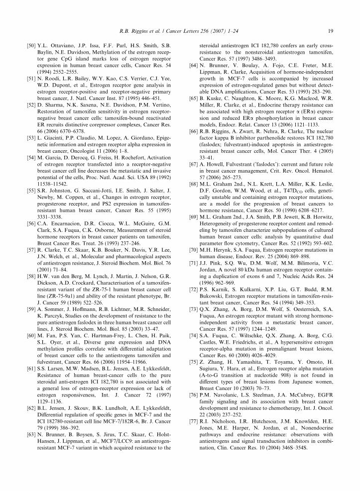
R.B. Riggins et al. / Cancer Letters 256 (2007) 1–24 19
[50] Y.L. Ottaviano, J.P. Issa, F.F. Parl, H.S. Smith, S.B.Baylin, N.E. Davidson, Methylation of the estrogen recep-tor gene CpG island marks loss of estrogen receptorexpression in human breast cancer cells, Cancer Res. 54(1994) 2552–2555.
[51] N. Roodi, L.R. Bailey, W.Y. Kao, C.S. Verrier, C.J. Yee,W.D. Dupont, et al., Estrogen receptor gene analysis inestrogen receptor-positive and receptor-negative primarybreast cancer, J. Natl. Cancer Inst. 87 (1995) 446–451.
[52] D. Sharma, N.K. Saxena, N.E. Davidson, P.M. Vertino,Restoration of tamoxifen sensitivity in estrogen receptor-negative breast cancer cells: tamoxifen-bound reactivatedER recruits distinctive corepressor complexes, Cancer Res.66 (2006) 6370–6378.
[53] L. Giacinti, P.P. Claudio, M. Lopez, A. Giordano, Epige-netic information and estrogen receptor alpha expression inbreast cancer, Oncologist 11 (2006) 1–8.
[54] M. Garcia, D. Derocq, G. Freiss, H. Rochefort, Activationof estrogen receptor transfected into a receptor-negativebreast cancer cell line decreases the metastatic and invasivepotential of the cells, Proc. Natl. Acad. Sci. USA 89 (1992)11538–11542.
[55] S.R. Johnston, G. Saccani-Jotti, I.E. Smith, J. Salter, J.Newby, M. Coppen, et al., Changes in estrogen receptor,progesterone receptor, and PS2 expression in tamoxifen-resistant human breast cancer, Cancer Res. 55 (1995)3331–3338.
[56] C.A. Encarnacion, D.R. Ciocca, W.L. McGuire, G.M.Clark, S.A. Fuqua, C.K. Osborne, Measurement of steroidhormone receptors in breast cancer patients on tamoxifen,Breast Cancer Res. Treat. 26 (1993) 237–246.
[57] R. Clarke, T.C. Skaar, K.B. Bouker, N. Davis, Y.R. Lee,J.N. Welch, et al., Molecular and pharmacological aspectsof antiestrogen resistance, J. Steroid Biochem. Mol. Biol. 76(2001) 71–84.
[58] H.W. van den Berg, M. Lynch, J. Martin, J. Nelson, G.R.Dickson, A.D. Crockard, Characterisation of a tamoxifen-resistant variant of the ZR-75-1 human breast cancer cellline (ZR-75-9a1) and ability of the resistant phenotype, Br.J. Cancer 59 (1989) 522–526.
[59] A. Sommer, J. Hoffmann, R.B. Lichtner, M.R. Schneider,K. Parczyk, Studies on the development of resistance to thepure antiestrogen faslodex in three human breast cancer celllines, J. Steroid Biochem. Mol. Biol. 85 (2003) 33–47.
[60] M. Fan, P.S. Yan, C. Hartman-Frey, L. Chen, H. Paik,S.L. Oyer, et al., Diverse gene expression and DNAmethylation profiles correlate with differential adaptationof breast cancer cells to the antiestrogens tamoxifen andfulvestrant, Cancer Res. 66 (2006) 11954–11966.
[61] S.S. Larsen, M.W. Madsen, B.L. Jensen, A.E. Lykkesfeldt,Resistance of human breast-cancer cells to the puresteroidal anti-estrogen ICI 182,780 is not associated witha general loss of estrogen-receptor expression or lack ofestrogen responsiveness, Int. J. Cancer 72 (1997)1129–1136.
[62] B.L. Jensen, J. Skouv, B.K. Lundholt, A.E. Lykkesfeldt,Differential regulation of specific genes in MCF-7 and theICI 182780-resistant cell line MCF-7/182R-6, Br. J. Cancer79 (1999) 386–392.
[63] N. Brunner, B. Boysen, S. Jirus, T.C. Skaar, C. Holst-Hansen, J. Lippman, et al., MCF7/LCC9: an antiestrogen-resistant MCF-7 variant in which acquired resistance to the
steroidal antiestrogen ICI 182,780 confers an early cross-resistance to the nonsteroidal antiestrogen tamoxifen,Cancer Res. 57 (1997) 3486–3493.
[64] N. Brunner, V. Boulay, A. Fojo, C.E. Freter, M.E.Lippman, R. Clarke, Acquisition of hormone-independentgrowth in MCF-7 cells is accompanied by increasedexpression of estrogen-regulated genes but without detect-able DNA amplifications, Cancer Res. 53 (1993) 283–290.
[65] B. Kuske, C. Naughton, K. Moore, K.G. Macleod, W.R.Miller, R. Clarke, et al., Endocrine therapy resistance canbe associated with high estrogen receptor a (ERa) expres-sion and reduced ERa phosphorylation in breast cancermodels, Endocr. Relat. Cancer 13 (2006) 1121–1133.
[66] R.B. Riggins, A. Zwart, R. Nehra, R. Clarke, The nuclearfactor kappa B inhibitor parthenolide restores ICI 182,780(faslodex; fulvestrant)-induced apoptosis in antiestrogen-resistant breast cancer cells, Mol. Cancer Ther. 4 (2005)33–41.
[67] A. Howell, Fulvestrant (’faslodex’): current and future rolein breast cancer management, Crit. Rev. Oncol. Hematol.57 (2006) 265–273.
[68] M.L. Graham 2nd., N.L. Krett, L.A. Miller, K.K. Leslie,D.F. Gordon, W.M. Wood, et al., T47DCO cells, geneti-cally unstable and containing estrogen receptor mutations,are a model for the progression of breast cancers tohormone resistance, Cancer Res. 50 (1990) 6208–6217.
[69] M.L. Graham 2nd., J.A. Smith, P.B. Jewett, K.B. Horwitz,Heterogeneity of progesterone receptor content and remod-eling by tamoxifen characterize subpopulations of culturedhuman breast cancer cells: analysis by quantitative dualparameter flow cytometry, Cancer Res. 52 (1992) 593–602.
[70] M.H. Herynk, S.A. Fuqua, Estrogen receptor mutations inhuman disease, Endocr. Rev. 25 (2004) 869–898.
[71] J.J. Pink, S.Q. Wu, D.M. Wolf, M.M. Bilimoria, V.C.Jordan, A novel 80 kDa human estrogen receptor contain-ing a duplication of exons 6 and 7, Nucleic Acids Res. 24(1996) 962–969.
[72] P.S. Karnik, S. Kulkarni, X.P. Liu, G.T. Budd, R.M.Bukowski, Estrogen receptor mutations in tamoxifen-resis-tant breast cancer, Cancer Res. 54 (1994) 349–353.
[73] Q.X. Zhang, A. Borg, D.M. Wolf, S. Oesterreich, S.A.Fuqua, An estrogen receptor mutant with strong hormone-independent activity from a metastatic breast cancer,Cancer Res. 57 (1997) 1244–1249.
[74] S.A. Fuqua, C. Wiltschke, Q.X. Zhang, A. Borg, C.G.Castles, W.E. Friedrichs, et al., A hypersensitive estrogenreceptor-alpha mutation in premalignant breast lesions,Cancer Res. 60 (2000) 4026–4029.
[75] Z. Zhang, H. Yamashita, T. Toyama, Y. Omoto, H.Sugiura, Y. Hara, et al., Estrogen receptor alpha mutation(A-to-G transition at nucleotide 908) is not found indifferent types of breast lesions from Japanese women,Breast Cancer 10 (2003) 70–73.
[76] P.M. Navolanic, L.S. Steelman, J.A. McCubrey, EGFRfamily signaling and its association with breast cancerdevelopment and resistance to chemotherapy, Int. J. Oncol.22 (2003) 237–252.
[77] R.I. Nicholson, I.R. Hutcheson, J.M. Knowlden, H.E.Jones, M.E. Harper, N. Jordan, et al., Nonendocrinepathways and endocrine resistance: observations withantiestrogens and signal transduction inhibitors in combi-nation, Clin. Cancer Res. 10 (2004) 346S–354S.
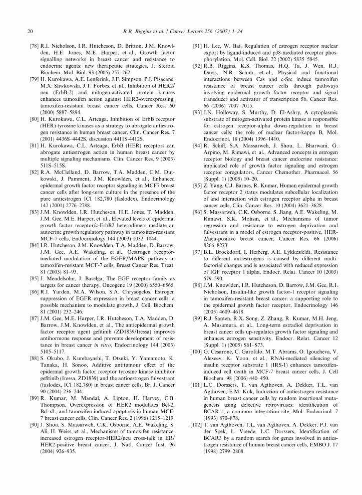
20 R.B. Riggins et al. / Cancer Letters 256 (2007) 1–24
[78] R.I. Nicholson, I.R. Hutcheson, D. Britton, J.M. Knowl-den, H.E. Jones, M.E. Harper, et al., Growth factorsignalling networks in breast cancer and resistance toendocrine agents: new therapeutic strategies, J. SteroidBiochem. Mol. Biol. 93 (2005) 257–262.
[79] H. Kurokawa, A.E. Lenferink, J.F. Simpson, P.I. Pisacane,M.X. Sliwkowski, J.T. Forbes, et al., Inhibition of HER2/neu (ErbB-2) and mitogen-activated protein kinasesenhances tamoxifen action against HER2-overexpressing,tamoxifen-resistant breast cancer cells, Cancer Res. 60(2000) 5887–5894.
[80] H. Kurokawa, C.L. Arteaga, Inhibition of ErbB receptor(HER) tyrosine kinases as a strategy to abrogate antiestro-gen resistance in human breast cancer, Clin. Cancer Res. 7(2001) 4436S–4442S, discussion 4411S-4412S.
[81] H. Kurokawa, C.L. Arteaga, ErbB (HER) receptors canabrogate antiestrogen action in human breast cancer bymultiple signaling mechanisms, Clin. Cancer Res. 9 (2003)511S–515S.
[82] R.A. McClelland, D. Barrow, T.A. Madden, C.M. Dut-kowski, J. Pamment, J.M. Knowlden, et al., Enhancedepidermal growth factor receptor signaling in MCF7 breastcancer cells after long-term culture in the presence of thepure antiestrogen ICI 182,780 (faslodex), Endocrinology142 (2001) 2776–2788.
[83] J.M. Knowlden, I.R. Hutcheson, H.E. Jones, T. Madden,J.M. Gee, M.E. Harper, et al., Elevated levels of epidermalgrowth factor receptor/c-ErbB2 heterodimers mediate anautocrine growth regulatory pathway in tamoxifen-resistantMCF-7 cells, Endocrinology 144 (2003) 1032–1044.
[84] I.R. Hutcheson, J.M. Knowlden, T.A. Madden, D. Barrow,J.M. Gee, A.E. Wakeling, et al., Oestrogen receptor-mediated modulation of the EGFR/MAPK pathway intamoxifen-resistant MCF-7 cells, Breast Cancer Res. Treat.81 (2003) 81–93.
[85] J. Mendelsohn, J. Baselga, The EGF receptor family astargets for cancer therapy, Oncogene 19 (2000) 6550–6565.
[86] R.I. Yarden, M.A. Wilson, S.A. Chrysogelos, Estrogensuppression of EGFR expression in breast cancer cells: apossible mechanism to modulate growth, J. Cell. Biochem.81 (2001) 232–246.
[87] J.M. Gee, M.E. Harper, I.R. Hutcheson, T.A. Madden, D.Barrow, J.M. Knowlden, et al., The antiepidermal growthfactor receptor agent gefitinib (ZD1839/Iressa) improvesantihormone response and prevents development of resis-tance in breast cancer in vitro, Endocrinology 144 (2003)5105–5117.
[88] S. Okubo, J. Kurebayashi, T. Otsuki, Y. Yamamoto, K.Tanaka, H. Sonoo, Additive antitumour effect of theepidermal growth factor receptor tyrosine kinase inhibitorgefitinib (Iressa, ZD1839) and the antioestrogen fulvestrant(faslodex, ICI 182,780) in breast cancer cells, Br. J. Cancer90 (2004) 236–244.
[89] R. Kumar, M. Mandal, A. Lipton, H. Harvey, C.B.Thompson, Overexpression of HER2 modulates Bcl-2,Bcl-xL, and tamoxifen-induced apoptosis in human MCF-7 breast cancer cells, Clin. Cancer Res. 2 (1996) 1215–1219.
[90] J. Shou, S. Massarweh, C.K. Osborne, A.E. Wakeling, S.Ali, H. Weiss, et al., Mechanisms of tamoxifen resistance:increased estrogen receptor-HER2/neu cross-talk in ER/HER2-positive breast cancer, J. Natl. Cancer Inst. 96(2004) 926–935.
[91] H. Lee, W. Bai, Regulation of estrogen receptor nuclearexport by ligand-induced and p38-mediated receptor phos-phorylation, Mol. Cell. Biol. 22 (2002) 5835–5845.
[92] R.B. Riggins, K.S. Thomas, H.Q. Ta, J. Wen, R.J.Davis, N.R. Schuh, et al., Physical and functionalinteractions between Cas and c-Src induce tamoxifenresistance of breast cancer cells through pathwaysinvolving epidermal growth factor receptor and signaltransducer and activator of transcription 5b, Cancer Res.66 (2006) 7007–7015.
[93] J.N. Holloway, S. Murthy, D. El-Ashry, A cytoplasmicsubstrate of mitogen-activated protein kinase is responsiblefor estrogen receptor-alpha down-regulation in breastcancer cells: the role of nuclear factor-kappa B, Mol.Endocrinol. 18 (2004) 1396–1410.
[94] R. Schiff, S.A. Massarweh, J. Shou, L. Bharwani, G.Arpino, M. Rimawi, et al., Advanced concepts in estrogenreceptor biology and breast cancer endocrine resistance:implicated role of growth factor signaling and estrogenreceptor coregulators, Cancer Chemother. Pharmacol. 56(Suppl. 1) (2005) 10–20.
[95] Z. Yang, C.J. Barnes, R. Kumar, Human epidermal growthfactor receptor 2 status modulates subcellular localizationof and interaction with estrogen receptor alpha in breastcancer cells, Clin. Cancer Res. 10 (2004) 3621–3628.
[96] S. Massarweh, C.K. Osborne, S. Jiang, A.E. Wakeling, M.Rimawi, S.K. Mohsin, et al., Mechanisms of tumorregression and resistance to estrogen deprivation andfulvestrant in a model of estrogen receptor-positive, HER-2/neu-positive breast cancer, Cancer Res. 66 (2006)8266–8273.
[97] B.L. Brockdorff, I. Heiberg, A.E. Lykkesfeldt, Resistanceto different antiestrogens is caused by different multi-factorial changes and is associated with reduced expressionof IGF receptor 1 alpha, Endocr. Relat. Cancer 10 (2003)579–590.
[98] J.M. Knowlden, I.R. Hutcheson, D. Barrow, J.M. Gee, R.I.Nicholson, Insulin-like growth factor-1 receptor signalingin tamoxifen-resistant breast cancer: a supporting role tothe epidermal growth factor receptor, Endocrinology 146(2005) 4609–4618.
[99] R.J. Santen, R.X. Song, Z. Zhang, R. Kumar, M.H. Jeng,A. Masamura, et al., Long-term estradiol deprivation inbreast cancer cells up-regulates growth factor signaling andenhances estrogen sensitivity, Endocr. Relat. Cancer 12(Suppl. 1) (2005) S61–S73.
[100] G. Cesarone, C. Garofalo, M.T. Abrams, O. Igoucheva, V.Alexeev, K. Yoon, et al., RNAi-mediated silencing ofinsulin receptor substrate 1 (IRS-1) enhances tamoxifen-induced cell death in MCF-7 breast cancer cells, J. CellBiochem. 98 (2006) 440–450.
[101] L.C. Dorssers, T. van Agthoven, A. Dekker, T.L. vanAgthoven, E.M. Kok, Induction of antiestrogen resistancein human breast cancer cells by random insertional muta-genesis using defective retroviruses: identification ofBCAR-1, a common integration site, Mol. Endocrinol. 7(1993) 870–878.
[102] T. van Agthoven, T.L. van Agthoven, A. Dekker, P.J. vander Spek, L. Vreede, L.C. Dorssers, Identification ofBCAR3 by a random search for genes involved in anties-trogen resistance of human breast cancer cells, EMBO J. 17(1998) 2799–2808.
R.B. Riggins et al. / Cancer Letters 256 (2007) 1–24 21
[103] A.H. Bouton, R.B. Riggins, P.J. Bruce-Staskal, Functionsof the adapter protein Cas: signal convergence and thedetermination of cellular responses, Oncogene 20 (2001)6448–6458.
[104] P. Defilippi, P. Di Stefano, S. Cabodi, p130cas: a versatilescaffold in signaling networks, Trends Cell Biol. 16 (2006)257–263.
[105] A. Brinkman, S. van der Flier, E.M. Kok, L.C. Dorssers,BCAR1, a human homologue of the adapter proteinp130Cas, and antiestrogen resistance in breast cancer cells,J. Natl. Cancer Inst. 92 (2000) 112–120.
[106] M.R. Burnham, P.J. Bruce-Staskal, M.T. Harte, C.L.Weidow, A. Ma, S.A. Weed, et al., Regulation of c-Srcactivity and function by the adapter protein Cas, Mol. Cell.Biol. 20 (2000) 5865–5878.
[107] R.B. Riggins, L.A. Quilliam, A.H. Bouton, Synergisticpromotion of c-Src activation and cell migration by Casand AND-34/BCAR3, J. Biol. Chem. 278 (2003)28264–28273.
[108] S. Cabodi, L. Moro, G. Baj, M. Smeriglio, P. Di Stefano, S.Gippone, et al., p130cas interacts with estrogen receptoralpha and modulates non-genomic estrogen signaling inbreast cancer cells, J. Cell Sci. 117 (2004) 1603–1611.
[109] M.D. Planas-Silva, K.N. Hamilton, Targeting c-Src kinaseenhances tamoxifen’s inhibitory effect on cell growth bymodulating expression of cell cycle and survival proteins,Cancer Chemother. Pharmacol., in press.
[110] L.N. Cowell, J.D. Graham, A.H. Bouton, C.L. Clarke,G.M. O’Neill, Tamoxifen treatment promotes phosphory-lation of the adhesion molecules, p130Cas/BCAR1, Fak andSrc, via an adhesion-dependent pathway, Oncogene 25(2006) 7597–7607.
[111] A.E. Ottenhoff-Kalff, G. Rijksen, E.A. van Beurden, A.Hennipman, A.A. Michels, G.E. Staal, Characterization ofprotein tyrosine kinases from human breast cancer:Involvement of the c-Src oncogene product, Cancer Res.52 (1992) 4773–4778.
[112] B.S. Verbeek, T.M. Vroom, S.S. Adriaansen-Slot, A.E.Ottenhoff-Kalff, J.G. Geertzema, A. Hennipman, et al., c-Src protein expression is increased in human breast cancer.An immunohistochemical and biochemical analysis, J.Pathol. 180 (1996) 383–388.
[113] S. van der Flier, C.M. Chan, A. Brinkman, M. Smid, S.R.Johnston, L.C. Dorssers, et al., BCAR1/p130Cas expressionin untreated and acquired tamoxifen-resistant human breastcarcinomas, Int. J. Cancer 89 (2000) 465–468.
[114] J.S. Biscardi, A.P. Belsches, S.J. Parsons, Characterizationof human epidermal growth factor receptor and c-Srcinteractions in human breast tumor cells, Mol. Carcinog. 21(1998) 261–272.
[115] A.P. Belsches-Jablonski, J.S. Biscardi, D.R. Peavy, D.A.Tice, D.A. Romney, S.J. Parsons, Src family kinases andHER2 interactions in human breast cancer cell growth andsurvival, Oncogene 20 (2001) 1465–1475.
[116] T. Gotoh, D. Cai, X. Tian, L.A. Feig, A. Lerner, p130casregulates the activity of AND-34, a novel Ral, Rap1, andR-Ras guanine nucleotide exchange factor, J. Biol. Chem.275 (2000) 30118–30123.
[117] D. Cai, K.N. Felekkis, R.I. Near, G.M. O’Neill, J.M. vanSeventer, E.A. Golemis, et al., The GDP exchange factorAND-34 is expressed in B cells, associates with HEF1, andactivates Cdc42, J. Immunol. 170 (2003) 969–978.
[118] K.N. Felekkis, R.P. Narsimhan, R. Near, A.F. Castro, Y.Zheng, L.A. Quilliam, et al., AND-34 activates phospha-tidylinositol 3-kinase and induces anti-estrogen resistance ina SH2 and GDP exchange factor-like domain-dependentmanner, Mol. Cancer Res. 3 (2005) 32–41.
[119] K. Felekkis, L.A. Quilliam, A. Lerner, Characterization ofAND-34 function and signaling, Methods Enzymol. 407(2005) 55–63.
[120] Y. Yu, L.A. Feig, Involvement of R-Ras and Ral GTPasesin estrogen-independent proliferation of breast cancer cells,Oncogene 21 (2002) 7557–7568.
[121] A. Toker, M. Yoeli-Lerner, AKT signaling and cancer:surviving but not moving on, Cancer Res. 66 (2006)3963–3966.
[122] D. Kim, J. Chung, AKT: versatile mediator of cell survivaland beyond, J. Biochem. Mol. Biol. 35 (2002) 106–115.
[123] R.A. Campbell, P. Bhat-Nakshatri, N.M. Patel, D. Con-stantinidou, S. Ali, H. Nakshatri, Phosphatidylinositol 3-kinase/AKT-mediated activation of estrogen receptoralpha: a new model for anti-estrogen resistance, J. Biol.Chem. 276 (2001) 9817–9824.
[124] B.N. Duong, S. Elliott, D.E. Frigo, L.I. Melnik, L. Vanhoy,S. Tomchuck, et al., AKT regulation of estrogen receptor btranscriptional activity in breast cancer, Cancer Res. 66(2006) 8373–8381.
[125] J. Faridi, L. Wang, G. Endemann, R.A. Roth, Expressionof constitutively active AKT-3 in MCF-7 breast cancer cellsreverses the estrogen and tamoxifen responsivity of thesecells in vivo, Clin. Cancer Res. 9 (2003) 2933–2939.
[126] L.A. DeGraffenried, W.E. Friedrichs, L. Fulcher, G.Fernandes, J.M. Silva, J.M. Peralba, et al., Eicosapenta-enoic acid restores tamoxifen sensitivity in breast cancercells with high AKT activity, Ann. Oncol. 14 (2003)1051–1056.
[127] A.S. Clark, K. West, S. Streicher, P.A. Dennis, Constitutiveand inducible AKT activity promotes resistance to chemo-therapy, trastuzumab, or tamoxifen in breast cancer cells,Mol. Cancer Ther. 1 (2002) 707–717.
[128] N.J. Jordan, J.M. Gee, D. Barrow, A.E. Wakeling, R.I.Nicholson, Increased constitutive activity of PKB/AKT intamoxifen resistant breast cancer MCF-7 cells, BreastCancer Res. Treat. 87 (2004) 167–180.
[129] T. Frogne, J.S. Jepsen, S.S. Larsen, C.K. Fog, B.L. Brock-dorff, A.E. Lykkesfeldt, Antiestrogen-resistant human breastcancer cells require activated protein kinase B/AKT forgrowth, Endocr. Relat. Cancer 12 (2005) 599–614.
[130] A. Brodie, G. Sabnis, D. Jelovac, Aromatase and breastcancer, J. Steroid Biochem. Mol. Biol. 102 (2006)97–102.
[131] Y. Mamane, E. Petroulakis, O. LeBacquer, N. Sonenberg,mTOR, translation initiation and cancer, Oncogene 25(2006) 6416–6422.
[132] L.A. DeGraffenried, W.E. Friedrichs, D.H. Russell, E.J.Donzis, A.K. Middleton, J.M. Silva, et al., Inhibition ofmTOR activity restores tamoxifen response in breast cancercells with aberrant AKT activity, Clin. Cancer Res. 10(2004) 8059–8067.
[133] A. Boulay, J. Rudloff, J. Ye, S. Zumstein-Mecker, T.O’Reilly, D.B. Evans, et al., Dual inhibition of mTOR andestrogen receptor signaling in vitro induces cell death inmodels of breast cancer, Clin. Cancer. Res. 11 (2005)5319–5328.
22 R.B. Riggins et al. / Cancer Letters 256 (2007) 1–24
[134] H. Carraway, M. Hidalgo, New targets for therapy inbreast cancer: mammalian target of rapamycin (mTOR)antagonists, Breast Cancer Res. 6 (2004) 219–224.
[135] S.R. Johnston, Targeting downstream effectors of epider-mal growth factor receptor/HER2 in breast cancer witheither farnesyltransferase inhibitors or mTOR antagonists,Int. J. Gynecol. Cancer 16 (Suppl. 2) (2006) 543–548.
[136] D.A. Lannigan, Estrogen receptor phosphorylation, Ste-roids 68 (2003) 1–9.
[137] L.C. Murphy, G.E. Weitsman, G.P. Skliris, E.M. Teh, L.Li, B. Peng, et al., Potential role of estrogen receptor alpha(ER alpha) phosphorylated at serine(118) in human breastcancer in vivo, J. Steroid Biochem. Mol. Biol. 102 (2006)139–146.
[138] S.F. Arnold, J.D. Obourn, H. Jaffe, A.C. Notides, Phos-phorylation of the human estrogen receptor on tyrosine 537in vivo and by Src family tyrosine kinases in vitro, Mol.Endocrinol. 9 (1995) 24–33.
[139] M.R. Yudt, D. Vorojeikina, L. Zhong, D.F. Skafar, S.Sasson, T.A. Gasiewicz, et al., Function of estrogenreceptor tyrosine 537 in hormone binding, DNA binding,and transactivation, Biochemistry 38 (1999) 14146–14156.
[140] K.E. Weis, K. Ekena, J.A. Thomas, G. Lazennec, B.S.Katzenellenbogen, Constitutively active human estrogenreceptors containing amino acid substitutions for tyrosine537 in the receptor protein, Mol. Endocrinol. 10 (1996)1388–1398.
[141] G.B. Tremblay, A. Tremblay, F. Labrie, V. Giguere,Ligand-independent activation of the estrogen receptorsalpha and beta by mutations of a conserved tyrosine can beabolished by antiestrogens, Cancer Res. 58 (1998) 877–881.
[142] I. Rogatsky, J.M. Trowbridge, M.J. Garabedian, Potenti-ation of human estrogen receptor alpha transcriptionalactivation through phosphorylation of serines 104 and 106by the cyclin A-CDK2 complex, J. Biol. Chem. 274 (1999)22296–22302.
[143] R. Michalides, H. van Tinteren, A. Balkenende, J.B. Ver-morken, J. Benraadt, J. Huldij, et al., Cyclin A is a prognosticindicator in early stage breast cancer with and withouttamoxifen treatment, Br. J. Cancer 86 (2002) 402–408.
[144] S.F. Arnold, J.D. Obourn, H. Jaffe, A.C. Notides, Serine167 is the major estradiol-induced phosphorylation site onthe human estrogen receptor, Mol. Endocrinol. 8 (1994)1208–1214.
[145] S. Glaros, N. Atanaskova, C. Zhao, D.F. Skafar, K.B.Reddy, Activation function-1 domain of estrogen receptorregulates the agonistic and antagonistic actions of tamox-ifen, Mol. Endocrinol. 20 (2006) 996–1008.
[146] V.S. Likhite, F. Stossi, K. Kim, B.S. Katzenellenbogen,J.A. Katzenellenbogen, Kinase-specific phosphorylation ofthe estrogen receptor changes receptor interactions withligand, deoxyribonucleic acid, and coregulators associatedwith alterations in estrogen and tamoxifen activity, Mol.Endocrinol. 20 (2006) 3120–3132.
[147] D. Chen, P.E. Pace, R.C. Coombes, S. Ali, Phosphoryla-tion of human estrogen receptor alpha by protein kinase Aregulates dimerization, Mol. Cell. Biol. 19 (1999)1002–1015.
[148] R.A. Wang, A. Mazumdar, R.K. Vadlamudi, R. Kumar,p21-activated kinase-1 phosphorylates and transactivatesestrogen receptor-alpha and promotes hyperplasia in mam-mary epithelium, EMBO J. 21 (2002) 5437–5447.
[149] S. Balasenthil, C.J. Barnes, S.K. Rayala, R. Kumar,Estrogen receptor activation at serine 305 is sufficient toupregulate cyclin D1 in breast cancer cells, FEBS Lett. 567(2004) 243–247.
[150] S.K. Rayala, P.R. Molli, R. Kumar, Nuclear p21-activatedkinase 1 in breast cancer packs off tamoxifen sensitivity,Cancer Res. 66 (2006) 5985–5988.
[151] S.K. Rayala, A.H. Talukder, S. Balasenthil, R. Tharakan,C.J. Barnes, R.A. Wang, et al., p21-activated kinase 1regulation of estrogen receptor-alpha activation involvesserine 305 activation linked with serine 118 phosphoryla-tion, Cancer Res. 66 (2006) 1694–1701.
[152] R. Michalides, A. Griekspoor, A. Balkenende, D. Verw-oerd, L. Janssen, K. Jalink, et al., Tamoxifen resistance bya conformational arrest of the estrogen receptor alpha afterPKA activation in breast cancer, Cancer Cell 5 (2004)597–605.
[153] P.B. Joel, A.M. Traish, D.A. Lannigan, Estradiol-inducedphosphorylation of serine 118 in the estrogen receptor isindependent of p42/p44 mitogen-activated protein kinase,J. Biol. Chem. 273 (1998) 13317–13323.
[154] D. Chen, T. Riedl, E. Washbrook, P.E. Pace, R.C.Coombes, J.M. Egly, et al., Activation of estrogen receptoralpha by S118 phosphorylation involves a ligand-dependentinteraction with TFIIH and participation of CDK7, Mol.Cell 6 (2000) 127–137.
[155] D. Chen, E. Washbrook, N. Sarwar, G.J. Bates, P.E.Pace, V. Thirunuvakkarasu, et al., Phosphorylation ofhuman estrogen receptor alpha at serine 118 by twodistinct signal transduction pathways revealed by phos-phorylation-specific antisera, Oncogene 21 (2002)4921–4931.
[156] S. Medunjanin, A. Hermani, B. De Servi, J. Grisouard, G.Rincke, D. Mayer, Glycogen synthase kinase-3 interactswith and phosphorylates estrogen receptor alpha and isinvolved in the regulation of receptor activity, J. Biol.Chem. 280 (2005) 33006–33014.
[157] D.J. Britton, I.R. Hutcheson, J.M. Knowlden, D. Barrow,M. Giles, R.A. McClelland, et al., Bidirectional cross talkbetween ER alpha and EGFR signalling pathways regulatestamoxifen-resistant growth, Breast Cancer Res. Treat. 96(2006) 131–146.
[158] G.E. Weitsman, L. Li, G.P. Skliris, J.R. Davie, K. Ung, Y.Niu, et al., Estrogen receptor-alpha phosphorylated atser118 is present at the promoters of estrogen-regulatedgenes and is not altered due to HER-2 overexpression,Cancer Res. 66 (2006) 10162–10170.
[159] K.J. Park, V. Krishnan, B.W. O’Malley, Y. Yamamoto,R.B. Gaynor, Formation of an IKK alpha-dependenttranscription complex is required for estrogen receptor-mediated gene activation, Mol. Cell 18 (2005) 71–82.
[160] C.E. Caldon, R.J. Daly, R.L. Sutherland, E.A. Musgrove,Cell cycle control in breast cancer cells, J. Cell. Biochem. 97(2006) 261–274.
[161] A.J. Butt, C.M. McNeil, E.A. Musgrove, R.L. Sutherland,Downstream targets of growth factor and oestrogensignalling and endocrine resistance: the potential roles ofc-Myc, cyclin D1 and cyclin E, Endocr. Relat. Cancer 12(Suppl. 1) (2005) S47–S59.
[162] C.J. Sherr, J.M. Roberts, CDK inhibitors: positive andnegative regulators of G1-phase progression, Genes Dev. 13(1999) 1501–1512.

R.B. Riggins et al. / Cancer Letters 256 (2007) 1–24 23
[163] C.J. Sherr, J.M. Roberts, Living with or without cyclins andcyclin-dependent kinases, Genes Dev. 18 (2004) 2699–2711.
[164] R. Hui, G.L. Finney, J.S. Carroll, C.S. Lee, E.A. Mus-grove, R.L. Sutherland, Constitutive overexpression ofcyclin D1 but not cyclin E confers acute resistance toantiestrogens in T-47D breast cancer cells, Cancer Res. 62(2002) 6916–6923.
[165] R.L. Kilker, M.W. Hartl, T.M. Rutherford, M.D. Planas-Silva, Cyclin D1 expression is dependent on estrogenreceptor function in tamoxifen-resistant breast cancer cells,J. Steroid Biochem. Mol. Biol. 92 (2004) 63–71.
[166] R.M. O’Regan, C. Osipo, E. Ariazi, E.S. Lee, K. Meeke, C.Morris, et al., Development and therapeutic options for thetreatment of raloxifene-stimulated breast cancer in athymicmice, Clin. Cancer Res. 12 (2006) 2255–2263.
[167] R.L. Kilker, M.D. Planas-Silva, Cyclin D1 is necessary fortamoxifen-induced cell cycle progression in human breastcancer cells, Cancer Res. 66 (2006) 11478–11484.
[168] R.L. Sutherland, E.A. Musgrove, Cyclins and breastcancer, J. Mammary Gland Biol. Neoplasia 9 (2004)95–104.
[169] J.S. Foster, D.C. Henley, S. Ahamed, J. Wimalasena,Estrogens and cell-cycle regulation in breast cancer, TrendsEndocrinol. Metab. 12 (2001) 320–327.
[170] S. Akli, K. Keyomarsi, Low-molecular-weight cyclin E: themissing link between biology and clinical outcome, BreastCancer Res. 6 (2004) 188–191.
[171] S. Akli, P.J. Zheng, A.S. Multani, H.F. Wingate, S. Pathak,N. Zhang, et al., Tumor-specific low molecular weightforms of cyclin E induce genomic instability and resistanceto p21, p27, and antiestrogens in breast cancer, Cancer Res.64 (2004) 3198–3208.
[172] N.K. Dhillon, M. Mudryj, Ectopic expression of cyclin E inestrogen responsive cells abrogates antiestrogen mediatedgrowth arrest, Oncogene 21 (2002) 4626–4634.
[173] S. Cariou, J.C. Donovan, W.M. Flanagan, A. Milic, N.Bhattacharya, J.M. Slingerland, Down-regulation ofp21WAF1/CIP1 or p27Kip1 abrogates antiestrogen-mediatedcell cycle arrest in human breast cancer cells, Proc. Natl.Acad. Sci. USA 97 (2000) 9042–9046.
[174] J.S. Carroll, D.K. Lynch, A. Swarbrick, J.M. Renoir, B.Sarcevic, R.J. Daly, et al., p27(Kip1) induces quiescenceand growth factor insensitivity in tamoxifen-treated breastcancer cells, Cancer Res. 63 (2003) 4322–4326.
[175] J. Liang, J.M. Slingerland, Multiple roles of the PI3K/PKB(AKT) pathway in cell cycle progression, Cell Cycle 2(2003) 339–345.
[176] S. Mukherjee, S.E. Conrad, c-Myc suppresses p21WAF1/CIP1
expression during estrogen signaling and antiestrogenresistance in human breast cancer cells, J. Biol. Chem. 280(2005) 17617–17625.
[177] J.C. Donovan, A. Milic, J.M. Slingerland, ConstitutiveMEK/MAPK activation leads to p27(Kip1) deregulationand antiestrogen resistance in human breast cancer cells, J.Biol. Chem. 276 (2001) 40888–40895.
[178] M. Dowsett, J. Houghton, C. Iden, J. Salter, J. Farndon, R.A’Hern, et al., Benefit from adjuvant tamoxifen therapy inprimary breast cancer patients according oestrogen recep-tor, progesterone receptor, EGF receptor and HER2 status,Ann. Oncol. 17 (2006) 818–826.
[179] S. Hiscox, L. Morgan, D. Barrow, C. Dutkowskil, A.Wakeling, R.I. Nicholson, Tamoxifen resistance in breast
cancer cells is accompanied by an enhanced motile andinvasive phenotype: Inhibition by gefitinib (’Iressa’,ZD1839), Clin. Exp. Metastasis 21 (2004) 201–212.
[180] Y. Kang, J. Massague, Epithelial-mesenchymal transitions:twist in development and metastasis, Cell 118 (2004)277–279.
[181] S. Hiscox, L. Morgan, T.P. Green, D. Barrow, J. Gee, R.I.Nicholson, Elevated Src activity promotes cellular invasionand motility in tamoxifen resistant breast cancer cells,Breast Cancer Res. Treat. 97 (2006) 263–274.
[182] J.M. Lee, S. Dedhar, R. Kalluri, E.W. Thompson, Theepithelial-mesenchymal transition: new insights in signaling,development, and disease, J. Cell Biol. 172 (2006) 973–981.
[183] P. Schraml, G. Schwerdtfeger, F. Burkhalter, A. Raggi, D.Schmidt, T. Ruffalo, et al., Combined array comparativegenomic hybridization and tissue microarray analysis sug-gest Pak1 at 11q13.5-q14 as a critical oncogene target inovarian carcinoma, Am. J. Pathol. 163 (2003) 985–992.
[184] E.C. Robanus-Maandag, C.A. Bosch, P.M. Kristel, A.A.Hart, I.F. Faneyte, P.M. Nederlof, et al., Association of c-Myc amplification with progression from the in situ to theinvasive stage in c-Myc-amplified breast carcinomas, J.Pathol. 201 (2003) 75–82.
[185] E. Tokunaga, Y. Kimura, K. Mashino, E. Oki, A.Kataoka, S. Ohno, et al., Activation of PI3K/AKTsignaling and hormone resistance in breast cancer, BreastCancer 13 (2006) 137–144.
[186] N. Shoman, S. Klassen, A. McFadden, M.G. Bickis, E.Torlakovic, R. Chibbar, Reduced PTEN expression pre-dicts relapse in patients with breast carcinoma treated bytamoxifen, Mod. Pathol. 18 (2005) 250–259.
[187] F.S. Kenny, R. Hui, E.A. Musgrove, J.M. Gee, R.W.Blamey, R.I. Nicholson, et al., Overexpression of cyclin D1messenger RNA predicts for poor prognosis in estrogenreceptor-positive breast cancer, Clin. Cancer Res. 5 (1999)2069–2076.
[188] M. Stendahl, A. Kronblad, L. Ryden, S. Emdin, N.O.Bengtsson, G. Landberg, Cyclin D1 overexpression is anegative predictive factor for tamoxifen response in post-menopausal breast cancer patients, Br. J. Cancer 90 (2004)1942–1948.
[189] P.N. Span, V.C. Tjan-Heijnen, P. Manders, L.V. Beex,C.G. Sweep, Cyclin-E is a strong predictor of endocrinetherapy failure in human breast cancer, Oncogene 22 (2003)4898–4904.
[190] C. Desmedt, F.E. Ouriaghli, V. Durbecq, A. Soree, M.A.Colozza, E. Azambuja, et al., Impact of cyclins E, neutro-phil elastase and proteinase 3 expression levels on clinicaloutcome in primary breast cancer patients, Int. J. Cancer119 (2006) 2539–2545.
[191] G. Pohl, M. Rudas, O. Dietze, S. Lax, E. Markis, R. Pirker,et al., High p27Kip1 expression predicts superior relapse-freeand overall survival for premenopausal women with early-stage breast cancer receiving adjuvant treatment with tamox-ifen plus goserelin, J. Clin. Oncol. 21 (2003) 3594–3600.
[192] G. Perez-Tenorio, F. Berglund, A. Esguerra Merca, B.Nordenskjold, L.E. Rutqvist, L. Skoog, et al., Cytoplasmicp21WAF1/CIP1 correlates with AKT activation and poorresponse to tamoxifen in breast cancer, Int. J. Oncol. 28(2006) 1031–1042.
[193] L.C. Murphy, Y. Niu, L. Snell, P. Watson, Phospho-serine-118 estrogen receptor-alpha expression is associated with

24 R.B. Riggins et al. / Cancer Letters 256 (2007) 1–24
better disease outcome in women treated with tamoxifen,Clin. Cancer Res. 10 (2004) 5902–5906.
[194] J. Bergqvist, G. Elmberger, J. Ohd, B. Linderholm, J.Bjohle, H. Hellborg, et al., Activated ERK1/2 and phos-phorylated oestrogen receptor alpha are associated withimproved breast cancer survival in women treated withtamoxifen, Eur. J. Cancer 42 (2006) 1104–1112.
[195] H. Yamashita, M. Nishio, S. Kobayashi, Y. Ando, H.Sugiura, Z. Zhang, et al., Phosphorylation of estrogenreceptor alpha serine 167 is predictive of response toendocrine therapy and increases postrelapse survival inmetastatic breast cancer, Breast Cancer Res. 7 (2005)R753–R764.
[196] V.J. Bardou, G. Arpino, R.M. Elledge, C.K. Osborne,G.M. Clark, Progesterone receptor status significantlyimproves outcome prediction over estrogen receptor statusalone for adjuvant endocrine therapy in two large breastcancer databases, J. Clin. Oncol. 21 (2003) 1973–1979.
[197] M. Ferno, O. Stal, B. Baldetorp, T. Hatschek, A.C.Kallstrom, P. Malmstrom, et al., Results of two or fiveyears of adjuvant tamoxifen correlated to steroid receptorand S-phase levels. South Sweden Breast Cancer Group,and South–East Sweden Breast Cancer Group, BreastCancer Res. Treat. 59 (2000) 69–76.
[198] M.J. Ellis, A. Coop, B. Singh, Y. Tao, A. Llombart-Cussac,F. Janicke, et al., Letrozole inhibits tumor proliferationmore effectively than tamoxifen independent of HER1/2expression status, Cancer Res. 63 (2003) 6523–6531.
[199] M.P. Jansen, J.A. Foekens, I.L. van Staveren, M.M.Dirkzwager-Kiel, K. Ritstier, M.P. Look, et al., Molec-ular classification of tamoxifen-resistant breast carcino-mas by gene expression profiling, J. Clin. Oncol. 23(2005) 732–740.
[200] M.P. Goetz, V.J. Suman, J.N. Ingle, A.M. Nibbe, D.W.Visscher, C.A. Reynolds, et al., A two-gene expressionratio of homeobox 13 and interleukin-17B receptor forprediction of recurrence and survival in women receivingadjuvant tamoxifen, Clin. Cancer Res. 12 (2006) 2080–2087.
[201] J.F. Reid, L. Lusa, L. De Cecco, D. Coradini, S. Veneroni,M.G. Daidone, et al., Limits of predictive models usingmicroarray data for breast cancer clinical treatment out-come, J. Natl. Cancer Inst. 97 (2005) 927–930.
[202] J. Frasor, F. Stossi, J.M. Danes, B. Komm, C.R. Lyttle,B.S. Katzenellenbogen, Selective estrogen receptor modu-lators: discrimination of agonistic versus antagonisticactivities by gene expression profiling in breast cancer cells,Cancer Res. 64 (2004) 1522–1533.
[203] J. Hur, J. Chesnes, K.R. Coser, R.S. Lee, P. Geck, K.J.Isselbacher, et al., The Bik BH3-only protein is induced inestrogen-starved and antiestrogen-exposed breast cancercells and provokes apoptosis, Proc. Natl. Acad. Sci. USA101 (2004) 2351–2356.
[204] T. Itoh, K. Karlsberg, I. Kijima, Y.C. Yuan, D. Smith, J.Ye, et al., Letrozole-, anastrozole- and tamoxifen-respon-sive genes in MCF-7aro cells: a microarray approach, Mol.Cancer Res. 3 (2005) 203–218.
[205] M. Becker, A. Sommer, J.R. Kratzschmar, H. Seidel, H.D.Pohlenz, I. Fichtner, Distinct gene expression patterns in atamoxifen-sensitive human mammary carcinoma xenograftand its tamoxifen-resistant subline MaCa 3366/TAM, Mol.Cancer Ther. 4 (2005) 151–168.
[206] M.L. Bowie, E.C. Dietze, J. Delrow, G.R. Bean, M.M.Troch, R.J. Marjoram, et al., Interferon-regulatory factor-1 is critical for tamoxifen-mediated apoptosis in humanmammary epithelial cells, Oncogene 23 (2004) 8743–8755.
[207] K. Xu, C.L. Geczy, IFN-gamma and TNF regulatemacrophage expression of the chemotactic S100 proteinS100A8, J. Immunol. 164 (2000) 4916–4923.
[208] S.S. Jeffrey, P.E. Lonning, B.E. Hillner, Genomics-basedprognosis and therapeutic prediction in breast cancer, J.Natl. Compr. Canc. Netw. 3 (2005) 291–300.
[209] C. Fan, D.S. Oh, L. Wessels, B. Weigelt, D.S. Nuyten, A.B.Nobel, et al., Concordance among gene-expression-basedpredictors for breast cancer, N. Engl. J. Med. 355 (2006)560–569.
[210] S. Paik, Methods for gene expression profiling in clinicaltrials of adjuvant breast cancer therapy, Clin. Cancer Res.12 (2006) 1019S–1023S.
[211] M. Dowsett, R.I. Nicholson, R.J. Pietras, Biologicalcharacteristics of the pure antiestrogen fulvestrant: over-coming endocrine resistance, Breast Cancer Res. Treat. 93Suppl. 1 (2005) S11–S18.
[212] S.R. Johnston, Clinical efforts to combine endocrine agentswith targeted therapies against epidermal growth factorreceptor/human epidermal growth factor receptor 2 andmammalian target of rapamycin in breast cancer, Clin.Cancer Res. 12 (2006) 1061S–1068S.
[213] J. Head, S.R. Johnston, New targets for therapy in breastcancer: farnesyltransferase inhibitors, Breast Cancer Res. 6(2004) 262–268.
[214] A. Leary, M. Dowsett, Combination therapy with aroma-tase inhibitors: the next era of breast cancer treatment?, BrJ. Cancer 95 (2006) 661–666.
[215] S. Chan, M.E. Scheulen, S. Johnston, K. Mross, F.Cardoso, C. Dittrich, et al., Phase II study of temsirolimus(CCI-779), a novel inhibitor of mTOR, in heavily pre-treated patients with locally advanced or metastatic breastcancer, J. Clin. Oncol. 23 (2005) 5314–5322.
[216] L. Perey, R. Paridaens, H. Hawle, K. Zaman, F. Nole, H.Wildiers, et al., Clinical benefit of fulvestrant in postmen-opausal women with advanced breast cancer and primaryor acquired resistance to aromatase inhibitors: final resultsof phase II Swiss Group for Clinical Cancer Research Trial(SAKK 21/00), Ann. Oncol. 18 (2007) 64–69.

Principal Investigator/Program Director (Last, First, Middle):
PHS 398/2590 (Rev. 09/04, Reissued 4/2006) Page Biographical Sketch Format Page
BIOGRAPHICAL SKETCH Provide the following information for the key personnel and other significant contributors in the order listed
on Form Page 2. Follow this format for each person. DO NOT EXCEED FOUR PAGES.
NAME Rebecca B. Riggins eRA COMMONS USER NAME rbriggins
POSITION TITLE Research Assistant Professor of Oncology
EDUCATION/TRAINING (Begin with baccalaureate or other initial professional education, such as nursing, and include postdoctoral training.)
INSTITUTION AND LOCATION DEGREE
(if applicable)
YEAR(s) FIELD OF STUDY
Hood College, Frederick MD, USA B.A. 1998 Biochemistry, cum laude University of Virginia, Charlottesville VA, USA Ph.D. 2003 Microbiology Georgetown University, Washington DC, USA postdoc 2003-2006 Oncology
A. Positions and Honors Positions 2007 – present Research Assistant Professor, Division of Molecular Endocrinology, Nutrition, and Obesity, Dept. of Oncology, Georgetown University, Washington DC Honors 7/2001 Selected for the Edward A. Smuckler Pathobiology of Cancer Workshop, Keystone CO
(sponsored by AACR) 7/2005 Selected for the 2005 Susan G. Komen Breast Cancer Career Mentoring Program for
Postdoctoral Fellows. 4th Annual Future of Breast Cancer Congress: Individualizing Breast Cancer Treatment, Southampton, Bermuda
Other Experience and Professional Memberships 2001 – present Member, American Association for Cancer Research (AACR) and Women in Cancer
Research (WICR) 2005 – present Manuscript reviewer, Cancer Research, Clinical Cancer Research and Nutrition & Cancer 2006 Grant reviewer, Dutch Cancer Society (KWF Kankerbestrijding) B. Publications (in chronological order) Peer-Reviewed Original Articles1.) R.B. Riggins, R.M. DeBerry, M.D. Toosarvandani, and A.H. Bouton. Association of Cas and Phosphatidylinositol 3-kinase (PI3K) in crk-transformed cells. Molecular Cancer Research, 1: 428-37 (2003). 2.) R.B. Riggins, L.A. Quilliam, and A.H. Bouton. Synergistic promotion of c-Src activation and cell migration by Cas and AND-34/BCAR3. J. Biological Chemistry, 278(30): 28264-73 (2003). 3.) K.B. Bouker, T.C. Skaar, D.R. Fernandez, K.A. O’Brien, R.B. Riggins, D. Cao and R. Clarke. Interferon regulatory factor-1 regulates the proapoptotic but not cell cycle arrest effects of the steroidal antiestrogen ICI 182,780 (Faslodex, Fulvestrant). Cancer Research, 64: 4030-39 (2004). 4.) R.B. Riggins, A. Zwart, R. Nehra, and R. Clarke. The NFκB inhibitor parthenolide restores ICI 182,780 (Faslodex; Fulvestrant)-induced apoptosis in antiestrogen-resistant breast cancer cells. Molecular Cancer Therapeutics, 4: 33-41 (2005). 5.) K.B. Bouker, T.C. Skaar, R.B. Riggins, D.S. Harburger, D.R. Fernandez, A. Zwart, A. Wang, and R. Clarke. Interferon regulatory factor-1 (IRF-1) exhibits tumor suppressor activities in breast cancer associated with caspase activation and induction of apoptosis. Carcinogenesis, 26: 1527-35 (2005).

Principal Investigator/Program Director (Last, First, Middle):
PHS 398/2590 (Rev. 09/04, Reissued 4/2006) Page Continuation Format Page
6.) *R.B. Riggins, K. Thomas, N. Schuh, S.S. Donelan, H. Ta, M.A. Gibson, M.A. Shupnik, C.M. Silva, R. Clarke, S.J. Parsons, and A.H. Bouton. Physical and functional interactions between Cas and c-Src induce Tamoxifen resistance of breast cancer cells through EGFR and STAT5b. Cancer Research, 66:7007-15 (2006). *article was featured in Cancer Research Highlights. 7.) K.B. Bouker, T.C. Skaar, D.S. Harburger, R.B. Riggins, D.R. Fernandez, A. Zwart, and R. Clarke. The A4396G Polymorphism in Interferon Regulatory Factor-1 is Frequently Expressed in Breast Cancer Cell Lines. Cancer Genetics and Cytogenetics, 175:61-4 (2007). 8.) R.S. Schrecengost, R.B. Riggins, K.S. Thomas, M.S. Guerrero, and A.H. Bouton. BCAR3 expression regulates breast cancer cell migration through promotion of p130Cas localization and membrane ruffling. Cancer Research, 67:6174-82 (2007). 9.) B.P. Gomez, R.B. Riggins, A.N. Shajahan, U. Klimach, A. Wang, A.C. Crawford, Y. Zhu, A. Zwart, M. Wang and R. Clarke. Human X-box binding protein-1 confers both estrogen-independence and antiestrogen resistance in breast cancer cell lines. FASEB Journal (2007 Jul 27, Epub ahead of print: http://www.fasebj.org/cgi/content/abstract/fj.06-7990comv1 ). Reviews and Book Chapters 1.) A.H. Bouton, R.B. Riggins, and P.J. Bruce-Staskal. Functions of the Adapter Protein Cas: Signal Convergence and the Determination of Cellular Responses. Oncogene, 20: 6448-58 (2001). 2.) L. Hilakivi-Clarke, C. Wang, M. Kalil, R. Riggins, and R.G. Pestell. Cell cycle control in breast cancer: the role of dietary intervention. Endocrine Related Cancer, 11 (4): 603-22 (2004). 3.) R.B. Riggins, A.H. Bouton, M.C. Liu, and R. Clarke. Antiestrogens, aromatase inhibitors, and apoptosis in breast cancer. Vitamins and Hormones, 71:201-37 (2005). 4.) R.B. Riggins, R.S. Schrecengost, M.S. Guerrero, and A.H. Bouton. Pathways to Tamoxifen Resistance. Cancer Letters 256: 1-24 (2007). 5.) A.N. Shajahan, R.B. Riggins, and R. Clarke. Apoptosis, cell death, and breast cancer. Breast Cancer: Prognosis, Treatment, and Prevention, 2nd edition, (in press, 2007). C. Research Support Completed Research Support (No # assigned) Riggins (PI) 7/2004 – 6/2005 Cancer Research Fellowship, Ladies Auxiliary to the Veterans of Foreign Wars, USA “Role of IRF-1 in Breast Cancer Antiestrogen Resistance” This project was designed to identify which apoptotic genes are affected by IRF-1 downregulation in breast cancer cells with acquired antiestrogen resistance. Role: PI/Postdoctoral Fellow PDF0503551 Clarke (PI) 5/2005 – 4/2007 Susan G. Komen Breast Cancer Foundation Postdoctoral Fellowship “IRF-1-dependent apoptosis and antiestrogen sensitivity in breast cancer” The primary goals of this project are to determine how IRF-1 controls apoptosis through the BCL2 gene family, and ascertain whether restoration of IRF-1 expression restores antiestrogen sensitivity to resistant breast cancer cells. Role: Postdoctoral Fellow BC051851 Riggins (PI) 9/2006 – 9/2007 Department of Defense Breast Cancer Research Program Concept Award “ERR Gamma: Does an Orphan Nuclear Receptor Link Steroid Hormone Biogenesis to Endocrine Resistance?” This study will determine whether ERRγ expression and activity affects cholesterol synthesis in breast cancer cells. Role: PI