NTA GPAT 2019 - rxacademic.inrxacademic.in/GPAT-2019.pdf · 2/1/2019 … ...
RIPERAUTONOMOUS
NAAC &
NBA (UG)
SIRO- DSIR
Raghavendra Institute of Pharmaceutical Education and Research - AutonomousK.R.Palli Cross, Chiyyedu, Anantapuramu, A. P- 515721
GPAT Online Class for B Pharm students
Human Anatomy and Physiology part -1(Digestive and Cardiovascular systems)
By
Dr. K. SOMASEKHAR REDDY, M. Pharm., Ph. D
Associate Professor and Head
Department of Pharmacology
Raghavendra Institute of Pharmaceutical Education and Research (RIPER) – Autonomous, Ananthapuramu
RIPERAUTONOMOUS
NAAC &
NBA (UG)
SIRO- DSIR
Raghavendra Institute of Pharmaceutical Education and Research - AutonomousK.R.Palli Cross, Chiyyedu, Anantapuramu, A. P- 515721
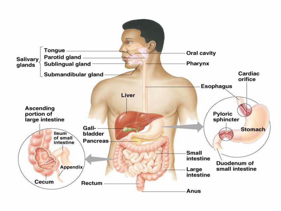
• The gastrointestinal tract (or alimentary canal) is anorgan system within humans and other animals whichtakes in food, digests, absorb nutrients, and expels theremaining waste as feces.
• The major organs of the digestive system
Mouth, Pharynx, Esophagus, Stomach, Small Intestine,Large Intestine, Rectum.
RIPERAUTONOMOUS
NAAC &
NBA (UG)
SIRO- DSIR
Raghavendra Institute of Pharmaceutical Education and Research - AutonomousK.R.Palli Cross, Chiyyedu, Anantapuramu, A. P- 515721
Accessory digestive organs
Liver, Gallbladder, Pancreas, Salivary gland.
Functions of GI tract
• Ingestion: taking of food into the alimentary tract
eating & drinking.
• Propulsion: mixes & moves the contents along the alimentary tract.
• Digestion:
• Mechanical breakdown of food e.g. mastication (chewing)
• Chemical digestion of food into small molecules by enzymes

RIPERAUTONOMOUS
NAAC &
NBA (UG)
SIRO- DSIR
Raghavendra Institute of Pharmaceutical Education and Research - AutonomousK.R.Palli Cross, Chiyyedu, Anantapuramu, A. P- 515721
Absorption: this is the process by which digested foodsubstances pass through the walls some organs of thealimentary canal into the blood for circulation.
Elimination: food substances that have been eaten butcannot be digested & absorbed are excreted from thealimentary canal as faeces by the process ofdefaecation.
RIPERAUTONOMOUS
NAAC &
NBA (UG)
SIRO- DSIR
Raghavendra Institute of Pharmaceutical Education and Research - AutonomousK.R.Palli Cross, Chiyyedu, Anantapuramu, A. P- 515721
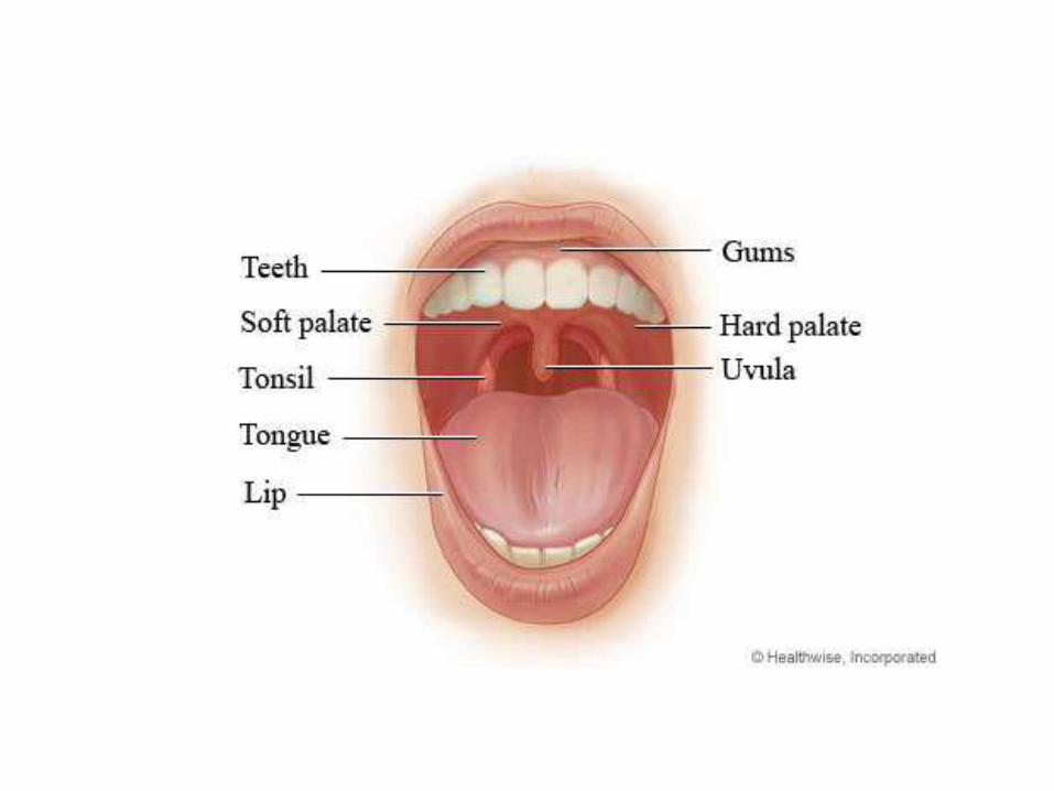
• MOUTH
• The mouth is the first portion of the alimentary canalthat receives food and produces saliva.
• The oral mucosa is the mucous membrane epitheliumlining the inside of the mouth.
• Relations
• Anteriorly-lips
• Posteriorly-continue with the oropharynx
RIPERAUTONOMOUS
NAAC &
NBA (UG)
SIRO- DSIR
Raghavendra Institute of Pharmaceutical Education and Research - AutonomousK.R.Palli Cross, Chiyyedu, Anantapuramu, A. P- 515721
• Laterally-muscles of cheeks
• Superiorly-bony hard palate
• Inferiorly-muscular tongue & the soft tissues of the
floor of the mouth
• The palate forms the roof of the mouth & is divided
into the anterior hard palate & posterior soft palate.
• The uvula is a curved fold of muscle covered with
mucous membrane,hanging down from the middle.
RIPERAUTONOMOUS
NAAC &
NBA (UG)
SIRO- DSIR
Raghavendra Institute of Pharmaceutical Education and Research - AutonomousK.R.Palli Cross, Chiyyedu, Anantapuramu, A. P- 515721
Tongue
• The tongue is a muscular organ in the mouth, that
manipulates food for mastication, and is used in the actof swallowing (Deglutition).
• It is of importance in the digestive system and is theprimary organ of taste in the gustatory system.
• The tongue's upper surface (dorsum) is covered by taste buds housed in numerous lingual papillae.
• The human tongue is divided into two parts, an oral part at the front and a pharyngeal part at the back.
RIPERAUTONOMOUS
NAAC &
NBA (UG)
SIRO- DSIR
Raghavendra Institute of Pharmaceutical Education and Research - AutonomousK.R.Palli Cross, Chiyyedu, Anantapuramu, A. P- 515721
• BLOOD SUPPLY
lingual artery
external carotid artery
• VENOUS DRAINAGE
lingual veins
internal jugular vein
• NERVE SUPPLY
hypoglossal nerve
Taste and sensation: glossopharyngeal nerve

RIPERAUTONOMOUS
NAAC &
NBA (UG)
SIRO- DSIR
Raghavendra Institute of Pharmaceutical Education and Research - AutonomousK.R.Palli Cross, Chiyyedu, Anantapuramu, A. P- 515721
Primary teeth
• Among deciduous (primary) teeth, ten are found in themaxilla (upper jaw) and ten in the mandible (lower jaw),for a total of 20. The dental formula for primary teeth is2.1.0.2/2.1.0.2.
Start to come in (erupt) at about 6 months of age
• In the primary set of teeth, two types of incisors –centrals and laterals, one canine & two types of molars– first and second.
• All primary teeth are normally later replaced with theirpermanent counterparts.
RIPERAUTONOMOUS
NAAC &
NBA (UG)
SIRO- DSIR
Raghavendra Institute of Pharmaceutical Education and Research - AutonomousK.R.Palli Cross, Chiyyedu, Anantapuramu, A. P- 515721
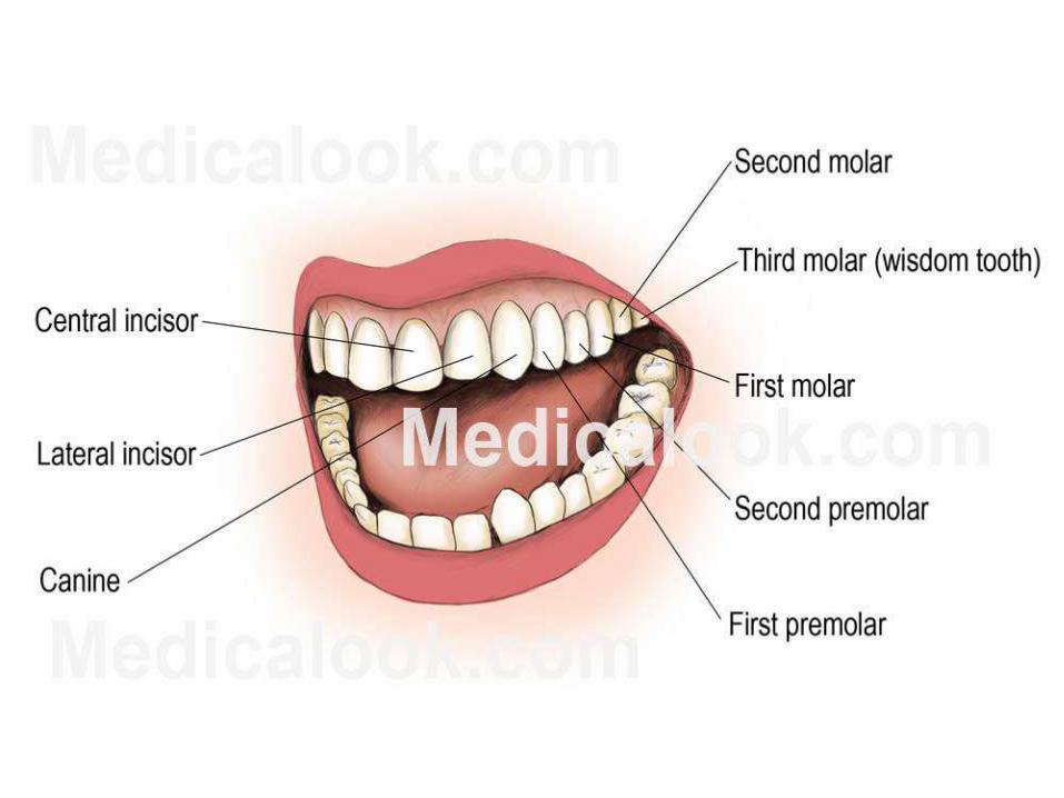
Permanent teeth
• Among permanent teeth, 16 are found in the maxilla
and 16 in the mandible, for a total of 32. The dental
formula is 2.1.2.3/2.1.2.3.
• Age 21, all 32 of the permanent teeth have usually
erupted.
The permanent teeth are the
• Two incisor (for cutting)-central incisor, lateral incisor
One canine (for tearing), Two premolar(for crushing)-first premolar, second premolar
RIPERAUTONOMOUS
NAAC &
NBA (UG)
SIRO- DSIR
Raghavendra Institute of Pharmaceutical Education and Research - AutonomousK.R.Palli Cross, Chiyyedu, Anantapuramu, A. P- 515721
• Three molar (for grinding)-first molar, second molar,
and third molar.
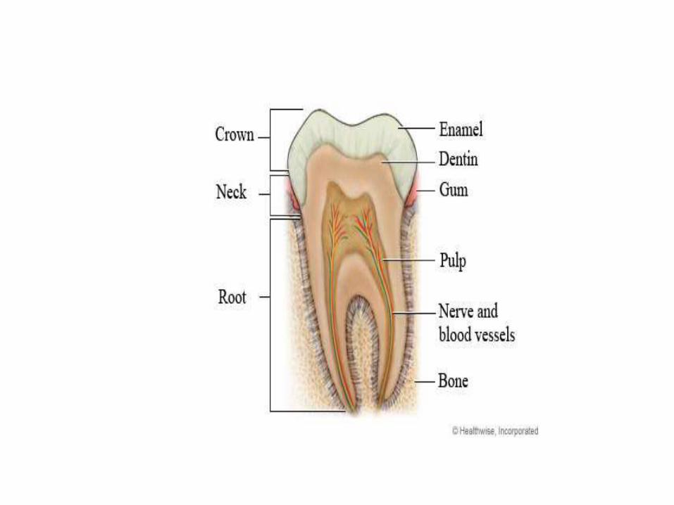
• PARTS
ENAMEL
• Enamel is the hardest and most highly mineralizedsubstance of the body.
• It is one of the four major tissues which make up thetooth, along with dentin, cementum, and dental pulp.
• 96% of enamel consists of mineral, with water andorganic material comprising the rest.
• The normal color of enamel varies from light yellow to
grayish white.
RIPERAUTONOMOUS
NAAC &
NBA (UG)
SIRO- DSIR
Raghavendra Institute of Pharmaceutical Education and Research - AutonomousK.R.Palli Cross, Chiyyedu, Anantapuramu, A. P- 515721
DENTIN
• Dentin is the substance between enamel or cementum
and the pulp chamber.
• The porous, yellow-hued material is made up of 70%inorganic materials, 20% organic materials, and 10%water by weight.
• Dentin is a mineralized connective tissue with an organic matrix of collagenous proteins.
RIPERAUTONOMOUS
NAAC &
NBA (UG)
SIRO- DSIR
Raghavendra Institute of Pharmaceutical Education and Research - AutonomousK.R.Palli Cross, Chiyyedu, Anantapuramu, A. P- 515721
Cementum
• Cementum is a specialized bone like substance coveringthe root of a tooth.
• Its coloration is yellowish and it is softer than dentinand enamel.
Dental pulp
• The dental pulp is the central part of the tooth filledwith soft connective tissue.
• This tissue contains blood vessels and nerves that enterthe tooth from a hole at the apex of the root.
RIPERAUTONOMOUS
NAAC &
NBA (UG)
SIRO- DSIR
Raghavendra Institute of Pharmaceutical Education and Research - AutonomousK.R.Palli Cross, Chiyyedu, Anantapuramu, A. P- 515721
SALIVARY GLANDS
• The salivary glands in are exocrine glands that producesaliva through a system of ducts.
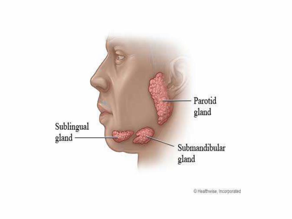
• Humans have 3 paired major salivary glands:
Parotid, submandibular and Sublingual as well hundredsof minor salivary glands.
• Parotid glands
• The two parotid glands are major salivary glandswrapped around the mandibular ramus in humans.
• The largest of the salivary glands.
RIPERAUTONOMOUS
NAAC &
NBA (UG)
SIRO- DSIR
Raghavendra Institute of Pharmaceutical Education and Research - AutonomousK.R.Palli Cross, Chiyyedu, Anantapuramu, A. P- 515721
They secrete saliva to
• facilitate mastication and swallowing, and amylase tobegin the digestion of starches.
• It enters the oral cavity via the parotid duct.
• Submandibular glands
• The submandibular glands are a pair of major salivaryglands located beneath the lower jaws, superior to thedigastric muscles.
• The secretion produced is a mixture of both serous fluidand mucus, and enters the oral cavity via thesubmandibular duct.
RIPERAUTONOMOUS
NAAC &
NBA (UG)
SIRO- DSIR
Raghavendra Institute of Pharmaceutical Education and Research - AutonomousK.R.Palli Cross, Chiyyedu, Anantapuramu, A. P- 515721
Sublingual glands
• The sublingual glands are a pair of major salivary
glands located inferior to the tongue, anterior to the
submandibular glands.
• Approximately 5% of saliva entering the oral cavitycomes from these glands.
• The secretion produced is mainly mucous in nature.
• Minor salivary glands
• There are 800 to 1,000 minor salivary glands locatedthroughout the oral cavity within the submucosa of theoral mucosa in the tissue of the buccal and lingualmucosa
RIPERAUTONOMOUS
NAAC &
NBA (UG)
SIRO- DSIR
Raghavendra Institute of Pharmaceutical Education and Research - AutonomousK.R.Palli Cross, Chiyyedu, Anantapuramu, A. P- 515721
BLOOD SUPPLY
External carotid artery
VENOUS DRAINAGE
Jugular veins
COMPOSITION OF SALIVA
About 1.5 litres of saliva is produced daily & it consists
of: Water, Mineral salts, An enzyme, Mucus, Lysozyme
Immunoglobulins.

RIPERAUTONOMOUS
NAAC &
NBA (UG)
SIRO- DSIR
Raghavendra Institute of Pharmaceutical Education and Research - AutonomousK.R.Palli Cross, Chiyyedu, Anantapuramu, A. P- 515721
• THE PHARYNX
• The pharynx is the part of the throat that is behind themouth and nasal cavity and above the esophagus andthe larynx, or the tubes going down to the stomach andthe lungs.
• The pharynx is the portion of the digestive tract thatreceives the food from your mouth.
• Branching off the pharynx is the esophagus, whichcarries food to the stomach.
RIPERAUTONOMOUS
NAAC &
NBA (UG)
SIRO- DSIR
Raghavendra Institute of Pharmaceutical Education and Research - AutonomousK.R.Palli Cross, Chiyyedu, Anantapuramu, A. P- 515721
THE ESOPHAGUS
• The esophagus or oesophagus, commonly known as thefood pipe or gullet, The esophagus is a muscular tubeconnecting the throat (pharynx) with the stomach.
• The esophagus runs behind the windpipe (trachea) andheart, and in front of the spine.
RIPERAUTONOMOUS
NAAC &
NBA (UG)
SIRO- DSIR
Raghavendra Institute of Pharmaceutical Education and Research - AutonomousK.R.Palli Cross, Chiyyedu, Anantapuramu, A. P- 515721
FUNCTIONS
• Formation of a bolus
• Swallowing
• Food is ingested through the mouth and whenswallowed passes first into the pharynx and then intothe esophagus.
• Reducing gastric reflux
• Constriction of the upper and lower esophagealsphincters help to prevent reflux (backflow) of gastriccontents and acid into the esophagus,
• Protecting the esophageal mucosa.
RIPERAUTONOMOUS
NAAC &
NBA (UG)
SIRO- DSIR
Raghavendra Institute of Pharmaceutical Education and Research - AutonomousK.R.Palli Cross, Chiyyedu, Anantapuramu, A. P- 515721
• STOMACH
• The stomach is a muscular organ located on the leftside of the upper abdomen. The stomach receives foodfrom the esophagus.
• As food reaches the end of the esophagus, it enters thestomach through a muscular valve called the loweresophageal sphincter.
RIPERAUTONOMOUS
NAAC &
NBA (UG)
SIRO- DSIR
Raghavendra Institute of Pharmaceutical Education and Research - AutonomousK.R.Palli Cross, Chiyyedu, Anantapuramu, A. P- 515721
GI structureMucosa (lumen side)Epithelial tissue
Submucosaelastic connective tissue contains lymph and bloodVesselsMuscularis externasmooth muscle layersSerosaOutermost lining of GI organs

RIPERAUTONOMOUS
NAAC &
NBA (UG)
SIRO- DSIR
Raghavendra Institute of Pharmaceutical Education and Research - AutonomousK.R.Palli Cross, Chiyyedu, Anantapuramu, A. P- 515721
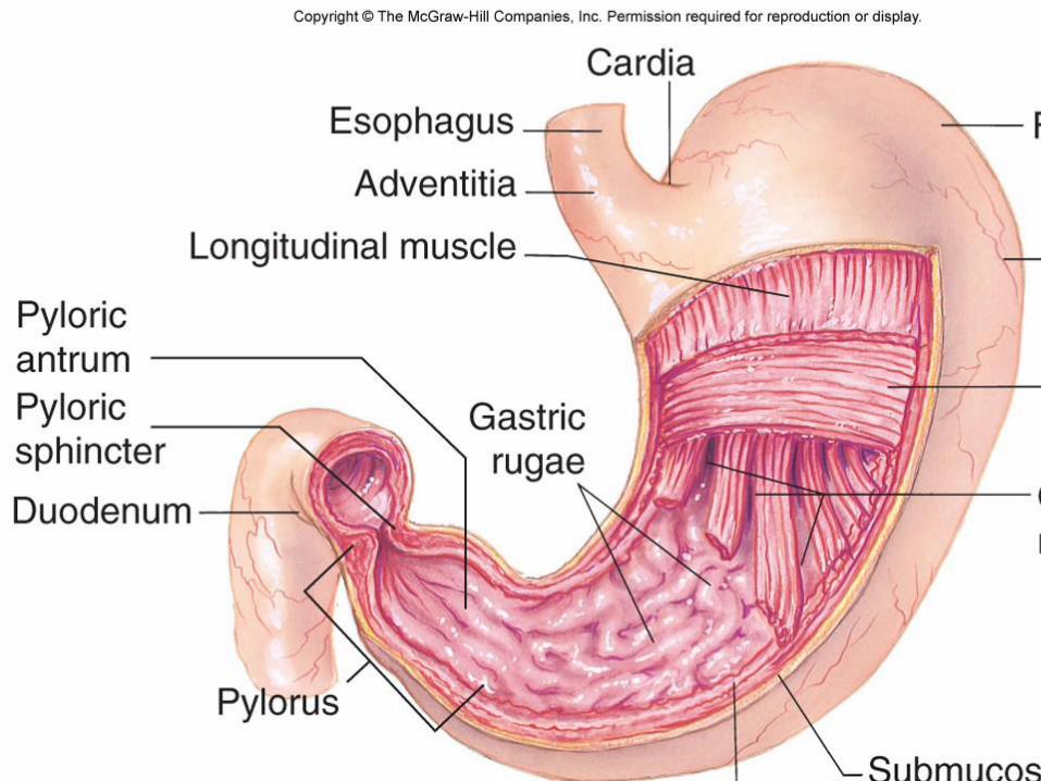
• A pouch-like organ primarily designed for food storage(for 2-4 hours) , some mechanical and chemicaldigestion also occur .
• Contains two sphincters at both ends to regulate foodmovement
• cardiac sphincter near the esophagus
• pyloric sphincter near the small intestine
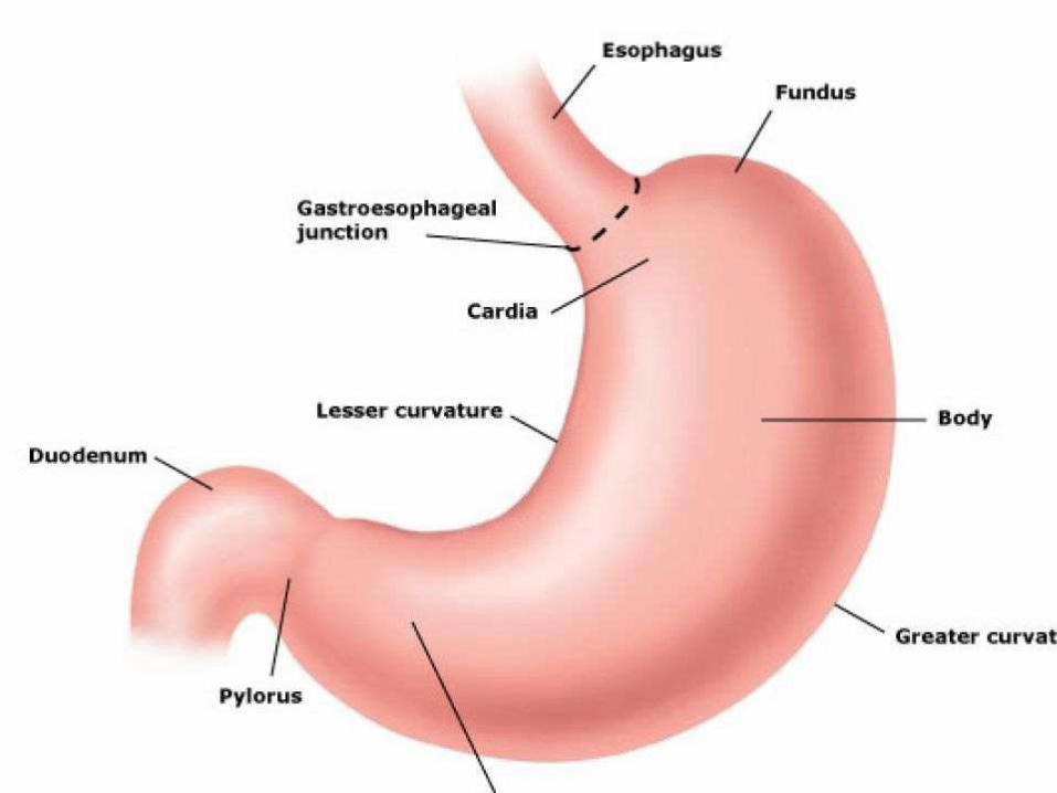
• Divided into 4 regions :
• cardiac stomach (or cardiac), fundic stomach (orfunded) , body of stomach, pyloric stomach (or Pylorus).
RIPERAUTONOMOUS
NAAC &
NBA (UG)
SIRO- DSIR
Raghavendra Institute of Pharmaceutical Education and Research - AutonomousK.R.Palli Cross, Chiyyedu, Anantapuramu, A. P- 515721
Contain thick folds called rugae at its layer, for providing
larger surface area for expansion, secretion, digestion ,and some absorption.
FUNCTIONS
Digestion
• The stomach releases proteases (protein-digestingenzymes such as pepsin) and hydrochloric acid, whichkills or inhibits bacteria and provides the acidic pH of 2for the proteases to work.
• Food is churned by the stomach through muscularcontractions of the wall called peristalsis.
RIPERAUTONOMOUS
NAAC &
NBA (UG)
SIRO- DSIR
Raghavendra Institute of Pharmaceutical Education and Research - AutonomousK.R.Palli Cross, Chiyyedu, Anantapuramu, A. P- 515721
Absorption
• some absorption of certain small moleculesnevertheless does occur in the stomach through itslining
• GASTRIC JUICE
• Gastric acid, gastric juice or stomach acid, is a digestivefluid formed in the stomach and is composed ofhydrochloric acid (HCl), potassium chloride (KCl) andsodium chloride (NaCl).
• The acid plays a key role in digestion of proteins, byactivating digestive enzymes, and making ingestedproteins unravel so that digestive enzymes break downthe long chains of amino acids.
RIPERAUTONOMOUS
NAAC &
NBA (UG)
SIRO- DSIR
Raghavendra Institute of Pharmaceutical Education and Research - AutonomousK.R.Palli Cross, Chiyyedu, Anantapuramu, A. P- 515721
Gastric Secretory Cells
• Chief cells: secrete pepsinogen (an inactive enzyme).
• Parietal cells: secrete hydrochloric and (HCl) and"intrinsic factor" (which helps absorption of vitamin B12in the intestines).
• Mucous cells: secrete mucus and alkaline substances tohelp neutralize HCl in the gastric juice .
• G cells: secrete a hormone called gastrin , whichstimulates the parietal cells and overall gastric.
RIPERAUTONOMOUS
NAAC &
NBA (UG)
SIRO- DSIR
Raghavendra Institute of Pharmaceutical Education and Research - AutonomousK.R.Palli Cross, Chiyyedu, Anantapuramu, A. P- 515721
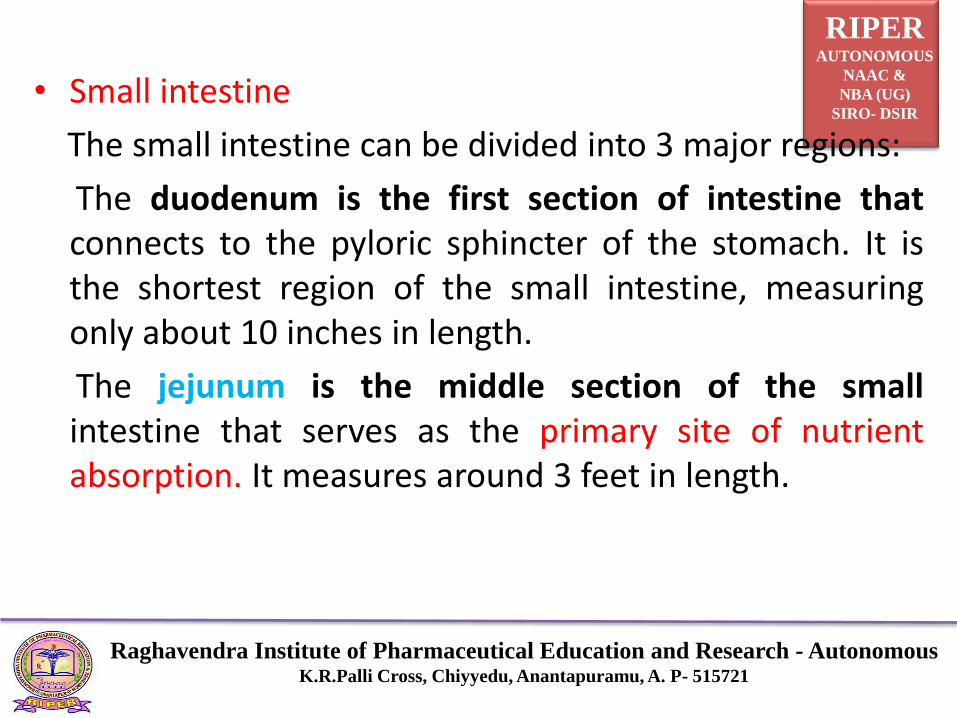
• Small intestine
The small intestine can be divided into 3 major regions:
The duodenum is the first section of intestine thatconnects to the pyloric sphincter of the stomach. It isthe shortest region of the small intestine, measuringonly about 10 inches in length.
The jejunum is the middle section of the smallintestine that serves as the primary site of nutrientabsorption. It measures around 3 feet in length.
RIPERAUTONOMOUS
NAAC &
NBA (UG)
SIRO- DSIR
Raghavendra Institute of Pharmaceutical Education and Research - AutonomousK.R.Palli Cross, Chiyyedu, Anantapuramu, A. P- 515721
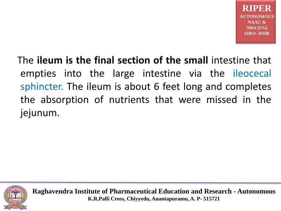
The ileum is the final section of the small intestine thatempties into the large intestine via the ileocecalsphincter. The ileum is about 6 feet long and completesthe absorption of nutrients that were missed in thejejunum.
RIPERAUTONOMOUS
NAAC &
NBA (UG)
SIRO- DSIR
Raghavendra Institute of Pharmaceutical Education and Research - AutonomousK.R.Palli Cross, Chiyyedu, Anantapuramu, A. P- 515721
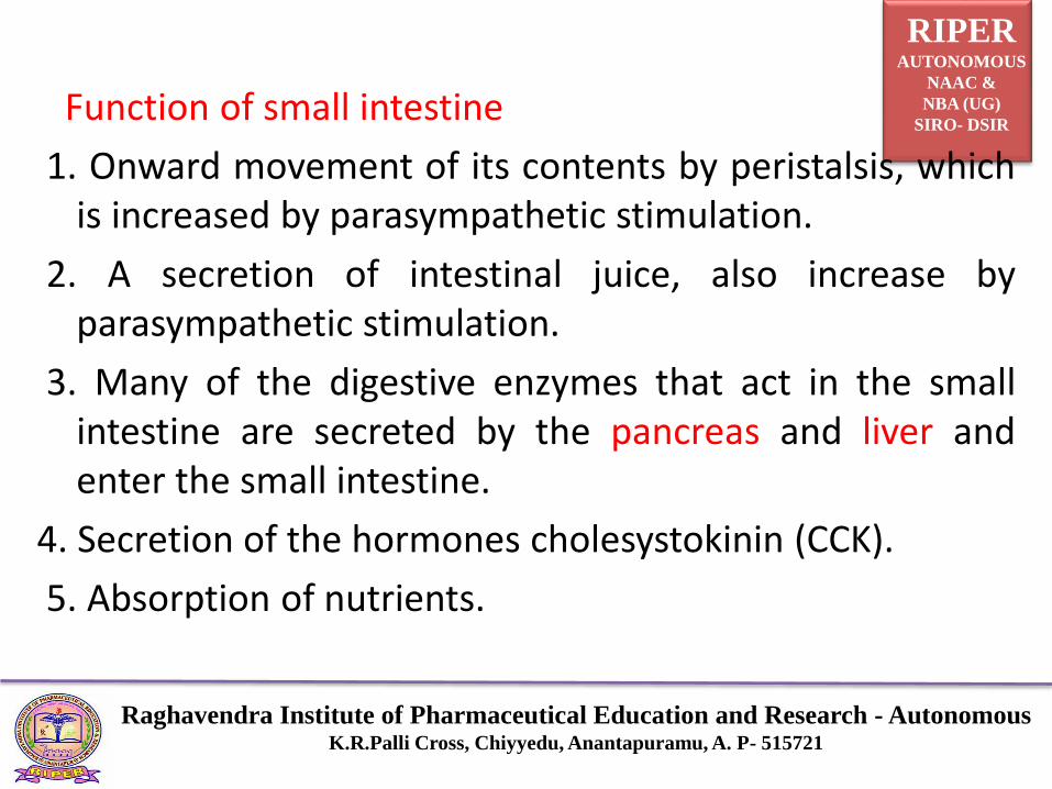
Function of small intestine
1. Onward movement of its contents by peristalsis, whichis increased by parasympathetic stimulation.
2. A secretion of intestinal juice, also increase byparasympathetic stimulation.
3. Many of the digestive enzymes that act in the smallintestine are secreted by the pancreas and liver andenter the small intestine.
4. Secretion of the hormones cholesystokinin (CCK).
5. Absorption of nutrients.

RIPERAUTONOMOUS
NAAC &
NBA (UG)
SIRO- DSIR
Raghavendra Institute of Pharmaceutical Education and Research - AutonomousK.R.Palli Cross, Chiyyedu, Anantapuramu, A. P- 515721
RIPERAUTONOMOUS
NAAC &
NBA (UG)
SIRO- DSIR
Raghavendra Institute of Pharmaceutical Education and Research - AutonomousK.R.Palli Cross, Chiyyedu, Anantapuramu, A. P- 515721
RIPERAUTONOMOUS
NAAC &
NBA (UG)
SIRO- DSIR
Raghavendra Institute of Pharmaceutical Education and Research - AutonomousK.R.Palli Cross, Chiyyedu, Anantapuramu, A. P- 515721
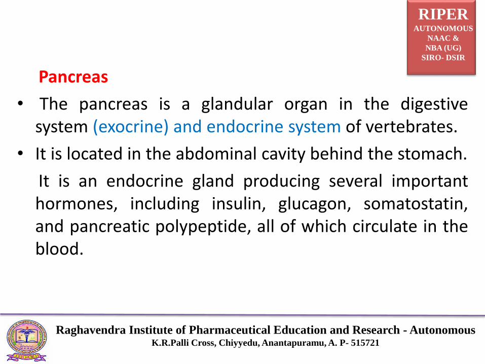
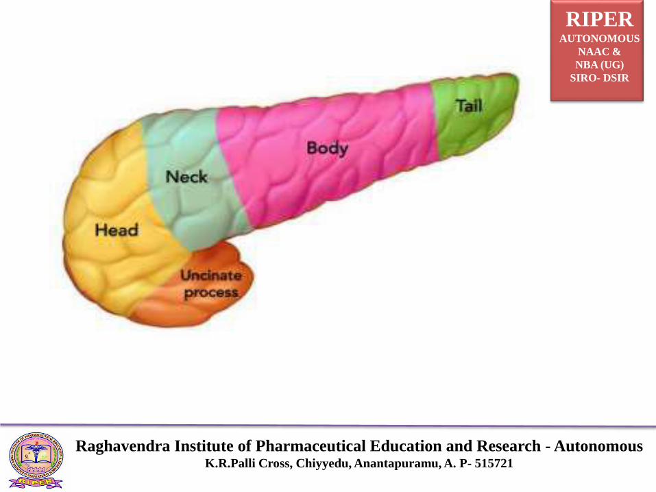
Pancreas
• The pancreas is a glandular organ in the digestivesystem (exocrine) and endocrine system of vertebrates.
• It is located in the abdominal cavity behind the stomach.
It is an endocrine gland producing several importanthormones, including insulin, glucagon, somatostatin,and pancreatic polypeptide, all of which circulate in theblood.
RIPERAUTONOMOUS
NAAC &
NBA (UG)
SIRO- DSIR
Raghavendra Institute of Pharmaceutical Education and Research - AutonomousK.R.Palli Cross, Chiyyedu, Anantapuramu, A. P- 515721
RIPERAUTONOMOUS
NAAC &
NBA (UG)
SIRO- DSIR
Raghavendra Institute of Pharmaceutical Education and Research - AutonomousK.R.Palli Cross, Chiyyedu, Anantapuramu, A. P- 515721
• Function
• The pancreas is involved in blood sugar control andmetabolism within the body.
• Within these islets are four main types of cells which areinvolved in the regulation of blood glucose levels.
• α alpha cells secrete glucagon (increase glucose inblood).
• β beta cells secrete insulin (decrease glucose in blood)
• δ delta cells secrete somatostatin (regulates/stops αand β cells) and
• γ (gamma) cells, secrete pancreatic polypeptide.
RIPERAUTONOMOUS
NAAC &
NBA (UG)
SIRO- DSIR
Raghavendra Institute of Pharmaceutical Education and Research - AutonomousK.R.Palli Cross, Chiyyedu, Anantapuramu, A. P- 515721

RIPERAUTONOMOUS
NAAC &
NBA (UG)
SIRO- DSIR
Raghavendra Institute of Pharmaceutical Education and Research - AutonomousK.R.Palli Cross, Chiyyedu, Anantapuramu, A. P- 515721
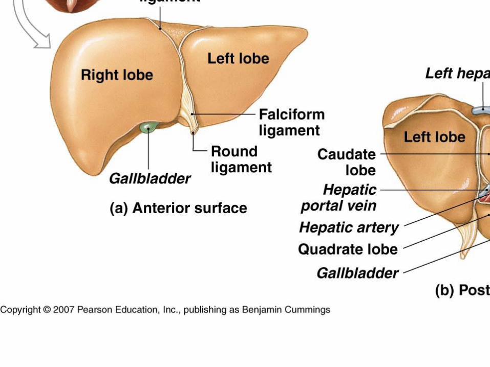
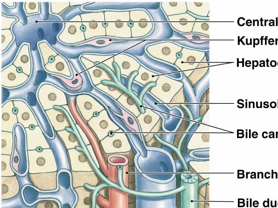
Liver
• Hepatocytes produce bile, which gets secreted into bilecanaliculi of lobule
• Bile canaliculi merge to form bile ducts which eventuallymerge to create the right & left hepatic ducts
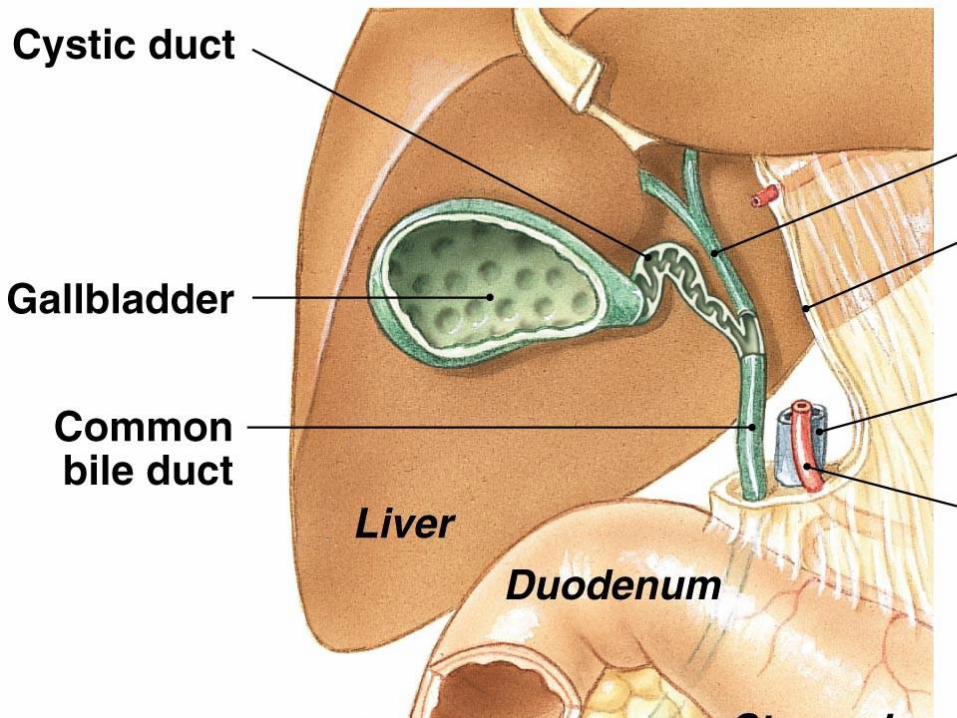
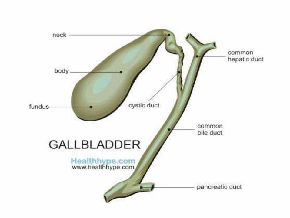
• Right & left hepatic ducts unite to form common hepaticduct which merges with cystic duct of gall bladder toform common bile duct which joins with pancreatic duct& enters the duodenum
• Gall bladder – hollow muscular sac under right lobe
of liver; stores & concentrates bile; releases bile throughcystic duct
RIPERAUTONOMOUS
NAAC &
NBA (UG)
SIRO- DSIR
Raghavendra Institute of Pharmaceutical Education and Research - AutonomousK.R.Palli Cross, Chiyyedu, Anantapuramu, A. P- 515721
• Bile released into duodenum functions in emulsificationof lipids, absorption of fats (due to presence of bilesalts), & excretion of bilirubin
RIPERAUTONOMOUS
NAAC &
NBA (UG)
SIRO- DSIR
Raghavendra Institute of Pharmaceutical Education and Research - AutonomousK.R.Palli Cross, Chiyyedu, Anantapuramu, A. P- 515721
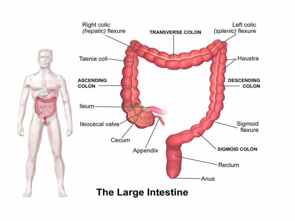
Large intestine
The large intestine, also known as the large bowel orcolon, is the last part of the gastrointestinal tract.
Water is absorbed here and the remaining wastematerial is stored as feces before being removed bydefecation.
RIPERAUTONOMOUS
NAAC &
NBA (UG)
SIRO- DSIR
Raghavendra Institute of Pharmaceutical Education and Research - AutonomousK.R.Palli Cross, Chiyyedu, Anantapuramu, A. P- 515721
• Sections of the colon are:
• The ascending colon including the cecum and
appendix
• The transverse colon including the colic flexures andtransverse mesocolon
• The descending colon
• The sigmoid colon – the s-shaped region of the largeintestine
RIPERAUTONOMOUS
NAAC &
NBA (UG)
SIRO- DSIR
Raghavendra Institute of Pharmaceutical Education and Research - AutonomousK.R.Palli Cross, Chiyyedu, Anantapuramu, A. P- 515721
• Cecum
• The cecum is the first section of the colon and
involved in the digestion, while the appendix is a
structure of the colon, not involved in digestion.
• The function of the appendix is uncertain containingIleocecal valve
• The ileocecal valve is a sphincter muscle valve thatseparates the small intestine and the large intestine.
• Its critical function is to limit the reflux of coloniccontents into the ileum.

RIPERAUTONOMOUS
NAAC &
NBA (UG)
SIRO- DSIR
Raghavendra Institute of Pharmaceutical Education and Research - AutonomousK.R.Palli Cross, Chiyyedu, Anantapuramu, A. P- 515721
• Transverse colon
The transverse colon is the part of the colon from the
hepatic flexure to the splenic flexure.
• Descending colon
The descending colon is the part of the colon from thesplenic flexure to the beginning of the sigmoid colon,descending colon is also called the distal gut.
One function of the descending colon in the digestivesystem is to store feces that will be emptied into therectum.
RIPERAUTONOMOUS
NAAC &
NBA (UG)
SIRO- DSIR
Raghavendra Institute of Pharmaceutical Education and Research - AutonomousK.R.Palli Cross, Chiyyedu, Anantapuramu, A. P- 515721
• Sigmoid colon
The sigmoid colon is the part of the large intestine afterthe descending colon and before the rectum.
The name sigmoid means S-shaped .
The walls of the sigmoid colon are muscular, andcontract to increase the pressure inside the colon,causing the stool to move into the rectum.
• Rectum
The rectum is the last section of the large intestine. Itholds the formed feces awaiting elimination viadefecation.
RIPERAUTONOMOUS
NAAC &
NBA (UG)
SIRO- DSIR
Raghavendra Institute of Pharmaceutical Education and Research - AutonomousK.R.Palli Cross, Chiyyedu, Anantapuramu, A. P- 515721
Anus
The anus is the external opening of the rectum.
Its function is to control the expulsion of feces.
Two sphincters control the exit of feces from the bodyduring an act of defecation. These are the internal analsphincter and the external anal sphincter, which arecircular muscles that normally maintain constriction ofthe orifice and which relaxes as required by normalphysiological functioning.
RIPERAUTONOMOUS
NAAC &
NBA (UG)
SIRO- DSIR
Raghavendra Institute of Pharmaceutical Education and Research - AutonomousK.R.Palli Cross, Chiyyedu, Anantapuramu, A. P- 515721
Functions
The large intestine absorbs water and any remainingabsorbable nutrients from the food before sending theindigestible matter to the rectum. The colon absorbsvitamins that are created by the colonic bacteria, suchas vitamin K.
Gut flora
The large intestine houses over 700 species of bacteriathat perform a variety of functions. The large intestineabsorbs some of the products formed by the bacteriainhabiting this region.
RIPERAUTONOMOUS
NAAC &
NBA (UG)
SIRO- DSIR
Raghavendra Institute of Pharmaceutical Education and Research - AutonomousK.R.Palli Cross, Chiyyedu, Anantapuramu, A. P- 515721
Cardiovascular system
Blood Vessels
The cardiovascular system has three types of bloodvessels:
Arteries (and arterioles) – carry blood away from theheart
Capillaries – where nutrient and gas exchange occurVeins (and venules) – carry blood toward the heart.
RIPERAUTONOMOUS
NAAC &
NBA (UG)
SIRO- DSIR
Raghavendra Institute of Pharmaceutical Education and Research - AutonomousK.R.Palli Cross, Chiyyedu, Anantapuramu, A. P- 515721
Arteries
Arteries and arterioles take blood away from the heart.The largest artery is the aorta. The middle layer of anartery wall consists of smooth muscle that can constrictto regulate blood flow and blood pressure.
Arterioles can constrict or dilate, changing bloodpressure.
RIPERAUTONOMOUS
NAAC &
NBA (UG)
SIRO- DSIR
Raghavendra Institute of Pharmaceutical Education and Research - AutonomousK.R.Palli Cross, Chiyyedu, Anantapuramu, A. P- 515721
The Capillaries
Capillaries have walls only one cell thick to allowexchange of gases and nutrients with tissue fluid.Capillary beds are present in all regions of the body butnot all capillary beds are open at the same time.
The Veins
Venules drain blood from capillaries, then join to formveins that take blood to the heart. Veins have muchless smooth muscle and connective tissue than arteries

RIPERAUTONOMOUS
NAAC &
NBA (UG)
SIRO- DSIR
Raghavendra Institute of Pharmaceutical Education and Research - AutonomousK.R.Palli Cross, Chiyyedu, Anantapuramu, A. P- 515721
Veins often have valves that prevent the backward flowof blood when closed. Veins carry about 70% of thebody’s blood and act as a reservoir during hemorrhage.
RIPERAUTONOMOUS
NAAC &
NBA (UG)
SIRO- DSIR
Raghavendra Institute of Pharmaceutical Education and Research - AutonomousK.R.Palli Cross, Chiyyedu, Anantapuramu, A. P- 515721
Structure of Vessels
• Arteries, veins and capillaries differ in structure.
• Three layers are found in arteries and veins.
• The outermost layer = tunica externa or adventitia. It is made of connective tissue fibers to reinforce the walls under pressure.
• The tunica media is the smooth muscle middle layer. It is much thicker in arteries than in veins and contains a thin layer of elastic tissue. This layer is under control of the ANS and maintains BP and blood distribution.
RIPERAUTONOMOUS
NAAC &
NBA (UG)
SIRO- DSIR
Raghavendra Institute of Pharmaceutical Education and Research - AutonomousK.R.Palli Cross, Chiyyedu, Anantapuramu, A. P- 515721
• The tunica intima lines the arteries and veins. It is asingle layer of squamous epithelial cells calledendothelium that lines the inner surface of the entirecardiovascular system.
RIPERAUTONOMOUS
NAAC &
NBA (UG)
SIRO- DSIR
Raghavendra Institute of Pharmaceutical Education and Research - AutonomousK.R.Palli Cross, Chiyyedu, Anantapuramu, A. P- 515721
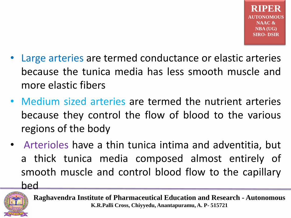
• Large arteries are termed conductance or elastic arteriesbecause the tunica media has less smooth muscle andmore elastic fibers
• Medium sized arteries are termed the nutrient arteriesbecause they control the flow of blood to the variousregions of the body
• Arterioles have a thin tunica intima and adventitia, buta thick tunica media composed almost entirely ofsmooth muscle and control blood flow to the capillarybed

RIPERAUTONOMOUS
NAAC &
NBA (UG)
SIRO- DSIR
Raghavendra Institute of Pharmaceutical Education and Research - AutonomousK.R.Palli Cross, Chiyyedu, Anantapuramu, A. P- 515721
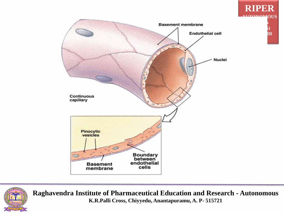
Capillary
Pre-capillary sphincter valves
Smooth muscle rings at the proximal end of the capillary
• Contraction decreases blood flow
• Relaxation increases blood flow
• Responsive to local changes in PaO2, PaCO2, pH and temperature
• Called exchange vessels because they are the site of gas, fluid, nutrient, and waste exchange
RIPERAUTONOMOUS
NAAC &
NBA (UG)
SIRO- DSIR
Raghavendra Institute of Pharmaceutical Education and Research - AutonomousK.R.Palli Cross, Chiyyedu, Anantapuramu, A. P- 515721
RIPERAUTONOMOUS
NAAC &
NBA (UG)
SIRO- DSIR
Raghavendra Institute of Pharmaceutical Education and Research - AutonomousK.R.Palli Cross, Chiyyedu, Anantapuramu, A. P- 515721
Venous system
• Transport deoxygenated blood back to the heart –exception: pulmonary vein
• Composed of the same layers as arteries, but arethinner Called capacitance or reservoir vessels because70% to 75% of the blood volume is contained in thevenous system
• Peripheral veins contain one-way valves
• Valves are formed by duplication of endothelial lining
• Found in veins >2mm in diameter
RIPERAUTONOMOUS
NAAC &
NBA (UG)
SIRO- DSIR
Raghavendra Institute of Pharmaceutical Education and Research - AutonomousK.R.Palli Cross, Chiyyedu, Anantapuramu, A. P- 515721
The Heart
The heart is a cone-shaped, muscular organlocated between the lungs behind the sternum.
The heart muscle forms the myocardium, withtightly interconnect cells of cardiac muscle tissue.
RIPERAUTONOMOUS
NAAC &
NBA (UG)
SIRO- DSIR
Raghavendra Institute of Pharmaceutical Education and Research - AutonomousK.R.Palli Cross, Chiyyedu, Anantapuramu, A. P- 515721
Layers of the heart
• The heart resides in the pericardium
A loose membranous sac
• Epicardium
Continuous with the pericardium
• Myocardium
Composed of bands of involuntary striated muscle fibers
• Endocardium
• Thin layer of tissue lining the inside of the heart
RIPERAUTONOMOUS
NAAC &
NBA (UG)
SIRO- DSIR
Raghavendra Institute of Pharmaceutical Education and Research - AutonomousK.R.Palli Cross, Chiyyedu, Anantapuramu, A. P- 515721
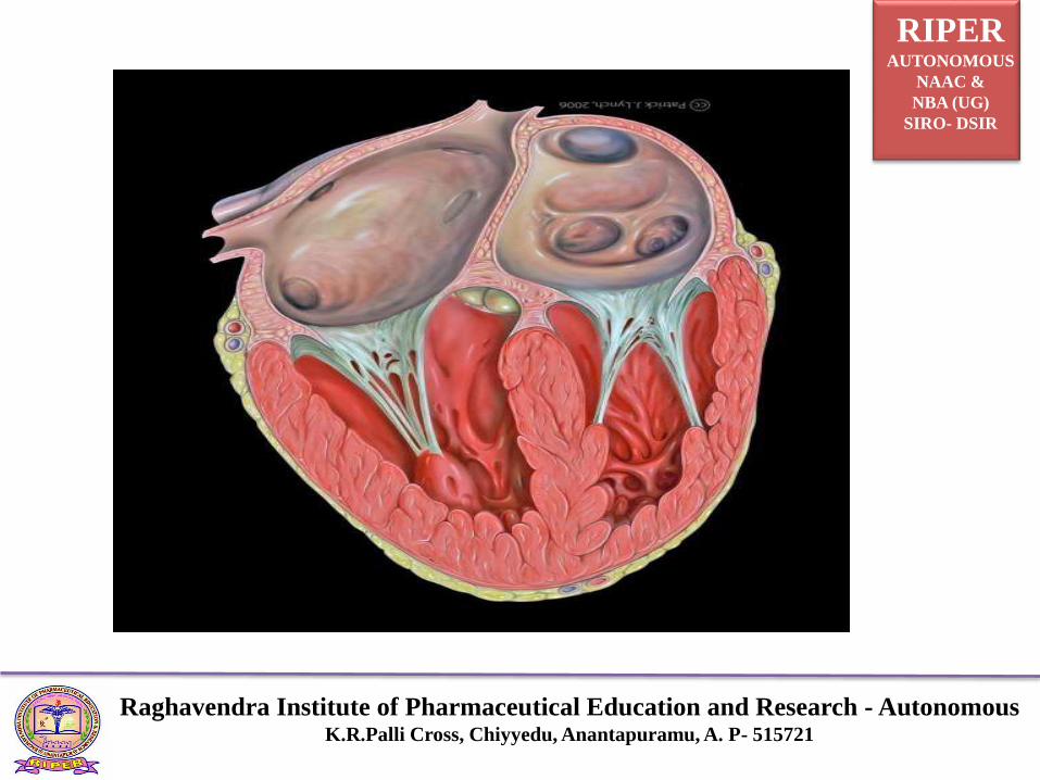
Chambers of the heart
Atria
Thin-walled upper chambers
Separated by atrial septum
Act as receiving chamber for blood returning from thebody and lungs
Ventricles
Lower chambers which make up the bulk of the muscle mass of the heart
Blood exits from the ventricles into arteries – pulmonary and aorta. The ventricles are referred to as discharging chambers
RIPERAUTONOMOUS
NAAC &
NBA (UG)
SIRO- DSIR
Raghavendra Institute of Pharmaceutical Education and Research - AutonomousK.R.Palli Cross, Chiyyedu, Anantapuramu, A. P- 515721

RIPERAUTONOMOUS
NAAC &
NBA (UG)
SIRO- DSIR
Raghavendra Institute of Pharmaceutical Education and Research - AutonomousK.R.Palli Cross, Chiyyedu, Anantapuramu, A. P- 515721
Valves of the heart
• The two valves that separate the atrial chambers abovefrom the ventricular chambers below are called theatrioventricular valves (AV).
• The right AV valve is called the tricupsid valve. Itconsists of three leaf-like valve components.
• The left AV valve is called the bicupsid or mitral valve.It consists of two leaf-like valve components.
• Both AV valves prevent the backflow of blood into theatria when the ventricles contract.
RIPERAUTONOMOUS
NAAC &
NBA (UG)
SIRO- DSIR
Raghavendra Institute of Pharmaceutical Education and Research - AutonomousK.R.Palli Cross, Chiyyedu, Anantapuramu, A. P- 515721
• String like structures called chordae tendineae attach the
AV valves to the walls of the ventricles via papillary
muscles extending from the floor of the ventricles.
• The semilunar valves (SL) valves are located between thetwo ventricular chambers and the arteries that carryblood away from the heart when systole occurs.
• The ventricles contract at the same time like the atria.The two SL valves open and close at the same time.
• The pulmonary SL valve is located at the beginning of thepulmonary artery and opens to allow blood to enterpulmonary circulation. The valve closes to preventbackflow of blood into the right ventricle.

RIPERAUTONOMOUS
NAAC &
NBA (UG)
SIRO- DSIR
Raghavendra Institute of Pharmaceutical Education and Research - AutonomousK.R.Palli Cross, Chiyyedu, Anantapuramu, A. P- 515721
Passage of blood through the heartHeart: superior and inferior vena cava →right atrium → tricuspid valve → right ventricle →pulmonary semilunar valve → pulmonary trunk andarteries to the lungs → pulmonary veins leaving the lungs→ left atrium → bicuspid valve → left ventricle → aorticsemilunar valve → aorta → to the body.
• The heart must perform a great amount of work bypumping through the pulmonary and systemic systems.
• It requires a constant supply of oxygen and nutrients via the coronary circulation to perform this task.
RIPERAUTONOMOUS
NAAC &
NBA (UG)
SIRO- DSIR
Raghavendra Institute of Pharmaceutical Education and Research - AutonomousK.R.Palli Cross, Chiyyedu, Anantapuramu, A. P- 515721
THANK YOU EVERY ONE
(Will be continued in next class)
By
Dr. K. Somasekhar Reddy
Mobile number: 9440730432