Automating Breast Ultrasound Workflow DICOM Compatible Automated Segmentation Digital Work Sheet...
28
Automating Breast Ultrasound Workflow DICOM Compatible Automated Segmentation Digital Work Sheet Documentation Point and Click Lesion Characteristics Previous Study Recall B-CAD
-
date post
19-Dec-2015 -
Category
Documents
-
view
226 -
download
1
Transcript of Automating Breast Ultrasound Workflow DICOM Compatible Automated Segmentation Digital Work Sheet...
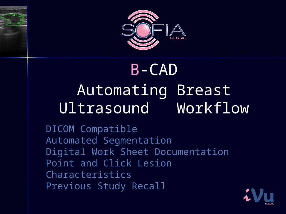
Automating Breast Ultrasound Workflow
DICOM Compatible Automated SegmentationDigital Work Sheet DocumentationPoint and Click Lesion CharacteristicsPrevious Study Recall
B-CAD
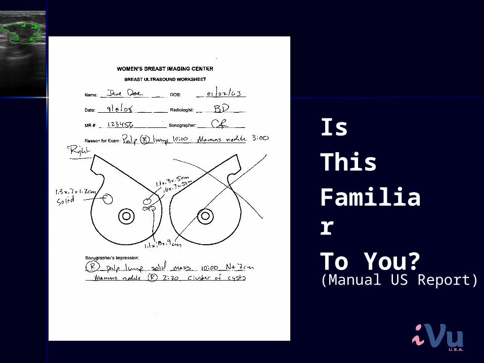
IsThis FamiliarTo You?(Manual US Report)
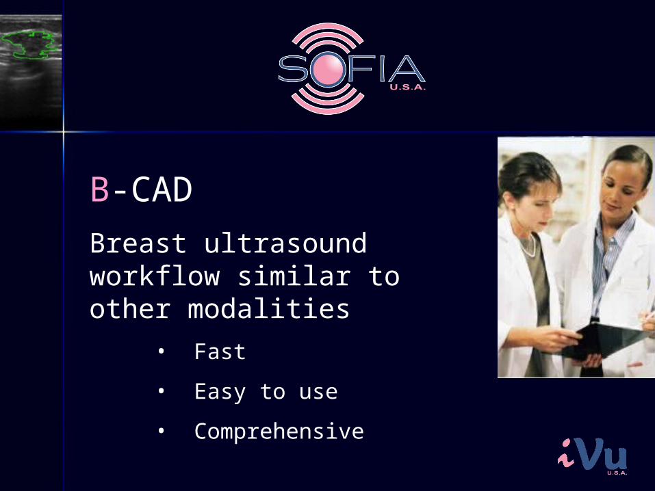
B-CAD
Breast ultrasound workflow similar to other modalities
• Fast
• Easy to use
• Comprehensive
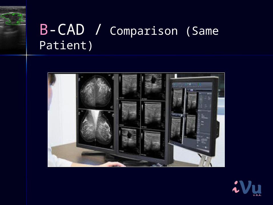
B-CAD / Comparison (Same Patient)
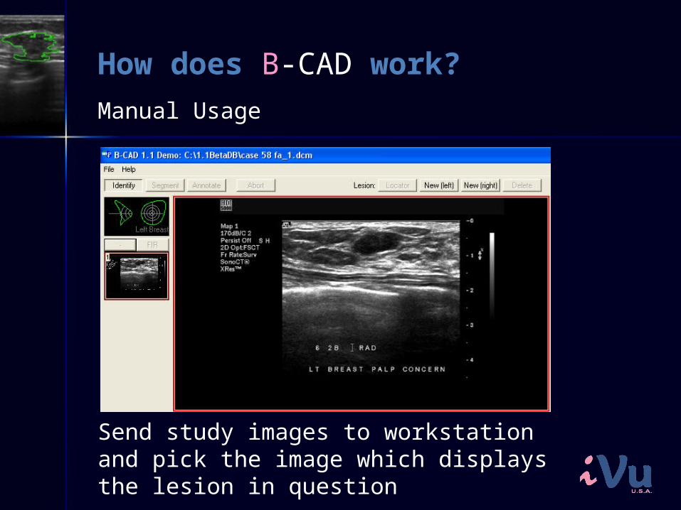
How does B-CAD work?
Send study images to workstation and pick the image which displays the lesion in question
Manual Usage
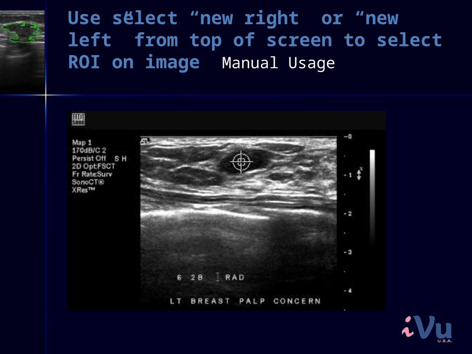
Use select “new right” or “new left” from top of screen to select ROI on image Manual Usage
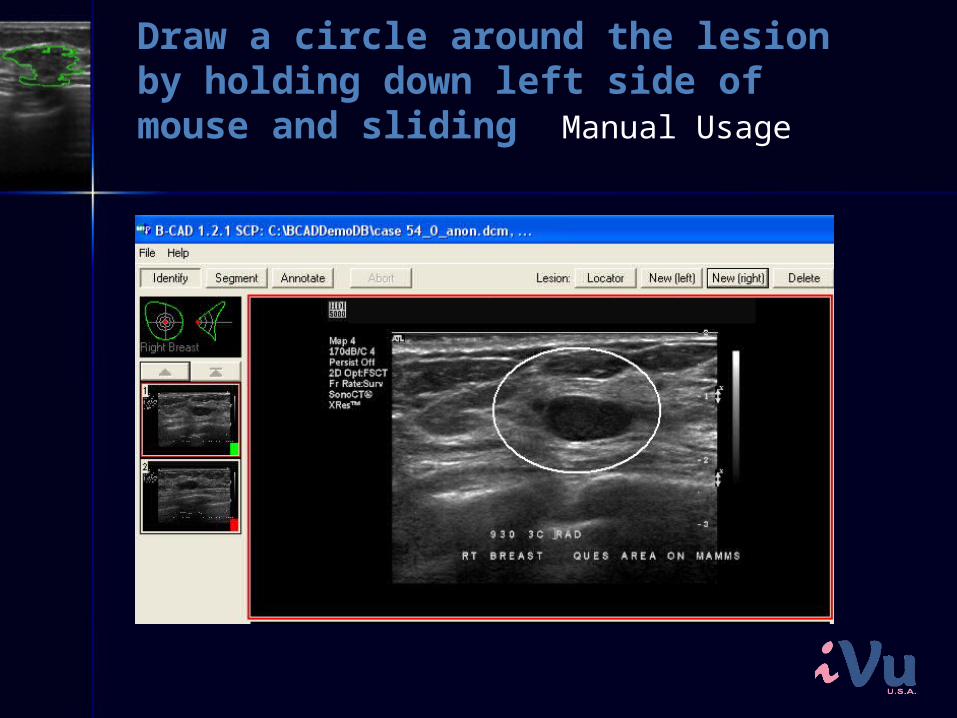
Draw a circle around the lesion by holding down left side of mouse and sliding Manual Usage
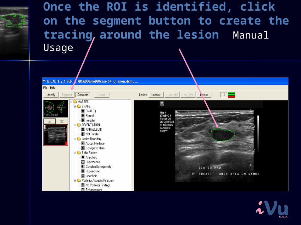
Once the ROI is identified, click on the segment button to create the tracing around the lesion Manual Usage
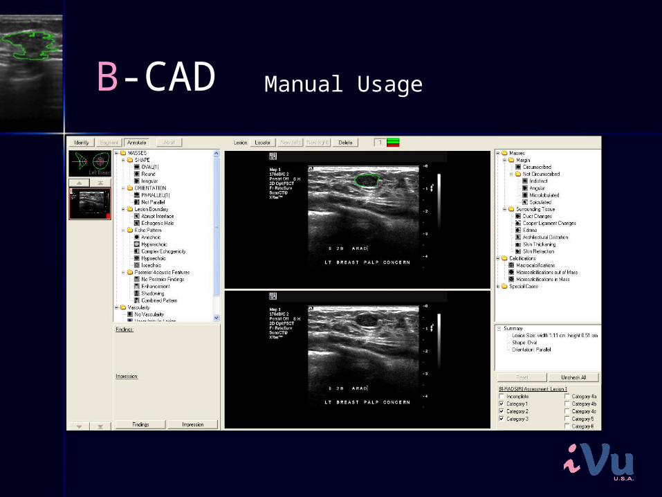
B-CAD Manual Usage
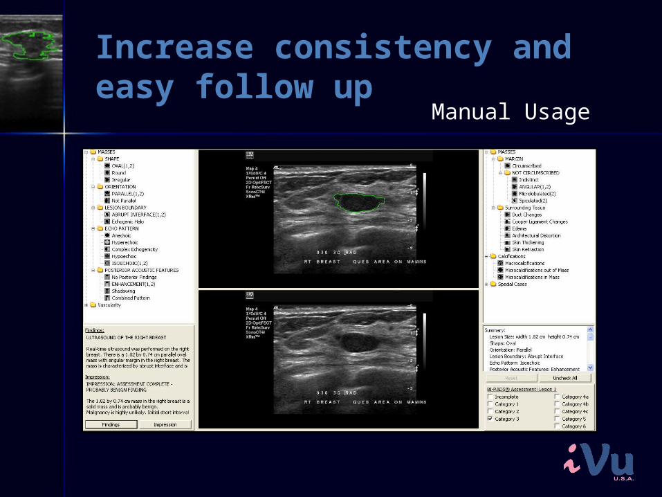
Increase consistency and easy follow up
Manual Usage
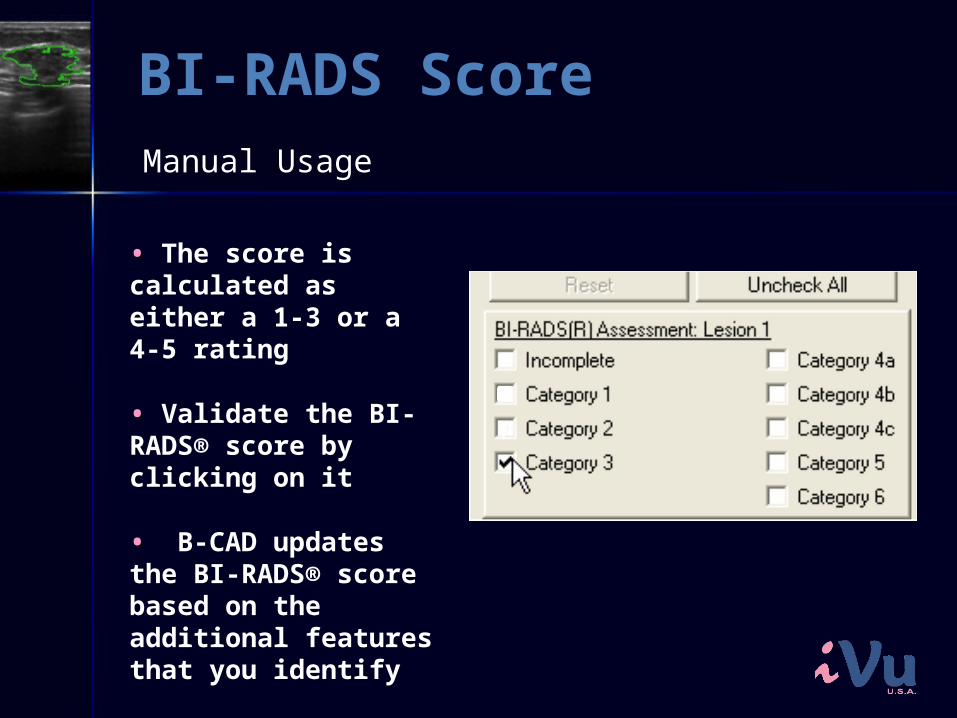
BI-RADS Score
•• The score is calculated as either a 1-3 or a 4-5 rating
•• Validate the BI-RADS® score by clicking on it
•• B-CAD updates the BI-RADS® score based on the additional features that you identify
Manual Usage
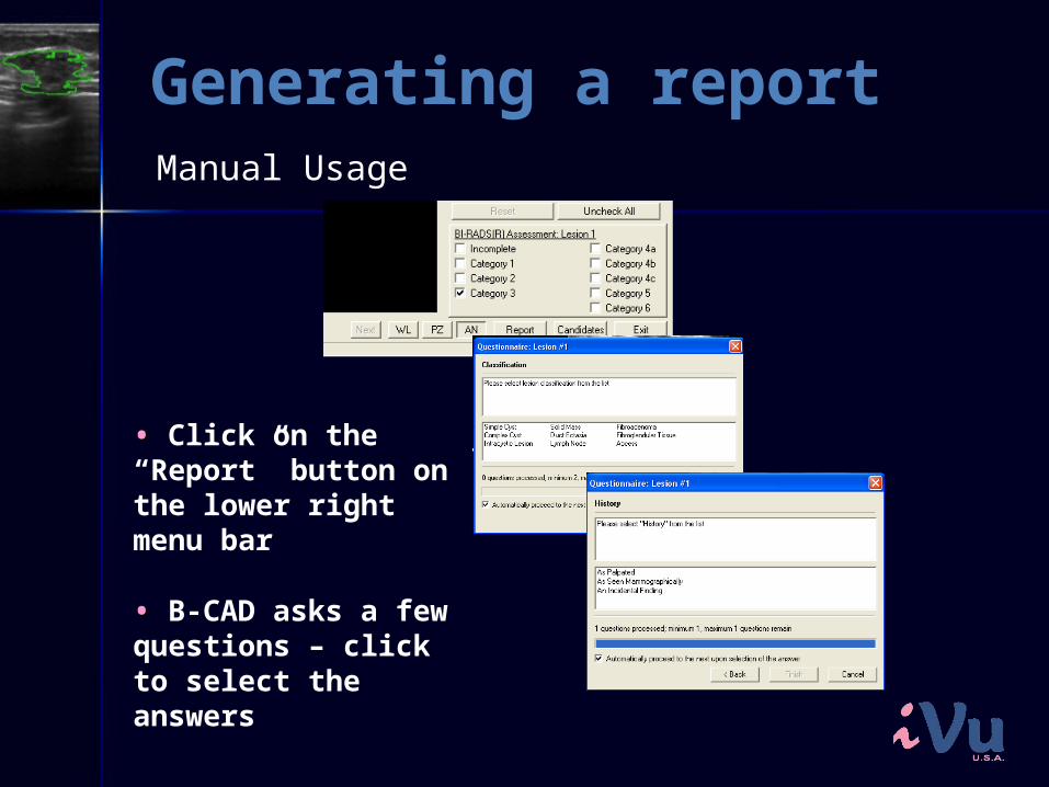
Generating a report
•• Click on the “Report” button on the lower right menu bar
•• B-CAD asks a few questions – click to select the answers
Manual Usage
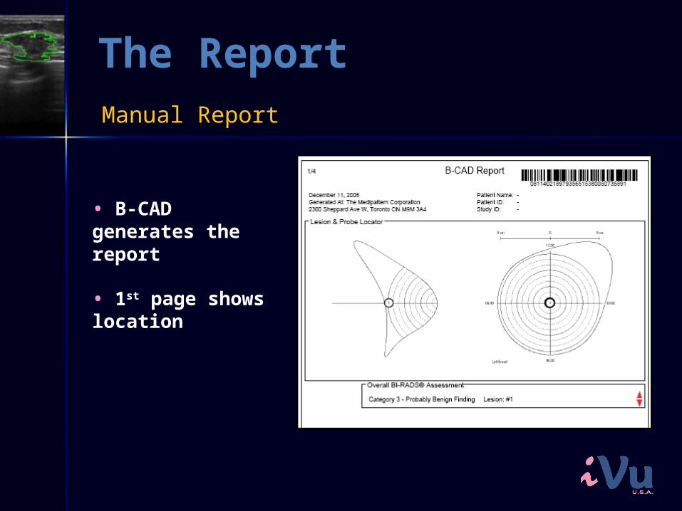
The Report
•• B-CAD generates the report
•• 1st page shows location
Manual Report
The Report
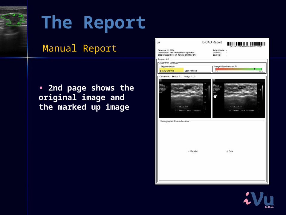
•• 2nd page shows the original image and the marked up image
Manual Report
The Report
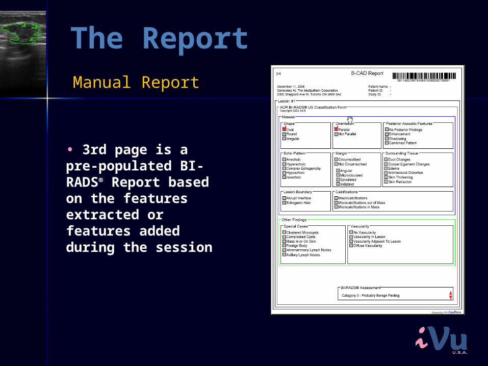
•• 3rd page is a pre-populated BI-RADS® Report based on the features extracted or features added during the session
Manual Report
The Report
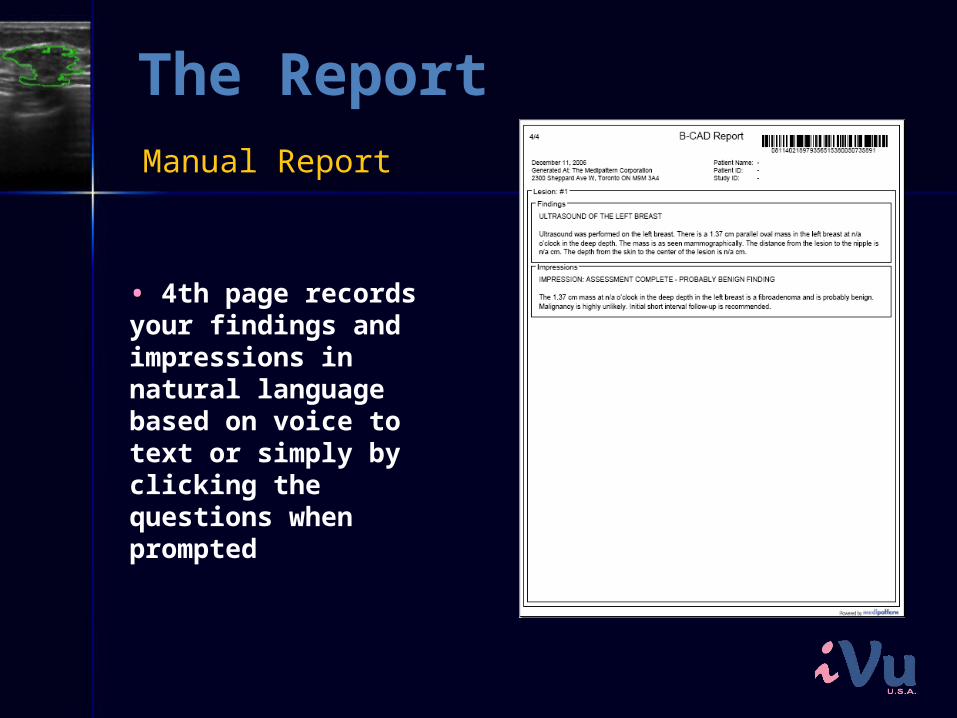
•• 4th page records your findings and impressions in natural language based on voice to text or simply by clicking the questions when prompted
Manual Report
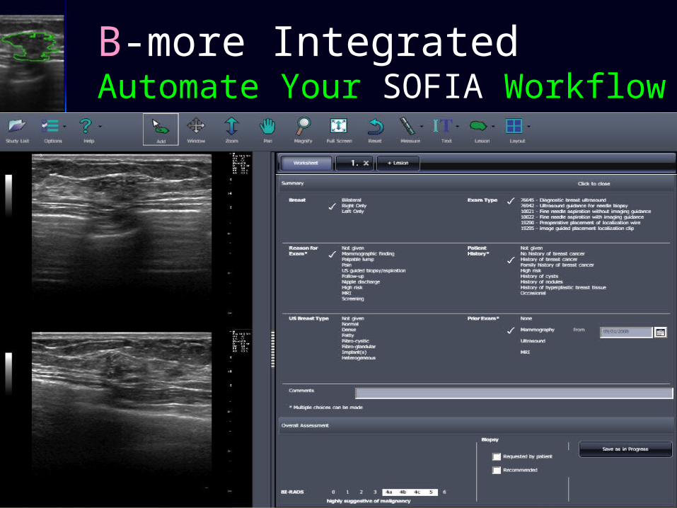
B-more IntegratedAutomate Your SOFIA Workflow
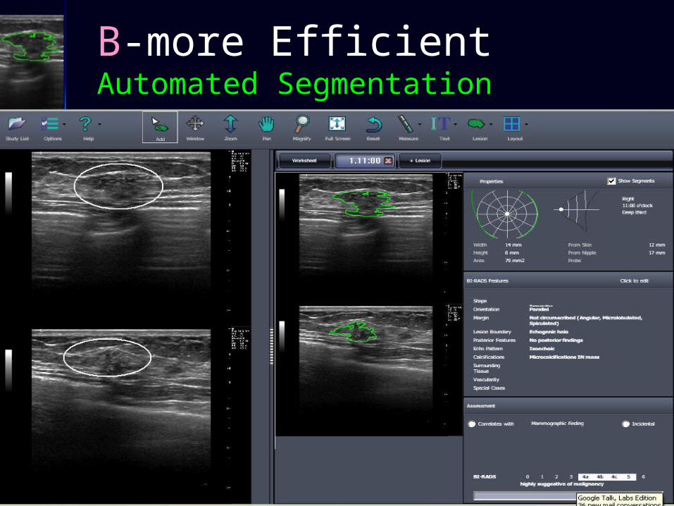
B-more EfficientAutomated Segmentation
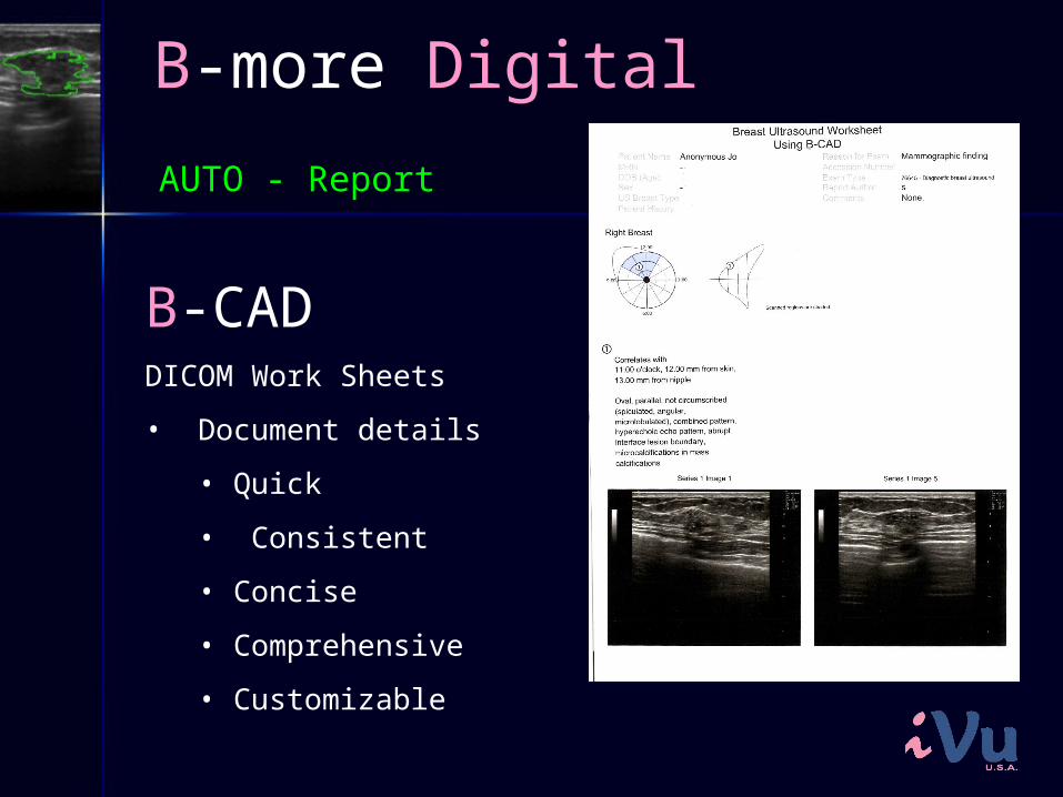
B-more Digital
B-CADDICOM Work Sheets
• Document details
• Quick
• Consistent
• Concise
• Comprehensive
• Customizable
AUTO - Report
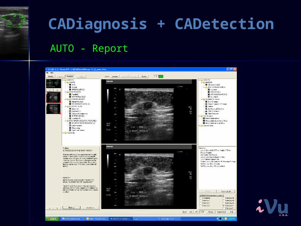
CADiagnosis + CADetection
AUTO - Report
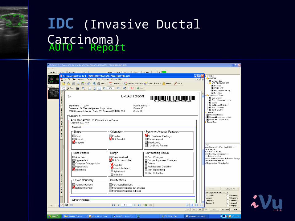
IDC (Invasive Ductal Carcinoma)
AUTO - Report
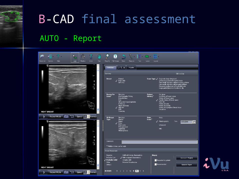
B-CAD final assessment
AUTO - Report
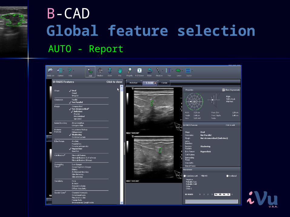
B-CAD Global feature selectionAUTO - Report
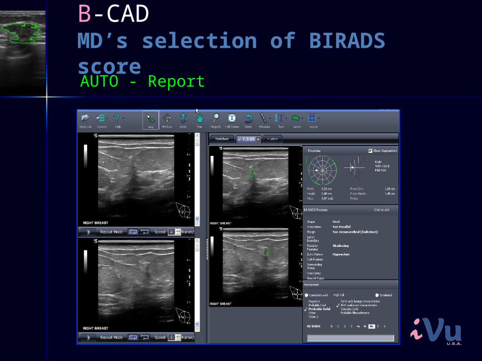
B-CAD MD’s selection of BIRADS scoreAUTO - Report
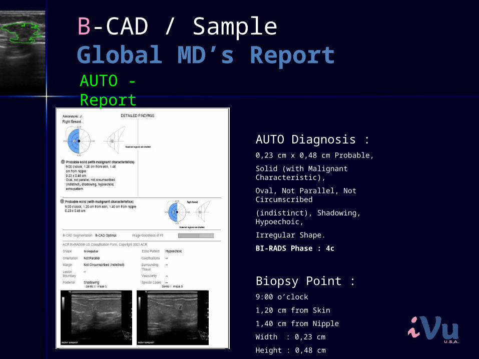
B-CAD / SampleGlobal MD’s ReportAUTO - Report
AUTO Diagnosis :0,23 cm x 0,48 cm Probable,
Solid (with Malignant Characteristic),
Oval, Not Parallel, Not Circumscribed
(indistinct), Shadowing, Hypoechoic,
Irregular Shape.
BI-RADS Phase : 4c
Biopsy Point : 9:00 o’clock
1,20 cm from Skin
1,40 cm from Nipple
Width : 0,23 cm
Height : 0,48 cm
Area : 0,07 cm²
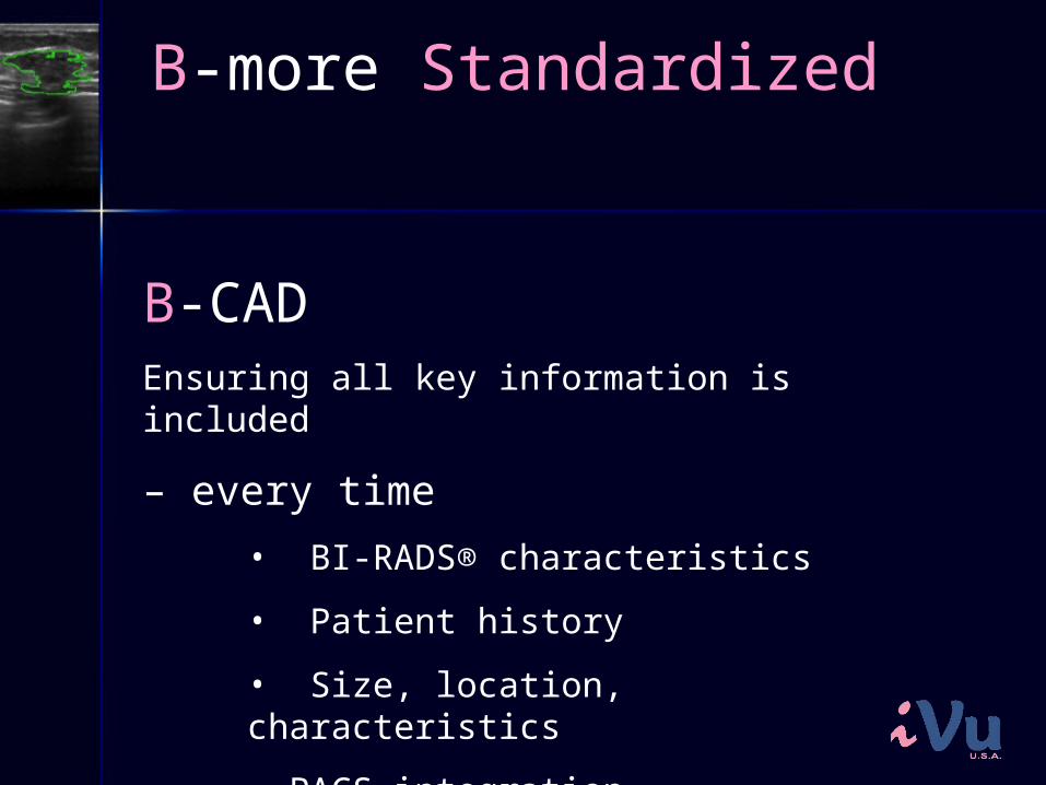
B-more Standardized
B-CADEnsuring all key information is included
– every time• BI-RADS® characteristics
• Patient history
• Size, location, characteristics
• PACS integration
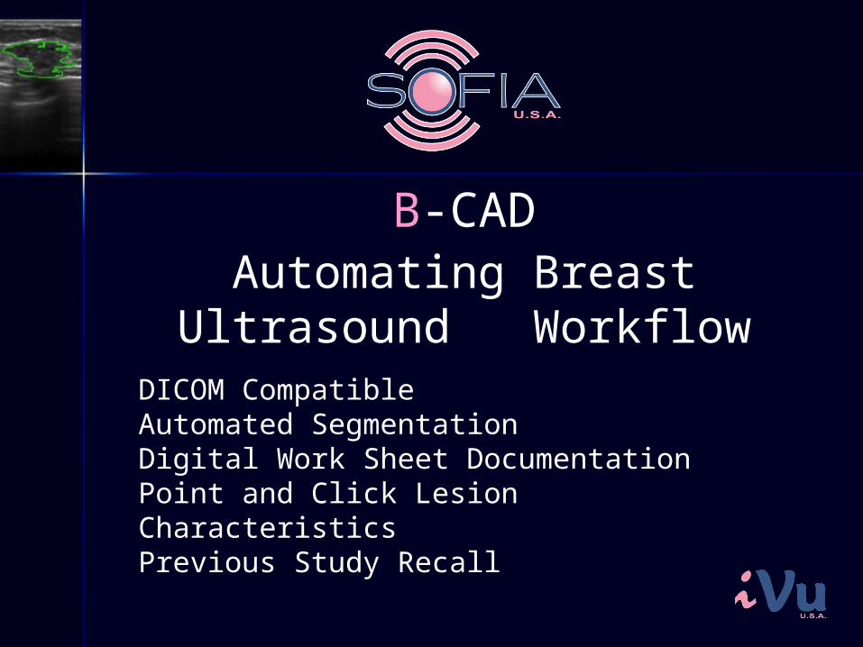
Automating Breast Ultrasound Workflow
DICOM Compatible Automated SegmentationDigital Work Sheet DocumentationPoint and Click Lesion CharacteristicsPrevious Study Recall
B-CAD

What’s in it for YOU!
•• Increased consistency•• Standardized reporting•• Automated Reporting•• Image optimization•• Comprehensive Paperless Workflow


![DICOM Conformance Statement9d48995e-cb8b-4ac4-ae9b... · 2020. 2. 20. · DICOM protocol. 1.5 References [DICOM PS 3 2006] The Digital Imaging and Communications in Medicine (DICOM)](https://static.fdocuments.in/doc/165x107/60e78a442d236e0f92518d06/dicom-conformance-statement-9d48995e-cb8b-4ac4-ae9b-2020-2-20-dicom-protocol.jpg)