Author's response to reviews High expression of P21 ...10.1186/1471...1 1 High expression of...
27
Author's response to reviews Title:High expression of P21-activated protein kinase 1 is an independent prognostic factor in primary and metastatic pancreatic cancer Authors: Juan Han ( [email protected]) Feng Wang ( [email protected]) Zhao-lei Zeng ( [email protected]) Jing Yang ( [email protected]) De-sen Wang ( [email protected]) Mei-yuan Liu ( [email protected]) Han Zhao ( [email protected]) Kai-yan Liu ( [email protected]) Jian-wei Liao ( [email protected]) Qing-feng Zou ( [email protected]) Rui-hua Xu ( [email protected]) Version:2Date:24 March 2014 Author's response to reviews: see over
Transcript of Author's response to reviews High expression of P21 ...10.1186/1471...1 1 High expression of...

Author's response to reviews
Title:High expression of P21-activated protein kinase 1 is an independentprognostic factor in primary and metastatic pancreatic cancer
Authors:
Juan Han ([email protected])Feng Wang ([email protected])Zhao-lei Zeng ([email protected])Jing Yang ([email protected])De-sen Wang ([email protected])Mei-yuan Liu ([email protected])Han Zhao ([email protected])Kai-yan Liu ([email protected])Jian-wei Liao ([email protected])Qing-feng Zou ([email protected])Rui-hua Xu ([email protected])
Version:2Date:24 March 2014
Author's response to reviews: see over

1
High expression of P21-activated protein kinase 1 is an independent prognostic 1
factor in primary and metastatic pancreatic cancer 2
3
Juan Han1,2†, Feng Wang1†, Zhao-lei Zeng3, Li-ren Li1 , Jing Yang3, De-sen Wang1, Mei-yuan Liu2, Han 4
Zhao2, Kai-yan Liu3, Jian-wei Liao3, Qing-feng Zou2*and Rui-hua Xu1* 5
6
* Corresponding authors: Qing-feng Zou [email protected] Rui-hua Xu [email protected] 7
† Equal contributors 8
1Department of Medical Oncology, Sun Yat-sen University Cancer Center, Guangzhou, Guangdong, 9
510060, China 10
2Section 3 of Internal Medicine, The Affiliated Tumor Hospital of Guangzhou Medical University, 78 11
Hengzhigang Road, Guangzhou 510095, Guangdong, China 12
3Sun Yat-sen University Cancer Center; State Key Laboratory of Oncology in South China; 13
Collaborative Innovation Center of Cancer Medicine, Guangzhou 510060, China 14
Email addresses: 15
Juan Han [email protected] - Feng Wang [email protected] - Zhao-lei Zeng 16
[email protected] Li-ren Li [email protected] - Jing Yang [email protected] - De-sen 17
Wang [email protected] - Mei-yuan Liu [email protected] - Han Zhao 18
2
[email protected] - Kai-yan Liu [email protected] - Jian-wei Liao [email protected] 19
- Qing-feng Zou [email protected] - Rui-hua Xu [email protected] 20
21
Abstract 22
Background: P21-activated protein kinase 1 (PAK1), a main downstream effector of small Rho 23
GTPases, is overexpressed in many malignancies. Its overexpression was associated with poor 24
prognosis in some tumor types including breast cancer, gastric cancer, colorectal cancer and so on. 25
However, the expression and clinical relevance of PAK1 expression in human pancreatic cancer remain 26
unknown. 27
Methods: The present study was designed to investigate the clinical and prognostic significance of 28
PAK1 expression in pancreatic carcinoma. We examined the expression of PAK1 by 29
immunohistochemistry in 72 primary pancreatic carcinoma samples and 20 liver metastatic samples. 30
The relationships between PAK1 and clinicopathological parameters and prognosis in primary and 31
metastatic pancreatic cancer were analyzed. 32
Results: Among the total 92 cases, primary pancreatic cancer samples had significantly higher rate 33
(38/72, 52.8%) of high PAK1 expression than liver metastatic samples (5/20, 25.0%) (p = 0.028). In 72 34
primary pancreatic cancer patients, high PAK1 expression was associated with younger age (p=0.038) 35
and moderately or well differentiated tumor (p=0.027). Moreover, a positive relationship was found 36

3
between high PAK1 expression and overall survival (OS) (p<0.005). Patients with high PAK1 37
expression had a better OS than those with low PAK1 expression. Univariate and multivariate analysis 38
by Cox regression including PAK1 and other prognostic pathological markers demonstrated high PAK1 39
immunostaining as an independent prognostic factor for survival in pancreatic cancer patients 40
(P<0.005). 41
Conclusions: For the first time, we report that PAK1 is a novel prognostic marker for pathologically 42
confirmed human pancreatic cancer. High PAK1 expression is significantly associated with better 43
survival in human pancreatic cancer patients. 44
Keywords: P21-activated protein kinase 1 (PAK1), Pancreatic cancer, Immunohistochemistry and 45
prognosis 46
47
Background 48
Pancreatic carcinoma (PC) is one of the most lethal human cancers. It is the fifth most common cancer 49
and the fourth leading cause of cancer-related mortality worldwide with a five-year survival rate less 50
than 5% in all stages [1,2]. Adenocarcinoma of the pancreatic ducts accounts for nearly 95% of all 51
pancreatic tumors [3]. Its median survival is only 3 to 6 months. The high mortality rate of pancreatic 52
cancer is largely attributed to the lack of reliable methods for early detection and its profound 53
resistance to the existing conventional therapies. Up to date, the only approach to cure this disease is 54

4
radical surgical resection which improves the five-year survival rate to approximately 20%-25% [4]. 55
However, due to the absence of early symptoms and robust diagnostic markers, only 10-20% of 56
pancreatic cancer patients present with potentially resectable disease. Most patients have lost the 57
chance of resection because of the very late and difficult diagnosis. Furthermore, even after extensive 58
curative pancreatectomy, patients often have high rate of liver metastasis, resulting in a poor prognosis. 59
Besides that, approximately 30% of patients have isolated local recurrence without evidence of 60
metastases. And 80% of patients have local recurrence within 2 years after potentially curative 61
resections [5]. Therefore, there is an urgent need to search for prognostic markers or therapeutic targets 62
for pancreatic cancer. 63
64
The Rho-family GTPases Rho Rac and Cdc42 regulate many intracellular processes through their 65
interaction with downstream effector proteins. The P21-associated kinases (PAKs) were first 66
discovered in a screen for proteins that interact with the small G-proteins Rac1 and Cdc42 in 1994 [6]. 67
As a set of evolutionarily conserved serine/threonine protein kinase, PAKs influences actin 68
polymerization by regulating cofilin and actin depolymerising factor (ADF), which are actin binding 69
proteins and regulate cytoskeletal dynamics by severing and depolymerizing actin filaments [7,8]. 70
Based on domain structure, sequence homology and regulation, PAKs have been classified mainly into 71
two groups: group I (PAK1 to PAK3) and group II (PAK4 to PAK6) [9]. The regulatory mechanism of 72

5
group I PAKs is considerably different from group II family members [10]. In group I PAKs, the 73
N-terminal domain as the p21-binding domain (PBD) binds small GTPases Rac and Cdc42 to cause 74
autophosphorylation of certain sites in the N-terminal inhibitory domain, leading to conformational 75
change that releases the autoinhibition and activates the kinase activity [11]. In addition, the 76
proline-rich region in group I PAKs is associated with binding to Nck, an adapter protein that is known 77
to be involved in the regulation of actin cytoskeletal dynamics [12]. In contrast with group I PAKs, 78
group II PAKs have very little sequence N-terminal to the PBD. Without obvious autoinhibitory regions 79
as in group I PAKs, the group II PAKs are constitutively active in cells. Crystal structures of 80
phosphorylated, active catalytic domains of all three group II PAKs reveal monomeric conformation 81
[13,14]. 82
83
P21-activated protein kinase1 (PAK1), a member of group I PAKs, is a main downstream effector of 84
small Rho GTPases. It plays an essential role in cell signaling and regulation of cellular functions 85
including cell motility, cytoskeletal rearrangement, angiogenesis, mitosis and survival. A study has 86
shown that PAK1 kinase activity is required for the Ras-induced transformation of fibroblast cells [15]. 87
Many studies also suggest that PAK1 may play a crucial role in cancer development. In breast cancer, 88
PAK1 expression and activity was demonstrated to be increased and correlates with a more malignant 89
phenotype. Experimental manipulation has also indicated that a constitutively active form of PAK1 90
6
could rapidly induce breast cancer cell proliferation and aggressive cell phenotypes including 91
anchorage-independent growth and mitotic defects [16]. Later investigations showed that in several 92
other human cancers the expression and activity of PAK1 has upregulated. In gastric cancer, 93
overexpression of PAK1 was observed and associated with metastasis and prognosis. Downregulation 94
of PAK1 expression reduced gastric cancer cell migration and invasion [17]. In another study of gastric 95
cancer, Liu et al. found that overexpression of PAK1 was associated with progression, metastasis and 96
prognosis probably by regulating the transcription of cyclin B1 through nuclear factor-κB (NF- κB) 97
[18]. In addition, overexpression of PAK1 was also found in colorectal cancer. It was significantly 98
increased in adenomas, invasive carcinomas, and lymph node metastases compared to normal colon 99
[19]. Therefore, PAK1 has gradually become a representative marker for cancers. 100
101
However, the role of PAK1 in pancreatic cancer remains largely unknown. One study in Balb/c mice 102
model of human pancreatic adenocarcinoma xenografts found overexpression of PAK1 in well to 103
moderately differentiated pancreatic cancer samples [20]. However, there are no reports on clinical 104
significance of PAK1 in human pancreatic cancer. Herein, we first investigated the expression status 105
and evaluated the prognostic significance of PAK1 in human pancreatic cancer. 106
107
Materials and methods 108

7
Patient information and tissue specimens 109
The study was approved by the Institutional Review Board and Human Ethics Committee of Sun 110
Yat-sen University Cancer Center and the Affiliated Tumor Hospital of Guangzhou Medical University. 111
Specimens of the present study contain 72 cases paraffin-embedded primary pancreatic cancer samples 112
and 20 cases liver metastatic tissues of pancreatic cancer which recruited from the Sun Yat-sen 113
University Cancer Center and the Affiliated Tumor Hospital of Guangzhou Medical University 114
(Guangzhou, China) between May 2005 and December 2012. Among the total of 92 cases, 72 patients 115
underwent surgical resection of pancreatic tumor, 9 patients underwent resection of liver metastatic 116
tumor and 11 patients had liver tissue biopsy. Histomorphology of all tumor specimens and regional 117
lymph nodes was confirmed with hematoxylin-eosin staining and diagnosed as pancreatic cancer 118
independently by two pathologists. The 7th Union International Cancer Control (UICC) TNM staging 119
system was applied after surgery combined with imaging manifestations [21] . Overall survival (OS) 120
was defined as the interval between the date of definite diagnosis and date of death or the last follow up. 121
The clinical and pathological features of 72 cases primary pancreatic carcinoma patients are 122
summarized in Table 1. 123
124
Immunohistochemistry (IHC) 125
Immunohistochemistry was carried out according to standard methods as described previously to study 126

8
altered protein expression in 72 primary and 20 metastatic pancreatic cancer tissues [22]. Briefly, the 127
tissue sections were deparaffinized in xylene at 37°C for 20 min and rehydrated in a series of graded 128
alcohols. To quench endogenous peroxidase activity the sections were then treated with 3% hydrogen 129
peroxide in methanol for 20 min at 37°C, and the antigens were retrieved in 10mM citrate buffer (pH 130
6.0) using a microwave oven, then sections were incubated overnight at 4°C with a 1:50 dilution of 131
rabbit anti-PAK1 (Cell Signaling, #2602) diluted in Dako Antibody Diluent (ZLI-9030). The next day 132
tissue sections were treated with anti-rabbit secondary antibody for 30 min and developed with 133
diaminobenzidine tetrahydrochloride (DAB) and counterstained with hematoxylin. The intensity of 134
staining and the percentage of the stained cells were estimated by two investigators who were blinded 135
to the patients’ data. According to the percentage of positive tumor cells one score was given as 136
follows:≤5% = 0, >5% to≤25% = 1, >25% to≤50% = 2, >50% to≤75% = 3, >75% = 4. Another score 137
was given according to the intensity of staining as negative = 0, weak = 1, moderate = 2, or strong = 3. 138
The final quantitation of each staining was obtained by multiplying the two scores. PAK1 expression 139
was classified as low expression for the final scores <4 and high expression for scores ≥4. 140
141
Statistical analysis 142
All statistical analyses were carried out using SPSS software (version 16.0; Chicago, IL). The χ2 test 143
was used for statistical analysis of interdependence between PAK1 expression and clinical data. 144

9
Survival curves were constructed using the Kaplan-Meier method and analyzed using the log-rank test. 145
Survival data were evaluated using univariate and multivariate Cox regression analyses. The risk ratio 146
and its 95% confidence interval were recorded for each clinicopathological parameter. P values <0.05 147
were considered statistically significant. 148
149
Results 150
PAK1 expression is higher in primary pancreatic cancer tissues than in metastatic tissues 151
PAK1 protein expression was significantly higher in 72 human primary pancreatic cancer tissues 152
compared to 20 liver metastatic tissues (Figure 1) by using immunohistochemistry. Positive staining 153
was observed in the cytoplasm of those human primary and metastatic pancreatic cancer samples. 154
According to the definition, scores less than four were defined as low PAK1 expression in the IHC 155
samples. Otherwise, they were considered as having high PAK1 expression. The rate of high PAK1 156
expression was significantly higher in primary pancreatic cancer samples (38/72, 52.8%) than the rate 157
in metastatic pancreatic cancer samples (5/20, 25.0%) (p = 0.028). 158
159
High PAK1 expression is associated with younger onset age and well differentiated tumor 160
According to the results of immunohistochemistry, we analysed PAK1 expression status in 72 primary 161
pancreatic cancer specimens which included 6 cases of clinical stage I (8.4%), 38 cases of stage II 162
10
(52.8%), 14 cases of stage III (19.4%) and 14 cases of stage IV (19.4%) pancreatic cancer. According 163
to the scoring system, low PAK1 expression was detected in 34/72 (47.2%) pancreatic carcinomas, 164
while high PAK1 expression was detected in 38/72 (52.8%). Our analyses showed that there was no 165
significant correlation between PAK1 protein expression and clinical characteristics such as gender or 166
clinical stage. However, PAK1 was associated with age (p=0.038) and pathologic differentiation (p= 167
0.027). The rate of high PAK1 expression was significantly higher in young patients (≤ 60 years) than 168
in senior patients (>60 years) (68.4% vs 31.6%, p=0.038). In addition, poor pathological differentiation 169
was associated with low PAK1 expression. On the contrary, high PAK1 expression was found more in 170
well differentiated samples (Figure 1). The rates of high PAK1 expression in poorly, moderately and 171
well differentiated samples were 39.0% (16/41), 71.4% (20/28) and 66.7% (2/3), respectively. Data 172
regarding PAK1 expression in relation to clinical and pathological parameters are summarized in Table 173
2. 174
175
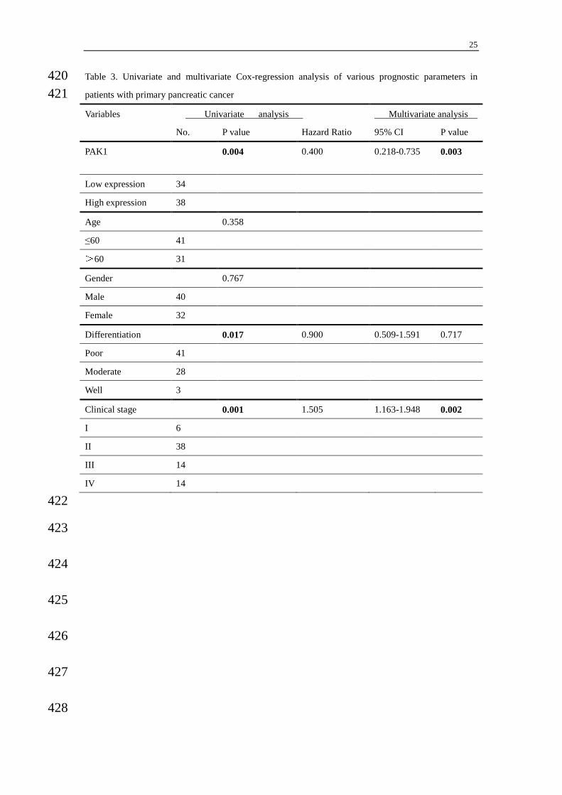
PAK1 expression is an independent prognostic factor 176
Survival analysis showed that OS was significantly different among 72 patients according to the 177
expression of PAK1 (P<0.005) (Figure 2) by using the Kaplan-Meier analysis and log-rank test. 178
Patients with high PAK1 expression had a significantly longer overall survival time (OS) than those 179
with low PAK1 expression (median OS 23.3 vs 12.0 months). In univariate analysis for primary 180

11
pancreatic cancer patients, PAK1 expression status (p=0.004), differentiation (p=0.017) and clinical 181
stage (p=0.001) were prognostic factors. While in multivariate analysis by Cox regression, PAK1 182
expression (p=0.003) and clinical stages (p=0.002) were two independent prognostic factors (Table 3). 183
Therefore, the present study indicated that PAK1 may be a novel prognostic factor for survival in 184
pancreatic cancer patients. 185
186
Discussion 187
P21-associated kinases (PAKs) are found to be increased in many malignancies and play an important 188
role in the regulation of cell morphogenesis, motility, mitosis and angiogenesis [23-28]. After 189
performing IHC in 72 primary pancreatic cancer samples and 20 liver metastatic samples in this study, 190
we found that PAK1 expression was lower in liver metastatic sites of pancreatic cancer compared to 191
primary pancreatic cancer tissues. The rate of high PAK1 expression in primary pancreatic cancer 192
samples was one fold higher than the rate in metastatic pancreatic cancer samples (52.8% vs 25.0%). 193
This result of current study is different from some previous studies done in other tumors. Kamai et al. 194
found PAK1 overexpression is associated with lymphovascular invasion and lymph node metastasis of 195
upper urinary tract cancer [29]. Ching et al. found that overexpression of PAK1 in human 196
hepatocellular carcinomas was associated with more aggressive tumor behavior and more advanced 197
tumor stages [30]. They also found PAK1 induced cancer metastasis may involve activation of c-Jun 198

12
NH2-terminal kinase (JNK) and phosphorylation of paxillin. In addition, another study demonstrated 199
that PAK1 induced colorectal cancer metastasis via extracellular signal-regulated kinase (ERK) 200
dependent phosphorylation of focal adhesion kinase (FAK) [31]. 201
202
Our study found PAK1 was associated with age (p<0.05) and pathologic differentiation (p<0.05). 203
Immunohistochemical results showed that poor pathological differentiation was correlated with lower 204
PAK1 expression and well pathological differentiation was correlated with higher PAK1 expression. 205
Some recent studies indicated that overexpression of PAK1 is associated with poor differentiation in 206
gastric cancer, colorectal cancer and breast cancer [17,31-32]. In addition, the subcellular localization 207
of PAK1 staining is related to clinicopathologic tumor parameters in breast cancer. Cytoplasmic PAK1 208
staining was strongly correlated with histological grade and the level of tumor cell proliferation,while 209
positive nuclear PAK1 staining was correlated with tumor cell proliferation [32]. In pancreatic cancer, 210
Makisumi K et al. immunized Balb/c mice with human pancreatic adenocarcinoma xenografts [20]. 211
However, they demonstrated that PAK1 showed a stronger positive immunostaining in well and 212
moderately differentiated pancreatic cancer compared to poorly differentiated pancreatic cancer. 213
Furthermore, acinar cells of normal fetal pancreas showed a strong positive PAK1 staining in 214
cytoplasm. While normal adult pancreas consisting of only islets of Langerhans showed a weak 215
staining of PAK1. So it suggests that PAK1 probably recognizes a certain category of oncofetal 216

13
antigens differentially expressed on pancreatic carcinomas. 217
218
Studies from other malignancies found that overexpression of PAK1 was associated with poor 219
prognosis in colorectal cancer, ovarian cancer, breast cancer and so on [19,32-34]. A recent study 220
revealed that PAK1 expression increased with the progression of colorectal cancer. The expression of 221
PAK1 in colon cancer cells promoted transformation through facilitating the ERK/MAPK 222
(mitogen-activated protein kinases) pathway and enhanced cell migration and survival by stimulating 223
AKT [35]. In some other studies, PAK1 genomic amplification found at 11q13 was prevalent in luminal 224
breast cancer and breast cancer cells. After inhibiting PAK1, cancer cells rapidly underwent apoptosis. 225
Furthermore, strong nuclear and cytoplasmic PAK1 expression was also prevalent in squamous 226
non-small cell lung carcinomas, and selective PAK1 inhibition was associated with delayed cell-cycle 227
progression in vitro and in vivo [36,37]. These studies have implicated the important role of PAK1 in 228
the regulation of cell motility and tumor cell invasiveness. 229
230
The expression of PAK1 in human pancreatic cancer has never been reported. This is the first study to 231
investigate the prognostic relevance of PAK1 expression in human primary and metastatic pancreatic 232
carcinoma. It’s striking that high PAK1 expression was correlated with a better survival outcome in 233
primary pancreatic cancer patients. The multivariate Cox model analysis identified PAK1 as an 234

14
independent prognostic factor in pancreatic cancer patients. Additionally, PAK1 expression was lower 235
in liver metastatic sites of pancreatic cancer compared to primary pancreatic cancer tissues. These 236
results indicate PAK1 may play a critical role in the initial phase of carcinogenesis rather than tumor 237
development or metastasis phase in pancreatic carcinoma. This may also indicated that PAK1 did not 238
promote tumor metastasis in pancreatic cancer patients. Nevertheless, the exact molecular mechanism 239
by which PAK1 involved in pancreatic cancer development and progression still remain unclear. Thus, 240
further investigations of PAK1 expression in pancreatic cancer will be needed to elucidate the precise 241
mechanism for its exact regulating pathway in vitro and in vivo. 242
243
Conclusion 244
For the first time, our study showed that higher PAK1 expression was found in primary pancreatic 245
cancer tissues compared to liver metastatic tissues of pancreatic cancer. High PAK1 expression was 246
associated with well differentiated tumors and closely correlated with better overall survival. High 247
expression of PAK1 was an independent positive prognostic factor for survival in pancreatic cancer 248
patients. 249
250
Abbreviations 251
AJCC: American Joint Committee on Cancer; c-JNK: c-Jun N-terminal kinase; 252

15
ADF: actin depolymerising factor; PBD: p21-GTPase-binding domain; 253
DAB: Diaminobenzidine tetrahydrochloride; IHC: Immunohistochemistry; 254
MAPK: mitogen-activated protein kinases; OS: Overall survival; 255
PAK1:P21-activated protein kinase 1; 256
UICC: Union International Cancer Control; ERK: extracellular signal-regulated kinase 257
258
Competing interests 259
The authors declare that they have no conflict of interest. 260
261
Authors’ contributions 262
HJ carried out experiments, analyzed the data, and participated in the experiments design and 263
manuscript writing. WF conceived the study and edited the manuscript. ZZL,LLR, LMY, WDS and 264
LKY participated in the clinical samples collection of the pancreatic cancer patients. YJ and LJW 265
contributed reagents and analysis tools. XRH and ZQF supervised and participated in data analysis and 266
interpretation, and manuscript writing. All authors read and approved the final manuscript. 267
268
Acknowledgements 269
These works were funded by National High Technology Research and Development Program of China 270

16
(863 Program), China (No.2012AA02A506); Natural Science Foundation of China(No.81372570); 271
National Natural Science Foundation of China (No.81372570); The Science and Technology 272
Department of Guangdong Province, China (No.2012B031800088); Medical Scientific Research 273
Foundation of Guangdong Province, China (No.C2011019). 274
275
Author details 276
1Department of Medical Oncology, Sun Yat-sen University Cancer Center, Guangzhou, Guangdong, 277
510060, China 278
2Section 3 of Internal Medicine, The Affiliated Tumor Hospital of Guangzhou Medical University, 78 279
Hengzhigang Road, Guangzhou 510095, Guangdong, China 280
3Sun Yat-sen University Cancer Center; State Key Laboratory of Oncology in South China; 281
Collaborative Innovation Center of Cancer Medicine, Guangzhou 510060, China 282
283
284
285
286
287
288
17
References 289
1. Jemal A, Bray F, Center MM, Ferlay J, Ward E, Forman D: Global cancer statistics. CA Cancer J 290
Clin 2011, 61(2):69-90. 291
2. Hariharan D, Saied A, Kocher HM: Analysis of mortality rates for pancreatic cancer across the 292
world . HPB (Oxford) 2008, 10(1):58-62. 293
3. Kloppel G, Lingenthal G, von Bulow M, Kern HF: Histological and fine structural features of 294
pancreatic ductal adenocarcinomas in relation to growth and prognosis: studies in 295
xenografted tumours and clinico-histopathological correlation in a series of 75 cases. 296
Histopathology 1985, 9(8):841-856. 297
4. Gaedcke J, Gunawan B, Grade M, Szoke R, Liersch T, Becker H, Ghadimi BM: The 298
mesopancreas is the primary site for R1 resection in pancreatic head cancer: relevance for 299
clinical trials . Langenbecks Arch Surg 2010, 395(4):451-458. 300
5. Strobel O, Hartwig W, Hackert T, Hinz U, Berens V, Grenacher L, Bergmann F, Debus J, Jager D, 301
Buchler M et al: Re-resection for isolated local recurrence of pancreatic cancer is feasible, 302
safe, and associated with encouraging survival. Ann Surg Oncol 2013, 20(3):964-972. 303
6. Manser E, Leung T, Salihuddin H, Zhao ZS, Lim L: A brain serine/threonine protein kinase 304
activated by Cdc42 and Rac1. Nature 1994, 367(6458):40-46. 305
7. Bokoch GM: Biology of the p21-activated kinases. Annu Rev Biochem 2003, 72:743-781. 306

18
8. Bamburg JR, Wiggan OP: ADF/cofilin and actin dynamics in disease. Trends Cell Biol 2002, 307
12(12):598-605. 308
9. Kumar R, Gururaj AE, Barnes CJ: p21-activated kinases in cancer. Nat Rev Cancer 2006, 309
6(6):459-471. 310
10. Eswaran J, Soundararajan M, Kumar R, Knapp S: UnPAKing the class differences among 311
p21-activated kinases. Trends Biochem Sci 2008, 33(8):394-403. 312
11. Bokoch GM: Biology of the p21-activated kinases. Annu Rev Biochem 2003, 72:743-781. 313
12. Zhao ZS, Manser E, Lim L: Interaction between PAK and nck: a template for Nck targets and 314
role of PAK autophosphorylation. Mol Cell Biol 2000, 20(11):3906-3917. 315
13. Pandey A, Dan I, Kristiansen TZ, Watanabe NM, Voldby J, Kajikawa E, Khosravi-Far R, Blagoev 316
B, Mann M: Cloning and characterization of PAK5, a novel member of mammalian 317
p21-activated kinase-II subfamily that is predominantly expressed in brain. Oncogene 2002, 318
21(24):3939-3948. 319
14. Eswaran J, Lee WH, Debreczeni JE, Filippakopoulos P, Turnbull A, Fedorov O, Deacon SW, 320
Peterson JR, Knapp S: Crystal Structures of the p21-activated kinases PAK4, PAK5, and 321
PAK6 reveal catalytic domain plasticity of active group II PAKs . Structure 2007, 322
15(2):201-213. 323
15. Tang Y, Chen Z, Ambrose D, Liu J, Gibbs JB, Chernoff J, Field J: Kinase-deficient Pak1 324
19
mutants inhibit Ras transformation of Rat-1 fibroblasts. Mol Cell Biol 1997, 17(8):4454-4464. 325
16. Vadlamudi RK, Adam L, Wang RA, Mandal M, Nguyen D, Sahin A, Chernoff J, Hung MC, 326
Kumar R: Regulatable expression of p21-activated kinase-1 promotes anchorage-independent 327
growth and abnormal organization of mitotic spindles in human epithelial breast cancer cells. 328
J Biol Chem 2000, 275(46):36238-36244. 329
17. Li LH, Luo Q, Zheng MH, Pan C, Wu GY, Lu YZ, Feng B, Chen XH, Liu BY: P21-activated 330
protein kinase 1 is overexpressed in gastric cancer and induces cancer metastasis. Oncol Rep 331
2012, 27(5):1435-1442. 332
18. Liu F, Li X, Wang C, Cai X, Du Z, Xu H, Li F: Downregulation of p21-activated kinase-1 333
inhibits the growth of gastric cancer cells involving cyclin B1. Int J Cancer 2009, 334
125(11):2511-2519. 335
19. Carter JH, Douglass LE, Deddens JA, Colligan BM, Bhatt TR, Pemberton JO, Konicek S, Hom J, 336
Marshall M, Graff JR: Pak-1 expression increases with progression of colorectal carcinomas to 337
metastasis. Clin Cancer Res 2004, 10(10):3448-3456. 338
20. Makisumi K, Takahashi K, Takako S, Sonoda S: Human pancreatic adenocarcinoma-associated 339
antigens defined by novel murine monoclonal antibodies Pak-1 and Pak-2. Gastroenterol Jpn 340
1990, 25(2):236-243. 341
21. Edge SB, Compton CC: The American Joint Committee on Cancer: the 7th edition of the 342
20
AJCC cancer staging manual and the future of TNM. Ann Surg Oncol 2010, 17(6):1471-1474. 343
22. Hong J, Hu K, Yuan Y, Sang Y, Bu Q, Chen G, Yang L, Li B, Huang P, Chen D et al: CHK1 344
targets spleen tyrosine kinase (L) for proteolysis in hepatocellular carcinoma. J Clin Invest 345
2012, 122(6):2165-2175. 346
23. Sells MA, Knaus UG, Bagrodia S, Ambrose DM, Bokoch GM, Chernoff J: Human p21-activated 347
kinase (Pak1) regulates actin organization in mammalian cells. Curr Biol 1997, 7(3):202-210. 348
24. Adam L, Vadlamudi R, KondaPaka SB, Chernoff J, Mendelsohn J, Kumar R: Heregulin regulates 349
cytoskeletal reorganization and cell migration through the p21-activated kinase-1 via 350
phosphatidylinositol-3 kinase. J Biol Chem 1998, 273(43):28238-28246. 351
25. Ching YP, Leong VY, Lee MF, Xu HT, Jin DY, Ng IO: P21-activated protein kinase is 352
overexpressed in hepatocellular carcinoma and enhances cancer metastasis involving c-Jun 353
NH2-terminal kinase activation and paxillin phosphorylation . Cancer Res 2007, 354
67(8):3601-3608. 355
26. Molli PR, Li DQ, Murray BW, Rayala SK, Kumar R: PAK signaling in oncogenesis. Oncogene 356
2009, 28(28):2545-2555. 357
27. Kumar R, Gururaj AE, Barnes CJ: p21-activated kinases in cancer. Nat Rev Cancer 2006, 358
6(6):459-471. 359
28. Vadlamudi RK, Adam L, Wang RA, Mandal M, Nguyen D, Sahin A, Chernoff J, Hung MC, 360

21
Kumar R: Regulatable expression of p21-activated kinase-1 promotes anchorage-independent 361
growth and abnormal organization of mitotic spindles in human epithelial breast cancer cells. 362
J Biol Chem 2000, 275(46):36238-36244. 363
29. Kamai T, Shirataki H, Nakanishi K, Furuya N, Kambara T, Abe H, Oyama T, Yoshida K: 364
Increased Rac1 activity and Pak1 overexpression are associated with lymphovascular 365
invasion and lymph node metastasis of upper urinary tract cancer. Bmc Cancer 2010, 10:164. 366
30. Ching YP, Leong VY, Lee MF, Xu HT, Jin DY, Ng IO: P21-activated protein kinase is 367
overexpressed in hepatocellular carcinoma and enhances cancer metastasis involving c-Jun 368
NH2-terminal kinase activation and paxillin phosphorylation . Cancer Res 2007, 369
67(8):3601-3608. 370
31. Li LH, Zheng MH, Luo Q, Ye Q, Feng B, Lu AG, Wang ML, Chen XH, Su LP, Liu BY: 371
P21-activated protein kinase 1 induces colorectal cancer metastasis involving ERK activation 372
and phosphorylation of FAK at Ser-910. Int J Oncol 2010, 37(4):951-962. 373
32. Holm C, Rayala S, Jirstrom K, Stal O, Kumar R, Landberg G: Association between Pak1 374
expression and subcellular localization and tamoxifen resistance in breast cancer patients. J 375
Natl Cancer Inst 2006, 98(10):671-680. 376
33. Siu MK, Wong ES, Chan HY, Kong DS, Woo NW, Tam KF, Ngan HY, Chan QK, Chan DC, 377
Chan KY et al: Differential expression and phosphorylation of Pak1 and Pak2 in ovarian 378

22
cancer: effects on prognosis and cell invasion. Int J Cancer 2010, 127(1):21-31. 379
34. Schraml P, Schwerdtfeger G, Burkhalter F, Raggi A, Schmidt D, Ruffalo T, King W, Wilber K, 380
Mihatsch MJ, Moch H: Combined array comparative genomic hybridization and tissue 381
microarray analysis suggest PAK1 at 11q13.5-q14 as a critical oncogene target in ovarian 382
carcinoma. Am J Pathol 2003, 163(3):985-992. 383
35. Huynh N, Liu KH, Baldwin GS, He H: P21-activated kinase 1 stimulates colon cancer cell 384
growth and migration/invasion via ERK- and AKT-dependent pathways. Biochim Biophys 385
Acta 2010, 1803(9):1106-1113. 386
36. Ong CC, Jubb AM, Haverty PM, Zhou W, Tran V, Truong T, Turley H, O'Brien T, Vucic D, 387
Harris AL et al: Targeting p21-activated kinase 1 (PAK1) to induce apoptosis of tumor cells. 388
Proc Natl Acad Sci U S A 2011, 108(17):7177-7182. 389
37. Rettig M, Trinidad K, Pezeshkpour G, Frost P, Sharma S, Moatamed F, Tamanoi F, Mortazavi F: 390
PAK1 kinase promotes cell motility and invasiveness through CRK-II serine phosphorylation 391
in non-small cell lung cancer cells. PLoS One 2012, 7(7):e42012. 392
393
394
395
396

23
Table 1. Clinical and pathological characteristics of 72 patients with primary pancreatic cancer 397 Number of cases (%)
Gender
Male 40(55.6)
Female 32(44.4)
Age (years)
≤60 41(56.9)
>60 31(43.1)
Clinical Stage
I 6(8.4)
II 38(52.8)
III 14(19.4)
IV 14(19.4)
T classification
T1 0(0)
T2 12(16.7)
T3 41(56.9)
T4 19(26.4)
N classification
N0 45(62.5)
N1 27(37.5)
M classification
M0 58(80.6)
M1 14(19.4)
Pathologic Differentiation
Poor 41(56.9)
Moderate 28(38.9)
Well 3(4.2)
Expression of PAK1
Low expression 34(47.2)
High expression 38(52.8)
398 399 400 401

24
Table 2. Correlation analysis between PAK1 expression and clinicopathological characteristics in 72 402 patients with primary pancreatic cancer. 403
Characteristics PAK1 Chi-square test P-value
Low or none No. cases (%) High No. cases (%)
Gender Male 18 (52.9% ) 22(57.9%) 0.673
Female 16(47.1%) 16(42.1%)
Age (years) ≤ 60 15(44.1%) 26(68.4%) 0.038
> 60 19(55.9%) 12(31.6%)
Pathologic Differentiation Poor 25(73.5%) 16(42.1%) 0.027
Moderate 8(23.5%) 20(52.6%)
Well 1(3.0%) 2(5.3%)
Clinical Stage I -III 29(85.3% ) 29(76.3%) 0.706
IV 5(14.7%) 9(23.7%)
T classification T1+T2 4(11.8%) 8(21.1%) 0.291
T3+T4 30(88.2%) 30(78.9%)
N classification No 20(58.8%) 25(65.8%) 0.542
Yes 14(41.2%) 13(34.2%)
M classification M0 29(85.3%) 29(76.3%) 0.337
M1 5(14.7%) 9(23.7%)
404 405 406 407 408 409 410 411 412 413 414 415 416 417 418 419
25
Table 3. Univariate and multivariate Cox-regression analysis of various prognostic parameters in 420 patients with primary pancreatic cancer 421
Variables Univariate analysis Multivariate analysis
No. P value Hazard Ratio 95% CI P value
PAK1 0.004 0.400 0.218-0.735 0.003
Low expression 34
High expression 38
Age 0.358
≤60 41
>60 31
Gender 0.767
Male 40
Female 32
Differentiation 0.017 0.900 0.509-1.591 0.717
Poor 41
Moderate 28
Well 3
Clinical stage 0.001 1.505 1.163-1.948 0.002
I 6
II 38
III 14
IV 14
422
423
424
425
426
427
428

26
Figure legends 429
Fig. 1 Immunohistochemistry of PAK1 expression in human primary pancreatic cancer tissues and 430
metastatic liver samples. Original magnifications: x100 (the left side) and x200 (the right side). (a) 431
Representative images showing very weak staining of cytoplasmic PAK1 staining (brown) in poorly 432
differentiated metastatic pancreatic cancer tissues. (b-d) Representative images showing an obvious 433
increasing trend staining of cytoplasmic PAK1 staining (brown) in poorly, moderate and well 434
differentiated primary pancreatic cancer tissues, respectively. 435
Fig. 2 Kaplan–Meier survival curves for primary pancreatic cancer patients with low PAK1 expression 436
(dotted line) versus high PAK1 expression (solid line). (a) The overall survival of patients (clinical 437
stages Ⅰ-Ⅳ) with low/high PAK1 expression. (b) The overall survival of patients (clinical stages Ⅰ-438
Ⅲ) with low/high PAK1 expression. (c) The overall survival of patients (clinical stages Ⅳ) with 439
low/high PAK1 expression. 440
441
442
443
444
445