Author’s Postprint version of: Bull AMJ, Andersen HN, Basso O, … · 2015-12-26 · Author’s...
12
Author’s Postprint version of: Bull AMJ, Andersen HN, Basso O, Target J, Amis AA. Incidence and mechanism of the pivot shift: An in vitro study. Clin. Orthop. Rel. Res. 363 , (1999), 219-231. This is not the final published version Publisher version: http://c-orthopaedicpractice.com/pt/re/cop/abstract.00003086-199906000- 00029.htm;jsessionid=Ls2S47Chs9cXcGDddkhPTsnGQ668z4Lw2r201Krhn2pDn9jrjTDT!- 1124491571!181195628!8091!-1
Transcript of Author’s Postprint version of: Bull AMJ, Andersen HN, Basso O, … · 2015-12-26 · Author’s...

Author’s Postprint version of: Bull AMJ, Andersen HN, Basso O, Target J, Amis AA. Incidence and mechanism of the pivot shift: An in vitro study. Clin. Orthop. Rel. Res. 363, (1999), 219-231. This is not the final published version Publisher version: http://c-orthopaedicpractice.com/pt/re/cop/abstract.00003086-199906000-00029.htm;jsessionid=Ls2S47Chs9cXcGDddkhPTsnGQ668z4Lw2r201Krhn2pDn9jrjTDT!-1124491571!181195628!8091!-1

Incidence and Mechanism of the Pivot Shift: An In Vitro Study
Bull, Anthony M.J. PhD; Andersen, Henrik N. MD, PhD; Basso, Oreste MD; Targett, John BS; Amis, Andrew A. PhD
Funded by an Engineering and Physical Sciences Research Council Award and the Arthritis Research Campaign. Corresponding Author: Anthony M.J. Bull, PhD, Biomechanics Section, Mechanical Engineering Department, Imperial College of Science, Technology and Medicine, Exhibition Road, London SW7 2BX, United Kingdom.
Abstract The aim of this study was to determine the incidence and mechanism of the pivot shift phenomenon in the normal and anterior cruciate ligament transected knee in vitro. Fifteen knees were tested under a range of valgus moments and iliotibial tract tensions when intact and after anterior cruciate ligament transection. Knee kinematics were measured and described in terms of tibial rotation as the knee flexed. Eight knees pivoted after anterior cruciate ligament transection. The mean pivot shift motion was an external tibial rotation of 17° (±11° standard deviation) over a range of 27° (±24°) knee flexion, at a mean flexion angle of 56° (±27°). Clinically, this corresponds to a reduction of an anteriorly subluxed lateral tibial plateau as the knee flexes. When intact, pivoting and nonpivoting knees had similar anteroposterior laxity, but after anterior cruciate ligament transection, the pivoting group had significantly greater laxity. The loading required to elicit the pivot shift was critical and variable between knees, which raises questions about comparing clinicians' techniques and results in assessing the buckling instability attributable to anterior cruciate ligament injury.
The pivot shift is the most widely recognized dynamic instability of the knee. It has been shown that the pivot shift correlates with reduced sports activity,25 degeneration of the cartilage,10,44 reinjury, meniscal damage,39 joint arthritis,39 and a history of instability symptoms.27
There are two main types of clinical tests to determine the presence of the pivot shift. In the reduction test, the knee is flexed from full extension under a valgus moment. A sudden reduction of the anteriorly subluxed lateral tibial plateau is seen as the pivot shift.16 The subluxation test is effectively the reverse of the reduction test.22 Various positions of the limb have been proposed to elicit the pivot shift, but
the findings are not all in agreement.4,22,24,32,38 Only 35% to 75% of patients whose knees pivot while the patient is under anesthesia will experience such pivot when awake.3,11,12,37
The pivot shift has a rotational component of the tibia about its long axis, and it has a translational component-anterior subluxation of the lateral tibial plateau followed by its sudden reduction. Isolated anterior cruciate ligament injury is sufficient to produce the pivot shift in as many as 100% of cases.12,21,23 Cadaveric studies have not always managed to produce the pivot shift anterior cruciate ligament transection.15,34 Tears or stretching of the lateral capsule can contribute to the pivot shift,15,31,32,37,47 and an isolated lateral capsule injury sometimes can cause the pivot shift.15,22,37 Injury to the medial collateral ligament reduces the likelihood of a pivot shift even with anterior cruciate ligament injury.12,18,33,35 Slocum et al 43 proposed that the iliotibial tract must be intact: in the reduction test, the iliotibial tract luxates posteriorly during flexion, externally rotating the tibia; in the subluxation test, the iliotibial tract fibers luxate anteriorly, assisting the sudden anterior subluxation of the lateral tibial plateau.14,23,30 Conversely, other studies found that iliotibial tract damage or relaxation increased the severity of the pivot shift.4,16,17,46,47 These apparent contradictions are highlighted by the reports of intersurgeon variation in the diagnosis of the pivot shift.8
Few researchers have measured three-dimensional knee kinematics after anterior cruciate ligament injury, and none have controlled the loading during dynamic measurement.19,26,34,38,41 Although the role of the geometry of the knee has been discussed and a hypothesis proposed, in which the convexity of the lateral tibial plateau contributes to the presence, absence, or magnitude of the

pivot shift,35 no clear conclusions have been reached from these discussions.
The objectives of this study were to determine the incidence and mechanism of the pivot shift phenomenon in the normal and anterior cruciate ligament transected knee in an in vitro model using kinematic and laxity measurements.
MATERIALS AND METHODS Specimen Preparation
Fifteen freshly frozen anatomic specimen knees from donors aged 60 years or more were used and checked before experimentation for gross abnormalities. Specimens were thawed at room temperature for 12 hours. Extracapsular soft tissues were removed. Because the foot was not present in the specimens, the lateral collateral ligament was compromised because in an intact knee the fibula is stabilized by the interosseous membrane and distally by its attachments at the ankle. To restore the functionality of the lateral collateral ligament, the fibula was fixed to the tibia by two bone screws. The femur and tibia were cut to a length of 180 mm and potted coaxially using polymethylmethacrylate. Between tests, the knees were wrapped in tissue paper soaked in water to maintain high relative humidity to avoid dehydration, as advocated by Viidik.48
Laxity Tests
Laxity is defined as the displacement of rotation produced because of an applied load or moment, respectively. The anteroposterior (AP) laxity was assessed at 20° and 90° flexion by an AP drawer test of ± 150 N at 100 mm per minute in an Instron tensile testing machine (Instron Ltd, High Wycombe, United Kingdom) in a 4 degree of freedom fixture,1 allowing the tibia freedom to undergo secondary coupled rotation and translations. The joints were preconditioned by 10 cycles before each test. To enable easy access to the anterior cruciate ligament, the patella was excised in each specimen. The effect of this was assessed by testing five knees in AP laxity at 20° and at 90° flexion with the extensor mechanism intact. The tests were repeated after the patella was excised.
Tibial rotations for fixed angles for flexion under constant moments of 2, 3, 5, and 8 Nm were measured in a 3 degree of freedom rig, allowing the femurs to translate axially. Each moment
was applied to the tibia for 30 seconds, which allowed creep to occur, before reading were taken at 20° and 90° knee flexion.
Experimental Setup and Measurement of the Pivot Shift Phenomenon
The femur was clamped with the lateral aspect facing down, so that the tibia flexed in a horizontal plane with full 6 degrees of freedom. Iliotibial tract loading was stimulated by hanging weights from a cord fixed to Gerdy's tubercle and routed over a pulley. Valgus moments were applied to the joint by hanging weights from an intramedullary tibial rod at 50 cm from the joint. An electromagnetic device (Flock of Birds, Ascension Technology, Burlington, VT) that measures translation and rotation with an error of ± 2% of the displacement interval in the configuration used 7 was used to measure full 6 degrees of freedom motion (Fig 1).6,7,9,36
Fig 1. Experimental setup to measure joint kinematics. The magnetic receivers are fixed rigidly to the femur and tibia using nylon posts screwed to the bone with bone screws. A third receiver is used as a digitizer. Testing Procedure
The knee was flexed passively (minimum, 120°) by hand from full extension at a constant rate for 5 seconds. This was repeated four times and the motion reduced to terms of tibial rotation versus angle of flexion. This was repeated for a series of loading conditions with 0, 10, 20, 30, 40, and 50 N on the iliotibial tract and 0, 5, and 10 Nm valgus moments. In the first nine knees, the iliotibial tract was loaded at two different orientations: parallel to and at 6° posterior to the line of the femur.
After the laxity and kinematics tests were completed, the anterior cruciate ligament was excised, and the tests were repeated.
Joint Geometry

Because the geometry of the lateral hemijoint has been proposed to be a contributor to the pivot shift,35 the geometry of the lateral hemijoint was measured and characterized. Knees were fixed at 20° flexion and dissected to the bone. The lateral tibial and femoral condyles were cut in a sagittal plane at their point of contact. The geometry of the articular surfaces was digitized from photographs and analyzed in Auto-CAD (Autodesk, Inc, San Rafael, CA) using three parameters: (1) the radius of a circle manually fitted to the femoral condyle at the point of contact with the tibia, taken to the most distal point of the femur; (2) the radius of a circle fitted to the tibial plateau; and (3) the slope of the tibial plateau. Parameters (2) and (3) were measured as follows: the edges of the anterior and posterior portions of the tibial plateau (Fig 2 points [alpha] and [beta]) were identified. In the software package AutoCAD, a circle manually was fitted to the surface. This circle was fixed to go through points [alpha] and [beta] and then fitted to the convex or concave geometry of the tibial plateau. Some tibial plateaus were flat, so a circle could not be fitted to them. The slope of the tibial plateau was defined as the angle between the line [alpha] and [beta] and the horizontal.
Fig 2. Lateral tibial plateau bone slice for geometric analysis. Two parameters are measured: the radius of a circle fitted to the tibial plateau and the slope of the tibial plateau. Statistics
Student's t test was used to test the hypotheses that (1) removal of the patella had no effect on AP laxity, (2) the anterior cruciate ligament transected knees have a larger AP laxity than intact knees, (3) pivoting knees have higher AP and rotary laxity than nonpivoting knees, and (4) there is a difference in the geometric parameters between the pivoting and nonpivoting knees, and these parameters affect the magnitude of the pivot shift.
Statistical significance was set at the 95% level, and the Bonferroni correction was used for multiple comparisons. For hypotheses 1, 2, and 3 the resultant critical p value, with Bonferroni correction, was 0.025. For hypothesis 4, the critical p value was 0.05.
RESULTS
Before anterior cruciate ligament section, one knee had the pivot shift. This knee had no evidence of previous injury or ligamentous laxity. The contralateral joint was unavailable for testing. After anterior cruciate ligament section, an additional seven knees had the pivot shift (Fig 3), of which four were pairs from the same donors. Two of these knees pivoted without the contralateral joint pivoting, and the contralateral joint of the final pivoting knee was not available for testing. Another knee had a reverse pivot shift (a sudden subluxation at high angles of flexion). The contralateral joint did not have the pivot shift or reverse pivot shift. All knees that pivoted after anterior cruciate ligament transection were included in the analysis as pivoting knees. This included the knee that pivoted when intact.
Fig 3. An example of the pivot in a single knee under a single loading condition. The pivot shift occurs at approximately 60° knee flexion and is a sudden external rotation (reduction) of an internally rotated (subluxed) lateral tibial plateau. The Dynamic Pivot Shift
The mean pivot shift motion can be described as a sudden external tibial rotation of 17° (± 11° standard deviation) over a range of 27°(±24°) knee flexion, at a mean flexion angle of 56° (±27°). The large standard deviations indicate the variability of the pivot shift.
The balance between applied valgus and iliotibial tract loads was variable and critical for each knee in producing the pivot shift and the angle at which the pivot shift and the angle at which the pivot shift occurred. It could be abolished by changing either the valgus moments or the iliotibial tract loads, or both, by one increment in most cases. For knees that had the pivot shift under more than one loading condition, the flexion range and magnitude of tibial rotation during the pivot shift remained fairly constant.
Significance of Iliotibial Tract Loading and Valgus Moment on the Presence or Absence and Magnitude of the Pivot Shift
The average iliotibial tract load required to elicit the pivot shift when the anterior cruciate ligament was transected was 30 N (± N standard deviation). At least 20 N iliotibial tract loading was required to produce the pivot shift motion. The average valgus torque required to elicit the pivot shift was 7 Nm (± 4 Nm).
The iliotibial tract loading was critical in all cases in which the pivot shift occurred. In most knees the iliotibial tract loading had two threshold values, between which the knee kinematics were highly consistent. These threshold values were not the same for the intact or injured knee. In some knees both threshold values were not observed. In these cases the knee kinematics were highly consistent for all of the iliotibial tract loading conditions. Changing the direction of iliotibial tract loading had the effect in some knees of moving the threshold values of iliotibial tract loading; however, there were no other significant effects on the knee kinematics. The valgus loading also was critical in all cases in which the pivot shift occurred. In some knees two threshold values were seen, in a manner similar to the iliotibial tract loading. This is discussed with the example of a single knee.
The effect of tibial rotary moment was measured for only two knees because this was identified as a possible contributor to the pivot shift at the end of the experiments. For the first knee, the pivot shift occurred at 8° flexion with 1 Nm external rotary moment applied to the tibia. Any decrease in the external rotary moment applied stopped the pivot shift from occurring. For the second knee the pivot shift occurred with 0.5 Nm external rotary moment and with no rotary moment. Internal rotary moment stopped the pivot shift, as did increased external rotary moment.
Knee Kinematics
The full 6 degree of freedom joint motion data (kinematics) was reduced to a description of the tibial internal and external rotation and knee joint flexion according to Bull and Amis.7 Knee kinematics varied markedly between knees and for the same knees under different loading conditions. In nine knees, anterior cruciate ligament transection increased the external tibial rotation.
The screw home phenomenon 20 was seen in some instances, but not for all knees or all loading conditions. For example, two knees showed the screw home phenomenon under all loading conditions, whereas seven knees showed no screw home phenomenon under any conditions.
Comparison of Knee Laxity in the Normal and Anterior Cruciate Ligament Transectioned Knees With and Without a Pivot Shift
There was no significant difference in the AP laxity with or without the patella, justifying the protocol (p = 0.12 at 20° knee flexion, p = 0.37 at 90°). The average increase in AP laxity attributable to anterior cruciate ligament transection was 12.0 mm at 20° knee flexion (p = 0.00) and 9.4 mm at 90° (p = 0.00, Table 1).
TABLE 1. Anteroposterior Laxity Results (± 150 N in mm)
There was no statistical difference between the intact AP laxity of the knees that pivoted after anterior cruciate ligament transection and those that did not pivot after anterior cruciate ligament transection (p = 0.31 at 20° flexion, p = 0.48 at 90° flexion). After injury, the knees that did not pivot had significantly less AP laxity at 90° flexion than knees that did pivot (p = 0.01), and at 20° flexion they also had less AP laxity; however, this difference did not reach statistical significance with this number of specimens (p = 0.08).
There was no statistical difference between the intact or injured rotary laxity results. This can be attributed to the small numbers in the full rotary laxity experiments and because the anterior cruciate ligament is only a secondary restraint to rotation of the tibia.
Ability of Three Anatomic Parameters of the Lateral Hemijoint to Predict the Presence or Absence and Magnitude of the Pivot Shift

There was no significant difference between the pivoting and nonpivoting knees for any of the measured or deduced geometric parameters (total knees analyzed = 13). Thus, these parameters could not predict the presence or absence and magnitude of the pivot shift. This may be related to different understandings of the pivot shift motion and is discussed later.
RESULTS
The great variability between specimen responses to forces intended to cause pivoting means that it is not meaningful to try to combine the results into a description of mean behavior because many aspects either did or did not occur. However, Knee K shows reasonably typical trends in the effect of different loading conditions and threshold loading values on the kinematics and thus was chosen as a preliminary to the discussion on all factors involved in the pivot shift motion.
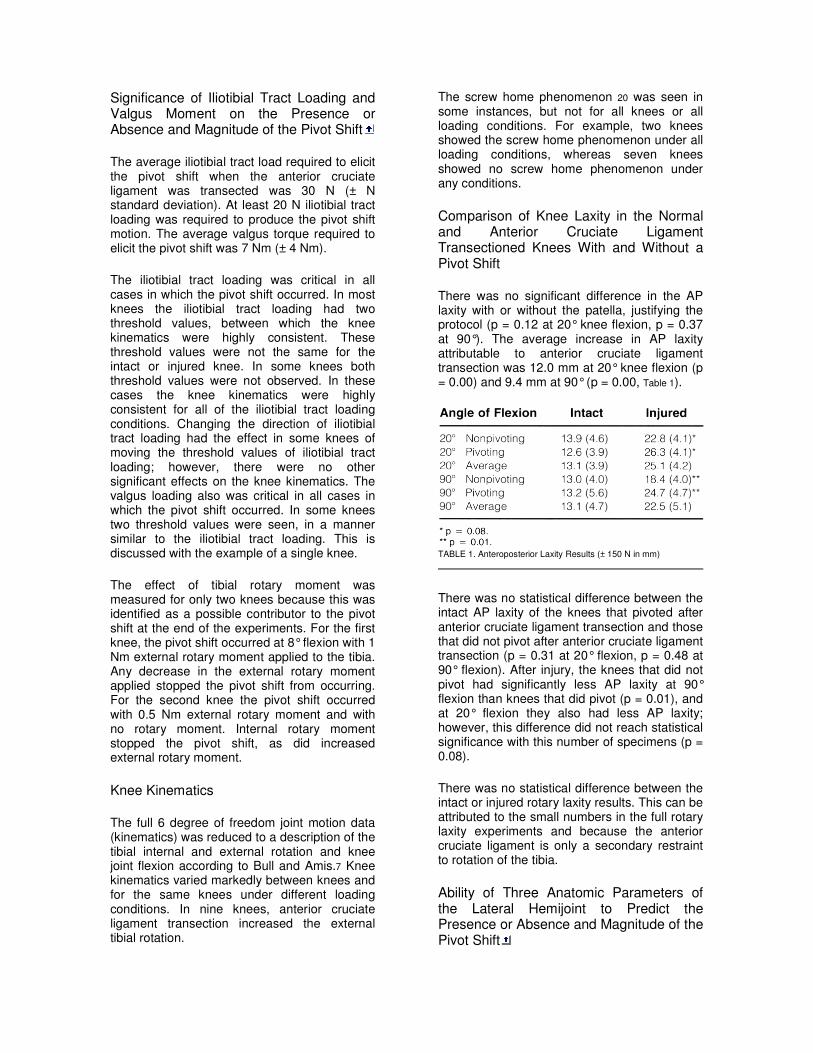
With no valgus moment (Fig 4A), the iliotibial tract loading had little effect above a threshold value between 0 and 10 N. In intact and injured cases, joint kinematics were similar when the iliotibial tract was tensed.
Fig 4A-D. Kinematics results for Knee K: the effect of varying the iliotibial tract loads at different valgus moments. (A) With no valgus moment and under all iliotibial tract loads the kinematics of the joint were similar. (B) With 4.8 Nm valgus moment there was increased external rotation of the injured knee above 20° flexion. (C) With 10 Nm valgus moment there were two distinct patterns of motion, with the pivot shift switching from one pattern to another. (D) With 13 Nm of valgus moment no pivot occurred. Again there were two distinct patterns of motion.
When 4.8 Nm valgus moment was applied (Fig
4B), there was increased external rotation of the injured knee beyond 20° flexion. Thus, a lower threshold value for the applied valgus moment to affect the motion of the injured knee is between 0 and 4.8 Nm.
When the valgus moment was increased to 10 Nm (Fig 4C), the intact and injured kinematics were affected. For low iliotibial tract loads (10 and 20 N) the intact knee had increased internal rotation in a similar manner to the kinematics shown by the joint with no valgus moment and iliotibial tract loading. Thus, in this situation, the lower iliotibial tract threshold value required to alter the kinematics was close to 30 N: the increase in valgus moment had increased the iliotibial tract loading required to reproduce the consistent kinematic pattern, as described previously. When the iliotibial tract loading was increased to 40 N the injured knee pivoted.
With a valgus moment of 13 Nm (Fig 4D), the injured joint did not pivot. Again, the joint showed similar kinematic characteristics for high iliotibial tract loading in the intact condition. However, the iliotibial tract loading required to achieve this consistent pattern once again was increased. There is a trend in which the lower iliotibial tract threshold value is increased for increased valgus moments. In this case the characteristics for the injured knee were consistent for all of the iliotibial tract loading conditions. The knee did not pivot but had increased internal tibial rotation. This can be compared with the clinical situation with a subluxed knee that does not reduce during the pivot shift test. It could be speculated that a higher iliotibial tract load might produce a pivot shift.
With 0 N loading on the iliotibial tract, joint kinematics were fairly consistent for all valgus moments (Fig 5A), with internal tibial rotation maintained throughout flexion. When the iliotibial tract load was increased to 10 N (Fig
5B), there were two distinct kinematics patterns: one pattern involved increasing internal rotation of the tibia throughout flexion (with high valgus moments); the second pattern involved an increase in internal tibial rotation only after approximately 90° flexion (with low valgus moments). In this case, the lower threshold value for valgus moment was breached somewhere between 4.8 and 10 Nm. As the iliotibial tract loading was increased (Fig 5C-D), more of the kinematics involved some increased external tibial rotation at high angles of flexion, and only some of the injured
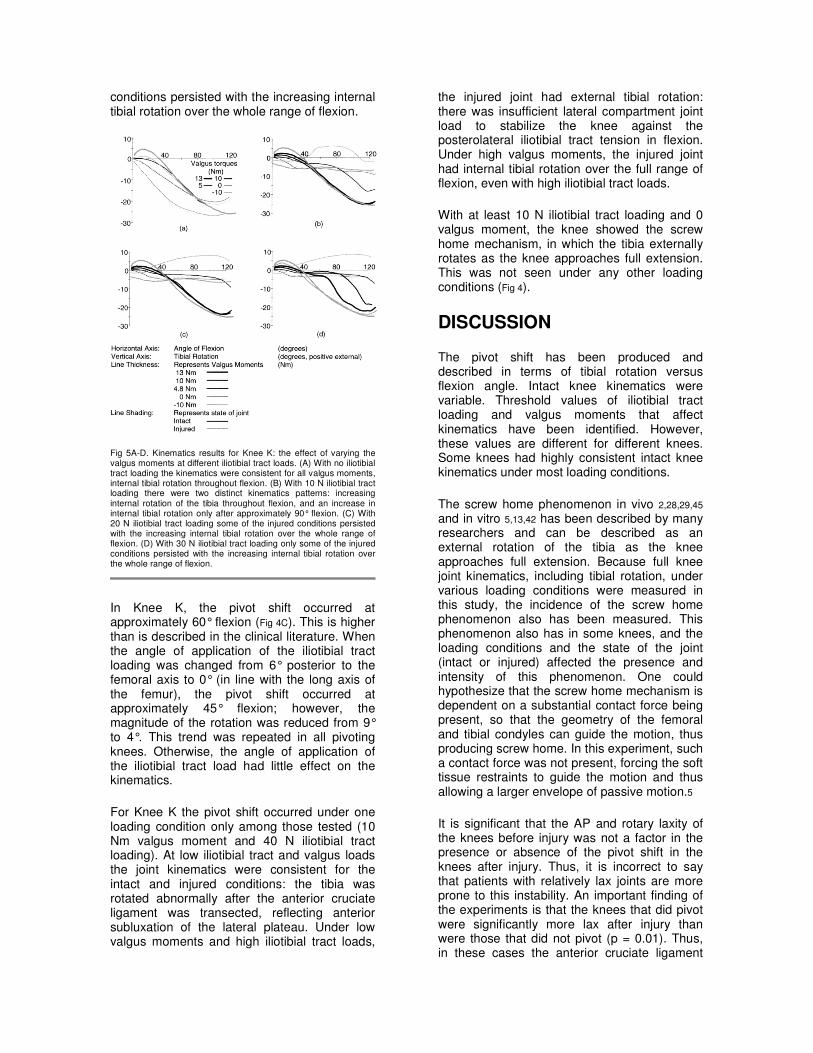
conditions persisted with the increasing internal tibial rotation over the whole range of flexion.
Fig 5A-D. Kinematics results for Knee K: the effect of varying the valgus moments at different iliotibial tract loads. (A) With no iliotibial tract loading the kinematics were consistent for all valgus moments, internal tibial rotation throughout flexion. (B) With 10 N iliotibial tract loading there were two distinct kinematics patterns: increasing internal rotation of the tibia throughout flexion, and an increase in internal tibial rotation only after approximately 90° flexion. (C) With 20 N iliotibial tract loading some of the injured conditions persisted with the increasing internal tibial rotation over the whole range of flexion. (D) With 30 N iliotibial tract loading only some of the injured conditions persisted with the increasing internal tibial rotation over the whole range of flexion.
In Knee K, the pivot shift occurred at approximately 60° flexion (Fig 4C). This is higher than is described in the clinical literature. When the angle of application of the iliotibial tract loading was changed from 6° posterior to the femoral axis to 0° (in line with the long axis of the femur), the pivot shift occurred at approximately 45° flexion; however, the magnitude of the rotation was reduced from 9° to 4°. This trend was repeated in all pivoting knees. Otherwise, the angle of application of the iliotibial tract load had little effect on the kinematics.
For Knee K the pivot shift occurred under one loading condition only among those tested (10 Nm valgus moment and 40 N iliotibial tract loading). At low iliotibial tract and valgus loads the joint kinematics were consistent for the intact and injured conditions: the tibia was rotated abnormally after the anterior cruciate ligament was transected, reflecting anterior subluxation of the lateral plateau. Under low valgus moments and high iliotibial tract loads,
the injured joint had external tibial rotation: there was insufficient lateral compartment joint load to stabilize the knee against the posterolateral iliotibial tract tension in flexion. Under high valgus moments, the injured joint had internal tibial rotation over the full range of flexion, even with high iliotibial tract loads.
With at least 10 N iliotibial tract loading and 0 valgus moment, the knee showed the screw home mechanism, in which the tibia externally rotates as the knee approaches full extension. This was not seen under any other loading conditions (Fig 4).
DISCUSSION
The pivot shift has been produced and described in terms of tibial rotation versus flexion angle. Intact knee kinematics were variable. Threshold values of iliotibial tract loading and valgus moments that affect kinematics have been identified. However, these values are different for different knees. Some knees had highly consistent intact knee kinematics under most loading conditions.
The screw home phenomenon in vivo 2,28,29,45 and in vitro 5,13,42 has been described by many researchers and can be described as an external rotation of the tibia as the knee approaches full extension. Because full knee joint kinematics, including tibial rotation, under various loading conditions were measured in this study, the incidence of the screw home phenomenon also has been measured. This phenomenon also has in some knees, and the loading conditions and the state of the joint (intact or injured) affected the presence and intensity of this phenomenon. One could hypothesize that the screw home mechanism is dependent on a substantial contact force being present, so that the geometry of the femoral and tibial condyles can guide the motion, thus producing screw home. In this experiment, such a contact force was not present, forcing the soft tissue restraints to guide the motion and thus allowing a larger envelope of passive motion.5
It is significant that the AP and rotary laxity of the knees before injury was not a factor in the presence or absence of the pivot shift in the knees after injury. Thus, it is incorrect to say that patients with relatively lax joints are more prone to this instability. An important finding of the experiments is that the knees that did pivot were significantly more lax after injury than were those that did not pivot (p = 0.01). Thus, in these cases the anterior cruciate ligament

exerted a proportionally greater contribution to the AP restraint than for those knees that did not pivot. An alternative way to describe this finding is that knees with relatively poor secondary restraints were prone to pivot. The secondary restraints to anterior draw are also the primary restraints to tibial rotation, the collateral ligaments,49 and associated capsular structures. One explanation for this experiment not producing a pivot in all knees tested is that these secondary structures were not stretched as they might be after an anterior cruciate ligament rupture in the clinical setting. In the clinical setting, patients with acute injury also may have a pivot shift without stretching of the secondary restraints. These are patients who have had an isolated anterior cruciate ligament injury for whom there has not been the time or type of activity for secondary structures to be stretched. Thus, these isolated injuries correspond to this experiment's in vitro model of anterior cruciate ligament insufficiency. It has been shown that a knee can be unstable in the AP direction and not show a pivot shift. Thus, the pivot shift is not the only indicator of severe anterior cruciate ligament instability.
The authors also found that one knee had the pivot shift before the anterior cruciate ligament was transected. It is not trivial to ascertain why this happened. The lateral tibial plateau was able to sublux anteriorly. This would imply that the soft tissue restraints to internal rotation or anterior displacement of the tibia were lax when intact. This was the case. This knee had the highest AP laxity of all knees at 20° flexion, but not at 90° flexion, although the anterior cruciate ligament was intact. Transecting the anterior cruciate ligament increased the AP laxity at 20° flexion by 62%.
Additional analysis of this knee was done, and the anterior cruciate ligament was reconstructed using a prosthetic device routed over the top of the lateral femoral condyle and tensioned to match the intact AP laxity at 20° flexion.2 After this reconstruction, the knee did not pivot. Thus, the authors cannot say that the reason the knee pivoted was the AP laxity of the joint, although it was related to the function of the anterior cruciate ligament because the reconstruction eliminated the pivot shift. It may have been possible that this anterior cruciate ligament had a prior incomplete injury that was not diagnosed in the laboratory. Such an incomplete ligament injury may have affected the rotary characteristics of the joint in a way that allowed the pivot shift to occur, even with good anterior stability. This only serves to highlight the three-dimensional nature of the
motion and the three-dimensional function of the anterior cruciate ligament.
Only eight of the 15 knees pivoted. Of cases published in the literature a higher percentage of knees pivot because of isolated anterior cruciate ligament injury. The authors think this may be because of a combination of factors. First, isolated cruciate ligament injury is difficult to confirm. Even arthroscopic diagnosis can miss stretching of secondary structures. Thus, the literature may be biased toward anterior cruciate ligament injuries with some injuries to other structures. The authors also have shown that knees that pivoted were more unstable in the AP direction than were knees that did not pivot, although the anterior cruciate ligament was transected. This may suggest that a certain level of anterior laxity must be present for the pivot shift to occur. In clinical examinations, the physician will apply varying loads by hand, hunting for the pivot shift conditions by feel, rather than the fixed loads applied in the laboratory.
The loading required to elicit the pivot shift was variable. One may speculate how this relates to the loads applied when the patient stands on the leg and the knee gives way. For example, how much valgus moment is applied to the knee during normal stance or walking? Does this compare with the loads applied in this experiment? What is the loading on the iliotibial tract during stance, and in which direction does the resultant force act relative to the femoral condyles and the anterior cruciate ligament?
These loads were known in these anatomic specimen experiments, but this needs to be correlated with the in vivo loads experienced when the patient feels the giving way. The variability of the loads required to elicit the pivot shift may explain why some patients with documented anterior cruciate ligament deficiency do not report giving way, whereas others have frequent giving way episodes for similar activities. In addition, in the clinical setting, there is a large axial compression force that may accentuate the stabilizing effect of any concavity on the tibial plateau. Additional experiments could include the application of such axial loads to bring the in vitro experiments closer to the in vivo conditions in which the patient's knee buckles and gives way. Without measuring the forces applied by the surgeons, one cannot say whether these axial loads are reproduced when the clinical pivot shift test is conducted. The experiments reproduced the clinical test and not the giving way of the clinical situation in which the

quadriceps forces and perhaps the forces in the hamstrings and other muscles around the knee joint occur. Additional work is needed to study the loading applied during the pivot shift test and the loads when the knee gives way.
Matsumoto 34 applied a similar range of loads on cadaveric joints, but he did not move the knee while taking measurements or define the iliotibial tract load. This may explain why only 35% of his specimens showed a pivot shift. Similarly, loading on the quadriceps has not been applied in the current work. In the current study, the extensor mechanism was removed. The authors have shown that the extensor mechanism does not provide any significant passive restraint to joint motion, although it has been shown by Reuben et al 41 that the pivot shift could be elicited in vitro by loading of the quadriceps alone. Reuben et al 41 did not publish their pivot shift kinematic data, and Matsumoto 34 also did not present the tibial rotations he measured, so additional comparisons between these two studies and the current study cannot be made.
Because the pivot shift phenomenon is a posterior reduction of the lateral tibial plateau with rotation about an axis medial to the center of the joint 34 at approximately 20° knee flexion, the geometry of the tibial and femoral condyles 35 and the stiffness of the medial collateral ligament all could be expected to influence the severity of the pivot shift phenomenon. Different measures of congruency were defined, and these were used to analyze the geometry of the lateral hemijoint in relation to the pivot shift. No correlation was found. It is possible that a more complex analysis that includes the medial hemijoint would discover this. A full three-dimensional geometric analysis also could be conducted to assess the relative importance of all lateral and medial geometric parameters. One of the reasons for this lack of correlation is related to the definition of the pivot shift. For some, the pivot shift is the reduction of the subluxed lateral tibial plateau as the knee flexes without regard to the suddenness of that motion; for others, the pivot shift is related to the speed of reduction of the lateral tibial plateau. The second understanding would require, one could hypothesize, a highly convex lateral tibial plateau for the femoral condyle to jump over during reduction. The first understanding would allow for a flatter tibial plateau, allowing the femoral condyle to slide back to the reduced position.
There are other structures that may affect the pivot shift, such as the lateral meniscus, the
secondary restraints to internal tibial rotation, and the active stabilizers of the knee joint. This was reported by Peterson et al,40 who showed that the pivot shift could be elicited by electrical stimulation of the popliteus muscle, which is an internal rotator of the tibia. Similarly, Noyes et al 38 cut the superficial fibers of the medial collateral ligament and the anterior cruciate ligament when conducting their experiments, and Matsumoto 34 sectioned the anterior cruciate ligament and the lateral capsule. Additional work should include analysis of different injuries and injury mechanisms.
Although this study has produced an in vitro pivot shift, this instability seen in the laboratory study cannot be compared easily with the clinical pivot shift sign because the consistent loading conditions achieved in the laboratory study are not reproduced in the clinical situation. The results presented here suggest that there are effectively two sets of loading conditions for the knee, each producing stable behavior: one set keeping the tibia subluxed in a pattern of internal rotation as the knee flexes, and the other set keeping in the tibia in the reduced position.
Figure 4C shows that the pivot shift is a sudden switch between these two relatively stable states. This observation suggests why a pivot shift could not be elicited in all specimens, the constant loads in each test tending to maintain one or other of the stable states. A corollary of this is that there is a documented 100% success rate in eliciting the pivot shift with the patient under anesthesia and in vivo, which depends on the application of varying forces, and one may speculate that the examiner does this intuitively to cause the tibia to move from one state to the other. The experiments described need to be correlated with the tests applied by clinicians in vivo, and perhaps a jig could be designed to allow for reproducible testing of the pivot shift. This has been addressed for drawer tests performed by the KT1000 machine (MEDmetric® Corporation San Diego, CA), for example.
From this study one may hypothesize that the pivot shift can be elicited by applying a compressive force to the lateral compartment of the knee, by applying a valgus torque, for example, in extension, internally rotating the tibia so that the lateral tibial plateau is in an anteriorly subluxed position, and flexing the joint. The pivot shift will be present if the combination of forces applied to the joint allow the tibia suddenly to reduce from its subluxed position. This reduction will be facilitated

additionally by releasing the international rotation torque on the tibia during flexion. Additional research could focus on developing a jig for the clinical setting to apply a varying rotation torque on the tibia and flex the joint while applying a valgus torque.
To link this work to clinical reality and ultimately to use the knowledge gained to help clinical diagnosis and operative techniques, this instrumentation should be taken into the operating theater to measure the kinematics of the pivot shift in vivo. By this means, the motions induced doing a pivot shift test in vivo can be matched with the motions in vitro. A more complex stage would be to measure the forces applied by the surgeon during the tests.
All of the knee specimens used in these experiments were old. Some of the discrepancies between the experimental results and the clinical condition may be a result of age related loss of strength and stiffness of the ligaments. For example, a medial collateral ligament that has low stiffness may cause the anterior subluxation of the lateral tibial plateau during the pivot shift to be combined with an anterior subluxation of the medial tibial plateau, thus changing the nature of the instability from a rotational instability to one that is predominantly translational.
General conclusions regarding the pivot shift are difficult to make. One knee unexpectedly pivoted before injury. In the work of Fetto and Marshall,15 some knees were seen that had positive pivot shifts, even with intact anterior cruciate ligaments (10%). An isolated anterior cruciate ligament injury was sufficient to produce the pivot shift under the experimental conditions of constant loading in an additional seven of 14 knees (eight of 15 knees).
The pivot shift cannot be predicted from the intact AP laxity and is not necessarily present in the contralateral knees. The contribution of the anterior cruciate ligament to anterior restraint was greater for the pivoting knees than for those that did not pivot. A knee can be unstable in the AP direction and not have a pivot shift. The pivot shift in vitro can be described as a sudden external tibial rotation of 3° to 38° at flexion angles over the whole range of flexion (range, 13°-93°). This does not correlate precisely with the clinical pivot shift as it is described in the literature because the clinical pivot shift is described as occurring between 20° to 40° flexion.17,43
Acknowledgments
The authors thank Kevin Pickett for assistance with the laboratory work.
References 1. Amis AA, Scammell BE: Autogenous reconstruction of the anterior cruciate ligament: Biomechanics of intraarticular, extraarticular and combined techniques. J Bone Joint Surg 75B:812-817, 1993.
2. Asai O: [The combination method for diagnosis of meniscus lesion in the knee (Translation by O Asai)]. Nippon Seikeigeka Gakkai Zasshi 7:625-633, 1981.
3. Bach Jr BR, Jones GT, Sweet FA, et al: Arthroscopy-assisted anterior cruciate ligament reconstruction using patellar tendon substitution. Two- to four-year follow-up results. Am J Sports Med 22:758-767, 1994.
4. Bach Jr BR, Warren RF, Wickiewicz TL: The pivot shift phenomenon: Results and description of a modified clinical test for anterior cruciate ligament insufficiency. Am J Sports Med 16:571-576, 1988.
5. Blankevoort L: Passive motion characteristics of the human knee joint: Experiments and computer simulations. PhD Thesis. University of Nijmegen, The Netherlands 1991.
6. Bull AMJ, Amis AA: Letter to the editor: Accuracy of an electromagnetic tracking device. J Biomech 30:857-858, 1997.
7. Bull AMJ, Amis AA: Accuracy of an electromagnetic measurement device and application to the measurement and description of knee joint motion. Proc Inst Mech Eng Part H 212:347-355, 1998.
8. Bull AMJ, Amis AA: The pivot shift phenomenon: A clinical and biomechanical perspective. The Knee 5:141-158, 1998.
9. Bull AMJ, Andersen HN, Basso O, et al: Three dimensional measurement of femoro-tibial motion pre-injured, anterior cruciate ligament-injured and post-reconstruction. Trans Eur Soc Biomech 10:368, 1996.
10. Conteduca F, Ferretti A, Mariani PP, et al: Chondromalacia and chronic anterior instabilities of the knee. Am J Sports Med 19:119-123, 1991.
11. Daniel DM, Stone ML, Riehl B: Ligament Surgery: The Evaluation of Results. In Daniel DM, Akeson W, O'Connor JJ (eds). Knee Ligaments, Structure, Function and Repair. New York, Raven Press Ltd 521-534, 1990.
12. Donaldson III WF, Warren RF, Wickiewicz T: A comparison of acute anterior cruciate ligament
examinations. Initial versus examination under anesthesia. Am J Sports Med 13:5-10, 1985.
13. Eckhoff DG, Smith D, Schecter R, et al: Automatic rotation (screw-home) in the cruciate deficient and prosthetic knee. Trans Orthop Res Soc 21:216, 1996.
14. Ellison AE: The pathogenesis and treatment of anterolateral rotatory instability. Clin Orthop 147:51-55, 1980.
15. Fetto JF, Marshall JL: Injury to the anterior cruciate ligament producing the pivot-shift sign. J Bone Joint Surg 61A:710-714, 1979.
16. Galway HR, Beaupre A, MacIntosh DL: Pivot shift: A clinical sign of symptomatic anterior cruciate deficiency. J Bone Joint Surg 54B:763-764, 1972.
17. Galway HR, MacIntosh DL: The lateral pivot shift: A symptom and sign of anterior cruciate ligament insufficiency. Clin Orthop 147:45-50, 1980.
18. Gerber C, Matter P: Biomechanical analysis of the knee after rupture of the anterior cruciate ligament and its primary repair. An instant-centre analysis of function. J Bone Joint Surg 65B:391-399, 1983.
19. Gillquist J, Messner K: Instrumented analysis of the pivot shift phenomenon after reconstruction of the anterior cruciate ligament. Int J Sports Med 16:484-488, 1995.
20. Hallen LG, Lindahl O: The 'screw-home' movement in the knee-joint. Acta Orthop Scand 37:97-106, 1966.
21. Harilainen A, Sandelin J, Osterman K, et al: Prospective preoperative evaluation of anterior cruciate ligament instability of the knee joint and results of reconstruction with patellar ligament. Clin Orthop 297:17-22, 1993.
22. Hughston JC, Andrews JR, Cross MJ, et al: Classification of knee ligament instabilities. Part I. The medial compartment and cruciate ligaments. J Bone Joint Surg 58A:159-172, 1976.
23. Jakob RP, Hassler H, Stäubli HU: Observations on rotatory instability of the lateral compartment of the knee. Experimental studies on the functional anatomy and the pathomechanism of the true and the reversed pivot shift sign. Acta Orthop Scand 191(Suppl):1-32, 1981.
24. Jakob RP, Stäubli HU, Deland JT: Grading the pivot shift. Objective tests with implications for treatment. J Bone Joint Surg 69B:294-299, 1987.
25. Kaplan N, Wickiewicz TL, Warren RF: Primary surgical treatment of anterior cruciate ligament ruptures. A long-term follow-up study. Am J Sports Med 18:354-358, 1990.
26. Kärrholm J, Elmqvist LG, Selvik G, et al: Chronic anterolateral instability of the knee. A
roentgen stereophotogrammetric evaluation. Am J Sports Med 17:555-563, 1989.
27. Kujala UM, Nelimarkka O, Koskinen SK: Relationship between the pivot shift and the configuration of the lateral tibial plateau. Arch Orthop Trauma Surg 111:228-229, 1992.
28. Kuriwaka Y: [A biomechanical study of osteoarthritis of the knee with special reference to the rotatory movement of the knee joint. (Translation by Y. Kunuaka)] Nippon Seikeigeka Gakkai Zasshi 56:713-726, 1982.
29. Lafortune MA, Cavanagh PR, Sommer III HJ, et al: Three-dimensional kinematics of the human knee during walking. J Biomech 25:347-357, 1992.
30. Larson RL: Physical examination in the diagnosis of rotatory instability. Clin Orthop 172:38-44, 1983.
31. Losee RE: Concepts of the pivot shift. Clin Orthop 172:45-51, 1983.
32. Losee RE, Johnson TR, Southwick WO: Anterior subluxation of the lateral tibial plateau. A diagnostic test and operative repair. J Bone Joint Surg 60A:1015-1030, 1978.
33. Lucie RS, Wiedel JD, Messner DG: The acute pivot shift: Clinical correlation. Am J Sports Med 12:189-191, 1984.
34. Matsumoto H: The Pivot Shift Phenomenon: A Biomechanical Study. PhD Thesis. University of Leeds, United Kingdom 1987.
35. Matsumoto H: Mechanism of the pivot shift. J Bone Joint Surg 72B:816-821, 1990.
36. Milne AD, Chess DG, Johnson JA, et al: Accuracy of an electromagnetic tracking device: A study of the optimal operating range and metal interference. J Biomech 29:791-793, 1996.
37. Norwood LA, Andrews JR, Meisterling RC, et al: Acute anterolateral rotatory instability of the knee. J Bone Joint Surg 61A:704-709, 1979.
38. Noyes FR, Grood ES, Cummings JF, et al: An analysis of the pivot shift phenomenon. Am J Sports Med 19:148-155, 1991.
39. Noyes FR, Mooar PA, Matthews DS, et al: The symptomatic anterior cruciate-deficient knee. Part 1: The long-term functional disability in athletically active individuals. J. Bone Joint Surg 65A:154-162, 1983.
40. Peterson L, Pitman MI, Gold J: The active pivot shift: The role of the popliteus muscle. Am J Sports Med 12:313-317, 1984.
41. Reuben JD, Rovick JS, Schrager RJ, et al: Three-dimensional dynamic motion analysis of the anterior cruciate ligament deficient knee joint. Am J Sports Med 17:463-471, 1989.

42. Schlepckow P: [Experimental studies of the kinematics of the stable and unstable human knee joint.] Experimentelle Untersuchungen zur Kinematik des stabilen and instabilen menschlichen Kniegelenkes. Z Orthop 127:711-715, 1989.
43. Slocum DB, James SL, Larson RL, et al: Clinical test for anterolateral rotary instability of the knee. Clin Orthop 118:63-69, 1976.
44. Tamea CD, Henning CE: Pathomechanics of the pivot shift maneuver. Am J Sports Med 9:31-37, 1981.
45. Tasker T, Waugh W: Articular changes associated with internal derangement of the knee. J Bone Joint Surg 64B:486-488, 1982.
46. Terry GC, Hughston JC, Rose RM: The anatomy of the iliopatellar band and iliotibial tract. Am J Sports Med 14:39-45, 1986.
47. Terry GC, Norwood LA, Hughston JC, et al: How iliotibial tract injuries of the knee combine with acute anterior cruciate ligament tears to influence abnormal anterior tibial displacement. Am J Sports Med 21:55-60, 1993.
48. Viidik A: Functional properties of collagenous tissues. Int Rev Connect Tissue Res 6:127-215, 1973.
49. Wang C, Walker PS: Rotatory laxity of the human knee joint. J Bone Joint Surg 56A:161-170, 1974.