Author Manuscript NIH Public Access a,b, a, c, Hsiu-Fang ... Breast Cancer Stem Cells Yanyan Lia,b,...
19
Sulforaphane, a Dietary Component of Broccoli/Broccoli Sprouts, Inhibits Breast Cancer Stem Cells Yanyan Li a,b , Tao Zhang a , Hasan Korkaya c , Suling Liu c , Hsiu-Fang Lee a , Bryan Newman a , Yanke Yu a , Shawn G. Clouthier c , Steven J. Schwartz b,* , Max S. Wicha c,* , and Duxin Sun a,* a Department of Pharmaceutical Sciences, College of Pharmacy, University of Michigan, USA b Department of Food Science and Technology, The Ohio State University, USA c Comprehensive Cancer Center, Department of Internal Medicine, University of Michigan, USA Abstract Purpose—The existence of cancer stem cells (CSCs) in breast cancer has profound implications for cancer prevention. In this study, we evaluated sulforaphane, a natural compound derived from broccoli/broccoli sprouts, for its efficacy to inhibit breast CSCs and its potential mechanism. Experimental Design—Aldefluor assay and mammosphere formation assay were used to evaluate the effect of sulforaphane on breast CSCs in vitro. A NOD/SCID xenograft model was employed to determine whether sulforaphane could target breast CSCs in vivo, as assessed by Aldefluor assay and tumor growth upon cell re-implantation in secondary mice. The potential mechanism was investigated utilizing Western blotting analysis and β-catenin reporter assay. Results—Sulforaphane (1~5 μM) decreased aldehyde dehydrogenase (ALDH)-positive cell population by 65%~80% in human breast cancer cells (P < 0.01), and reduced the size and number of primary mammospheres by 8~125-fold and 45%~75% (P < 0.01), respectively. Daily injection with 50 mg/kg sulforaphane for two weeks reduced ALDH-positive cells by more than 50% in NOD/ SCID xenograft tumors (P = 0.003). Sulforaphane eliminated breast CSCs in vivo, thereby abrogating tumor growth after re-implantation of primary tumor cells into the secondary mice (P < 0.01). Western blotting analysis and β-catenin reporter assay showed that sulforaphane down-regulated Wnt/β-catenin self-renewal pathway. Conclusions—Sulforaphane inhibits breast CSCs and down-regulates Wnt/β-catenin self-renewal pathway. These findings support the use of sulforaphane for chemoprevention of breast cancer stem cells and warrant further clinical evaluation. Keywords sulforaphane; breast cancer stem cell; aldehyde dehydrogenase; mammosphere; NOD/SCID mouse *Correspondence authors: Duxin Sun, Ph.D. (requests for reprints), Associate Professor, Department of Pharmaceutical Sciences, University of Michigan, 428 Church Street, Room 2020, Ann Arbor, MI 48109, Tel: 734-615-8740; Fax: 734-615-6162; [email protected]. Max S. Wicha, M.D. Professor, Department of Internal Medicine; Director, University of Michigan Comprehensive Cancer Center, 1500 East Medical Center Dr., Room 6302, Ann Arbor, MI 48109, Tel: 734-936-1831; Fax: 734-615-3947; [email protected]. Steven J. Schwartz, Ph.D. Professor, Department of Food Science and Technology, The Ohio State University, 2015 Fyffe Ct., 235 Parker Food Science & Technology Building, Columbus, OH 43210, Tel: 614-292-2934; Fax: 614-292-4233; [email protected]. NIH Public Access Author Manuscript Clin Cancer Res. Author manuscript; available in PMC 2011 May 1. Published in final edited form as: Clin Cancer Res. 2010 May 1; 16(9): 2580–2590. doi:10.1158/1078-0432.CCR-09-2937. NIH-PA Author Manuscript NIH-PA Author Manuscript NIH-PA Author Manuscript
Transcript of Author Manuscript NIH Public Access a,b, a, c, Hsiu-Fang ... Breast Cancer Stem Cells Yanyan Lia,b,...
Sulforaphane, a Dietary Component of Broccoli/Broccoli Sprouts,Inhibits Breast Cancer Stem Cells
Yanyan Lia,b, Tao Zhanga, Hasan Korkayac, Suling Liuc, Hsiu-Fang Leea, Bryan Newmana,Yanke Yua, Shawn G. Clouthierc, Steven J. Schwartzb,*, Max S. Wichac,*, and Duxin Suna,*a Department of Pharmaceutical Sciences, College of Pharmacy, University of Michigan, USAb Department of Food Science and Technology, The Ohio State University, USAc Comprehensive Cancer Center, Department of Internal Medicine, University of Michigan, USA
AbstractPurpose—The existence of cancer stem cells (CSCs) in breast cancer has profound implicationsfor cancer prevention. In this study, we evaluated sulforaphane, a natural compound derived frombroccoli/broccoli sprouts, for its efficacy to inhibit breast CSCs and its potential mechanism.
Experimental Design—Aldefluor assay and mammosphere formation assay were used to evaluatethe effect of sulforaphane on breast CSCs in vitro. A NOD/SCID xenograft model was employed todetermine whether sulforaphane could target breast CSCs in vivo, as assessed by Aldefluor assayand tumor growth upon cell re-implantation in secondary mice. The potential mechanism wasinvestigated utilizing Western blotting analysis and β-catenin reporter assay.
Results—Sulforaphane (1~5 μM) decreased aldehyde dehydrogenase (ALDH)-positive cellpopulation by 65%~80% in human breast cancer cells (P < 0.01), and reduced the size and numberof primary mammospheres by 8~125-fold and 45%~75% (P < 0.01), respectively. Daily injectionwith 50 mg/kg sulforaphane for two weeks reduced ALDH-positive cells by more than 50% in NOD/SCID xenograft tumors (P = 0.003). Sulforaphane eliminated breast CSCs in vivo, thereby abrogatingtumor growth after re-implantation of primary tumor cells into the secondary mice (P < 0.01).Western blotting analysis and β-catenin reporter assay showed that sulforaphane down-regulatedWnt/β-catenin self-renewal pathway.
Conclusions—Sulforaphane inhibits breast CSCs and down-regulates Wnt/β-catenin self-renewalpathway. These findings support the use of sulforaphane for chemoprevention of breast cancer stemcells and warrant further clinical evaluation.
Keywordssulforaphane; breast cancer stem cell; aldehyde dehydrogenase; mammosphere; NOD/SCID mouse
*Correspondence authors: Duxin Sun, Ph.D. (requests for reprints), Associate Professor, Department of Pharmaceutical Sciences,University of Michigan, 428 Church Street, Room 2020, Ann Arbor, MI 48109, Tel: 734-615-8740; Fax: 734-615-6162;[email protected]. Max S. Wicha, M.D. Professor, Department of Internal Medicine; Director, University of Michigan ComprehensiveCancer Center, 1500 East Medical Center Dr., Room 6302, Ann Arbor, MI 48109, Tel: 734-936-1831; Fax: 734-615-3947;[email protected]. Steven J. Schwartz, Ph.D. Professor, Department of Food Science and Technology, The Ohio State University,2015 Fyffe Ct., 235 Parker Food Science & Technology Building, Columbus, OH 43210, Tel: 614-292-2934; Fax: 614-292-4233;[email protected].
NIH Public AccessAuthor ManuscriptClin Cancer Res. Author manuscript; available in PMC 2011 May 1.
Published in final edited form as:Clin Cancer Res. 2010 May 1; 16(9): 2580–2590. doi:10.1158/1078-0432.CCR-09-2937.
NIH
-PA Author Manuscript
NIH
-PA Author Manuscript
NIH
-PA Author Manuscript
IntroductionBroccoli and broccoli sprouts contain large amounts of glucosinolates (1). Numerous studieshave substantiated the chemoprevention effect of increasing cruciferous vegetable intakeagainst cancer, which has been attributed to the activity of various isothiocyanates that areenzymatically hydrolyzed from glucosinolates (2). Sulforaphane was found to be convertedfrom glucoraphanin, a major glucosinolate in broccoli/broccoli sprouts (3). Thechemoprevention properties of sulforaphane against cancer are through both “blocking” and“suppressing” effects (2). The “blocking” function of sulforaphane is achieved throughinhibiting Phase 1 metabolism enzymes that convert procarcinogens to carcinogens andinducing Phase 2 metabolism enzymes that promote excretion of carcinogens (2). Subsequentstudies revealed the “suppressing” effects of sulforaphane in modulating diverse cellularactivities to inhibit the growth of transformed cells (2,4). The ability of sulforaphane to induceapoptosis and cell cycle arrest is associated with regulation of many molecules including Bcl-2family proteins, caspases, p21, cyclins and cdks (4). Sulforaphane was also shown to suppressangiogenesis and metastasis by down-regulating VEGF, HIF-1α, MMP-2 and MMP-9 (4).
Accumulating evidence has shown that many types of cancer, including breast cancer, areinitiated from and maintained by a small population of cancer stem cells (CSCs) (5,6). Thisminor population produces the tumor bulk through continuous self-renewal and differentiation,which may be regulated by similar signaling pathways occurring in normal stem cells (5–8).Several pathways including Wnt/β-catenin, Hedgehog, and Notch have been identified to becritical to the self-renewal behavior of CSCs (7,9,10). Furthermore, CSCs have been suggestedto contribute to tumor resistance/relapse because chemotherapy and radiation therapy areincapable of eradicating them (6,11,12). Thus, targeting these self-renewal pathways mayprovide an effective strategy to target CSCs and thereby overcome tumor resistance and reducerelapse (5). Several dietary compounds, such as curcumin (13,14), quercetin andepigallocatechin-gallate (15), were found to be potentially against CSC self-renewal.
Wnt/β-catenin signaling is one of the key pathways that promotes self-renewal of breast CSCs(5). Activation of Wnt target genes are mediated by β-catenin, which translocates into nucleusand binds to the transcription factors T cell factor/lymphoid enhancer factor (TCF/LEF) (5,16). The level of intracellular β-catenin is modulated by a multi-protein complex consisting ofglycogen synthase kinase3β (GSK3β), adenomatous polyposis coli, casein kinase1α and axin(TCF/LEF) (5,16,17). GSK3β promotes the ubiquitin-proteasome degradation of β-catenin byphosphorylating three specific amino acids, Ser33/Ser37/Thr41, on β-catenin (17).
Sulforaphane was shown to target pancreatic tumor-initiating cells in a very recent report(18). In the present study, we examined the efficacy of sulforaphane against breast CSCs inboth breast cancer cell lines and breast cancer xenografts. We demonstrated that sulforaphaneeliminated breast CSCs in vivo, which was reflected by the inhibition of tumor growth inrecipient mice that were inoculated with tumor cells derived from sulforaphane-treated primaryxenografts. Furthermore, since sulforaphane was reported to induce down-regulation of β-catenin in human cervical carcinoma HeLa and hepatocarcinoma HepG2 cells (19), weinvestigated the suppressing activity of sulforaphane on Wnt/β-catenin pathway.
Materials and MethodsCell Lines and Reagents
Human breast cancer cell lines, MCF7 and SUM159, were obtained from American TypeCulture Collection and from Dr. Stephen Ethier (Karmanos Cancer Center), respectively. Thesource of SUM159 cell line is primary breast anaplastic carcinoma. This cell line is ERnegative, PR negative, and does not have Her2 over-expression. Both cell lines were tested
Li et al. Page 2
Clin Cancer Res. Author manuscript; available in PMC 2011 May 1.
NIH
-PA Author Manuscript
NIH
-PA Author Manuscript
NIH
-PA Author Manuscript

and authenticated in their origin sources. Authentication of these cell lines includedmorphology analysis, growth curve analysis, isoenzyme analysis, short tandem repeat analysis,and mycoplasma detection. Both cell lines were passaged in our laboratory for fewer than sixmonths after receipt. To maintain the integrity of collections, stocks of the earliest-passagecells have been stored, and cell lines have been carefully maintained in culture as describedbelow. MCF7 was maintained in RPMI1640 medium (Invitrogen, Carlsbad, CA) supplementedwith 10% fetal bovine serum (Fisher Scientific, Pittsburgh, PA), 1% antibiotic-antimycotic(Invitrogen, Carlsbad, CA), and 5 μg/ml insulin (Sigma-Aldrich, St Louis, MO). SUM159 wasmaintained in Ham’s F12 medium (Invitrogen, Carlsbad, CA) supplemented with 5% fetalbovine serum, 1% antibiotic-antimycotic, 5 μg/ml insulin, 1μg/ml hydrocortisone (Sigma-Aldrich, St Louis, MO), and 4 μg/ml gentamicin (Invitrogen, Carlsbad, CA).
Sulforaphane was obtained from LKT Laboratories (St. Paul, MN). Propidium iodide (PI) wasfrom Invitrogen (Carlsbad, CA). LiCl was purchased from Fisher Scientific (Pittsburgh, PA);BIO (GSK3 inhibitor IX) was from Calbiochem (EMD Biosciences, San Diego, CA); andMG132 was obtained from Assay Designs (Stressgen, Ann Arbor, MI).
Antibodies to β-catenin, phospho-β-catenin Ser33/Ser37/Thr41, phospho-GSK3β Ser9, andGSK3β were purchased from Cell Signaling Technology (Danvers, MA). Antibodies to cyclinD1 and β-actin were acquired from Santa Cruz Biotechnology (Santa Cruz, CA).
MTS Cell Proliferation AssayMCF7 and SUM159 were seeded in 96-well microplates at a density of 3,000~5,000 cells perwell. Cells were treated with increasing concentrations of sulforaphane as indicated. After 48hr incubation cell viability was assessed by MTS assay (Promega, Madison, WI) according tomanufacturer’s instruction. The number of living cells is directly proportional to the absorbanceat 490 nm of a formazan product reduced from MTS by living cells.
Caspase-3 Activity AssayCells were treated with different concentrations of sulforaphane and collected after 24 hrs. Thecaspase-3 activity assay was based on the manufacturer’s instruction of Caspase-3/CPP32Fluorometric Assay Kit (Biovision Research Products, Mountain View, CA). Cellular proteinwas extracted with the supplied lysis buffer, followed by determination of protein concentrationusing BCA Protein Assay Reagents (Pierce, Rockford, IL). The cleavage of DEVD-AFC, asubstrate of caspase-3, was quantified by using a fluorescence microtiter plate reader with a400 nm excitation filter and a 505 nm emission filter.
Mammosphere Formation AssayStem/progenitor cells are enriched in mammospheres of breast cancer cells (20), based on theunique ability of stem/progenitor cells to grow and form spheres in serum-free medium (21).Mammosphere culture was performed as previously described (22,23) in a serum-freemammary epithelium basal medium (Lonza Inc., Walkersville, MD) supplemented with B27(Invitrogen), 1% antibiotic-antimycotic, 5 μg/ml insulin, 1μg/ml hydrocortisone, 4 μg/mlgentamicin, 20 ng/mL EGF (Sigma-Aldrich, St Louis, MO), 20 ng/mL bFGF (Sigma-Aldrich,St Louis, MO), and 1:25,000,000 β-mercaptoethanol (Sigma-Aldrich, St Louis, MO). Singlecells prepared from mechanical and enzymatic dissociation were plated in six-well ultra-lowattachment plates (Corning, Acton, MA) at a density of 500~1,000 cells per milliliter in primaryculture and 100~500 cells per milliliter in the following passages. Different concentrations ofsulforaphane were added to primary culture, while the second and third passages were grownin the absence of drug. After 7 days of culture, the number of mammospheres was countedunder Nikon Eclipse TE2000-S microscope and the photos were acquired with MetaMorph7.6.0.0.
Li et al. Page 3
Clin Cancer Res. Author manuscript; available in PMC 2011 May 1.
NIH
-PA Author Manuscript
NIH
-PA Author Manuscript
NIH
-PA Author Manuscript
Aldefluor AssayA cell population with a high Aldehyde dehydrogenase (ALDH) enzyme activity waspreviously reported to enrich mammary stem/progenitor cells (23). Aldefluor assay wasperformed according to manufacturer’s guidelines (StemCell Technologies, Vancouver, BC,Canada). Single cells obtained from cell cultures or xenograft tumors were incubated inAldefluor assay buffer containing an ALDH substrate, bodipy-aminoacetaldehyde (BAAA, 1μmol/L per 1,000,000 cells), for 40~50 min at 37 °C. As a negative control, a fraction of cellsfrom each sample was incubated under identical condition in the presence of ALDH inhibitor,diethylaminobenzaldehyde (DEAB). Flow cytometry was used to measure ALDH-positive cellpopulation.
Primary NOD/SCID Mouse ModelAll experimentation involving mice were conducted in accordance with standard protocolapproved by the University Committee on the Use and Care of Animals (UCUCA) at Universityof Michigan. SUM159 cells (2,000,000) mixed with Matrigel (BD Biosciences, San Jose, CA)were injected to the mammary fat pads of 5-week-old female NOD/SCID mice (The JacksonLaboratory, Bar Harbor, ME) as previously described (24). Tumors were measured with acaliper, and the volume was calculated using V=1/2 (width2 × length). Two weeks after cellinjection, the mice were randomly separated into two groups, one group intraperitoneallyinjected with control (0.9% NaCl solution) and the other group with 50 mg/kg sulforaphane(dissolved in 0.9% NaCl solution) daily for two weeks.
Dissociation of TumorsAt the end of drug treatment, the mice were humanely euthanized and tumors were harvested.Tumor tissues were dissociated mechanically and enzymatically to obtain single cellsuspension as previously described (25). Briefly, tumors were minced by scalpel and incubatedin medium 199 (Invitrogen, Carlsbad, CA) mixed with collagenase/hyaluronidase (StemCellTechnologies, Vancouver, BC, Canada) at 37 °C for 15~20 min. The tissues were furtherdissociated by pipet trituration, and then passed through 40 μm nylon mesh to produce singlecell suspension, which was used for Aldeflour assay and flow cytometry.
Secondary NOD/SCID Mouse ModelLiving cells from the dissociated tumors were sorted out by Fluorescence-activated cell sorting(FACS). Two groups of mice (four in group 1 and three in group 2) were implanted with tumorcells separately. Each secondary NOD/SCID mouse was inoculated with 50,000 cells fromcontrol mouse tumors in one side of inguinal mammary fat pad and another 50,000 cells fromsulforaphane-treated tumors in the contralateral mammary fat pad. The growth of tumors wasmonitored; and tumor volumes were measured twice a week. Mice were humanely euthanizedwhen the larger one of the two tumors reached 300~500 mm3.
Western Blotting AnalysisCells were treated with sulforaphane at varying concentrations for indicated time periods infigure legends. Cells were harvested, lysed in RIPA buffer (20 mM Tris-HCl, 150 mM NaCl,1% NP-40, 5 mM EDTA, 1 mM Na3VO4, pH 7.5) supplemented with a protease inhibitorcocktail (Pierce, Rockford, IL) and a phosphatase inhibitor (Calbiochem, EMD Biosciences,San Diego, CA), and incubated on ice for 30 min. Cell lysate was centrifuged at 14,000 rpmfor 15 min, and the supernatant was recovered. Protein concentration was determined withBCA Protein Assay Reagents (Pierce, Rockford, IL). Equal amounts of protein were subjectto SDS-PAGE, and transferred to PVDF membrane (BioRad, Richmond, CA). The membranewas then incubated with appropriate antibodies.
Li et al. Page 4
Clin Cancer Res. Author manuscript; available in PMC 2011 May 1.
NIH
-PA Author Manuscript
NIH
-PA Author Manuscript
NIH
-PA Author Manuscript

TOP-dGFP Lentiviral β-Catenin Reporter AssayTCF/LEF-1 (TOP-dGFP, FOP-dGFP) lentiviral reporter system was kindly gifted by Dr.Wiessman at Ludwig Center, Stanford University School of Medicine (26). Cells were infectedwith TOP-dGFP or control reporter FOP-dGFP with mutated TCF/LEF-1 binding sites. TOP-dGFP MCF7 and FOP-dGFP MCF7 cells were maintained in the same RPMI1640 medium asMCF7 cells. MCF7, TOP-dGFP MCF7 and FOP-dGFP MCF7 cells were cultured in the sameserum-free mammary epithelium basal medium as mammospheres in six-well ultra-lowattachment plates at a density of 1,000~1,500 cells per milliliter for 5 days. Single cells preparedfrom the primary spheres were incubated in medium containing 5 μM sulforaphane or/and 0.5μM BIO for 48 hrs. After dissociation, single cell suspension was subject to flow cytometryanalysis for dGFP-positive cell population. Parental MCF7 cells served as a control forautofluorescence. The photos of mammospheres were taken with Nikon Eclipse TE2000-Smicroscope and acquired with MetaMorph 7.6.0.0.
Statistical AnalysisStatistical differences were determined using two-tailed Student t-test. Data are presented asmean ± SD (n ≥3).
ResultsSulforaphane Inhibits Proliferation and Induces Apoptosis of Breast Cancer Cells
Sulforaphane was previously shown to inhibit proliferation (27) and induce apoptosis (28) inbreast cancer cells. We first evaluated the anti-proliferative effects of sulforaphane in twohuman breast cancer cell lines, SUM159 and MCF7, by MTS assay. Cells were treated withincreasing concentrations of sulforaphane for 48 hrs; and the ratio of viable cells of treatmentrelative to control is plotted in Figure 1A. Cell survival decreased as the concentration ofsulforaphane increased, with the IC50 around 10 μM for SUM159 and 16 μM for MCF7.Caspase-3 fluorometric assay showed that sulforaphane (10 μM) significantly (P = 0.005)induced activation of caspase-3 (Figure 1B).
Sulforaphane Inhibits Breast Cancer Stem/Progenitor Cells in VitroIt has been demonstrated that mammary stem/progenitor cells are enriched in non-adherentspherical clusters of cells, termed mammospheres (22). These cells are capable of yieldingsecondary spheres and differentiating along multiple lineages (22). In order to evaluate whethersulforaphane could suppress the formation of mammospheres in vitro, we exposed primaryMCF7 and SUM159 spheres to varying concentrations of sulforaphane and then cultured themtwo additional passages in the absence of drug. As shown in Figure 2A and 2B, sulforaphaneinhibited the formation of primary spheres. Not only the number of spheres declined by 45%~75% (P < 0.01) (Figure 2A), but also the size of spheres was reduced by 8~125-fold (Figure2B). Furthermore, a significant decrease in the number of sphere-forming cells in subsequentpassages indicated a reduced self-renewal capacity of these stem/progenitor cells (Figure 2C)(22). MCF7 Cells initially propagated in the presence of 5 μM sulforaphane barely producedsecondary spheres, with no cells passaged to third generation (Figure 2C). It is worth notingthat the concentrations of sulforaphane that were capable of suppressing mammosphereformation (IC50 around 0.5~1 μM for both SUM159 and MCF7 spheres) were approximately10-fold lower than those exhibiting anti-proliferative effects in MTS assay (IC50 around 10μM for SUM159 and 16 μM for MCF7).
In breast carcinomas, a cell population with high aldehyde dehydrogenase (ALDH) activity asassessed by the Aldefluor assay has been demonstrated to enrich tumorigenic stem/progenitorcells (23). This cell population is capable of self-renewal and generating tumors resembling
Li et al. Page 5
Clin Cancer Res. Author manuscript; available in PMC 2011 May 1.
NIH
-PA Author Manuscript
NIH
-PA Author Manuscript
NIH
-PA Author Manuscript

the parental tumor (23). Since SUM159 has a relatively high percentage of ALDH-positivecells, we selected SUM159 to examine whether sulforaphane inhibits the tumor-initiatingALDH-positive cells in vitro. As shown in Figure 3A, 1 μM sulforaphane significantlydecreased the ALDH-positive population of SUM159 cells by over 65% (P = 0.008), while 5μM produced greater than an 80% reduction of ALDH-positive population (P < 0.008).Representative flow cytometry dot plots are presented in Figure 3B. These data showed thatsulforaphane inhibited the ALDH-positive cells at similar concentrations to those inhibitedmammosphere formation and at 10-fold lower concentrations than those inhibited cancer cellsas determined by MTS assay.
Therefore, these findings demonstrate sulforaphane in reducing the breast cancer stem/progenitor cell population in vitro. An interesting observation is that sulforaphane was able toinhibit stem/progenitor cells at the concentrations (0.5~ 5 μM) that hardly affected the bulkpopulation of cancer cells, implying that sulforaphane is likely to preferentially target stem/progenitor cells compared to the differentiated cancer cells.
Sulforaphane Eliminates Breast Cancer Stem Cells in VivoIn order to determine whether sulforaphane could target breast CSCs in vivo, we utilized axenograft model of SUM159 cells in NOD/SCID mice. Two weeks after cell inoculation,animals were daily injected with 50 mg/kg sulforaphane. After two weeks of treatment, tumorsin sulforaphane-treated mice were 50% of the size of 0.9% NaCl solution control animals (P= 0.018) (Figure 4A), while sulforaphane had no apparent toxicity as determined by bodyweight (Figure 4B). Tumors were isolated from animals and the tumor cells were analyzed byAldefluor assay. As shown in Figure 4C and 4D, sulforaphane reduced ALDH-positivepopulation by more than 50% compared to that from control mice (P = 0.003).
Although the decreased ALDH-positive cell population in sulforaphane-treated tumorssuggests that sulforaphane may target breast cancer stem/progenitor cells, the ability of residualcancer cells to initiate tumors upon re-implantation in secondary mice is a more definitive assay(6). Therefore, we examined the growth of secondary tumors in NOD/SCID mice inoculatedwith primary tumor cells obtained from primary xenografts. In order to avoid potentialvariations due to mouse heterogeneity, each recipient mouse was injected with 50,000 cellsobtained from sulforaphane-treated tumors in one side of inguinal mammary fat pad andanother 50,000 cells obtained from control tumors in the contralateral fat pad. The resultsshowed that cancer cells from control animals exhibited rapid tumor re-growth, reaching a finaltumor size ranging from 300 to 500 mm3 in secondary NOD/SCID mice. However, the cancercells obtained from sulforaphane-treated mice largely failed to produce any tumors in recipientmice up to 33 days after implantation (Figure 5A). Figure 5A & 5B showed that tumor cellsderived from sulforaphane-treated mice only gave rise to one small tumor (6 mm3) out of 7inoculations at day 19, while control tumor cells yielded tumors as early as day 7 (P < 0.01).All control inoculations produced tumors by day 15 (Figure 5B). These results suggest thatsulforaphane was able to eliminate breast CSCs in primary xenografts, thereby abrogating there-growth of tumors in secondary mice. Taken together with the in vivo Aldefluor assay results,these findings suggest that sulforaphane targets breast CSCs with high potency.
Sulforaphane Down-regulates Wnt/β-Catenin Pathway in Breast Cancer CellsNext, we investigated the mechanisms that may contribute to the effects of sulforaphane onbreast CSCs. Wnt/β-catenin pathway is known to be an important regulator of stem cell self-renewal (8). Since sulforaphane was reported to down-regulate β-catenin in human cervicalcarcinoma and hepatocarcinoma cell lines (19), we examined whether β-catenin and Wnt/β-catenin downstream targets are down-regulated by sulforaphane in human breast cancer cells.As shown in Figure 6A, sulforaphane decreased the protein level of β-catenin by up to 85% in
Li et al. Page 6
Clin Cancer Res. Author manuscript; available in PMC 2011 May 1.
NIH
-PA Author Manuscript
NIH
-PA Author Manuscript
NIH
-PA Author Manuscript

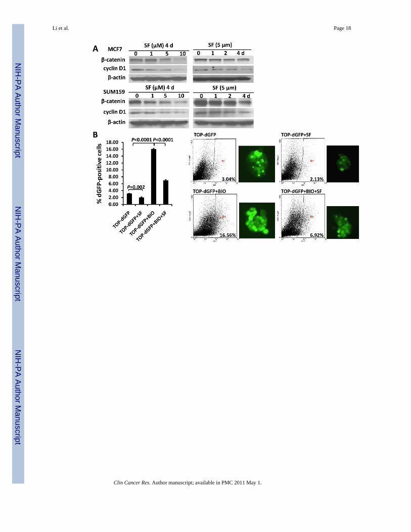
MCF7 and SUM159 cells; and the expression of cyclin D1, one of the Wnt/β-catenin targetgenes, declined by up to 77% as well. To further confirm that the down-regulation of β-cateninprotein level decreased its transcriptional activity, we utilized a TCF/LEF TOP-dGFP lentiviralreporter system. The β-catenin activates TCF/LEF in nucleus, driving the transcription ofdestabilized GFP (dGFP) gene; and the dGFP expression was analyzed by fluorescencemicroscopy and quantified by flow cytometry. As determined by flow cytometry,approximately 3% of transfected cells are dGFP-positive, and 5 μM sulforaphane reduced thispopulation by 30% ~40% (P = 0.002) (Figure 6B).
The intracellular level of β-catenin is regulated by its phosphoryaltion status and subsequentproteasomal degradation. When β-catenin is phosphorylated at Ser33/Ser37/Thr41 byGSK3β, it is immediately subject to ubiquitin-proteasome degradation (17). Phospharylationof GSK3β at Ser9 may decrease the activity of GSK3β, thereby stabilizing β-catenin (29,30).Thus, we used a proteasome inhibitor, MG132, to block proteasome function and observed anaccumulation of phospho-β-catenin (Ser33/Ser37/Thr41) in response to sulforaphane (Figure6C, upper panel). The sulforaphane-induced β-catenin phosphorylation was reversed whenLiCl, a GSK3β inhibitor, was present (Figure 6C, upper panel) (31). As shown in Figure 6B,0.5 μM BIO, another specific GSK3β inhibitor (31,32), enhanced the dGFP-positive cellpopulation by more than 5-fold (P < 0.0001), and sulforaphane (5 μM) decreased thispopulation by over 60% in the presence of BIO (P < 0.0001). Furthermore, our resultdemonstrated a decreased level of phospho-GSK3β (Ser9) by up to 74% in cells with increasingconcentrations of sulforaphane (Figure 6C, middle panel). LiCl was demonstrated to inactivateGSK3β through Ser9 phosphorylation, which in turn reduce phosphorylation of β-catenin atSer33/Ser37/Thr41 and its degradation (31,32). As shown in the bottom panel of Figure 6C,sulforaphane was able to attenuate LiCl-induced GSK3β phosphorylation and β-cateninaccumulation.
Taken together, these data suggest that the down-regulation of Wnt/β-catenin self-renewalpathway might contribute to the inhibitory effects of sulforaphane on breast CSCs. Thiswarrants further studies to establish the conclusive role of this down-regulation in inhibitionof breast CSCs by sulforaphane.
DiscussionThe anti-cancer efficacy of sulforaphane, a natural compound derived from broccoli/broccolisprouts, has been evaluated in various cancers. For instance, oral or intraperitonealadministration of sulforaphane inhibited the tumor growth in prostate PC-3 and pancreaticPanc-1 xenografts (33,34). The risk of premenopausal breast cancer was shown to be inverselyassociated with broccoli consumption (35). The orally administered sulforaphane reachedmammary gland and increased the detoxification enzyme activity (36). Additionally, it hasbeen suggested that sulforaphane may have the potential to act against tumor resistance andrelapse/recurrence (37). A very recent study demonstrated the effectiveness of sulforaphane inabrogating pancreatic tumor resistance to TRAIL by interfering with NF-κB induced anti-apoptotic signaling (18). Another study indicated that sulforaphane could overcomedoxorubicin resistance and restore apoptosis induction in cells (38). These findings provide astrong rationale for investigating the chemoprevention property of sulforaphane or broccoli/broccoli sprouts in clinical trials.
Increasing evidence supports the cancer stem cell theory, which states that a variety of cancersare driven and sustained by a small proportion of CSCs (8). The concept of CSCs has profoundclinical implications for cancer therapeutics and prevention (8,39). Recent studies indicate thatCSCs have the capacity to drive tumor resistance and relapse/recurrence (40,41). Lack ofefficacy of current chemotherapies in advance and metastatic disease requires novel approaches
Li et al. Page 7
Clin Cancer Res. Author manuscript; available in PMC 2011 May 1.
NIH
-PA Author Manuscript
NIH
-PA Author Manuscript
NIH
-PA Author Manuscript

to specifically target CSC population (8,42,43). Thus, therapies that are directed against bothdifferentiated cancer cells and CSCs may provide advantages to treat these diseases.Researchers have found that several dietary compounds are promising chemoprevention agentsagainst CSCs, such as curcumin (13,14). Therefore, based on the chemopreventive activity ofsulforaphane and the implications of CSC theory, we have utilized both in vitro and in vivosystems to determine whether sulforaphane acts against breast CSCs.
Several techniques have been developed to isolate and characterize breast CSCs in vitro.Mammosphere culture was first used to isolate and expand mammary stem/progenitor cells byDontu et al. (22), based on the ability of stem/progenitor cells to grow in serum-free suspension,while differentiated cells fail to survive under the same condition (21). By employing thistechnique, we have demonstrated that sulforaphane (0.5~5 μM) significantly suppressed themammospheres formation of both SUM159 and MCF7 cells (Figure 2). Another technique isto utilize cell makers, e.g., CD44+CD24−/lowlin− and ALDH-positive (21,23,25), to distinguishmammary stem/progenitor cells from differentiated cancer cells. It has been reported that asfew as 500 ALDH-positive cells were able to generate a breast tumor within 40 days, while50,000 ALDH-negative cells failed to form tumor (23). ALDH-positive andCD44+CD24−/lowlin− were identified a small overlap that has the highest tumorigenic capacity,generating tumors from as few as 20 cells (23). In contrast, ALDH-positive cells without theCD44+CD24−/lowlin− marker were able to produce tumors from 1,500 cells, whereas 50,000CD44+CD24−/lowlin− ALDH-negative cells did not (23). Thus, we utilized Aldefluor assay toevaluate the ability of sulforaphane to target breast cancer stem/progenitor cells. We havedemonstrated that sulforaphane (1~5 μM) could inhibit the tumor-initiating ALDH-positivecells in vitro by 65% to 80% (Figure 3). Of special note, concentrations of sulforaphane whichinhibit stem/progenitor cells in both mammosphere formation assay and Aldefluor assay hadonly minimal effects on the bulk population of breast cancer cell lines, which implies thepreferential targeting of stem/progenitor cells by sulforaphane.
The injection of human breast cancer cells into the mammary fat pad of immune-deficientNOD/SCID mice provides a reliable and sensitive in vivo system for studying human breastcancer (25,44). We demonstrated that sulforaphane was able to target breast CSCs in vivo byusing this xenograft model. Daily injection of sulforaphane for two weeks suppressed tumorgrowth in primary NOD/SCID mice and reduced ALDH-positive cell population of the tumorsby ~50% (Figure 4). More importantly, we found that the tumor cells derived fromsulforaphane-treated mice were not able to form secondary tumors in recipient mice up to 33days (Figure 5). There are two possible reasons that may explain the difference between the50% reduction of ALDH-positive population and the failure of tumor growth in secondarymice. One is that although ALDH-positive cells are enriched with stem/progenitor cells, notall ALDH-positive cells have tumor-initiating capacity. Another possible reason is theexperimental setting we used for the primary NOD/SCID mice. We inoculated 2,000,000SUM159 cells into the primary NOD/SCID mice, and treated them with the drug after twoweeks of cell inoculation, both of which could lead to an under-estimation of the effect ofsulforaphane on ALDH-positive cell population. However, the ability of CSCs to self-renewand differentiate as determined by reimplantation of primary tumor cells in secondary animalsis a more definitive functional assay (6). These are consistent with the in vitro observation thatsulforaphane preferentially targeted cancer stem/progenitor cells instead of bulk cellpopulation. The preference of sulforaphane in killing CSCs may be significant forchemoprevention.
The well-known curcumin was shown to interfere with self-renewal pathways, Wnt and Notch,in colon and pancreatic cancer cells respectively (13,14). Apple-derived quercetin and greentea epigallocatechin-gallate were reported to regulate key elements of Wnt and Notch pathwaysin human colon cancer cells (15). Park et al. previously reported that β-catenin was down-
Li et al. Page 8
Clin Cancer Res. Author manuscript; available in PMC 2011 May 1.
NIH
-PA Author Manuscript
NIH
-PA Author Manuscript
NIH
-PA Author Manuscript

regulated in HeLa and HepG2 cells (19). In consistent with this study, we demonstrated thatsulforaphane was able to down-regulate Wnt/β-catenin self-renewal pathway in breast cancercells, and sulforaphane-induced β-catenin phosphorylation (Ser33/Ser37/Thr41) andproteasome degradation was possibly through activation of GSK3β (Figure 6). Myzak et al.reported that sulforaphane increased β-catenin activity without altering its protein level inHDAC1-transfected HEK293 cells (45). The differences among the studies could arise fromdistinct cell lines and treatment conditions.
As a chemoprevention agent, sulforaphane possesses many advantages, such as highbioavailability and low toxicity (4). Sulforaphane from broccoli extracts is efficiently andrapidly absorbed in human small intestine, and distributed throughout the body (2,46). Plasmaconcentrations of sulforaphane equivalents peaked 0.94~2.27 μM in humans 1 hr after a singledose of 200 μmol broccoli sprout isothiocyanates (mainly sulforaphane) (47). A recent pilotstudy detected an accumulation of sulforaphane in human breast tissue, with 1.45 ± 1.12 pmol/mg for the right breast and 2.00 ± 1.95 pmol/mg for the left, in eight women who consumedbroccoli sprout preparation containing 200 μmol sulforaphane about 1 hr before the surgery(36). These concentrations of sulforaphane are expected to be effective against breast CSCs,based on our in vitro results. Although sulforaphane itself has not been evaluated in humans,broccoli sprouts were tested for toxicity in clinical trials (4). A Phase I trial showed that broccolisprouts caused no significant toxicity when administered orally at 8-hr intervals for 7 days as25 μmol isothiocyanates (mainly sulforaphane) (48). In another study, it was well tolerated in200 adults who consumed broccoli sprout solution containing 400 μmol glucoraphanin(precursor of sulforaphane) nightly for 2 weeks (49). Additionally, sulforaphane atconcentrations below 10 μM did not show significant effect on cell cycle arrest and apoptosisinduction of human non-transformed T-lymphocytes (50).
In conclusion, we have demonstrated that sulforaphane was able to target breast CSCs asdetermined by the mammosphere formation assay, Aldefluor assay, and tumor growth uponreimplantation in secondary mice. Furthermore, our study identified the down-regulation ofWnt/β-catenin self-renewal pathway by sulforaphane as one of the possible mechanisms forits efficacy. These studies support the use of sulforaphane for breast cancer chemoprevention.These findings provide a strong rationale for preclinical and clinical evaluation of sulforaphaneor broccoli/broccoli sprouts for breast cancer therapies.
Translational Relevance
Sulforaphane, the natural compound derived from broccoli/broccoli sprouts, has beenproved to possess anti-cancer activity. This study demonstrates that sulforaphane inhibitsbreast cancer stem cells in vitro and in vivo, which provides a strong rationale for futureclinical evaluation of sulforaphane or extract of broccoli/broccoli sprouts for breast cancerchemoprevention. Breast cancer is initiated from and maintained by a small population ofbreast cancer stem cells. Currently available chemotherapy and radiation therapy areincapable of suppressing cancer stem cell population. Aldefluor assay and mammosphereformation assay showed that sulforaphane inhibited breast cancer stem cells in vitro. NOD/SCID mouse model exhibited that sulforaphane eliminated breast cancer stem cells invivo.
AcknowledgmentsGrant support: This work was supported by the National Institutes of Health (RO1 CA120023); University ofMichigan Cancer Center Research Grant (Munn); and University of Michigan Cancer Center Core Grant to DuxinSun.
Li et al. Page 9
Clin Cancer Res. Author manuscript; available in PMC 2011 May 1.
NIH
-PA Author Manuscript
NIH
-PA Author Manuscript
NIH
-PA Author Manuscript

We would like to thank Dr. Stephen P. Ethier at Karmanos Cancer Center for the SUM159 cell line. We also wouldlike to thank Dr. Irving L. Weissman at Ludwig Center, Stanford University School of Medicine for kindly providingthe TCF/LEF-1 (TOP-dGFP, FOP-dGFP) lentiviral reporter system.
References1. Zhang Y, Talalay P, Cho CG, Posner GH. A major inducer of anticarcinogenic protective enzymes
from broccoli: isolation and elucidation of structure. Proc Natl Acad Sci U S A 1992;89:2399–403.[PubMed: 1549603]
2. Clarke JD, Dashwood RH, Ho E. Multi-targeted prevention of cancer by sulforaphane. Cancer Lett2008;269:291–304. [PubMed: 18504070]
3. Fahey JW, Zhang Y, Talalay P. Broccoli sprouts: an exceptionally rich source of inducers of enzymesthat protect against chemical carcinogens. Proc Natl Acad Sci U S A 1997;94:10367–72. [PubMed:9294217]
4. Zhang Y, Tang L. Discovery and development of sulforaphane as a cancer chemopreventivephytochemical. Acta Pharmacol Sin 2007;28:1343–54. [PubMed: 17723168]
5. Liu S, Dontu G, Wicha MS. Mammary stem cells, self-renewal pathways, and carcinogenesis. BreastCancerRes 2005;7:86–95.
6. Korkaya H, Paulson A, Charafe-Jauffret E, et al. Regulation of mammary stem/progenitor cells byPTEN/Akt/beta-catenin signaling. PLoS Biol 2009;7:e1000121. [PubMed: 19492080]
7. Liu S, Dontu G, Mantle ID, et al. Hedgehog signaling and Bmi-1 regulate self-renewal of normal andmalignant human mammary stem cells. Cancer Res 2006;66:6063–71. [PubMed: 16778178]
8. Reya T, Morrison SJ, Clarke MF, Weissman IL. Stem cells, cancer, and cancer stem cells. Nature2001;414:105–11. [PubMed: 11689955]
9. Dontu G, Jackson KW, McNicholas E, Kawamura MJ, Abdallah WM, Wicha MS. Role of Notchsignaling in cell-fate determination of human mammary stem/progenitor cells. Breast Cancer Res2004;6:R605–15. [PubMed: 15535842]
10. Smalley MJ, Dale TC. Wnt signalling in mammalian development and cancer. Cancer MetastasisRev 1999;18:215–30. [PubMed: 10728985]
11. Shafee N, Smith CR, Wei S, et al. Cancer stem cells contribute to cisplatin resistance in Brca1/p53-mediated mouse mammary tumors. Cancer Res 2008;68:3243–50. [PubMed: 18451150]
12. Hambardzumyan D, Squatrito M, Holland EC. Radiation resistance and stem-like cells in braintumors. Cancer Cell 2006;10:454–6. [PubMed: 17157785]
13. Wang Z, Zhang Y, Banerjee S, Li Y, Sarkar FH. Notch-1 down-regulation by curcumin is associatedwith the inhibition of cell growth and the induction of apoptosis in pancreatic cancer cells. Cancer2006;106:2503–13. [PubMed: 16628653]
14. Jaiswal AS, Marlow BP, Gupta N, Narayan S. Beta-catenin-mediated transactivation and cell-celladhesion pathways are important in curcumin (diferuylmethane)-induced growth arrest and apoptosisin colon cancer cells. Oncogene 2002;21:8414–27. [PubMed: 12466962]
15. Pahlke G, Ngiewih Y, Kern M, Jakobs S, Marko D, Eisenbrand G. Impact of quercetin and EGCGon key elements of the Wnt pathway in human colon carcinoma cells. J Agric Food Chem2006;54:7075–82. [PubMed: 16968065]
16. Clevers H. Wnt/beta-catenin signaling in development and disease. Cell 2006;127:469–80. [PubMed:17081971]
17. Liu C, Li Y, Semenov M, et al. Control of beta-catenin phosphorylation/degradation by a dual-kinasemechanism. Cell 2002;108:837–47. [PubMed: 11955436]
18. Kallifatidis G, Rausch V, Baumann B, et al. Sulforaphane targets pancreatic tumour-initiating cellsby NF-kappaB-induced antiapoptotic signalling. Gut 2009;58:949–63. [PubMed: 18829980]
19. Park SY, Kim GY, Bae SJ, Yoo YH, Choi YH. Induction of apoptosis by isothiocyanate sulforaphanein human cervical carcinoma HeLa and hepatocarcinoma HepG2 cells through activation ofcaspase-3. Oncol Rep 2007;18:181–7. [PubMed: 17549366]
20. Ponti D, Costa A, Zaffaroni N, et al. Isolation and in vitro propagation of tumorigenic breast cancercells with stem/progenitor cell properties. Cancer Res 2005;65:5506–11. [PubMed: 15994920]
Li et al. Page 10
Clin Cancer Res. Author manuscript; available in PMC 2011 May 1.
NIH
-PA Author Manuscript
NIH
-PA Author Manuscript
NIH
-PA Author Manuscript

21. Charafe-Jauffret E, Monville F, Ginestier C, Dontu G, Birnbaum D, Wicha MS. Cancer stem cells inbreast: current opinion and future challenges. Pathobiology 2008;75:75–84. [PubMed: 18544962]
22. Dontu G, Abdallah WM, Foley JM, et al. In vitro propagation and transcriptional profiling of humanmammary stem/progenitor cells. Genes Dev 2003;17:1253–70. [PubMed: 12756227]
23. Ginestier C, Hur MH, Charafe-Jauffret E, et al. ALDH1 is a marker of normal and malignant humanmammary stem cells and a predictor of poor clinical outcome. Cell Stem Cell 2007;1:555–67.[PubMed: 18371393]
24. Luo M, Fan H, Nagy T, et al. Mammary epithelial-specific ablation of the focal adhesion kinasesuppresses mammary tumorigenesis by affecting mammary cancer stem/progenitor cells. Cancer Res2009;69:466–74. [PubMed: 19147559]
25. Al-Hajj M, Wicha MS, Benito-Hernandez A, Morrison SJ, Clarke MF. Prospective identification oftumorigenic breast cancer cells. Proc Natl Acad Sci U S A 2003;100:3983–8. [PubMed: 12629218]
26. Reya T, Duncan AW, Ailles L, et al. A role for Wnt signalling in self-renewal of haematopoietic stemcells. Nature 2003;423:409–14. [PubMed: 12717450]
27. Azarenko O, Okouneva T, Singletary KW, Jordan MA, Wilson L. Suppression of microtubuledynamic instability and turnover in MCF7 breast cancer cells by sulforaphane. Carcinogenesis2008;29:2360–8. [PubMed: 18952594]
28. Pledgie-Tracy A, Sobolewski MD, Davidson NE. Sulforaphane induces cell type-specific apoptosisin human breast cancer cell lines. Mol Cancer Ther 2007;6:1013–21. [PubMed: 17339367]
29. Pap M, Cooper GM. Role of glycogen synthase kinase-3 in the phosphatidylinositol 3-Kinase/Aktcell survival pathway. J Biol Chem 1998;273:19929–32. [PubMed: 9685326]
30. Cohen P, Frame S. The renaissance of GSK3. Nat Rev Mol Cell Biol 2001;2:769–76. [PubMed:11584304]
31. Hedgepeth CM, Conrad LJ, Zhang J, Huang HC, Lee VM, Klein PS. Activation of the Wnt signalingpathway: a molecular mechanism for lithium action. Dev Biol 1997;185:82–91. [PubMed: 9169052]
32. Klein PS, Melton DA. A molecular mechanism for the effect of lithium on development. Proc NatlAcad Sci U S A 1996;93:8455–9. [PubMed: 8710892]
33. Pham NA, Jacobberger JW, Schimmer AD, Cao P, Gronda M, Hedley DW. The dietaryisothiocyanatesulforaphane targets pathways of apoptosis, cell cycle arrest, and oxidative stress inhuman pancreatic cancer cells and inhibits tumor growth in severe combined immunodeficient mice.Mol Cancer Ther 2004;3:1239–48. [PubMed: 15486191]
34. Singh AV, Xiao D, Lew KL, Dhir R, Singh SV. Sulforaphane induces caspase-mediated apoptosisin cultured PC-3 human prostate cancer cells and retards growth of PC-3 xenografts in vivo.Carcinogenesis 2004;25:83–90. [PubMed: 14514658]
35. Ambrosone CB, McCann SE, Freudenheim JL, Marshall JR, Zhang Y, Shields PG. Breast cancer riskin premenopausal women is inversely associated with consumption of broccoli, a source ofisothiocyanates, but is not modified by GST genotype. J Nutr 2004;134:1134–8. [PubMed:15113959]
36. Cornblatt BS, Ye L, Dinkova-Kostova AT, et al. Preclinical and clinical evaluation of sulforaphanefor chemoprevention in the breast. Carcinogenesis 2007;28:1485–90. [PubMed: 17347138]
37. Myzak MC, Dashwood RH. Chemoprotection by sulforaphane: keep one eye beyond Keap1. CancerLett 2006;233:208–18. [PubMed: 16520150]
38. Fimognari C, Nusse M, Lenzi M, Sciuscio D, Cantelli-Forti G, Hrelia P. Sulforaphane increases theefficacy of doxorubicin in mouse fibroblasts characterized by p53 mutations. Mutat Res2006;601:92–101. [PubMed: 16843502]
39. Kakarala M, Wicha MS. Implications of the cancer stem-cell hypothesis for breast cancer preventionand therapy. J Clin Oncol 2008;26:2813–20. [PubMed: 18539959]
40. Sakariassen PO, Immervoll H, Chekenya M. Cancer stem cells as mediators of treatment resistancein brain tumors: status and controversies. Neoplasia 2007;9:882–92. [PubMed: 18030356]
41. Tang C, Chua CL, Ang BT. Insights into the cancer stem cell model of glioma tumorigenesis. AnnAcad Med Singapore 2007;36:352–7. [PubMed: 17549283]
42. Lippman ME. High-dose chemotherapy plus autologous bone marrow transplantation for metastaticbreast cancer. N Engl J Med 2000;342:1119–20. [PubMed: 10760313]
Li et al. Page 11
Clin Cancer Res. Author manuscript; available in PMC 2011 May 1.
NIH
-PA Author Manuscript
NIH
-PA Author Manuscript
NIH
-PA Author Manuscript

43. Williams SD, Birch R, Einhorn LH, Irwin L, Greco FA, Loehrer PJ. Treatment of disseminated germ-cell tumors with cisplatin, bleomycin, and either vinblastine or etoposide. N Engl J Med1987;316:1435–40. [PubMed: 2437455]
44. Dick JE. Breast cancer stem cells revealed. Proc Natl Acad Sci U S A 2003;100:3547–9. [PubMed:12657737]
45. Myzak MC, Karplus PA, Chung FL, Dashwood RH. A novel mechanism of chemoprotection bysulforaphane: inhibition of histone deacetylase. Cancer Res 2004;64:5767–74. [PubMed: 15313918]
46. Petri N, Tannergren C, Holst B, et al. Absorption/metabolism of sulforaphane and quercetin, andregulation of phase II enzymes, in human jejunum in vivo. Drug Metab Dispos 2003;31:805–13.[PubMed: 12756216]
47. Ye L, Dinkova-Kostova AT, Wade KL, Zhang Y, Shapiro TA, Talalay P. Quantitative determinationof dithiocarbamates in human plasma, serum, erythrocytes and urine: pharmacokinetics of broccolisprout isothiocyanates in humans. Clin Chim Acta 2002;316:43–53. [PubMed: 11750273]
48. Shapiro TA, Fahey JW, Dinkova-Kostova AT, et al. Safety, tolerance, and metabolism of broccolisprout glucosinolates and isothiocyanates: a clinical phase I study. Nutr Cancer 2006;55:53–62.[PubMed: 16965241]
49. Kensler TW, Chen JG, Egner PA, et al. Effects of glucosinolate-rich broccoli sprouts on urinary levelsof aflatoxin-DNA adducts and phenanthrene tetraols in arandomized clinical trial in He Zuo township,Qidong, People’s Republic of China. Cancer Epidemiol Biomarkers Prev 2005;14:2605–13.[PubMed: 16284385]
50. Fimognari C, Nusse M, Berti F, Iori R, Cantelli-Forti G, Hrelia P. Isothiocyanates as novel cytotoxicand cytostatic agents: molecular pathway on human transformed and non-transformed cells. BiochemPharmacol 2004;68:1133–8. [PubMed: 15313410]
Li et al. Page 12
Clin Cancer Res. Author manuscript; available in PMC 2011 May 1.
NIH
-PA Author Manuscript
NIH
-PA Author Manuscript
NIH
-PA Author Manuscript
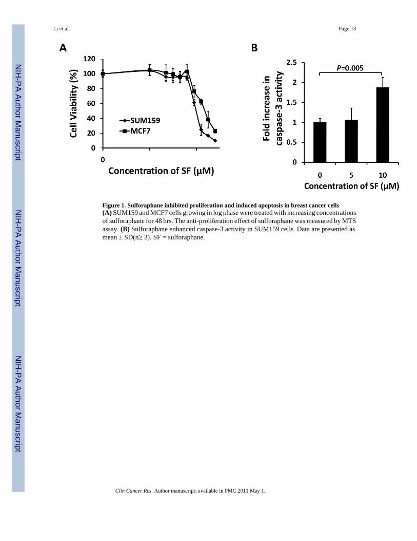
Figure 1. Sulforaphane inhibited proliferation and induced apoptosis in breast cancer cells(A) SUM159 and MCF7 cells growing in log phase were treated with increasing concentrationsof sulforaphane for 48 hrs. The anti-proliferation effect of sulforaphane was measured by MTSassay. (B) Sulforaphane enhanced caspase-3 activity in SUM159 cells. Data are presented asmean ± SD(n≥ 3). SF = sulforaphane.
Li et al. Page 13
Clin Cancer Res. Author manuscript; available in PMC 2011 May 1.
NIH
-PA Author Manuscript
NIH
-PA Author Manuscript
NIH
-PA Author Manuscript

Figure 2. Inhibitory effect of sulforaphane on mammosphere formationMCF7 and SUM159 cells were cultured in mammosphere forming conditions. (A) Primarymammospheres were incubated with sulforaphane (0.5, 1, and 5 μM) or DMSO for 7 days.Sulforaphane treatment reduced the number of primary mammospheres. (B) Sulforaphanereduced the size of primary mammospheres(magnification × 100). The size of mammosphereswas estimated using V = (4/3)π R3. (C) In the absence of drug, the 2nd and 3rd passages thatwere derived from sulforaphane-treated primary mammospheres yielded smaller numbers ofspheres in comparison with control. Data are presented as mean ± SD(n = 3). *, P < 0.05; **,P < 0.01. SF = sulforaphane.
Li et al. Page 14
Clin Cancer Res. Author manuscript; available in PMC 2011 May 1.
NIH
-PA Author Manuscript
NIH
-PA Author Manuscript
NIH
-PA Author Manuscript

Figure 3. Inhibitory effect of sulforaphane on ALDH-positive cell populationSUM159 cells were treated with sulforaphane (1 and 5 μM) or DMSO for 4 days, and subjectto Aldefluor assay and flow cytometry analysis. (A) Sulforaphane decreased the percentage ofALDH-positive cells. Data are presented as mean ± SD(n =3). (B) A set of representative flowcytometry dot plots. R2 covers the region of ALDH-positive cells. SF = sulforaphane; ALDH= aldehyde dehydrogenase.
Li et al. Page 15
Clin Cancer Res. Author manuscript; available in PMC 2011 May 1.
NIH
-PA Author Manuscript
NIH
-PA Author Manuscript
NIH
-PA Author Manuscript
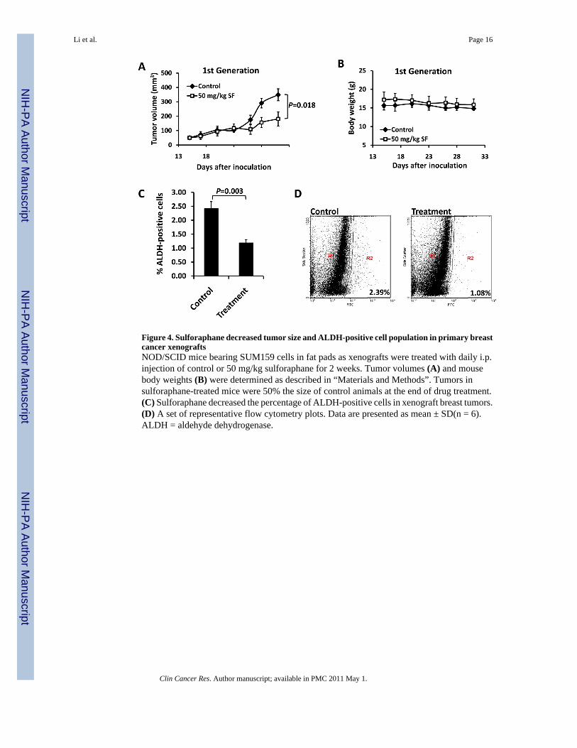
Figure 4. Sulforaphane decreased tumor size and ALDH-positive cell population in primary breastcancer xenograftsNOD/SCID mice bearing SUM159 cells in fat pads as xenografts were treated with daily i.p.injection of control or 50 mg/kg sulforaphane for 2 weeks. Tumor volumes (A) and mousebody weights (B) were determined as described in “Materials and Methods”. Tumors insulforaphane-treated mice were 50% the size of control animals at the end of drug treatment.(C) Sulforaphane decreased the percentage of ALDH-positive cells in xenograft breast tumors.(D) A set of representative flow cytometry plots. Data are presented as mean ± SD(n = 6).ALDH = aldehyde dehydrogenase.
Li et al. Page 16
Clin Cancer Res. Author manuscript; available in PMC 2011 May 1.
NIH
-PA Author Manuscript
NIH
-PA Author Manuscript
NIH
-PA Author Manuscript
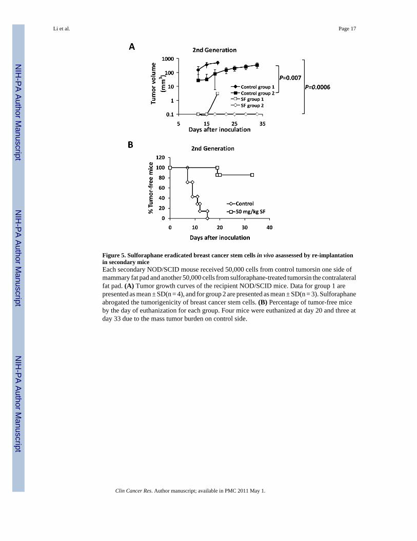
Figure 5. Sulforaphane eradicated breast cancer stem cells in vivo asassessed by re-implantationin secondary miceEach secondary NOD/SCID mouse received 50,000 cells from control tumorsin one side ofmammary fat pad and another 50,000 cells from sulforaphane-treated tumorsin the contralateralfat pad. (A) Tumor growth curves of the recipient NOD/SCID mice. Data for group 1 arepresented as mean ± SD(n = 4), and for group 2 are presented as mean ± SD(n = 3). Sulforaphaneabrogated the tumorigenicity of breast cancer stem cells. (B) Percentage of tumor-free miceby the day of euthanization for each group. Four mice were euthanized at day 20 and three atday 33 due to the mass tumor burden on control side.
Li et al. Page 17
Clin Cancer Res. Author manuscript; available in PMC 2011 May 1.
NIH
-PA Author Manuscript
NIH
-PA Author Manuscript
NIH
-PA Author Manuscript
Li et al. Page 18
Clin Cancer Res. Author manuscript; available in PMC 2011 May 1.
NIH
-PA Author Manuscript
NIH
-PA Author Manuscript
NIH
-PA Author Manuscript

Figure 6. Sulforaphane down-regulated Wnt/β-catenin self-renewal pathway(A) Sulforaphane decreased protein levels of β-catenin and cyclin D1 in both SUM159 andMCF7 cell lines. (B) TOP-dGFP reporter lentivirus-infected MCF7 mammospheres weretreated with indicated compounds (0.5 μM BIO and 5 μM sulforaphane) either alone or incombination for 2 days. Sulforaphane decreased the percentage of dGFP-positive cells by 30%~40%. BIO increased this population, while sulforaphane decreased it by over 60% in thepresence of BIO. Representative flow cytometry results of TOP-dGFP mammospheres andtheir pictures under fluorescence microscope(magnification × 100)are shown on the right.(C) Sulforaphane promoted β-catenin phosphorylation at Ser33/37/Thr41, while LiClsuppressed the phosphorylation by inactivating GSK3β (upper panel). Sulforaphane decreasedphospho-GSK3β (Ser9)level, whereas total GSK3β remained unchanged(middle panel). LiClincreased protein level of β-catenin by phosphorylating/inactivating GSK3β at Ser9, whilesulforaphane attenuated LiCl-induced GSK3β phosphorylation and β-catenin accumulation(bottom panel). SF = sulforaphane; dGFP = destabilized green fluorescent protein.
Li et al. Page 19
Clin Cancer Res. Author manuscript; available in PMC 2011 May 1.
NIH
-PA Author Manuscript
NIH
-PA Author Manuscript
NIH
-PA Author Manuscript