Microbial Metabolism and Colonization of Iron and Sulfur ...

fpls-07-00458 April 6, 2016 Time: 17:36 # 1
ORIGINAL RESEARCHpublished: 08 April 2016
doi: 10.3389/fpls.2016.00458
Edited by:Jesú Mercado-Blanco,
Consejo Superior de InvestigacionesCientíficas, Spain
Reviewed by:Stanislav Kopriva,
University of Cologne, GermanyAna Pineda,
Nederlands Instituut voor Ecologie –Koninklijke Nederlandse Akademievan Wetenschappen, Netherlands
Aurélien Bailly,University of Zurich, Switzerland
*Correspondence:Paul W. Paré
Specialty section:This article was submitted to
Plant Biotic Interactions,a section of the journal
Frontiers in Plant Science
Received: 14 December 2015Accepted: 24 March 2016
Published: 08 April 2016
Citation:Aziz M, Nadipalli RK, Xie X, Sun Y,
Surowiec K, Zhang J-L and Paré PW(2016) Augmenting Sulfur Metabolismand Herbivore Defense in Arabidopsis
by Bacterial Volatile Signaling.Front. Plant Sci. 7:458.
doi: 10.3389/fpls.2016.00458
Augmenting Sulfur Metabolism andHerbivore Defense in Arabidopsis byBacterial Volatile SignalingMina Aziz1,2, Ranjith K. Nadipalli1, Xitao Xie1, Yan Sun1, Kazimierz Surowiec1,Jin-Lin Zhang3 and Paul W. Paré1*
1 Department of Chemistry and Biochemistry, Texas Tech University, Lubbock, TX, USA, 2 Center for Plant Lipid Research,University of North Texas, Denton, TX, USA, 3 College of Pastoral Agriculture Science and Technology, Lanzhou University,Lanzhou, China
Sulfur is an element necessary for the life cycle of higher plants. Its assimilation andreduction into essential biomolecules are pivotal factors determining a plant’s growthand vigor as well as resistance to environmental stress. While certain soil microbescan enhance ion solubility via chelating agents or oxidation, microbial regulation ofplant-sulfur assimilation has not been reported. With an increasing understandingthat soil microbes can activate growth and stress tolerance in plants via chemicalsignaling, the question arises as to whether such beneficial bacteria also regulatesulfur assimilation. Here we report a previously unidentified mechanism by which thegrowth-promoting rhizobacterium Bacillus amyloliquefaciens (GB03) transcriptionallyactivates genes responsible for sulfur assimilation, increasing sulfur uptake andaccumulation in Arabidopsis. Transcripts encoding for sulfur-rich aliphatic and indolicglucosinolates are also GB03 induced. As a result, GB03-exposed plants with elevatedglucosinolates exhibit greater protection against the generalist herbivore, Spodopteraexigua (beet armyworm, BAW). In contrast, a previously characterized glucosinolatemutant compromised in the production of both aliphatic and indolic glucosinolatesis also compromised in terms of GB03-induced protection against insect herbivory.As with in vitro studies, soil-grown plants show enhanced glucosinolate accumulationand protection against BAW feeding with GB03 exposure. These results demonstratethe potential of microbes to enhance plant sulfur assimilation and emphasize thesophisticated integration of microbial signaling in plant defense.
Keywords: plant growth-promoting rhizobacteria (PGPR), Bacillus amyloliquefaciens GB03, bacterial volatileorganic compounds (VOCs), glucosinolates (GSL), sulfur assimilation, plant-defense priming
Abbreviations: 1MOI3M, 1-methoxyindol-3-ylmethyl glucosinolate; 4MOI3M, 4-methoxyindol-3-ylmethyl glucosinolate;4-MSOB, 4-methylsulfinylbutyl glucosinolate; 4-MTB, 4-methylthiobutyl glucosinolate; 5-MSOP, 5-methylsulfinylpentylglucosinolate; 6-MSOH, 6-methylsulfinylhexyl glucosinolate; 7-MSOH, 7-methylsulfinylheptyl glucosinolate; 7-MTH, 7-methylthioheptyl glucosinolate; 8-MSOO, 8-methylsulfinyloctyl glucosinolate; 8-MTO, 8-methylthiooctyl glucosinolate;APK, APS kinase; APR, APS reductase; APS, adenosine 5′-phosphosulfate; ATPS, ATP sulfurylase; BAW, beet armyworm;DMDS, dimethyl disulfide; BCAT, branched-chain amino acid amino transferase; CFU, colony forming unit; CYP,cytochromes P6450; DDW, double-distilled water; ESI-MS, electrospray ionization-mass spectrometry; FMOGS−OX, flavinmonooxygenase; GST, glutathione-s-transferase; I3M, indol-3-ylmethyl glucosinolate; IGMT, indole glucosinolate methyltransferase; IPMDH, isopropyl malate dehydrogenase; IPMI, isopropyl malate isomerase; MAM, methylthioalkyl malatesynthase; MSG, methylsulfinylalkyl glucosinolates; MTG, methylthioalkyl glucosinolates; PAPS, 3′-phosphoadenosine5′-phosphosulfate; PDA, photodiode array; PGPR, plant growth-promoting rhizobacteria; RT-PCR, reverse-transcriptionPCR; SOT, sulfotransferase; SUR, super root; TSA, tryptic soy agar; UHPLC, ultra-HPLC; VOCs, volatile organic compounds.
Frontiers in Plant Science | www.frontiersin.org 1 April 2016 | Volume 7 | Article 458

fpls-07-00458 April 6, 2016 Time: 17:36 # 2
Aziz et al. Beneficial Bacteria-Enhanced Sulfur Metabolism
INTRODUCTION
Sulfur, a crucial element for plants, is ubiquitous in proteins,present in the antioxidant tripeptide glutathione, the Cys-rich peptides phytochelatins that function in heavy metalsdetoxification and thioredoxins that are the major disulfidereductases responsible for maintaining the reduced state ofproteins inside cells (Arnér and Holmgren, 2000; Cobbett, 2000).Sulfur can also be present in chloroplastic membrane lipids aswell as certain coenzymes/vitamins (Falk et al., 2007). Sulfur istaken up by plants as inorganic sulfate via sulfate transporters andincorporated into APS by ATPS (Mugford et al., 2009). APS isthen sequentially reduced by APR and sulfite reductase to sulfiteand sulfide, and subsequently incorporated into O-acetylserineto form the sulfur containing amino acid cysteine. APS canalso be phosphorylated to PAPS by the action of APK. PAPSis the sulfate donor for the formation of sulfated metabolitesincluding glucosinolates, select flavonoids, phytosulfokines, andcertain hormones.
From an ecological context, sulfur metabolites function inplant defense against pathogens and herbivores (Falk et al.,2007). Defensin and thionin peptides are sulfur-containingantimicrobial defenses with widespread plant distribution(Broekaert et al., 1995), whereas anti-feedant glucosinolates arelimited to the Brassicale order (Falk et al., 2007). Brassicacrops including cabbage, broccoli, cauliflower (Brassica oleracea)and rapeseed (B. napus) as well as Arabidopsis are rich inglucosinolates. In addition to these amino acid derivativesfunctioning in plant defense, glucosinolates are a nutritionalsource of sulfur and possess cancer-preventive properties(Sønderby et al., 2010). Glucosinolates are classified based ontheir amino acid precursor with aliphatic glucosinolates derivedfrom Met, Ala, Leu, Ile, or Val; indolic glucosinolates derivedfrom Trp; and aromatic glucosinolates derived from Phe or Tyr(Kliebenstein et al., 2001).
With plant damage, glucosinolates are rapidly converted intoan array of toxic derivatives that can obfuscate phytochemicalanalysis. Enzymatically generated glucosinolate derivativesincluding isothiocyanates, epithionitriles, nitriles, andthiocyanates are produced in proportion to the amount of leafdamage as well as the reaction time (Halkier and Gershenzon,2006; Wittstock and Burow, 2010; Winde and Wittstock, 2011).Therefore quantifying the pool of original glucosinolates requiresdeactivating the myrosinase enzyme before glucosinolates areenzymatically converted (Koroleva et al., 2000; Andréasson andJørgensen, 2003; Zhao et al., 2008; Winde and Wittstock, 2011).
In addition to constitutive glucosinolate accumulation servingin chemical defense against herbivore damage, soil-bornemicrobes such as mycorrhizal fungi and PGPR can induceplant defense responses (van Loon, 2007; van Wees et al.,2008; Yang et al., 2009; Pineda et al., 2010, 2012). PGPR arenaturally occurring soil microorganisms that colonize rootsand stimulate plant growth. Such bacteria are applied to awide range of agricultural crops for the purpose of growthenhancement, including increased seed germination, plantweight, harvest yields, and disease resistance (Kloepper et al.,1980, 1991, 1999). Bacillus subtilis (GB03), recently re-named
as B. amyloliquefaciens is a commercially available PGPR strainthat can be introduced into the soil at the time of planting viaseed coating since spores are stable over time (Choi et al., 2014).Unlike many plant-growth promoting rhizobacterial strains thatactivate plant growth by directly producing and releasing indole-3-acetic acid and/or gibberellins, GB03 emits a bouquet ofvolatile metabolites, devoid of classic phytohormones that arecapable of triggering plant growth promotion (Ryu et al., 2003;Paré et al., 2005). These VOCs have been shown to activatedifferential expression of approximately 600 transcripts relatedto cell wall modifications, primary and secondary metabolism,stress responses, hormone regulation, and iron homeostasis (Ryuet al., 2003; Farag et al., 2006; Zhang et al., 2007). This Arabidopsisprofiling of GB03-induced transcripts has resulted in a newparadigm for PGPR-mediated iron uptake. While some soilmicrobes are proposed to enhance iron mobility and uptakesolely via production of bacterial siderophores (Neilands andLeong, 1986; Bar-Ness et al., 1992; Briat, 1992; Glick et al.,1999; Sharma et al., 2003), GB03 enhances Arabidopsis ironaccumulation via activation of the plant’s own iron acquisitionmachinery including the iron uptake-related genes FRO2 andIRT1 that encode for ferric reductase and iron transport enzymes,respectively (Zhang et al., 2009). GB03 also transcriptionallyregulates the Fe-deficiency-induced transcription factor 1 (FIT1)that is necessary and sufficient for ferric reductase and irontransporter induction (Zhang et al., 2009). More recently, anupstream iron acquisition-related transcription factor MYB72has been shown to be transcriptionally induced in Arabidopsis bybacterial VOCs with activation of the iron uptake-related genesFIT1, FRO2, and IRT1 (Zamioudis et al., 2015).
The current study reports a novel mechanism in which thegrowth-promoting rhizobacterium B. amyloliquefaciens strainGB03 induces Arabidopsis sulfur assimilation and accumulationby inducing the plant’s own sulfur assimilation machinery.Moreover, the impact of GB03 in regulating primary andsecondary sulfur metabolites to enhance plant defense againstherbivory is examined.
MATERIALS AND METHODS
Plant Material and TreatmentsArabidopsis thaliana seeds were surface sterilized and stratifiedfor 2 days at 4◦C in the absence of light. Seeds were plantedin plastic Petri dishes (100 × 15 mm) containing a centralpartition (I-plates; Fisher Scientific), covered Magenta boxes(75 mm × 75 mm × 100 mm) or standard Petri dishes(150 mm × 15 mm), based on the specific experimentalrequirements. The bacterial culture is inoculated on theunplanted side of the partitioned plate, a glass vial (4 dr.) or aplastic plate (35 mm × 10 mm). All chambers contained half-strength MS solid media prepared according to Murashige andSkoog (1962) with 1.5% (w/v) sucrose and 0.8% (w/v) agar (exceptwhere noted otherwise). Plants were grown under a 14-/10-hlight/dark cycle with metal halide and high pressure sodiumlamps for a total light intensity of 200 µmol photons m−2 s−1;temperature was 21 ± 4◦C and relative humidity 40 ± 10%. For
Frontiers in Plant Science | www.frontiersin.org 2 April 2016 | Volume 7 | Article 458
fpls-07-00458 April 6, 2016 Time: 17:36 # 3
Aziz et al. Beneficial Bacteria-Enhanced Sulfur Metabolism
plant growth, the media surface was oriented horizontally forI-plates and Magenta boxes and vertically for the larger plateswith media agar increased to 1.5% (w/v).
Bacillus amyloliquefaciens (GB03) was streaked onto TSAplates and incubated at 28◦C in the absence of light for 24 h. Cellswere harvested in double distilled water (DDW) to yield 109 CFUmL−1, as determined by optical density (OD600 = 0.7). Two daysafter seed germination, the bacterial suspension culture or DDW(25 µL for plates and 50 µL for Magenta boxes) was added tothe non-plant portion of the chamber. Vials containing bacterialculture were replaced with fresh culture every 14 days.
For soil experiments, bacterial liquid cultures were mixed withsterile growing mix (Sunshine LC1 Mix; Sun Gro Horticulture,Canada) to a final PGPR concentration of 108 – 109 CFU/g soil.For water control, the bacterial suspension was replaced withsterile DDW. Seeds were sown in growing mix and fertilizedweekly using 13:13:13 (N:P:K) fertilizer.
Semi-quantitative RT-PCRPlants were harvested 48- or 72-h after GB03 or water treatment.Total RNA was extracted using RNeasy plant mini kit (Qiagen,Valencia, CA, USA) with genomic DNA contamination excludedby DNase digestion. First strand cDNA was synthesized from 3-5 µg total RNA using MuMLV-RT (Fisher Scientific, Houston,TX, USA); primer sequences are shown (Table 1). The PCRreaction included an initial 3 min denaturation at 94◦C, followedby 30 s at 94◦C, 30 s at 54◦C and 1 min at 72◦C with 24–27 cycles (based on the optimized linear range for each pair ofspecific primers), a final 10 min extension at 72◦C (T100 ThermalCycler, Bio-Rad, Hercules, CA, USA). No-reverse-transcriptioncontrols were included with the PCR runs to confirm theabsence of DNA contamination. Agarose gel electrophoresiswere imaged with a Kodak Gel Logic 100 Imaging System(Fisher Scientific, Houston, TX, USA) and quantified usingImage J 1.33u1 (National Institute of Health, USA). TUB8 andUBQ10 were employed for normalization as they were uniformlyexpressed in all tissues examined.
Total Sulfur DeterminationShoots and roots were separated, oven-dried, pulverized, andconverted to dry-ash by heating at 550◦C for 3 h in thepresence of Ag2O and NaHCO3 based on Kalra (1998). Driedtissue was then neutralized, diluted, and analysis via a bariumchloride-gelatin turbidimetric assay (Tabatabai and Bremner,1970). Standards were prepared as tissue material and diluted to afinal concentration of 0–32 µg mL−1. Total sulfur was quantifiedspectrophotometrically at 420 nm based on a sulfur standardcurve.
35SO4−2 Uptake Assay
For sulfate uptake measurements, plants were germinated onnylon mesh and grown vertically on media-containing plateswith GB03 or water exposure for 11 days. Radio-labeling wasinitiated by submerging the roots into liquid media containing37 MBq L−1 35SO4
−2 (Perkin–Elmer). After incubation for
1http://rsb.info.nih.gov/ij/
TABLE 1 | Sequence of primers employed in the semi-quantitative RT-PCRanalysis.
Gene Name Primer Sequence (5′ to 3′)
ATPS1 Forward: GTTTCCTTCCCTTCCAAATC
Reverse: GAGCCAGTTTCCAGCATTAG
ATPS3 Forward: GAATGAAACAGCACGAGAAG
Reverse: CCAGGGCACATAAATCCATC
ATPS2 Forward: ATGCTGTTTTTGCGTTTCAG
Reverse: ACGGCTTGTTGTTTTGCTTC
ATPS4 Forward: GCGTATGAGACAGCACGAG
Reverse: AACCAACACCTTCCAACCAG
APR1 Forward: AGGTTTGGATGGTGGAGTTG
Reverse: CATAAAGCACGACGATCCAAG
APR2 Forward: CGAATCTTGGGTTACTCGTG
Reverse: CCTCCTTGATGTTCCCTTTG
APR3 Forward: GAGATGGTGGTGGGAAGATG
Reverse: TGGAACGAGACTGGATGGTC
APK1 Forward: TCCACCACCGTGAGATATGA
Reverse: ATCCGCAAAAAGCTTAGCAA
APK2 Forward: TGGCACGAGAGTTCGATATG
Reverse: CAGCACTACCTCGCAATTCA
CYP79F1 Forward: TCCATGGCATCAATCACTCTAC
Reverse: CATCAACATTCCAACCTCTCAA
SUR1 Forward: TCGTGCTGCTTACAGTGGTC
Reverse: ACACAGGGGATGTCCTTGAG
FMOGS−OX3 Forward: ACCAATGTCCCGAGAGAAAGTA
Reverse: GGAACGGAAATCTTCTCGTATG
UBQ10 Forward: CGATTACTCTTGAGGTGGAG
Reverse: AGACCAAGTGAAGTGTGGAC
TUB8 Forward: CGTGGATCACAGCAATACAGAGCC
Reverse: CCTCCTGCACTTCCACTTCGTCTTC
30 min, roots were briefly rinsed with non-labeled medium toremove apoplastic radioactivity (modified protocol from Kataokaet al., 2004; Maruyama-Nakashita et al., 2004; Yoshimoto et al.,2007). After blotting, shoots and roots were weighed separately,transferred to scintillation vials and covered with 1 mL of 0.1 MHCl. Overnight-extracted samples were mixed with universalscintillation cocktail (4 mL; Fisher Scientific) and incorporatedradioactivity measured by liquid scintillation counting.
Cysteine MeasurementsWhole plant tissue (0.1 g) was ground in liquid nitrogen andthiols were acid extracted using ice-chilled 0.1 N HCl (200 µL).The homogenate was centrifuged at 12,000 × g for 10 minat 4◦C. Supernatant aliquots were neutralized with 200 mMHEPES (pH 12.4), reduced with dithiothreitol and sulfhydrylgroups derivatized with monobromobimane (VWR). Separation,detection and quantification of fluorescent adducts was based onSchupp and Rennenberg (1988).
Glucosinolates AnalysisPlants were shoot and root separated, frozen in liquid nitrogenand lyophilized. Tissue (20–50 mg) was extracted for 15 minin boiling aqueous 7.5 mM Pb(OAc)2/Ba(OAc)2 (4 mL) with
Frontiers in Plant Science | www.frontiersin.org 3 April 2016 | Volume 7 | Article 458

fpls-07-00458 April 6, 2016 Time: 17:36 # 4
Aziz et al. Beneficial Bacteria-Enhanced Sulfur Metabolism
0.57 µmol internal standard (sinigrin, Sigma–Aldrich) basedon Reintanz et al. (2001). At room temperature, samples weregently shaken for 30 min, centrifuged at 4000 × g for 10 minand the supernatant was loaded on DEAE Sephadex A-25column (120 mg, Sigma–Aldrich). Resin was rinsed with aqueousmethanol (67%) and water and subsequently incubated with50 µL sulfatase solution overnight (Graser et al., 2000). Theresulting desulfoglucosinolates were eluted with 60% aqueousmethanol (800 µL) and water (800 µL). The pooled extract wasevaporated to dryness in vacuo and the residue was dissolved inHPLC-grade water (100 µL).
Desulfoglucosinolates were separated by HPLC on a DionexUltimate 3000 UHPLC system equipped with auto-sampler,column oven, and diode array detector. A C18 reversed phasecolumn (Acclaim 120 mm × 3.0 mm, 150 mm × 3.0 mm i.d.,3-µm particle size) was run with a 400 µL/min flow rate at 25◦C;the injection volume was 10 µL. Elution was performed with agradient (solvent A water; B acetonitrile) of 1.5 to 5% solvent B(6 min), 5 to 7% solvent B (2 min), 7 to 21% solvent B (10 min),21 to 29% solvent B (5 min), and 29 to 57% solvent B (14 min),followed by a cleaning cycle (57 to 93% solvent B for 3 min,6 min of hold, 93 to 1.5% solvent B for 3 min with a 5 min hold).Compounds were monitored at 229 nm.
Desulfoglucosinolates were identified by HPLC-PDA-MSbased on method of Kusznierewicz et al. (2013). Samples wereanalyzed on a LCQ Fleet HPLC system equipped with PALautosampler, Surveyor PDA detector, and Surveyor MS pumpusing an Alltima C18 reversed phase column (250 mm× 2.1 mmi.d., 5-µm particle size) with a 200 µL/min flow rate. Theinjection volume was 10 µL. Elution was performed with agradient (solvent A water/0.1% formic acid; B acetonitrile/0.1%formic acid) of 1.5% solvent B (3 min) 1.5 to 13% solvent B(15 min), 13 to 33% solvent B (12 min), 33 to 57% solvent B(7 min), followed by a cleaning cycle (57 to 93% solvent B for3 min, 6 min of hold, 93 to 1.5% solvent B for 3 min with a5 min hold). Compounds were monitored by PDA at 229 nm,then subsequently by ESI-MS (LCQ Fleet Ion Trap MS) operatedin positive ion mode, an acquisition time of 40 min with scanningfrom m/z 150 to 800 amu.
Previously reported desulfoglucosinolates were identifiedby MS via characteristic [M+H]+ and [M+Na]+ peaksexcept for 3-methylsulfinylpropyl glucosinolate (3MSOP) whichcould not be identified because of poor resolution. Positionalisomers 4MOI3M and 1MOI3M with equivalent masses weredifferentiated based on retention time comparisons withliterature values (Reintanz et al., 2001). Glucosinolates werequantified based on response factors established for individualdesulfoglucosinolates relative to the internal standard at 229 nm(Brown et al., 2003).
Herbivore FeedingSpodoptera exigua (BAW) eggs were purchased from Benzonresearch (Carlisle, PA, USA). After hatching, neonate larvae weretransferred to feed on artificial media for 6 days with a transferto fresh media every 2–3 days. Since an acclimation periodis required whenever larvae are transferred from one diet toanother, 1 day before the experiment, third-instar larvae were
transferred to feed on non-experimental wild-type Arabidopsisplants (Mewis et al., 2005). After this pre-feeding, larvae ofthe same developmental stage were weighted and transferredto 29-day-old GB03- or water-treated plants (one larva/plant);the initial average weight of larvae was recorded for bothGB03 and water treatments. Shoot biomass was recorded after56 h of feeding. Additional GB03-treated and untreated plantswere reserved to serve as undamaged controls. Plants wereharvested, rinsed, and weighted. Milligrams eaten per plantwere calculated based on the weight difference between BAWeaten and uneaten plants. The quadruple glucosinolate knock-outmutant (myb28 myb29 cyp79b2 cyp79b3) was treated the same asCol-0.
For soil experiments, herbivore weights were collected.Neonate larvae were transferred to 28-day-old GB03- orwater-treated plants with a transfer to fresh plants every 2–3 days. Larvae weight was measured at 7 and 9 days afterfeeding.
Statistical AnalysisFor herbivore feeding experiments, statistical analyses wereperformed using R software2. First, a Levene’s test wasperformed to check the homogeneity of variance (Levene,1960); homogeneous variance was achieved after transformingthe data into the corresponding square root. Then, two-way ANOVAs were performed separately for wild-type andknock-out mutant lines. Tukey’s method was used to do pair-wise comparisons of means and an “lsmeans” package wasused for means’ grouping. For all other experiments, pair-wise comparison of means was performed using Excel 2007with significant difference between treatments was based onStudent’s t-test at P-values ≤ 0.05. The number of biologicalreplicates is shown in each figure legend with minimum of threereplicates.
RESULTS
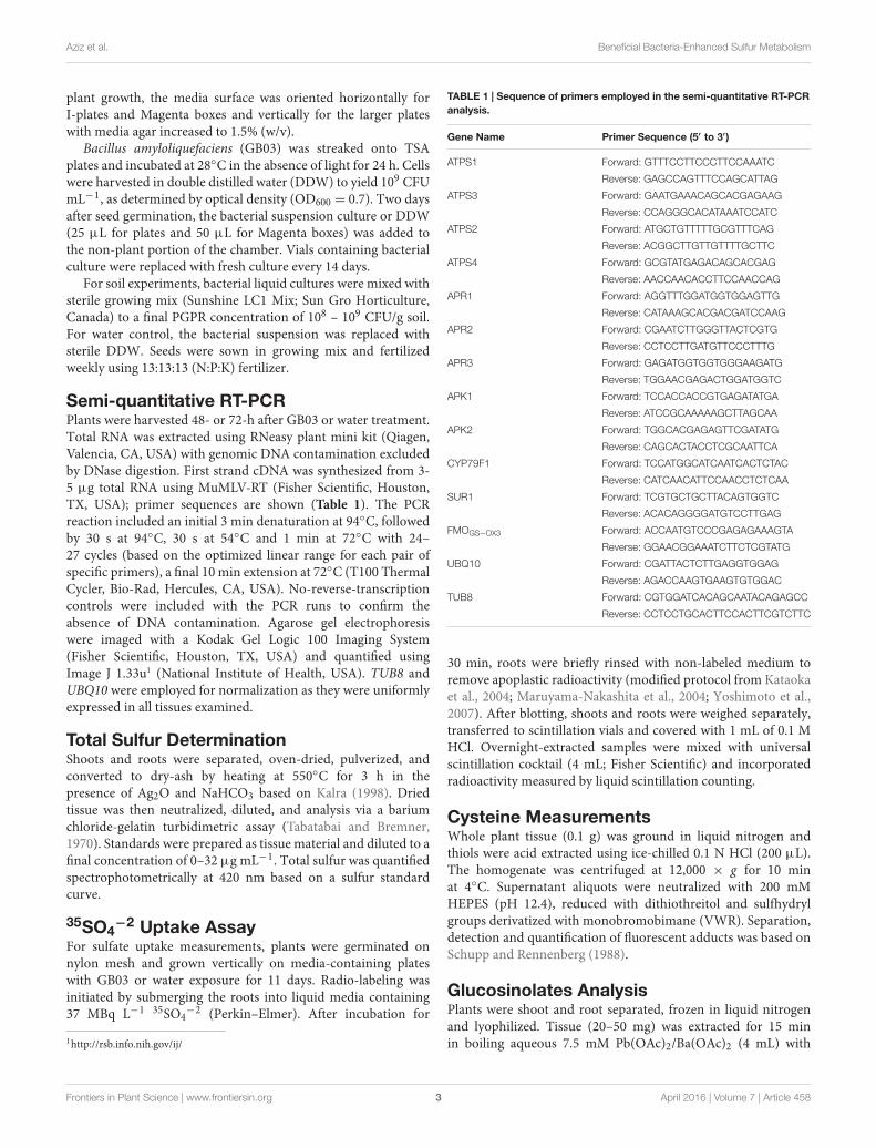
Elevated Sulfate Assimilation with GB03ExposureThe sulfate assimilation pathway with previously identifiedgenes is depicted in Figure 1A. Mining whole-plant microarraydata of GB03-exposed Arabidopsis seedlings identified sulfate-assimilation gene induction for ATPS and APR. Of the fourATPS and the three APR isozymes present, ATPS1 and ATPS3as well as APR1 and APR2 were found to be induced at72 h post GB03 exposure (Supplementary Figure 1). RT-PCRanalysis confirmed GB03 induction for ATPS1, ATPS2, andATPS3 and all APR genes (Figure 1B). Another branch ofsulfur assimilation involves APS conversion to PAPS by APK.There are four functional APK isoforms in Arabidopsis, amongthem APK1 and APK2 are the most active isoforms (Mugfordet al., 2009). From the microarray data, both APK1 and APK2are GB03 up regulated relative to controls (Supplementary
2www.R-project.org
Frontiers in Plant Science | www.frontiersin.org 4 April 2016 | Volume 7 | Article 458
fpls-07-00458 April 6, 2016 Time: 17:36 # 5
Aziz et al. Beneficial Bacteria-Enhanced Sulfur Metabolism
FIGURE 1 | Transcriptional regulation of Arabidopsis sulfur assimilation genes by GB03. Depicted sulfate assimilation pathway adapted from Mugford et al.(2009) (A). Semi-quantitative RT-PCR analysis of whole-plant sulfur assimilation gene expression at 72 h post GB03 treatment (B); data are the averages of threebiological replicates with error bars representing standard error. The amino acid cysteine increases with GB03 treatment (solid line) relative to the water controls(dashed lines; C); an asterisk (∗) indicate statistically significant difference between treatments (t-test, P-value ≤ 0.05, n = 6, mean ± SE). Sulfur assimilation pathwayincludes SULTR, sulfate transporter; ATPS, ATP sulfurlyase; APS, adenosine 5′-phosphosulfate; APR, APS reductase; SiR, sulfite reductase; OASTL; O-acetylserine(thiol) lyase; GSH, glutathione; APK, APS kinase; PAPS, 3′-phosphoadenosine 5′-phosphosulfate.
Figure 1). APK transcript induction confirmation via RT-PCR analysis showed GB03 induction only in shoots (APK1,1.3 ± 0.08; APK2, 1.5 ± 0.05). In addition, the aminoacid cysteine, a precursor of many organic sulfur metabolitesincreased 28 ± 11, 32 ± 8, 37 ± 10, and 93 ± 15 %with GB03 exposure at 5, 7, 9, and 14 days, respectively(Figure 1C).
GB03 Enhances Sulfur Accumulation andUptakeAs sulfate assimilation and reduction genes were GB03induced, sulfur accumulation was examined. While total sulfuraccumulation per tissue weight decreased ca. twofold in shoots11 days post GB03 exposure, shoot sulfur accumulation perplant increased ca. 75% (Figure 2A). In roots, increasesof ca. 50-fold and ca. 100-fold on a dry-weight and per-plant basis, respectively, were observed (Figure 2B). Tobetter characterize the process of inducible sulfur metabolism,plant sulfur movement was monitored with radioactive sulfate(35SO4
−2) to examine sulfur uptake and translocation. Althoughthere was a ca. 30% reduction in total sulfur uptake pertissue weight, GB03 exposure enhanced total sulfur uptakeper plant by ca. twofold, relative to untreated controls,
within 30 min of radio-labeling (Figure 3A). Shoot sulfurtranslocation per tissue weight was ca. twofold less withGB03 treatment; however, similar translocation rate per plantwas observed for both GB03 and controls (Figure 3B).And in roots, sulfur uptake and retention was higher withGB03 exposure on both a tissue weight and per-plant basis(Figure 3C).
Since select bacterial volatiles such as 2,3-butandiol havebeen previously shown to induce growth promotion andinduced systemic resistance in Arabidopsis (Ryu et al.,2003, 2004), an array of 2,3-butandiol concentrations wereassayed to examine for enhanced sulfur accumulationalbeit no sulfur-associated changes were detected (datanot shown). Similarly, collected bacterial volatiles re-introduced to plants also did not enhance sulfuraccumulation.
GB03 Induces GlucosinolateBiosynthetic TranscriptsThe aliphatic and indolic glucosinolate biosynthetic pathwayswith previously identified genes is depicted in Figures 4A,B,respectively. Mining microarray data for transcripts encodingglucosinolate biosynthesis revealed that the majority of aliphatic
Frontiers in Plant Science | www.frontiersin.org 5 April 2016 | Volume 7 | Article 458

fpls-07-00458 April 6, 2016 Time: 17:36 # 6
Aziz et al. Beneficial Bacteria-Enhanced Sulfur Metabolism
FIGURE 2 | Sulfur accumulation in Arabidopsis with GB03 exposure.Shoot (A) and root (B) sulfur accumulation in 13-day-old plants that areGB03- (black bars) or water-treated (white bars) on a dry-weight and per-plantbasis. Root sulfur values for water-treated plants are 1.36 ± 0.68 nmol/mgDW and 0.4 ± 0.05 nmol/plant, although values are not perceivable in thefigure. An asterisk (∗) indicates statistically significant difference betweentreatments (t-test, P-value ≤ 0.05, n = 4, mean ± SE).
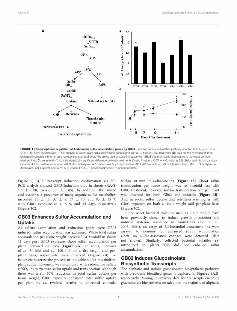
pathway genes are GB03 induced (Supplementary Figure 2A).For indolic glucosinolate biosynthesis, microarray data showedtranscript induction limited to GSTF9, SUR1, UGT74B1,and SOT16 (Supplementary Figure 2B). GSTF9 is a GSTwhich is responsible for the conjugation of the activatedaldoximes to the sulfur donor glutathione, where the resultingS-alkylthiohydroximates are converted to thiohydroximatesby a carbon-sulfur lyase, SUR1. Thiohydroximates are inturn S-glucosylated by the glucosyltransferases UGT74B1to form desulfoglucosinolates. Finally, desulfoglucosinolatesare sulfated to the corresponding glucosinolates by thesulfotransferase SOT16. Monitoring select shoot and roottranscripts separately by RT-PCR confirmed gene inductionwith CYP79F1 induction in shoots ca. threefold, while rootinduction was ca. 30% (Figures 4C,D). CYP79F1 catalyzesthe first committed step in biosynthesis of the aliphaticglucosinolate core structure that involves conversion ofamino acids to corresponding aldoximes (a rate-limitingstep in glucosinolates biosynthesis; Mikkelsen and Halkier,2003). FMOGS−OX3, a gene that encodes one of the fiveflavin monooxygenases responsible for S-oxygenation ofaliphatic glucosinolates resulting in conversion of MTGto MSG (Sønderby et al., 2010) was induced in shoots
FIGURE 3 | Sulfur uptake and translocation monitored by radioactivesulfate (35SO4
−2 30 min pulse) in Arabidopsis with GB03 exposure.Whole plant sulfur uptake (A), shoot sulfur translocation (B) and sulfur rootretention (C) is shown in 13-day-old plants that are GB03- (black bars) orwater-treated (white bars) on a fresh-weight and per-plant basis. The value ofsulfur retained in the roots for water-treated plants is 8.6 ± 0.98 pmol/30 min/plant, although values are not perceivable in the figure. An asterisk (∗)indicates statistically significant difference between treatments (t-test,P-value ≤ 0.05, n ≥ 4, mean ± SE).
within 48 h while root induction was delayed to 72 h(Figures 4C,D). SUR1 gene expression was induced ca.threefold in shoots (Figures 4C,D). To link transcriptionalregulation with downstream glucosinolate accumulation,qualitative and quantitative glucosinolate analysis wasperformed.
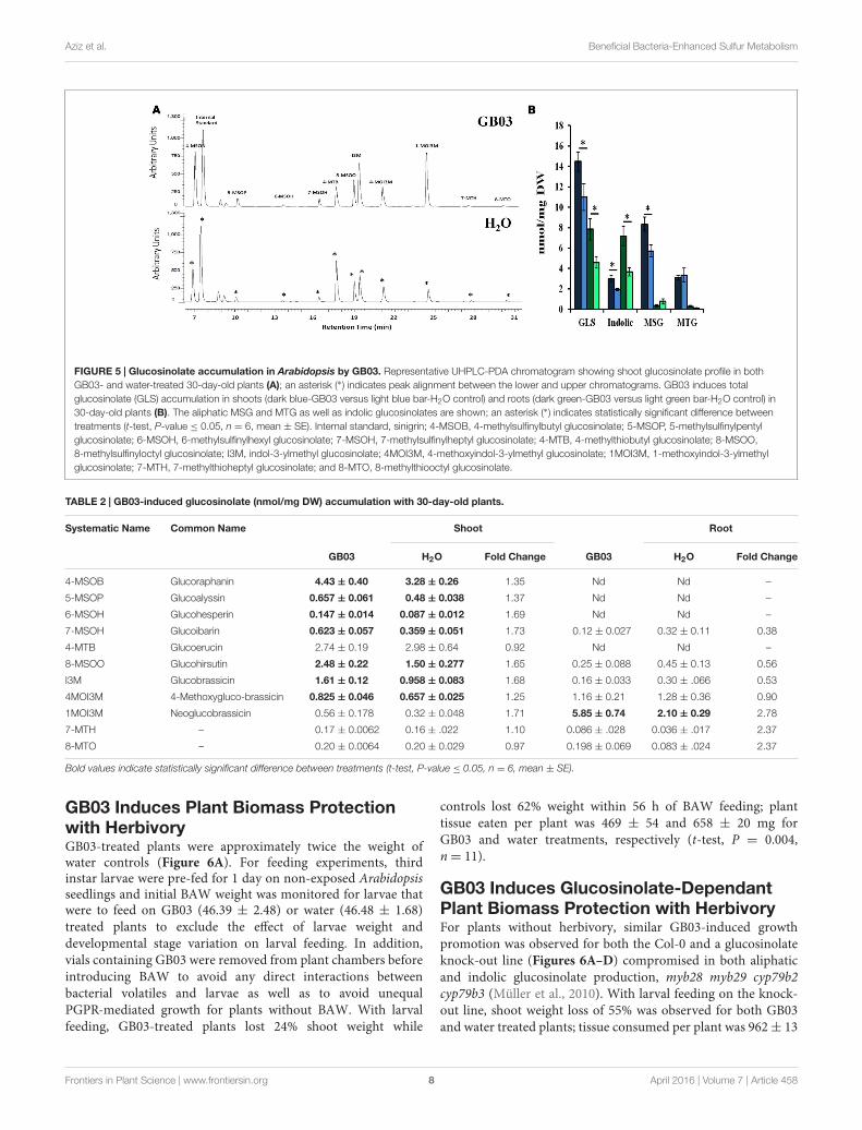
GB03 Induces GlucosinolateAccumulationDesulfoglucosinolates were separated by HPLC based on relativepolarity, with MSG eluting first in increasing order of theirside-chain length, followed by indolic glucosinolates; long chainMTGs eluted last off the column (Figure 5A). GB03 exposureresulted in ca. 33 and 70% greater glucosinolate accumulationin shoots and roots, respectively (Figure 5B). Specifically, GB03
Frontiers in Plant Science | www.frontiersin.org 6 April 2016 | Volume 7 | Article 458
fpls-07-00458 April 6, 2016 Time: 17:36 # 7
Aziz et al. Beneficial Bacteria-Enhanced Sulfur Metabolism
FIGURE 4 | Glucosinolate biosynthesis transcriptional regulation in Arabidopsis by GB03. Depicted aliphatic (A) and indolic (B) glucosinolate biosyntheticpathways adapted from Sønderby et al. (2010). Semi-quantitative RT-PCR analysis of CYP79F1 and FMOGS−OX3 gene expression in both shoots (C) and roots (D)at 48 and 72 h, and SUR1 at 72 h post GB03 exposure; an asterisk (*) indicates statistically significant difference between treatments (t-test, P-value ≤ 0.05, n = 3,mean ± SE).
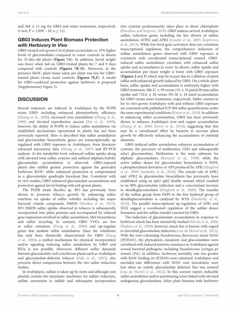
increased indolic glucosinolates in shoots (55%) and roots(twofold) while MSGs were induced in shoots by 45%. MTGaccumulation differences with regard to tissue or GB03 treatmentwas not observed. In shoots, I3M was the most GB03 inducedindolic glucosinolate (68%; Table 2); while among MSG, therewas a 35, 37, 69, 73, and 65% GB03 induction of 4-MSOB,
5-MSOP, 6-MSOH, 7-MSOH, and 8-MSOO, respectively. Forroots, the most abundant glucosinolate, 1MOI3M, increasedthreefold with no other statistically significant accumulationchanges. Glucosinolate accumulation was not induced with plantexposure to 2,3-butandiol or collected bacterial volatiles (data notshown).
Frontiers in Plant Science | www.frontiersin.org 7 April 2016 | Volume 7 | Article 458
fpls-07-00458 April 6, 2016 Time: 17:36 # 8
Aziz et al. Beneficial Bacteria-Enhanced Sulfur Metabolism
FIGURE 5 | Glucosinolate accumulation in Arabidopsis by GB03. Representative UHPLC-PDA chromatogram showing shoot glucosinolate profile in bothGB03- and water-treated 30-day-old plants (A); an asterisk (∗) indicates peak alignment between the lower and upper chromatograms. GB03 induces totalglucosinolate (GLS) accumulation in shoots (dark blue-GB03 versus light blue bar-H2O control) and roots (dark green-GB03 versus light green bar-H2O control) in30-day-old plants (B). The aliphatic MSG and MTG as well as indolic glucosinolates are shown; an asterisk (∗) indicates statistically significant difference betweentreatments (t-test, P-value ≤ 0.05, n = 6, mean ± SE). Internal standard, sinigrin; 4-MSOB, 4-methylsulfinylbutyl glucosinolate; 5-MSOP, 5-methylsulfinylpentylglucosinolate; 6-MSOH, 6-methylsulfinylhexyl glucosinolate; 7-MSOH, 7-methylsulfinylheptyl glucosinolate; 4-MTB, 4-methylthiobutyl glucosinolate; 8-MSOO,8-methylsulfinyloctyl glucosinolate; I3M, indol-3-ylmethyl glucosinolate; 4MOI3M, 4-methoxyindol-3-ylmethyl glucosinolate; 1MOI3M, 1-methoxyindol-3-ylmethylglucosinolate; 7-MTH, 7-methylthioheptyl glucosinolate; and 8-MTO, 8-methylthiooctyl glucosinolate.
TABLE 2 | GB03-induced glucosinolate (nmol/mg DW) accumulation with 30-day-old plants.
Systematic Name Common Name Shoot Root
GB03 H2O Fold Change GB03 H2O Fold Change
4-MSOB Glucoraphanin 4.43 ± 0.40 3.28 ± 0.26 1.35 Nd Nd –
5-MSOP Glucoalyssin 0.657 ± 0.061 0.48 ± 0.038 1.37 Nd Nd –
6-MSOH Glucohesperin 0.147 ± 0.014 0.087 ± 0.012 1.69 Nd Nd –
7-MSOH Glucoibarin 0.623 ± 0.057 0.359 ± 0.051 1.73 0.12 ± 0.027 0.32 ± 0.11 0.38
4-MTB Glucoerucin 2.74 ± 0.19 2.98 ± 0.64 0.92 Nd Nd –
8-MSOO Glucohirsutin 2.48 ± 0.22 1.50 ± 0.277 1.65 0.25 ± 0.088 0.45 ± 0.13 0.56
I3M Glucobrassicin 1.61 ± 0.12 0.958 ± 0.083 1.68 0.16 ± 0.033 0.30 ± .066 0.53
4MOI3M 4-Methoxygluco-brassicin 0.825 ± 0.046 0.657 ± 0.025 1.25 1.16 ± 0.21 1.28 ± 0.36 0.90
1MOI3M Neoglucobrassicin 0.56 ± 0.178 0.32 ± 0.048 1.71 5.85 ± 0.74 2.10 ± 0.29 2.78
7-MTH – 0.17 ± 0.0062 0.16 ± .022 1.10 0.086 ± .028 0.036 ± .017 2.37
8-MTO – 0.20 ± 0.0064 0.20 ± 0.029 0.97 0.198 ± 0.069 0.083 ± .024 2.37
Bold values indicate statistically significant difference between treatments (t-test, P-value ≤ 0.05, n = 6, mean ± SE).
GB03 Induces Plant Biomass Protectionwith HerbivoryGB03-treated plants were approximately twice the weight ofwater controls (Figure 6A). For feeding experiments, thirdinstar larvae were pre-fed for 1 day on non-exposed Arabidopsisseedlings and initial BAW weight was monitored for larvae thatwere to feed on GB03 (46.39 ± 2.48) or water (46.48 ± 1.68)treated plants to exclude the effect of larvae weight anddevelopmental stage variation on larval feeding. In addition,vials containing GB03 were removed from plant chambers beforeintroducing BAW to avoid any direct interactions betweenbacterial volatiles and larvae as well as to avoid unequalPGPR-mediated growth for plants without BAW. With larvalfeeding, GB03-treated plants lost 24% shoot weight while
controls lost 62% weight within 56 h of BAW feeding; planttissue eaten per plant was 469 ± 54 and 658 ± 20 mg forGB03 and water treatments, respectively (t-test, P = 0.004,n= 11).
GB03 Induces Glucosinolate-DependantPlant Biomass Protection with HerbivoryFor plants without herbivory, similar GB03-induced growthpromotion was observed for both the Col-0 and a glucosinolateknock-out line (Figures 6A–D) compromised in both aliphaticand indolic glucosinolate production, myb28 myb29 cyp79b2cyp79b3 (Müller et al., 2010). With larval feeding on the knock-out line, shoot weight loss of 55% was observed for both GB03and water treated plants; tissue consumed per plant was 962± 13
Frontiers in Plant Science | www.frontiersin.org 8 April 2016 | Volume 7 | Article 458
fpls-07-00458 April 6, 2016 Time: 17:36 # 9
Aziz et al. Beneficial Bacteria-Enhanced Sulfur Metabolism
and 364 ± 11 mg for GB03 and water treatments, respectively(t-test, P = 1.89E – 20, n ≥ 11).
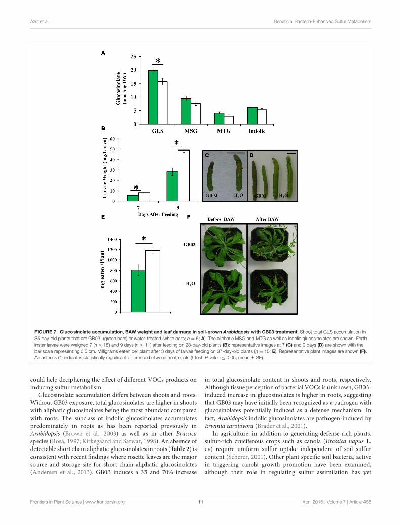
GB03 Induces Plant Biomass Protectionwith Herbivory In VivoGB03-treated soil-grown Col-0 plants accumulate ca. 25% higherlevels of glucosinolates compared to water controls in shootsfor 35-day-old plants (Figure 7A). In addition, larval weightwas lower when fed on GB03-treated plants for 7 and 9 dayscompared with controls (Figures 7B–D). Moreover, in thepresence BAW, plant tissue eaten per plant was less for GB03-treated plants versus water controls (Figures 7E,F). A modelfor GB03-conferred protection against herbivory is proposed(Supplementary Figure 3).
DISCUSSION
Several responses are induced in Arabidopsis by the PGPRstrain GB03 including enhanced photosynthetic efficiency(Zhang et al., 2008), increased iron assimilation (Zhang et al.,2009) and elevated reproductive success (Xie et al., 2009),however, the ability of PGPR to induce sulfur assimilation viaestablished mechanisms operational in plants has not beenpreviously reported. Here is described that sulfur assimilationand glucosinolate biosynthetic genes are transcriptionally upregulated with GB03 exposure in Arabidopsis, from literatureextracted microarray data (Zhang et al., 2007) and RT-PCRanalysis. At the metabolite level, enhanced sulfate uptake alongwith elevated total sulfur, cysteine and sulfated aliphatic/indolicglucosinolate accumulation is observed. GB03-exposedplants also exhibit greater protection against the generalistherbivore BAW, while enhanced protection is compromisedin a glucosinolate quadruple knockout line. Consistent within vitro studies, GB03 enhanced glucosinolate accumulation andprotection against larval feeding with soil-grown plants.
The PGPR strain Bacillus sp. B55 has previously beenshown to promote tobacco growth by enhancing sulfurnutrition via uptake of sulfur volatiles including the majorbacterial volatile component, DMDS (Meldau et al., 2013).B55 DMDS sulfur uptake observed in tobacco is subsequentlyincorporated into plant proteins and accompanied by reducedgene expression involved in sulfur assimilation, Met biosynthesisand sulfur recycling. In contrast, GB03 VOCs are lowin sulfur emissions (Farag et al., 2006) and up-regulategenes that mediate sulfur assimilation. Since the volatilomehas only been chemically characterized for GB03 (Faraget al., 2006), a unified mechanism for chemical incorporationand/or signaling inducing sulfur metabolism by GB03 andB55a is not possible. Moreover, different sulfur demandsbetween glucosinolate-rich cruciferous plants such as Arabidopsisand glucosinolate-deficient tobacco (Falk et al., 2007), alsoprevents direct comparisons between the two sulfur inductionstudies.
In Arabidopsis, sulfate is taken up by roots and although rootplastids contain the enzymatic machinery for sulfate reduction,sulfate conversion to sulfide and subsequent incorporation
into cysteine predominantly takes place in shoot chloroplasts(Davidian and Kopriva, 2010). GB03 induces several Arabidopsissulfate reduction genes including the key drivers of sulfateassimilation ATPS1 and APR2 (Loudet et al., 2007; Koprivovaet al., 2013). While low-level gene activation does not constitutetranscriptional regulation, the comprehensive induction ofsulfate assimilation genes observed with GB03 exposure isconsistent with coordinated transcriptional control. GB03-induced sulfur assimilation correlates with enhanced sulfuruptake and accumulation in roots. In shoots, sulfur uptake andaccumulation per tissue weight is lower with GB03 exposure(Figures 2 and 3) which may be in part due to a dilution of plantsulfur with enhanced growth induced by GB03. On a whole-plantbasis, sulfur uptake and accumulation is uniformly higher withGB03 treatment: 286.37± 59 versus 151± 35 pmol/30 min sulfuruptake and 732.6 ± 20 versus 391.36 ± 10 nmol accumulationfor GB03 versus water treatments, respectively. Sulfur content infor in vitro grown Arabidopsis with and without GB03 exposureare consistent with published ICP-MS sulfur quantification underthe same experimental conditions (Kwon et al., 2010). In additionto enhancing sulfur accumulation, GB03 has been previouslyshown to enhance Arabidopsis iron and copper accumulation(Zhang et al., 2009; Kwon et al., 2010), suggesting that thismay be a coordinated effort by bacteria to increase plantgrowth by effectively enhancing the accumulation of essentialelements.
GB03-induced sulfur assimilation enhances accumulation ofcysteine, the precursor of methionine, GSH and subsequentlyselect glucosinolates. Methionine is the main substrate foraliphatic glucosinolates (Ravanel et al., 1998) while theactive sulfate donor for glucosinolate biosynthesis is PAPS,a phosphorylated derivative of APS produced by APK (Mugfordet al., 2009; Sønderby et al., 2010). The crucial role of APK1and APK2 in glucosinolate biosynthesis has previously beenestablished using an apk1 apk2 double mutant which resultedin an 80% glucosinolate reduction and a concomitant increasein desulfoglucosinolates (Mugford et al., 2009). The transferof the sulfate group from PAPS to the free hydroxyl group ofdesulfoglucosinolates is catalyzed by SOTs (Sønderby et al.,2010). The parallel transcriptional up-regulation of APKs andSOTs suggest a coordinated regulation of the sulfate donorformation and the sulfate transfer reaction by GB03.
The induction of glucosinolate accumulation in response toherbivore attack has been extensively studied (Mewis et al., 2006;Hopkins et al., 2009), however, much less is known with regardto microbial glucosinolate induction (van de Mortel et al., 2012).With the root-colonizing Pseudomonas fluorescens strain SS101(Pf.SS101), the phytoalexin camalexin and glucosinolates werecorrelated with induced systemic resistance in Arabidopsis againstseveral bacterial pathogens, including Pseudomonas syringae pvtomato (Pst). In addition, herbivore mortality rate was greaterwith BAW feeding on Pf.SS101-root-colonized Arabidopsis andmortality-rate differences with SS101 root inoculation werelost when an indolic glucosinolate deficient line was assayed(van de Mortel et al., 2012). In this current report, induciblesulfur assimilation and/or partitioning is/are linked with elevatedendogenous glucosinolates, foliar plant biomass with herbivory
Frontiers in Plant Science | www.frontiersin.org 9 April 2016 | Volume 7 | Article 458

fpls-07-00458 April 6, 2016 Time: 17:36 # 10
Aziz et al. Beneficial Bacteria-Enhanced Sulfur Metabolism
FIGURE 6 | Arabidopsis protection against the generalist herbivore Spodoptera exigua (BAW) by GB03. Twenty nine-day-old wild-type (A,B) andglucosinolate quadruple myb28 myb29 cyp79b2 cyp79b3 mutant plants (C,D) without (–BAW) and with (+BAW) larval feeding (third instar) for 56 h. GB03-treated(green bars) and H2O-control plants (white bars) are shown. Data were analyzed by two-way ANOVA; different letters indicate statistically significant differencesbetween treatments (Tukey’s test, P-value ≤ 0.01, n ≥ 11, mean ± SE). Representative plant images (B,D) are shown.
and larval weight. By monitoring enhanced plant protectionagainst BAW feeding by bacterial volatiles albeit devoid ofdirect plant–bacteria contact, induced plant defense responsesindependent of potential confounding bacterial anti-feedanteffects can be identified. Without BAW larvae present, GB03induced plant growth in both the wild-type and glucosinolatemutant line (Figure 6), indicating that glucosinolates play no rolein GB03-triggered growth promotion. However, greater GB03-induced growth promotion in the mutant line compared to Col-0(Figure 6C) may be in part due to additional energy available forgrowth promotion without glucosinolate biosynthesis operative.With BAW herbivory, GB03-treated Col-0 plants lost less shootweight than water controls (Figures 6A,B), indicating GB03-induction of plant defense(s). With such GB03 plant protectionagainst larval feeding compromised in the glucosinolate mutantline (Figures 6C,D), a causal relationship is established betweenGB03-enhanced glucosinolate accumulation and conferred plantprotection. Interestingly, in the mutant line without glucosinolatedefenses present, larval-consumed plant tissue per plant wasgreater with versus without GB03 exposure; tissue consumedper plant was 962 ± 13 and 364 ± 11 mg for GB03 versuswater treatments, respectively. Future experiments will examineif GB03-induced plants contain greater amounts of young leavesthat have yet to accumulate non-glucosinolate based chemicaldefenses or if such plants dilute non-inducible chemical defensesmaking the GB03-induced glucosinolate mutant line morepalatable for feeding larvae.
Soil-grown GB03-treated plants exhibited enhancedglucosinolate accumulation and plant protection against
BAW is consistent with I-plate experiments; however, elicitationdifferences limit direct comparisons between in vitro andin vivo systems. For example, chemical signaling is confinedto bacterial VOCs in vitro while non-volatile metabolites canalso serve as potential signaling molecules in the in vivo soilsystem. Moreover, although the soil is sterilized before plantingand bacterial inoculation, the non-sterile environment inwhich soil-grown plants are exposed is conducive to bacterialproliferate of leaves and roots by other bacterial strains besidesGB03. Down-stream signaling pathways can also be differentiallyregulated in media and soil systems. For example, ethylenesignaling is operative with in vivo PGPR signaling but not within vitro growth promotion (Ryu et al., 2005). Future studieswill examine several mutant lines to elucidate which of thedifferent plant signaling pathways are involved in elicitingenhanced sulfur metabolism and protection against herbivoresby GB03 both in vitro and in vivo. Moreover, since it has beenwidely recognized that the plant hormone jasmonic acid (JA)plays a crucial role in plant defense against pathogens andherbivores as well as in glucosinolate accumulation (van Damet al., 2004; van Dam and Oomen, 2008), JA mutant lines will beassayed.
Since the growth promotion signal 2,3-butandiol (Ryu et al.,2003, 2004) as well as collected GB03 VOCs re-introducedto plants do not exhibit enhanced sulfur assimilation orglucosinolate accumulation a more effective absorbent maybe necessary to trap biologically active bacterial volatiles.Alternatively, as the genome sequence of GB03 has been recentlyidentified (Choi et al., 2014), testing different GB03 mutant lines
Frontiers in Plant Science | www.frontiersin.org 10 April 2016 | Volume 7 | Article 458
fpls-07-00458 April 6, 2016 Time: 17:36 # 11
Aziz et al. Beneficial Bacteria-Enhanced Sulfur Metabolism
FIGURE 7 | Glucosinolate accumulation, BAW weight and leaf damage in soil-grown Arabidopsis with GB03 treatment. Shoot total GLS accumulation in35-day-old plants that are GB03- (green bars) or water-treated (white bars; n = 5; A). The aliphatic MSG and MTG as well as indolic glucosinolates are shown. Forthinstar larvae were weighed 7 (n ≥ 18) and 9 days (n ≥ 11) after feeding on 28-day-old plants (B); representative images at 7 (C) and 9 days (D) are shown with thebar scale representing 0.5 cm. Milligrams eaten per plant after 3 days of larvae feeding on 37-day-old plants (n = 10; E). Representative plant images are shown (F).An asterisk (∗) indicates statistically significant difference between treatments (t-test, P-value ≤ 0.05, mean ± SE).
could help deciphering the effect of different VOCs products oninducing sulfur metabolism.
Glucosinolate accumulation differs between shoots and roots.Without GB03 exposure, total glucosinolates are higher in shootswith aliphatic glucosinolates being the most abundant comparedwith roots. The subclass of indolic glucosinolates accumulatespredominately in roots as has been reported previously inArabidopsis (Brown et al., 2003) as well as in other Brassicaspecies (Rosa, 1997; Kirkegaard and Sarwar, 1998). An absence ofdetectable short chain aliphatic glucosinolates in roots (Table 2) isconsistent with recent findings where rosette leaves are the majorsource and storage site for short chain aliphatic glucosinolates(Andersen et al., 2013). GB03 induces a 33 and 70% increase
in total glucosinolate content in shoots and roots, respectively.Although tissue perception of bacterial VOCs is unknown, GB03-induced increase in glucosinolates is higher in roots, suggestingthat GB03 may have initially been recognized as a pathogen withglucosinolates potentially induced as a defense mechanism. Infact, Arabidopsis indolic glucosinolates are pathogen-induced byErwinia carotovora (Brader et al., 2001).
In agriculture, in addition to generating defense-rich plants,sulfur-rich cruciferous crops such as canola (Brassica napus L.cv) require uniform sulfur uptake independent of soil sulfurcontent (Scherer, 2001). Other plant specific soil bacteria, activein triggering canola growth promotion have been examined,although their role in regulating sulfur assimilation has yet
Frontiers in Plant Science | www.frontiersin.org 11 April 2016 | Volume 7 | Article 458
fpls-07-00458 April 6, 2016 Time: 17:36 # 12
Aziz et al. Beneficial Bacteria-Enhanced Sulfur Metabolism
to be characterized (Kloepper et al., 1988; Bertrand et al.,2001). Commercial canola inoculants have been developedthat oxidize elemental sulfur to the sulfate form that is morereadily taken up by plants. Such bacterial inoculants areagriculturally relevant since elemental sulfur, an industrial by-product, is economically viable for regenerating sulfur deficientsoils. Here, GB03 transcriptionally induces sulfate assimilationand coordinates this process with enhanced sulfate uptake aswell as elevated sulfur, cysteine, and glucosinolate accumulation.In addition to the role of glucosinolates in plant defense,select sulfur metabolites possess cancer-preventive properties.For humans, isothiocyanates derived from the hydrolysis ofMSG are potent cancer-preventive agents (Hansen et al., 2007;Li et al., 2008). The cancer-preventive properties of MSG havebeen targeted for elevated production by plant breeders ofcruciferous crops (Li et al., 2008) and here they are shownto be selectively induced by GB03, relative to other aliphaticglucosinolates.
AUTHOR CONTRIBUTIONS
MA designed the project, performed experiments, collecteddata, analyzed results, and wrote up the study; RN performedexperiments, collected data, and analyzed results; XX designed
the project, performed experiments, collected data, and analyzeddata; YS collected data; KS collected data; J-LZ designed theproject and analyzed the results; and PP designed the project,analyzed results, and wrote up the study.
FUNDING
Financial support was provided in part by the Robert WelchFoundation (D-1478).
ACKNOWLEDGMENTS
We would like to thank Prof. Barbara Ann Halkier atthe University of Copenhagen for the generous gift of theglucosinolates mutant lines, Dr. Mohamed Ali Farag at CairoUniversity for the technical assistance with MS analysis, andGamage Pemantha Lakraj at Texas Tech University for assistancewith the statistical analyses.
SUPPLEMENTARY MATERIAL
The Supplementary Material for this article can be found onlineat: http://journal.frontiersin.org/article/10.3389/fpls.2016.00458
REFERENCESAndersen, T. G., Nour-Eldin, H. H., Fuller, V. L., Olsen, C. E., Burow, M., and
Halkier, B. A. (2013). Integration of biosynthesis and long-distance transportestablish organ-specific glucosinolate profiles in vegetative Arabidopsis. PlantCell 25, 3133–3145. doi: 10.1105/tpc.113.110890
Andréasson, E., and Jørgensen, L. B. (2003). “Localization of plant myrosinases andglucosinolates,” in Integrative Phytochemistry: From Ethnobotany to MolecularEcology, 1st Edn, ed. J. T. Romeo (Amsterdam: Elsevier), 79–99.
Arnér, E. S. J., and Holmgren, A. (2000). Physiological functions of thioredoxin andthioredoxin reductase. Eur. J. Biochem. 267, 6102–6109. doi: 10.1046/j.1432-1327.2000.01701
Bar-Ness, E., Hadar, Y., Chen, Y., Shanzer, A., and Libman, J. (1992). Iron uptakeby plants from microbial siderophores: a study with 7-nitrobenz-2 oxa-1,3-diazole-desferrioxamine as fluorescent ferrioxamine B analog. Plant Physiol. 99,1329–1335. doi: 10.1104/pp.100.1.451
Bertrand, H., Nalin, R., Bally, R., and Cleyet-Marel, J. C. (2001). Isolationand identification of the most efficient plant growth-promoting bacteriaassociated with canola (Brassica napus). Biol. Fertil. Soils 33, 152–156. doi:10.1007/s003740000305
Brader, G., Tas, E., and Palva, E. T. (2001). Jasmonate-dependent inductionof indole glucosinolates in Arabidopsis by culture filtrates of the non-specific pathogen Erwinia carotovora. Plant Physiol. 126, 849–860. doi:10.1104/pp.126.2.849
Briat, J. F. (1992). Iron assimilation and storage in prokaryotes. J. Gen. Microbiol.138, 2475–2483. doi: 10.1099/00221287-138-12-2475
Broekaert, W. F., Terras, F. R. G., Cammue, B. P. A., and Osborn, R. W. (1995).Plant defensins: novel antimicrobial peptides as components of the host defensesystem. Plant Physiol. 108, 1353–1358. doi: 10.1104/pp.108.4.1353
Brown, P. D., Tokuhisa, J. G., Reichelt, M., and Gershenzon, J. (2003). Variationof glucosinolate accumulation among different organs and developmentalstages of Arabidopsis thaliana. Phytochemistry 62, 471–481. doi: 10.1016/S0031-9422(02)00549-6
Choi, S.-K., Jeong, H., Kloepper, J. W., and Ryu, C.-M. (2014). Genomesequence of Bacillus amyloliquefaciens GB03, an active ingredient of the first
commercial biological control product. Genome Announc. 2, e1092-14. doi:10.1128/genomeA.01092-14
Cobbett, C. (2000). Phytochelatins and their roles in heavy metal detoxification.Plant Physiol. 123, 825–832. doi: 10.1104/pp.123.3.825
Davidian, J.-C., and Kopriva, S. (2010). Regulation of sulfate uptake andassimilation—the same or not the same? Mol. Plant 3, 314–325. doi:10.1093/mp/ssq001
Falk, K. L., Tokuhisa, J. G., and Gershenzon, J. (2007). The effect of sulfur nutritionon plant glucosinolate content: physiology and molecular mechanisms. PlantBiol. 9, 573–581. doi: 10.1055/s-2007-965431
Farag, M. A., Ryu, C.-M., Sumner, L. W., and Paré, P. W. (2006). GC-MS SPMEprofiling of rhizobacterial volatiles reveals prospective inducers of growthpromotion and induced systemic resistance in plants. Phytochemistry 67, 2262–2268. doi: 10.1016/j.phytochem.2006.07.021
Glick, B. R., Patten, C. N., Holguin, G., and Penrose, D. M. (1999). Biochemicaland Genetic Mechanisms used by Plant Growth Promotion Bacteria. London:Imperial College Press, 1–13.
Graser, G., Schneider, B., Oldham, N. J., and Gershenzon, J. (2000). Themethionine chain elongation pathway in the biosynthesis of glucosinolatesin Eruca sativa (Brassicaceae). Arch. Biochem. Biophys. 378, 411–419. doi:10.1006/abbi.2000.1812
Halkier, B. A., and Gershenzon, J. (2006). Biology and biochemistryof glucosinolates. Ann. Rev. Plant Biol. 57, 303–333. doi:10.1146/annurev.arplant.57.032905.105228
Hansen, B. G., Kliebenstein, D. J., and Halkier, B. A. (2007). Identification of aflavin-monooxygenase as the S-oxygenating enzyme in aliphatic glucosinolatebiosynthesis in Arabidopsis. Plant J. 50, 902–910. doi: 10.1111/j.1365-313X.2007.03101
Hopkins, R. J., van Dam, N. M., and van Loon, J. J. A. (2009). Role of glucosinolatesin insect–plant relationships and multitrophic interactions. Ann. Rev. Entomol.54, 57–83. doi: 10.1146/annurev.ento.54.110807.090623
Kalra, Y. P. (1998). Handbook of Standard Methods of Plant Analysis. Boca Raton,FL: CRC Press.
Kataoka, T., Hayashi, N., Yamaya, T., and Takahashi, H. (2004). Root-to-shoottransport of sulfate in Arabidopsis. Evidence for the role of SULTR3;5 as a
Frontiers in Plant Science | www.frontiersin.org 12 April 2016 | Volume 7 | Article 458
fpls-07-00458 April 6, 2016 Time: 17:36 # 13
Aziz et al. Beneficial Bacteria-Enhanced Sulfur Metabolism
component of low-affinity sulfate transport system in the root vasculature. PlantPhysiol. 136, 4198–4204. doi: 10.1104/pp.104.045625
Kirkegaard, J. A., and Sarwar, M. (1998). Biofumigation potential of Brassicas. I.Variation in glucosinolate profiles of diverse field-grown Brassicas. Plant Soil201, 71–89. doi: 10.1023/A:1004364713152
Kliebenstein, D. J., Kroymann, J., Brown, P., Figuth, A., Pedersen, D.,Gershenzon, J., et al. (2001). Genetic control of natural variation in Arabidopsisglucosinolate accumulation. Plant Physiol. 126, 811–825.
Kloepper, J. W., Hume, D. J., Scher, F. M., Singleton, C., Tipping, B., Aliberte, M. I.,et al. (1988). Plant growth-promoting rhizobacteria on canola (rapeseed). PlantDis. 72, 42–46. doi: 10.1094/PD-72-0042
Kloepper, J. W., Leong, J., Teintze, M., and Schroth, M. N. (1980). Enhanced plantgrowth by siderophores produced by plant growth promoting rhizobacteria.Nature 286, 885–886. doi: 10.1038/286885a0
Kloepper, J. W., Rodriguez-Kábana, R., Zehnder, G. W., Murphy, J. F., Sikora, E.,and Fernandez, C. (1999). Plant root-bacterial interactions in biological controlof soilborne diseases and potential extension to systemic and foliar diseases.Australas. Plant Pathol. 28, 21–26. doi: 10.1071/AP99003
Kloepper, J. W., Zablotowicz, R. M., Tipping, E. M., and Lifshitz, R. (1991).“Plant growth promotion mediated by bacterial rhizosphere colonizers,” in TheRhizosphere and Plant Growth, ed. D. L. Keisler and P. B. Cregan (Dordrecht:Kluwer Academic Publishers), 315–326.
Koprivova, A., Giovannetti, M., Baraniecka, P., Lee, B.-R., Grondin, C., Loudet, O.,et al. (2013). Natural variation in the ATPS1 isoform of ATP sulfurylasecontributes to the control of sulfate levels in Arabidopsis. Plant Physiol. 163,1133–1141. doi: 10.1104/pp.113.225748
Koroleva, O. A., Davies, A., Deeken, R., Thorpe, M. R., Tomos, A. D., andHedrich, R. (2000). Identification of a new glucosinolate-rich cell type inArabidopsis flower stalk. Plant Physiol. 124, 599–608. doi: 10.1104/pp.124.2.599
Kusznierewicz, B., Iori, R., Piekarska, A., Namiesnik, J., and Bartoszek, A.(2013). Convenient identification of desulfoglucosinolates on the basis ofmass spectra obtained during liquid chromatography–diode array–electrosprayionisation mass spectrometry analysis: method verification for sprouts ofdifferent Brassicaceae species extracts. J. Chromatogr. A 1278, 108–115. doi:10.1016/j.chroma.2012.12.075
Kwon, Y. S., Ryu, C.-M., Lee, S., Park, H. B., Han, K. S., Lee, J. H., et al. (2010).Proteome analysis of Arabidopsis seedlings exposed to bacterial volatiles. Planta232, 1355–1370. doi: 10.1007/s00425-010-1259-x
Levene, H. (1960). “Contributions to probability and statistics: essays in honor ofharold hotelling,” in Mathematics of Computation, eds I. Olkin, S. G. Ghurye, W.Hoeffding, W. G. Madow, and H. B. Mann (Palo Alto, CA: Stanford UniversityPress), 278–292.
Li, J., Hansen, B. G., Ober, J. A., Kliebenstein, D. J., and Halkier, B. A.(2008). Subclade of flavin-monooxygenases involved in aliphatic glucosinolatebiosynthesis. Plant Physiol. 148, 1721–1731. doi: 10.1104/pp.108.125757
Loudet, O., Saliba-Colombani, V., Camilleri, C., Calenge, F., Gaudon, V.,Koprivova, A., et al. (2007). Natural variation for sulfate content in Arabidopsisthaliana is highly controlled by APR2. Nat. Genet. 39, 896–900. doi:10.1038/ng2050
Maruyama-Nakashita, A., Nakamura, Y., Yamaya, T., and Takahashi, H. (2004).A novel regulatory pathway of sulfate uptake in Arabidopsis roots: implicationof CRE1/WOL/AHK4-mediated cytokinin-dependent regulation. Plant J. 38,779–789. doi: 10.1111/j.1365-313X.2004.02079.x
Meldau, D. G., Meldau, S., Hoang, L. H., Underberg, S., Wünsche, H., and Baldwin,I. T. (2013). Dimethyl disulfide produced by the naturally-associated bacteriumBacillus sp B55 promotes Nicotiana attenuata growth by enhancing sulfurnutrition. Plant Cell 25, 2731–2747. doi: 10.1105/tpc.113.114744
Mewis, I., Appel, H. M., Hom, A., Raina, R., and Schultz, J. C. (2005). Majorsignaling pathways modulate Arabidopsis glucosinolate accumulation andresponse to both phloem-feeding and chewing insects. Plant Physiol. 138,1149–1162. doi: 10.1104/pp.104.053389
Mewis, I., Tokuhisa, J. G., Schultz, J. C., Appel, H. M., Ulrichs, C., andGershenzon, J. (2006). Gene expression and glucosinolate accumulationin Arabidopsis thaliana in response to generalist and specialistherbivores of different feeding guilds and the role of defense signalingpathways. Phytochemistry 67, 2450–2462. doi: 10.1016/j.phytochem.2006.09.004
Mikkelsen, M. D., and Halkier, B. A. (2003). Metabolic engineering of valine-and isoleucine-derived glucosinolates in Arabidopsis expressing CYP79D2 fromCassava. Plant Physiol. 131, 773–779. doi: 10.1104/pp.013425
Mugford, S. G., Yoshimoto, N., Reichelt, M., Wirtz, M., Hill, L., Mugford, S. T.,et al. (2009). Disruption of adenosine-5′-phosphosulfate kinase in Arabidopsisreduces levels of sulfated secondary metabolites. Plant Cell 21, 910–927. doi:10.1105/tpc.109.065581
Müller, R., de Vos, M., Sun, J. Y., Sønderby, I. E., Halkier, B. A., Wittstock, U.,et al. (2010). Differential effects of indole and aliphatic glucosinolates onlepidopteran herbivores. J. Chem. Ecol. 36, 905–913. doi: 10.1007/s10886-010-9825-z
Murashige, T., and Skoog, F. (1962). A revised medium for rapid growthand bioassays with tobacco tissue cultures. Physiol. Plant. 15, 473–497. doi:10.1111/j.1399-3054.1962.tb08052
Neilands, J. B., and Leong, S. A. (1986). Siderophores in relation toplant growth and disease. Annu. Rev. Plant Physiol. 37, 187–208. doi:10.1146/annurev.pp.37.060186.001155
Paré, P. W., Farag, M. A., Krishnamachari, V., Zhang, H., Ryu, C.-M., and Kloepper,J. W. (2005). Elicitors and priming agents initiate plant defense responses.Photosynth. Res. 85, 149–159. doi: 10.1007/s11120-005-1001
Pineda, A., Zheng, S.-J., van Loon, J. J. A., and Dicke, M. (2012). Rhizobacteriamodify plant–aphid interactions: a case of induced systemic susceptibility. PlantBiol. 14, 83–90. doi: 10.1111/j.1438-8677.2011.00549.x
Pineda, A., Zheng, S.-J., van Loon, J. J. A., Pieterse, C. M. J., and Dicke, M. (2010).Helping plants to deal with insects: the role of beneficial soil-borne microbes.Trends Plant Sci. 15, 507–514. doi: 10.1016/j.tplants.2010.05.007
Ravanel, S., Gakiére, B., Job, D., and Douce, R. (1998). The specific features ofmethionine biosynthesis and metabolism in plants. Proc. Natl. Acad. Sci. U.S.A.95, 7805–7812. doi: 10.1073/pnas.95.13.7805
Reintanz, B., Lehnen, M., Reichelt, M., Gershenzon, J., Kowalczyk, M.,Sandberg, G., et al. (2001). Bus, a bushy Arabidopsis CYP79F1 knockout mutantwith abolished synthesis of short-chain aliphatic glucosinolates. Plant Cell 13,351–367. doi: 10.2307/3871281
Rosa, E. A. S. (1997). Daily variation in glucosinolate concentrations in the leavesand roots of cabbage seedlings in two constant temperature regimes. J. Sci. FoodAgric. 73, 364–368. doi: 10.1002/(SICI)1097-0010(199703)73:3<364::AID-JSFA742>3.0.CO;2-O
Ryu, C.-M., Farag, M. A., Hu, C. H., Reddy, M. S., Paré, P. W., andKloepper, J. W. (2004). Volatile emission from rhizobacteria elicits inducedsystemic resistance in Arabidopsis thaliana. Plant Physiol. 134, 1017–1026. doi:10.1104/pp.103.026583
Ryu, C.-M., Farag, M. A., Hu, C. H., Reddy, M. S., Wei, H. X., Paré, P. W., et al.(2003). Bacterial volatiles promote growth in Arabidopsis. Proc. Natl. Acad. Sci.U.S.A. 100, 4927–4932. doi: 10.1073/pnas.0730845100
Ryu, C.-M., Hu, C.-H., Locy, R. D., and Kloepper, J. W. (2005). Studyof mechanisms for plant growth promotion elicited by rhizobacteria inArabidopsis thaliana. Plant Soil 268, 285–292. doi: 10.1007/s11104-004-0301-9
Scherer, H. W. (2001). Sulfur in crop production – invited paper. Eur. J. Agron. 14,81–111. doi: 10.1016/S1161-0301(00)00082-4
Schupp, R., and Rennenberg, H. (1988). Diurnal changes in the glutathione contentof spruce needles (Picea abies L.). Plant Sci. 57, 113–117. doi: 10.1016/0168-9452(88)90076-3
Sharma, A., Johri, B. N., Sharma, A. K., and Glick, B. R. (2003). Plant growth-promoting bacterium Pseudomonas sp. strain GRP3 influences iron acquisitionin mung bean (Vigna radiata L. Wilzeck). Soil Biol. Biochem. 35, 887–894. doi:10.1016/S0038-0717(03)00119-6
Sønderby, I. E., Geu-Flores, F., and Halkier, B. A. (2010). Biosynthesis ofglucosinolates – gene discovery and beyond. Trends Plant Sci. 15, 283–290. doi:10.1016/j.tplants.2010.02.005
Tabatabai, M. A., and Bremner, J. M. (1970). A simple turbidimetric methodof determining total sulfur in plant materials. Agron. J. 62, 805–807. doi:10.2134/agronj1970.00021962006200060038x
van Dam, N. M., and Oomen, M. W. A. T. (2008). Root and shoot jasmonicacid applications differentially affect leaf chemistry and herbivore growth. PlantSignal. Behav. 3, 91–98. doi: 10.4161/psb.3.2.5220
van Dam, N. M., Witjes, L., and Svatos, A. (2004). Interactions betweenaboveground and belowground induction of glucosinolates in two wild
Frontiers in Plant Science | www.frontiersin.org 13 April 2016 | Volume 7 | Article 458

fpls-07-00458 April 6, 2016 Time: 17:36 # 14
Aziz et al. Beneficial Bacteria-Enhanced Sulfur Metabolism
Brassica species. New Phytol. 161, 801–810. doi: 10.1111/j.1469-8137.2004.00984.x
van de Mortel, J. E., de Vos, R. C. H., Dekkers, E., Pineda, A., Guillod, L.,Bouwmeester, K., et al. (2012). Metabolic and transcriptomic changes inducedin Arabidopsis by the rhizobacterium Pseudomonas fluorescens SS101. PlantPhysiol. 160, 2173–2188. doi: 10.1104/pp.112.207324
van Loon, L. C. (2007). Plant responses to plant growth-promoting rhizobacteria.Eur. J. Plant Pathol. 119, 243–254. doi: 10.1007/s10658-007-9165-1
van Wees, S. C. M., van der Ent, S., and Pieterse, C. M. J. (2008). Plant immuneresponses triggered by beneficial microbes. Curr. Opin. Plant Biol. 11, 443–448.doi: 10.1016/j.pbi.2008.05.005
Winde, I., and Wittstock, U. (2011). Insect herbivore counter-adaptations to theplant glucosinolate-myrosinase system. Phytochemistry 72, 1566–1575. doi:10.1016/j.phytochem.2011.01.016
Wittstock, U., and Burow, M. (2010). Glucosinolate breakdown in Arabidopsis:mechanism, regulation and biological significance. Arabidopsis Book 8:e0134.doi: 10.1199/tab.0134
Xie, X., Zhang, H., and Paré, P. W. (2009). Sustained growth promotion inArabidopsis with long-term exposure to the beneficial soil bacterium Bacillussubtilis (GB03). Plant Signal. Behav. 4, 948–953. doi: 10.4161/psb.4.10.9709
Yang, J., Kloepper, J. W., and Ryu, C.-M. (2009). Rhizosphere bacteriahelp plants tolerate abiotic stress. Trends Plant Sci. 14, 1–4. doi:10.1016/j.tplants.2008.10.004
Yoshimoto, N., Inoue, E., Watanabe-Takahashi, A., Saito, K., and Takahashi, H.(2007). Posttranscriptional regulation of high-affinity sulfate transportersin Arabidopsis by sulfur nutrition. Plant Physiol. 145, 378–388. doi:10.1104/pp.107.105742
Zamioudis, C., Korteland, J., Pelt, J. A. V., Hamersveld, M. V., Dombrowski, N.,Bai, Y., et al. (2015). Rhizobacterial volatiles and photosynthesis-related signals
coordinate MYB72 expression in Arabidopsis roots during onset of inducedsystemic resistance and iron-deficiency responses. Plant J. 84, 309–322. doi:10.1111/tpj.12995
Zhang, H., Kim, M.-S., Krishnamachari, V., Payton, P., Sun, Y., Grimson, M., et al.(2007). Rhizobacterial volatile emissions regulate auxin homeostasis and cellexpansion in Arabidopsis. Planta 226, 839–851. doi: 10.1007/s00425-007-0530-2
Zhang, H., Sun, Y., Xie, X., Kim, M.-S., Dowd, S. E., and Paré, P. W.(2009). A soil bacterium regulates plant acquisition of iron via deficiency-inducible mechanisms. Plant J. 58, 568–577. doi: 10.1111/j.1365-313X.2009.03803.x
Zhang, H., Xie, X., Kim, M.-S., Kormyeyev, D. A., Holaday, S., and Paré,P. W. (2008). Soil bacteria augment Arabidopsis photosynthesis by decreasingglucose sensing and abscisic acid levels in planta. Plant J. 56, 264–273. doi:10.1111/j.1365-313X.2008.03593.x
Zhao, Z., Zhang, W., Stanley, B. A., and Assmann, S. M. (2008). Functionalproteomics of Arabidopsis thaliana guard cells uncovers new stomatal signalingpathways. Plant Cell 20, 3210–3226. doi: 10.1105/tpc.108.063263
Conflict of Interest Statement: The authors declare that the research wasconducted in the absence of any commercial or financial relationships that couldbe construed as a potential conflict of interest.
Copyright © 2016 Aziz, Nadipalli, Xie, Sun, Surowiec, Zhang and Paré. Thisis an open-access article distributed under the terms of the Creative CommonsAttribution License (CC BY). The use, distribution or reproduction in other forumsis permitted, provided the original author(s) or licensor are credited and that theoriginal publication in this journal is cited, in accordance with accepted academicpractice. No use, distribution or reproduction is permitted which does not complywith these terms.
Frontiers in Plant Science | www.frontiersin.org 14 April 2016 | Volume 7 | Article 458


![SULFUR METABOLISM IN GLYCINE MAX [L.] MERR ...](https://static.fdocuments.in/doc/165x107/6169ec7e11a7b741a34cdf60/sulfur-metabolism-in-glycine-max-l-merr-.jpg)