AUDITORY MIDDLE LATENCY EVOKED RESPONSE Lecture 8 1.
34
AUDITORY MIDDLE LATENCY EVOKED RESPONSE Lecture 8 1
-
Upload
ilene-fleming -
Category
Documents
-
view
225 -
download
0
Transcript of AUDITORY MIDDLE LATENCY EVOKED RESPONSE Lecture 8 1.

1
AUDITORY MIDDLE LATENCY EVOKED RESPONSE
Lecture 8

2
Introduction
Auditory middle latency evoked response was historically used to estimate hearing thresholds.
But ABR became the preferred technique especially to be used with infants.
ABR itself have also many limitations in that regard especially for its lacking frequency specificity.

3
Introduction
MLR consists of a series of auditory evoked potentials that occur between 10-50 msec.
These responses follows the early evoked potentials and occur before the late evoked responses.
It is characterized by waves Na, Pa, Nb and P1 (some times Pb or P50).

4

5
Introduction
Na occurs at around 25 msec: is a negative peak.
Pa occurs at around 30 msec: is a positive peak.
Nb: a second negative peak and occurs between Pa and P1.
P1 occurs at around 50 msec: a positive peak.
P0: is a small positive peak that some times were noticed in some recordings.

6
Anatomical origins
Many studies suggest that the origins of the Pa component is in the thalamus and the primary auditory area.
Pb is though to occur in the secondary (association) auditory areas.
But anatomy of these components might differ depending on the location of the electrodes.
The above origins are suggested when the noninverting electrode is placed in the midline (Cz or Fz).
If noninverting electrode is placed at the side locations, then the origin reflects the anatomical origins of that side.

7
Test protocol
Stimulus parameters:
Transducer: can use either insert or a suprauaral headphones.
Type: Click: can be used for neurodiagnosis but is not
good for hearing thresholds estimation. Tone bursts: usually long plateau TBs are good
for hearing thresholds estimation. Use of lower frequencies TBs is essential to record Pb.

8
Stimulus parameters
Duration: Click: 0.1 msec. TBs: 2 cycles of rise/fall, 10 msec or longer
plateau time. Shorter rise/fall time produces AMLR with
larger amplitude. Longer plateau time produces larger Nb-Pb
amplitude but has no effect on early peaks. Studies has found that lower frequency TBs
produces larger amplitude AMLR especially for the Pb component.
9
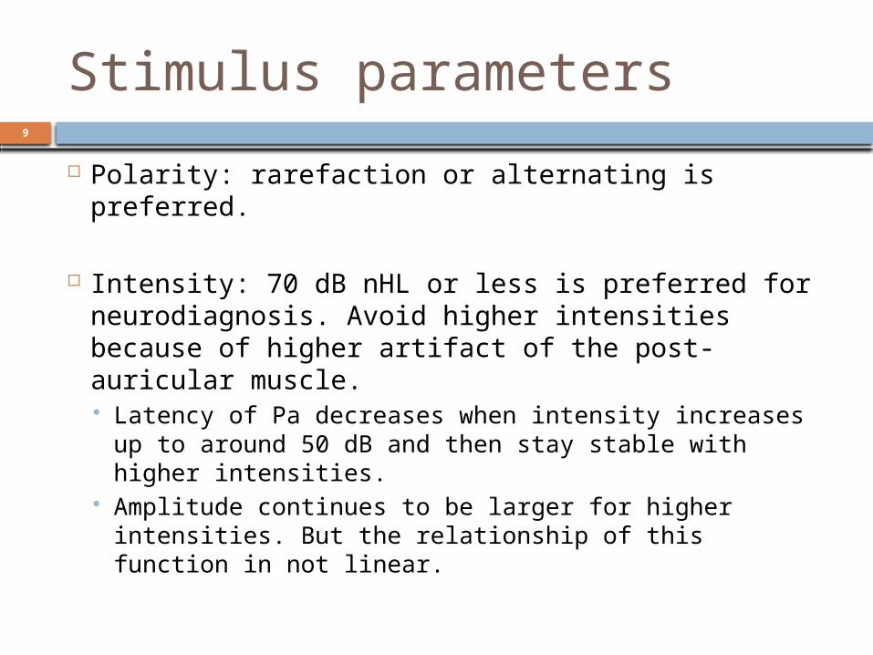
Stimulus parameters
Polarity: rarefaction or alternating is preferred.
Intensity: 70 dB nHL or less is preferred for neurodiagnosis. Avoid higher intensities because of higher artifact of the post-auricular muscle. Latency of Pa decreases when intensity increases up
to around 50 dB and then stay stable with higher intensities.
Amplitude continues to be larger for higher intensities. But the relationship of this function in not linear.

10
Stimulus parameters
Rate: rate slower than 7.1/sec is preferred. For infants even slower rate of 1/sec or 0.5/sec may be necessary to successfully record AMLR. Why? Pa amplitude remains stable for rates of 1-15/sec in
normal adults. Pb amplitude get reduced significantly when rate is
increased. Maximum Pb amplitude in some studies found to be
with a rate of 0.125/sec. Waves latency is significantly shorter for slower rates. As we go from peripheral to central, the refractory
period of the AP becomes longer. Accordingly longer inter-stimulus intervals (ISI) is required.

11
Stimulus parameters
Stimulus paradigm: Unlike short latency AEPs in which a single stimulus
(such as click) is presented at regular intervals, in AMLR several paradigms can be used for different functions. Like: Pairs of click (double click paradigm) that can be gated in
different ways. It can be simply done by presenting two identical clicks or TBs of
the same frequency. The first stimulus (S1) is followed relatively soon after by a second
(S2) stimulus (less than 500 msec) and then a longer interval (8-10 msec) separates the stimulus pair from the subsequent pairs.
This paradigm is used to study the amplitude of Pb. The ability of the brain to inhibit or habituate to the irrelevant (S2)
is reflected in Pb amplitude decrease. Used to study patients with schizophrenia or dementia.

12
Stimulus parameters
Stimulus paradigm: Odd ball paradigm: two stimuli that are
different in frequency or amplitude. One of them is frequent and the other is rare. Pb amplitude is increased.
Polarity: is not so critical as in early AEP.

13
Stimulus parameters
Monaural versus binaural stimulation:
The amplitude of the AMLR is larger with binaural stimulation but is less the total sum of the two monaural stimulation.
Pa latency is longer in binaural.
Right versus left ear: Essentially there are no major differences. Pb tends to be larger and more detected for
the right ear stimulation.

14
Recording parameters
Analysis time: although most AMLR components usually occur between 10 and 50 msec, an analysis time of a 100 msec is chosen.
Some times the late peak Pb might be delayed.

15
Recording parameters
Electrodes: Traditionally, AMLR is recorded using single channel like
ABR montage.
Recently, however, more than one channel of recording is suggested for neurodiagnosis purposes.
Since part of the AMLR response comes from the primary auditory area in the temporal lobes, the midline recording may be not sufficient to give good idea about that structure
Instead, several channels by placing noninverting electrodes over C3 and C4 temporal-parietal region.

16
Recording parameters
Electrodes:
For example if there is a lesion in the parietal lobe, the midline recording might miss it.
Recording simultaneously more than one channel usually give more information when using for neurodiagnosis.

17
Recording parameters
Filters: For the high pass setting it is suggested to use 10
-15 Hz. If higher cutoff (50 or more) frequency is used some
time Pb peak disappears suggesting that later AMLR components are of low frequency.
Very low cutoff frequency (less than 5 Hz) is not desirable because the Na-Pa response might not be detected.
Low pass setting: If simultaneously recorded with ABR then 1500 Hz
cutoff. If alone, a cutoff of 200 Hz is used.

18
Recording parameters
Notch filter: never used because it removes important information from the AMLR.

19
Analysis and interpretation
Normal AMLR:
Na-Pa-Nb-Pb complex. Peaks are less sharp (rounded) as compared with
early AEP. Watch for this configuration. Are all of these peaks available? Latency of these waves is less important than in
early AEP. Amplitude is more important than latency. Amplitude seem to be more sensitive to dysfunction
as we move centrally. In CNS.

20
Analysis and interpretation
Amplitude calculation is usually done from baseline- positive peak and baseline-negative peak.

21
Analysis and interpretation
How to say this normal or not?
It is not an easy task.
There is considerable inter-subjects variation.
As well as many nonpathologic factors affects AMLR waveform.
There is no normal values of the amplitude agreed upon.
Recording parameters are crucial and can make big change in AMLR waves, amplitude and configuration.

22
Analysis and interpretation
But generally speaking rather than comparing amplitude between subjects, comparing the amplitude of the different channels of the same subject might be more accurate.
Studies found that decrease of amplitude of the Pa amplitude for 50% or more in one channel as compared to other channels might be related to auditory dysfunction of the thalamo-cortical region.

23
Analysis and interpretation
The amplitude ratio also has been found to be a good indicator of auditory function when using double stimuli or odd ball paradigms.
So the Pb from S2 tends to be smaller that that of S1. So S2/S1 amplitude ratio is less than 1.0 if the auditory pathway is normal.
With odd ball paradigm, the deviant (rare) stimulus produces larger Pb amplitude so S2/S1 amplitude ratio tends to be larger than 1.0 for normal auditory pathway.

24
Nonpathologic factors
Age: AMLR can be recorded in infants with Na is
most prominent component. But it is not always possible to record AMLR
from infants. The probability of recording AMLR increases with age up to 10 years.
Latency of AMLR components increases in geriatrics population.

25
Nonpathologic factors
Gender: Slightly longer waves latency in males.
Sleep: Can be recorded in adults in REM sleep only
but not in deep sleep. More difficult to be recorded in children
during sleep.

26
Nonpathologic factors
Myogenic artifact: Seriously affects the recording of AMLR
especially the post auricular muscle (PAM) and the jaw muscles.
Sedation: Highly affects AMLR recording. Light sedation: AMLR might be present but with
significant amplitude decrease and poor morphology.
Anesthesia: suppresses AMLR.

27
Nonpathologic factors
Attention: Repetitive presentation of the same stimulus leads to
diminish of the AMLR amplitude especially Pb.
It is not due to neural fatigue as in ABR, rather it is due to automatic inhibition or irrelevant sensory input or sensory gating.
In odd ball paradigm this phenomenon is opposite, amplitude of Pb increase as a result to the rare stimulus.
Our brains hate routine……….looking for some thing new!

28
Nonpathologic factors
Body temperature:
AMLR latency decrease with hyperthermia but surprisingly amplitude is reduced as well.

29
Nonpathologic factors
Drugs:
Sedatives of different kinds affects on AMLR.
Neuromuscular blockers: AMLR can be successfully recorded.
Psychotherapeutic agents: affects mainly the late components like Pb.

30
Nonpathologic factors
Nicotine: Increases Na-Pa amplitude. This effect is more in males than in
females.
Acupuncture: Polarity of the P0-Na flipped. Amplitude of Na-Pa-Nb-Pb increased
significantly.

31
Clinical applications of AMLR
Hearing thresholds estimation: Pa can be detected to around 10 dB of behavioral threshold. Provides the following advantages over ABR:
Pa component is larger that wave V of ABR, which requires less averaging due to the high S/N ratio.
AMLR can be successfully recorded to long tone bursts, which gives more frequency specificity.
Disadvantages: Can not be effectively recorded during sleep or
sedation.

32
Clinical applications of AMLR
Neurodiagnosis: several studies documented changes in AMLR for different CNS lesions.
Brain tumors: severely distort or eliminate waves of AMLR.
Stroke affecting temporal lobe: reduces or eliminates Pa wave.
Alzheimer’s disease: some reported smaller Pb amplitude.

33
Clinical applications of AMLR
Auditory processing disorders: Increased latency and reduced amplitude of Na-
Pa. Autism: no significant changes.
Down syndrome: longer latency for Na.
Parkinson’s disease: delayed Pb.
Stuttering: abnormalities in Pb.

34
Clinical applications of AMLR
Was used to monitor depth of anesthesia:
As the patient moves from being conscious to being unconscious AMLR amplitude decreases or disappears especially Pb.