Atypical emotion recognition from bodies is associated with...
37
1 Accepted in Journal of Experimental Psychology: Human Perception and Performance, 19 th January, 2018 Atypical emotion recognition from bodies is associated with perceptual difficulties in healthy aging James Chard 1 , Rosanna Edey 1 , Daniel Yon 1 , Jennifer Murphy 2 , Geoffrey Bird 3 and Clare Press 1 1 Department of Psychological Sciences, Birkbeck, University of London 2 MRC Social, Genetic and Developmental Psychiatry Centre, Institute of Psychiatry, Psychology and Neuroscience, King’s College London 3 Department of Experimental Psychology, University of Oxford Corresponding author: [email protected] Department of Psychological Sciences, Birkbeck, University of London, Malet Street, London, United Kingdom WC1E 7HX Word count = 5500
Transcript of Atypical emotion recognition from bodies is associated with...

1
Accepted in Journal of Experimental Psychology: Human Perception and Performance, 19th
January, 2018
Atypical emotion recognition from bodies is associated with perceptual
difficulties in healthy aging
James Chard 1, Rosanna Edey 1, Daniel Yon 1, Jennifer Murphy 2, Geoffrey Bird 3 and Clare
Press 1
1 Department of Psychological Sciences, Birkbeck, University of London
2 MRC Social, Genetic and Developmental Psychiatry Centre, Institute of Psychiatry,
Psychology and Neuroscience, King’s College London
3 Department of Experimental Psychology, University of Oxford
Corresponding author: [email protected]
Department of Psychological Sciences, Birkbeck, University of London, Malet Street, London,
United Kingdom WC1E 7HX
Word count = 5500

2
Abstract
A range of processes are required for recognizing others’ affective states. It is particularly
important that we process the perceptual cues providing information about these states.
These experiments tested the hypothesis that difficulties with affective state identification in
older adults (OAs) arise, at least partly, from deficits in perceptual processing. To this end we
presented ‘point light display’ whole body stimuli to healthy OAs and comparison younger
adults (YAs) in three signal detection experiments. We examined the ability of OAs to
recognize visual bodily information – posture and kinematics – and whether impaired
recognition of affective states can be explained by deficits in processing these cues. OAs
exhibited reduced sensitivity to postural cues (Experiment 1) but not to kinematic cues
(Experiment 2) in affectively-neutral stimuli. Importantly, they also exhibited reduced
sensitivity only to affective states conveyed predominantly through posture (Experiment 3) –
i.e., the cue they were impaired in perceiving. These findings highlight how affective state
identification difficulties in OAs may arise from problems in perceptual processing, and
demonstrate more widely how it is essential to consider the contribution of perceptual
processes to emotion recognition.
Keywords: Emotion recognition; vision; body perception; healthy aging

3
Public significance statement
This study demonstrates that perceptual impairments in healthy aging contribute to
difficulties recognizing others’ emotional state from the way that they move. For instance, if
older adults cannot perceive accurately that another’s limbs are relaxed, they cannot use this
information to determine that they are feeling happy rather than tense. These findings
highlight how it is essential to consider the contribution of perceptual processes when
theorizing about emotion recognition, both in healthy aging and other populations.

4
1. Introduction
A range of processes are required for recognizing the affective states of others (Happé, Cook
& Bird, 2017), many of which are directly involved in identifying that a certain hidden ‘internal’
state (e.g., anger) was the driving force behind another individual’s observed behavior.
However, it is also particularly important that we process the perceptual cues providing
information about these states. A variety of cues provide this information, including the
lexical content and intonation of our speech, our facial expressions and our body language –
both our posture and the kinematics of our movements. For example, perception of relaxed
limbs can signal happiness, while perception of fast, jerky movements can signal anger
(Wallbott, 1998; Montepare et al., 1999; Dael et al., 2012). If we are insensitive to a certain
perceptual cue (e.g., relaxed limbs in another) we will be unable to use this information to
determine another’s internal state, and to use this state attribution for effective social
understanding and communication.
Older adults (OAs) exhibit impairments in recognizing affective states from facial expressions
(Calder et al., 2003; MacPherson et al., 2006; Keightly et al., 2006; Kessels et al., 2014) and
whole-body movements (Montepare et al., 1999; Ruffman et al., 2009; Spencer et al., 2016),
which are thought to result in a cascade of problems in social understanding and
communication and hence exacerbate social difficulties associated with isolation (Happe et
al., 1998; Shankar et al., 2011; Luo et al., 2012). These difficulties with emotion recognition
are hypothesized (e.g., Sullivan & Ruffman, 2004; Ruffman et al., 2008) to arise from
neurophysiological changes in the ‘social brain’ – involving regions such as the orbitofrontal
cortex, cingulate cortex and amygdala – i.e., the network implicated in the ‘accurate
perception of the dispositions and intentions of other individuals’ (p. 367; Brothers, 2002). It
5
appears to follow from this account (e.g., Ruffman et al., 2008) that problems with social
cognition are caused directly by problems in post-perceptual mechanisms for computing
internal states.
However, at least some of the age-related deficits in emotion recognition may result not from
post-perceptual processes but from changes in perceptual processing. Alongside anatomical
changes that influence perceptual processing – such as a decline in the senescent optics of
the eye (Elliott et al., 2009), thinning of retinal nerve fiber (Parikh et al., 2007) and cortical
changes in visual regions (Brewer & Barton, 2014) – aging is associated also with more
cognitive perceptual changes such as difficulties with ‘configural’ sensory processing,
requiring integration across ‘local-level’ features. For instance, OAs exhibit smaller ‘global
precedence’ effects, such that the speed advantage typically observed in recognizing the
global form of objects in comparison with local features is reduced in OAs (e.g., Oken et al.,
1999; Slavin et al., 2002; Lux et al., 2008; Insch et al., 2012).
The present experiments examined the ability of OAs to recognize visual information that is
critical for emotion recognition from body movements – specifically posture and kinematics
– and whether impaired recognition of affective states can be explained, at least partly, by
deficits in processing these cues. We used a signal detection paradigm, allowing dissociation
of signal sensitivity from response biases (Kingdom & Prins, 2010), in contrast with previous
studies of emotion recognition in OAs which have typically used accuracy measures. All
experiments presented point light displays (PLDs) where major joints of the human body are
represented by a point of light against a uniform background (Johansson, 1973). These
displays are widely used in the study of body perception because they allow presentation of
kinematic and postural information while removing other cues such as facial expressions.

6
Experiment 1 required participants to detect a postural feature of the stimuli, and Experiment
2 a kinematic feature. Experiment 3 required detection of affective states, presenting
affective states that have been found to rely differentially on postural and kinematic
information. We predicted that OAs would predominantly exhibit deficits in detecting those
affective states conveyed through cues they were impaired in perceiving, which would
suggest that impairments in perceptual processes may account, at least partly, for
atypicalities in emotion recognition.
2. Experiment 1
Experiment 1 required participants to detect a postural feature of the PLD walker. They were
asked to report whether one arm (e.g., right) of the walker was flexed at a more acute angle
at the elbow than the other (e.g., left). To do so, participants needed to assess the position of
the dot representing the wrist on one side of the body relative to those representing other
body parts – particularly the elbow and shoulder on the same side, and the equivalent body
parts on the other side of the body. We presented ‘Postural Difference’ trials, where the
described difference was present and ‘No Postural Difference’ trials where it was not (i.e.,
both elbows were equally flexed). Although the PLDs were in motion in Experiment 1, the
manipulation did not affect other aspects of implied movement so, for example, walking
speed was the same for Postural Difference and No Postural Difference trials.
Across all experiments, sensitivity to probed stimuli was calculated as d’, which indicates the
extent to which participants are more likely to report the presence of a probed stimulus when
it is present than when it is absent, i.e., the difference between the z-scores of the hit rate
(HR; proportion of Postural Difference trials correctly identified) and false alarm rate (FAR;

7
proportion of No Postural Difference trials wrongly identified as Postural Difference trials;
; p. 153; Kingdom & Prins, 2010). We report results in relation to
sensitivity below (response bias findings are presented in Supplementary Materials).
2.1 Methods
2.1.1 Participants
Two groups participated in Experiment 1, 30 YAs aged 35 or under (M = 27.5, SD = 4.7, 21
females) and 27 OAs aged 60 or older (M = 73.5, SD = 7.5, 17 females). One OA was excluded
from analysis due to a large negative d’, making the participant a statistical outlier and
indicating confusion over task demands. The sample size was determined in all experiments
reported in this manuscript such that we would have at least 80% power to detect a medium-
sized group x condition interaction effect (ηp2 = 0.06, alpha = 0.05), in line with previous
studies of emotion recognition (Ruffman et al., 2008; the number of participants undertaking
all three experiments was also past this threshold). This requirement led to the calculation
that we would require at least 24 in each group to detect effects. Note that in all experiments
we report more than 24 in each group because we tested all who responded to our
recruitment drive within a specified time-frame.
In Experiment 1, as in all experiments reported in this paper, participants had normal or
corrected-to-normal vision according to self-report. The experiments were carried out in
accordance with the ethical standards laid down in the 1964 Declaration of Helsinki and
approved by the Birkbeck, University of London Ethics Committee.
We obtained Weschler Abbreviated Scale of Intelligence (WASI) scores for two subtests
(matrix reasoning and vocabulary) for 26 OAs and 28 YAs in Experiment 1. Raw OA scores (M

8
= 70.5, SD = 7.1; FSIQ2 equivalent = 128.3) did not differ significantly from raw YA scores (M
= 71.9, SD = 5.4, FSIQ2 equivalent = 122.71): t(52) = 0.84, p = 0.41.
2.1.2 Stimuli
In Experiment 1, and in other experiments reported in this paper, stimuli were PLD videos
adapted from those developed by Nakaerts et al. (2012; see also Edey et al., 2017).
Experiment 1 used PLD videos of two actors (one male, one female) in affectively-neutral
states shown from two different viewpoints (coronal [0°] or intermediate to coronal and
sagittal [45°]) and played at a rate of 40 frames per second (mean velocity = 3.91 pixels/frame
[SD = 1.69]; mean acceleration = 1.30 pixels/frame2 [SD = .21]). All videos in Experiment 1
were two second clips and, in No Postural Difference trials, the angle of flexion at the elbow
was equivalent for right and left arms.
Postural Difference trials adapted the videos such that the average angle of flexion at the
elbow of one arm was greater than the other arm. For each frame of each video, the angle
was calculated between the elbow and wrist, and elbow and shoulder. Coordinates for a
revised wrist position were then established based on rotating its position relative to the
elbow by a proportion of the original angle. This manipulation maintained the appearance of
a natural arm swing in that the precise angles of flexion at both elbows varied systematically
across the video, but generated a more acute angle between the points representing the
wrist, elbow, and shoulder in one arm than the other. Fig. 1 illustrates the difference between
Postural Difference and No Postural Difference trials by giving three equivalent example
frames from Experiment 1 (see also Supplementary Videos). Two versions of each Postural
1 Raw scores provide a more appropriate comparison in the present context of comparing between age groups because FSIQ2 scores are normalized by age group, marginally increasing the score for the older participants.

9
Difference video were produced, differing in the extent of arm flexion and therefore signal
strength. Small signal videos reduced the apparent angle between the shoulder, elbow and
wrist by 10%, and large signal videos by 15%, over the course of the video.
Fig. 1 – Example frames from videos used in Experiment 1 (frames 1, 21, and 34) in the No Postural Difference (top) and Postural Difference (bottom) conditions. Color and lines are used to highlight the arm position at equivalent frames; in the Postural Difference stimulus the red arm on the right of the image is flexed at a more acute angle than the yellow arm on average across the video. In the actual videos, all PLDs were white on black, without connecting lines. The question presented in this example was, ‘Was the arm on the right of the screen more bent?’

10
This combination of two actors, two viewing angles, left and right arm flexion versions, and
large and small postural signals, generated 16 Postural Difference videos. There were four No
Postural Difference videos, corresponding to the two actors and two viewing angles.
2.1.3 Procedure
In Experiment 1, and all other experiments reported in this paper, participants were seated
in a dimly lit room at an approximate distance of 40 cm from a 24 inch LCD computer monitor
(resolution = 1920 x 1200 pixels; refresh rate = 60 Hz). The experiments were conducted in
MATLAB® using the Cogent graphics toolbox.
On each trial, participants were shown a PLD video and then asked either ‘Was the arm on
the right of the screen more bent?’ or, ‘Was the arm on the left of the screen more bent?’.
Participants did not know which of the two questions they would be asked during the stimulus
presentation. Participants responded ‘yes’ or ‘no’ using left and right keys, respectively.
Participants were shown their answer, and prompted to change their response or press a key
to continue. Participants saw no videos containing a signal other than the probed target
signal, e.g., where the arm on the right was flexed to a greater extent but the left arm was
probed.
Trials were presented to each participant in two blocks of 56. Within each block, each Postural
Difference video was presented twice, and each No Postural Difference video was presented
six times, resulting in 32 Postural Difference trials and 24 No Postural Difference trials (see
Supplementary Materials for a discussion of methodological decisions with respect to trial
numbers). Presentation order was fully randomized within each block.

11
2.2 Results and discussion
One sample t-tests confirmed that d’ was significantly positive for both YAs (M = 0.87, SD =
0.45; t(29) = 10.58, p < 0.001) and OAs (M = 0.62, SD = 0.32; t(26) = 10.07, p < 0.001) indicating
that both groups were able to distinguish Postural Difference from No Postural Difference
trials. We conducted a mixed ANOVA on the d’ data with size of the postural signal (large or
small based on the extent of implied arm flexion) as a within-participants factor, and age
group as a between-participants factor (see Supplementary Materials for tables of descriptive
and inferential data; the findings in all three experiments did not interact with the block or
the question asked so analyses are reported collapsed across these factors). Unsurprisingly,
there was a main effect of size of the postural signal, confirming that the signal was harder to
detect when the extent of implied arm flexion was lower (F(1,55) = 44.10, p < 0.001, ηp2 =
0.45). Importantly, there was also a main effect of age group, with YAs more sensitive to
differences in posture than OAs (F(1,55) = 6.30, p = 0.01, ηp2 = 0.10; see Fig. 2A). There was no
interaction between the size of postural signal and age group (F(1,55) = 1.86, p = 0.18).
These findings therefore demonstrated that OAs were less sensitive than YAs to postural body
features.

12
Fig. 2 – Mean sensitivity (d’) in YAs and OAs in the three experiments. Error bars represent ± 1 SE of the mean and individual points represent performance for each participant. (A) Experiment 1. Sensitivity to postural signal. (B) Experiment 2. Sensitivity to kinematic signal. (C) Experiment 3. Sensitivity to anger, sadness and happiness.
3. Experiment 2
The findings from Experiment 1 demonstrate that OAs exhibited lower sensitivity to postural
body features. Reduced performance in Experiment 1 is unlikely to be due to a decline in
intellectual capabilities (Salthouse, 2005; Salthouse, 2012) – WASI scores were matched
between OAs and YAs and the OA impairment was numerically smaller in the more

13
demanding version of the experimental task (see Fig. 2). However, as already noted, OAs
exhibit reduced functioning in several aspects of visual processing and it is important to
ascertain the specificity of the effect, especially given that the visual acuity was assessed
simply according to self-report. We therefore designed Experiment 2 such that task demands
were broadly similar to Experiment 1, but participants were required to detect whether the
velocity of the walker in the PLD increased or decreased across the time-course of the video.
Participants thereby identified a kinematic feature of the stimuli, rather than a postural
feature.
3.1 Methods
3.1.1 Participants
Two groups participated in Experiment 2, 39 YAs aged 35 or under (M = 27.5, SD = 4.7 years,
26 females) and 39 OAs aged 60 or older (M = 70.7, SD = 6.9 years, 26 females).
We obtained WASI scores for 27 OAs and 28 YAs in Experiment 2. Raw OA scores (M = 70.4,
SD = 6.9; FSIQ2 equivalent = 128.0) did not differ significantly from raw YA scores (M = 71.5,
SD = 4.9, FSIQ2 equivalent = 122.3; t(53) = 0.67, p = 0.50).
3.1.2 Stimuli
No Kinematic Difference trials presented unadapted videos identical to those presented as
No Postural Difference trials in Experiment 1. In Kinematic Difference trials the same PLD
videos were manipulated so that the velocity of the PLD figure either steadily increased or
decreased during the second half of the video, while leaving posture unchanged. To generate
the appearance of a gradual change in velocity, the coordinates of each point in each frame
in the second half were recalculated according to a power function such that they appeared
14
increasingly ahead of (or behind) the original while remaining on the same trajectory (see
Supplementary Videos; also note that it has been suggested that acceleration cannot be
directly detected over short time periods [Brouwer et al., 2002] and therefore participants
may in fact use velocity information as the basis for discriminations, but our hypotheses do
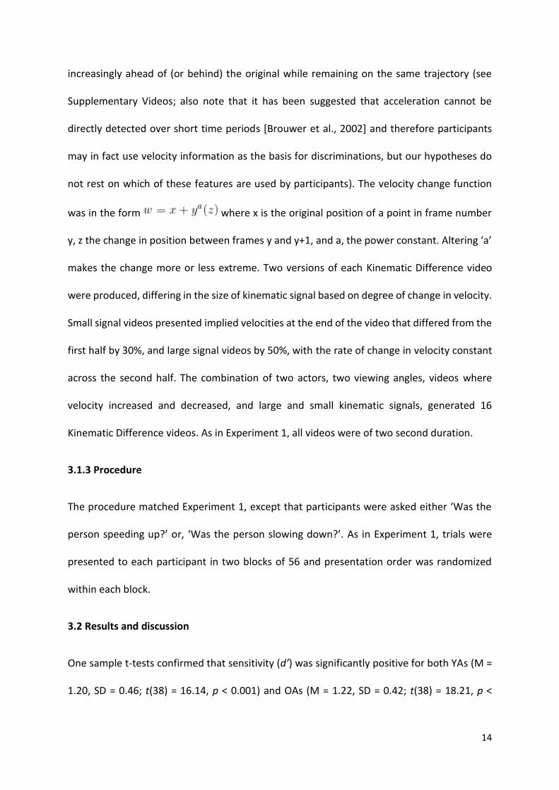
not rest on which of these features are used by participants). The velocity change function
was in the form where x is the original position of a point in frame number
y, z the change in position between frames y and y+1, and a, the power constant. Altering ‘a’
makes the change more or less extreme. Two versions of each Kinematic Difference video
were produced, differing in the size of kinematic signal based on degree of change in velocity.
Small signal videos presented implied velocities at the end of the video that differed from the
first half by 30%, and large signal videos by 50%, with the rate of change in velocity constant
across the second half. The combination of two actors, two viewing angles, videos where
velocity increased and decreased, and large and small kinematic signals, generated 16
Kinematic Difference videos. As in Experiment 1, all videos were of two second duration.
3.1.3 Procedure
The procedure matched Experiment 1, except that participants were asked either ‘Was the
person speeding up?’ or, ‘Was the person slowing down?’. As in Experiment 1, trials were
presented to each participant in two blocks of 56 and presentation order was randomized
within each block.
3.2 Results and discussion
One sample t-tests confirmed that sensitivity (d’) was significantly positive for both YAs (M =
1.20, SD = 0.46; t(38) = 16.14, p < 0.001) and OAs (M = 1.22, SD = 0.42; t(38) = 18.21, p <
15
0.001), indicating that both groups were able to distinguish Kinematic Difference from No
Kinematic Difference trials. We conducted a mixed ANOVA on d’ with size of kinematic signal
(large or small based on extent of implied change in velocity) as a within-participants factor,
and age group as a between-participants factor (see Supplementary Materials for tables of
descriptive and inferential data). Unsurprisingly, there was a main effect of size of kinematic
signal, confirming that participants were more sensitive to the signal when there was greater
velocity change (F(1,76) = 274.27, p < 0.001, ηp2 = 0.78). Importantly, there was no significant
main effect of age group, with OAs and YAs exhibiting equivalent sensitivity towards changes
in velocity (F(1,76) = 0.03, p = 0.87; see Fig. 2B). The interaction between age group and size
of kinematic signal was also not significant (F(1,76) = 2.39, p = 0.13).
To assess whether the non-significant difference between OA and YA sensitivity towards
changes in velocity reflected the absence of an effect rather than a lack of statistical power,
we calculated a Bayes Factor (BF01), representing the ratio of evidence for the null model over
evidence for the alternative model. BF01 > 3 has been assumed to provide good evidence to
support the null (Jeffreys, 1939; Lee & Wagenmakers, 2014). Conducting a Bayesian
independent samples t-test in JASP (Love et al., 2015) revealed evidence for the null
hypothesis over the alternative that OAs and YAs have differential sensitivity to changes in
velocity (BF01 = 4.18). Experiment 2 therefore demonstrates equivalent performance between
OAs and YAs with similar stimuli and task requirements to Experiment 1, but in a task that
required detection of a kinematic rather than postural cue.
16
4. Experiment 3
Based on the findings in Experiments 1 and 2, we hypothesized that OAs would exhibit
impairments in detecting affective states conveyed primarily through postural information
but would be relatively preserved in detecting those conveyed primarily through kinematics.
In other words, they would exhibit impairments when detecting affective states conveyed
through the cues that they have relative difficulty perceiving. This hypothesis was examined
in Experiment 3 where we studied the ability of OAs to recognize happy, angry and sad
affective states from PLDs.
Previous studies have indicated that the identification of some affective states relies more
heavily on kinematic cues such as velocity and acceleration whereas others can be identified
more easily from postural information. The specific pattern of these dependencies will likely
differ depending upon the stimulus set – and certainly also between bodily and facial cues –
but previous work in YAs has revealed much about the sources of information observers use
to make affective state judgments in the present stimulus set. Edey et al. (2017) found that
velocity cues were of greater importance when detecting anger (rapid, jerky movement) and
sadness (slow, sluggish movement) than when detecting happiness in these stimuli, given that
judgments were influenced to a greater extent by removal of the cues (see also Barliya et al.,
2013; and note that variation in acceleration tracked the variation in velocity). Additionally,
when the kinematic cues were removed from these stimuli leaving only postural cues,
participants detected happiness more readily than anger or sadness (happiness relative to
sadness [t(86) = 2.8, p = 0.006] and anger [t(86) = 3.6, p = 0.001]), suggesting that happiness
detection in these stimuli relied more upon postural features than anger or sadness detection.
We therefore predicted based on Experiments 1 and 2 that OAs would exhibit impaired
17
detection of happiness due to deficient posture processing, and relatively intact detection of
anger and sadness, due to intact kinematic processing.
4.1 Methods
4.1.1 Participants
Two groups participated in Experiment 3, 46 YAs aged 35 or under (M = 27.7, SD = 4.8 years,
32 females) and 37 OAs aged 60 or older (M = 71.8, SD = 7.2 years, 23 females). The results of
three OAs were excluded because d’s could not be calculated due to 100% false alarm rates
or 0% hit rates in at least one condition2.
We obtained WASI scores for 25 OAs and 30 YAs in Experiment 3. Raw OA scores (M = 70.8,
SD = 6.9; FSIQ2 equivalent = 129.0) did not differ significantly from raw YA scores (M = 71.1,
SD = 6.8, FSIQ2 equivalent = 121.5; t(53) = 0.18, p = 0.86).
To ensure groups were matched for other traits that may be associated with deficits in
emotion recognition, we also obtained scores on the Toronto Alexithymia Scale (TAS-20) and
the Beck Depression Inventory (BDI) for 25 OAs and 18 YAs. Scores did not differ according to
age group in relation to the TAS-20 (OA M = 45.20, SD = 8.80; YA M = 45.39, SD = 7.19; t(41)
= 0.08, p = 0.94) or BDI (OA M = 7.80, SD = 5.09; YA M = 8.72, SD = 6.44; t(41) = 0.52, p = 0.60).
4.1.2 Stimuli
Affectively Neutral trials presented the same four PLDs used in Experiments 1 and 2. Affective
State trials presented other stimuli from the same original set (Nackaerts et al., 2012) but
where the actors conveyed happiness, sadness, or anger. The sad PLD moved with low
2 Of those excluded, all three had 100% false alarm rates in the happy condition, and one of these also had 0% hit rates in sad and angry conditions (i.e. classified PLDs as happy at every opportunity, and never classified PLDs as sad or angry).

18
velocity and acceleration, taking fewer steps per second than the affectively-neutral walker
(sad: mean velocity = 2.03 pixels/frame [SD = .73], mean acceleration = .73 pixels/frame2 [SD
= .09]; neutral: mean velocity = 3.91 pixels/frame [SD = 1.69], mean acceleration = 1.30
pixels/frame2 [SD = .21]). In contrast, the happy (mean velocity = 5.91 pixels/frame [SD =
2.54]; mean acceleration = 1.99 pixels/frame2 [SD = .40]) and angry walkers (mean velocity =
6.97 [SD = 2.87]; mean acceleration = 2.49 pixels/frame2 [SD = .37]) both moved with higher
velocity and acceleration, but where the difference relative to affectively neutral walkers was
especially exaggerated in the angry PLD (see Supplementary Videos). Half of the videos were
trimmed to equalize step cycle (two cycles) and half to equalize duration (two seconds).
This combination of two actors, two viewing angles, and equalization by duration and step
cycle, generated eight Affective State videos per affective state, while there were four
Affectively Neutral videos.
4.1.3 Procedure
The procedure matched Experiments 1 and 2, except that participants were asked to consider
which affective state, if any, was conveyed in the PLD. They were told that this state could be
angry, sad, happy, or none of these. After each video, participants were asked either ‘Was the
person happy?’, ‘Was the person sad?’ or ‘Was the person angry?’. Giving participants a two
alternative forced choice task departed from typical emotion recognition studies in the
literature because we aimed to isolate sensitivity from response biases, and such a design is
recommended for orthogonalisation (see Yeshurun et al., 2008).
Trials were presented to each participant in two blocks of 84 PLD videos (16 Affective State
and 12 Affectively Neutral trials per affective state) and presentation order was randomized
within each block, so participants were not aware when watching a video which affective

19
state would be probed. Like in Experiments 1 and 2 participants saw no videos containing a
signal other than the target signal, for example trials in which the person was happy and they
were asked whether they were angry.
4.2 Results and discussion
One sample t-tests confirmed that d’ was significantly positive for both YAs and OAs for all
three affective states tested, indicating that both groups were able to distinguish Affective
State from Affectively Neutral trials (all ts > 2.66, all ps < 0.007). We carried out a mixed
ANOVA on the d’ data, with target affective state (happy, sad, or angry) as the within-
participants factor and age group as the between-participants factor (see Supplementary
Materials for tables of descriptive and inferential data). Greenhouse-Geisser corrections were
applied where appropriate. There was a significant main effect of affective state (F(2,162) =
138.06, p < 0.001, ηp2 = 0.63), with participants across age groups being most sensitive in the
sad condition and least sensitive in the happy condition. Importantly, this main effect was
qualified by a significant interaction between affective state and age group (F(2,162) = 9.62,
p = 0.001, ηp2 = 0.11). Follow-up tests indicated that OAs were significantly less sensitive to
happiness than the YAs (t(81) = 3.05, p = 0.003), equally sensitive to anger (t(81)= -1.18, p =
0.24) and, interestingly, more sensitive to sadness (t(81) = -2.20, p = 0.03; see Fig. 2C).
Although, at a group level, d’s were significantly positive for both groups in all three
conditions, the happy condition was most difficult for both groups and some participants had
negative d’ (in addition to the exclusions noted above, 7 YAs and 12 OAs fell into this
category). Since all participants with negative d’ in the happy condition had significantly
positive d’ in the sad and angry conditions, it is unlikely that these arose from confusion over
the task instructions. However, it is possible that, while some of those with negative d’ were
20
insensitive to informative visual cues, others may have been sensitive to the cues but
categorized neutral PLDs as happy and vice versa. However, even excluding all 19 participants
with negative d’s (all in the happy condition), there remained a significant interaction
between age group and target affective state (F(2,124) = 4.62, p = 0.02, ηp2 = 0.07), and follow-
up tests indicated OAs remained significantly less sensitive to happy PLDs than YAs (t(62) =
2.31, p = 0.02).
To summarize, OAs were impaired in detecting those affective states thought to be conveyed
predominantly through the cues they were shown to be impaired in perceiving in Experiments
1 and 2 (posture; i.e., happiness) but not in detecting those conveyed primarily through the
cues they were shown to process similarly to YAs (kinematics; i.e., sadness and anger).
5. Cross-experiment comparisons
By design, most of our participants completed all three experiments. This subset included 29
YAs (M = 27.3, SD = 4.7 years, 21 females) and 24 OAs (M = 74.8, SD = 6.8 years, 14 females).
Among this subset, the patterns of significance (both main effects and interactions) were the
same as with the full samples, and there was a task (i.e., Experiment) by age group interaction
(see Supplementary Materials). Additionally, data from these participants enabled us to carry
out partial correlations to verify the assumptions underlying Experiment 3. These correlation
analyses verified that within our dataset, happiness perception relied more heavily upon
postural than kinematic cues, and anger and sadness perception more heavily upon kinematic
cues. However, we note that these comparisons are relative rather than absolute given that
our study was not optimally powered for detecting such correlational effects and therefore
21
null effects should be treated with caution. The details of these analyses are provided in the
Supplementary Materials.
6. General discussion
The present experiments tested the hypothesis that perceptual disturbances may contribute
to OAs’ deficits in emotion recognition, using ‘point light display’ body movement stimuli. The
data demonstrated difficulty processing postural cues in OAs relative to YAs (Experiment 1),
alongside intact processing of kinematic cues (Experiment 2). In support of our hypothesis,
the OAs also exhibited difficulty recognizing only the affective state (happiness) conveyed
predominantly through the cue towards which Experiment 1 had demonstrated them to be
impaired in processing (posture; Experiment 3).
These findings are therefore consistent with the hypothesis that difficulties in recognizing
affective states in OAs relate to reduced sensitivity to the perceptual cues signaling those
states. In fact, not only were the emotion recognition deficits larger for those emotions
predominantly conveyed by perceptual cues they were impaired in processing, they were
absent for emotions predominantly conveyed by intact perceptual cues. This pattern may
appear inconsistent with the popular hypothesis that deterioration in the ‘social brain’ –
involving the orbitofrontal cortex, cingulate cortex and amygdala – is responsible for broad
deficits in emotion recognition in OAs (e.g., Ruffman et al., 2008). Given that this is the
network implicated in the ‘accurate perception of the dispositions and intentions of other
individuals’ (p. 367; Brothers, 2002), it appears to follow from a strong version of the ‘social
brain’ account that problems with emotion recognition are caused directly by problems in

22
post-perceptual mechanisms for computing internal states. Under this interpretation, this
specific pattern of impairments in Experiment 3 would not have been predicted. However,
given that the account is somewhat underspecified at the cognitive level, it is also possible
that one could use the present findings to flesh out the account and suggest that the ‘social
brain’ deteriorates due to reduced perceptual input across age.
One could speculate that the deficit OAs show in postural processing is related to difficulties
with visual configural processing. For instance, OAs exhibit smaller ‘global precedence’
effects, such that the speed advantage typically observed in recognizing the global form of
objects in comparison with local features is reduced in OAs (Oken et al., 1999; Slavin et al.,
2002; Lux et al., 2008; Insch et al., 2012; see also Murray et al., 2010; Slessor et al., 2012;
Spencer et al., 2016). Postural information requires computing the relative position of
effectors – in the case of Experiment 1, the position of the dot representing the wrist on one
side of the body relative to those representing other body parts – and therefore deficits
processing configural information would yield posture perception difficulties. Although
perception of kinematic features may often require configural processing, the task presented
in Experiment 2 likely did not. Specifically, participants could perform the required judgment
by focusing on any single point on an arm or leg. Therefore, under this hypothesis, the present
findings would indicate that deficits in perceiving posture will typically be found in OAs
because the nature of this cue is typically configural, but problems in perceiving kinematics
may depend upon whether the kinematic feature required configural processing (see Di
Domenico et al., 2015; Grainger et al., 2017). Possible reasons for age-related changes in
configural processing include narrowing of the attentional field based on retinal deterioration
(Kosslyn et al., 1998) and changes in patterns of hemispheric asymmetry (Dolcos et al., 2002).

23
Future research should further examine the functional and neural basis of the sensory deficit
generating this pattern across postural and kinematic cues. It could also consider how far
effects generalize to more naturalistic environments than those provided by sparse PLDs
(although it is of note that previous studies have indicated OAs do not have difficulties in
interpreting PLDs per se, including those conveying emotional information – see Ruffman et
al., 2009).
Our findings highlight a methodological issue in relation to previous literature suggesting
relatively emotion-general deficits in recognition from facial, vocal and bodily cues (e.g.,
Ruffman et al., 2008; Insch et al., 2012; Spencer et al., 2016; note that the previous literature
has typically found intact recognition of disgust). This literature has not allowed for a specific
assessment of sensitivity to the signal, with the majority of studies requiring participants to
label the affective state presented, from multiple response options, and calculating the
percentage accuracy. These procedures allow it to be inferred that individuals have difficulties
in correctly labeling emotions, but cannot determine whether these difficulties reflect poor
signal sensitivity or response biases (see Isaacowitz et al., 2007, for a discussion of this issue).
For instance, several studies have indicated intact performance for happiness recognition
and, given a possible ‘positivity bias’ in OAs (Carstensen & Mikels, 2005), it is particularly
important to dissociate sensitivity from bias effects in this context. However, future work
must examine whether OA deficits in sensitivity to affective states are only determined by
impaired perception of cues towards those states, or whether there are other contributing
factors.
More broadly, these findings highlight how difficulties in perceptual processing can generate
problems in emotion recognition. Although the requirement for perceptual processing in
24
emotion recognition is widely acknowledged, its specific contribution to patterns in
processing is often neglected, with studies considering only broad visual acuity via self-report
both in aging studies and those examining other populations. For instance, difficulties in
emotion recognition in autism may not stem from lower empathy, as has classically been
assumed (Baron-Cohen, 2009), but rather perceptual atypicalities (Brewer et al., 2016; see
also Cracco et al., 2016; Hayes et al., 2015). In line with the present findings, it has also
recently been found that individuals with developmental prosopagnosia – a deficit
hypothesized by some to result from atypical configural perceptual processing (Avidan et al.,
2011) – also have emotion recognition difficulties (Biotti & Cook, 2016).
In conclusion, the present findings suggest that difficulties in recognizing affective states from
bodily cues in OAs may be related to difficulties in perceiving the perceptual cues signaling
those states. These findings demonstrate more widely how it is essential to consider the
contribution of perceptual processes to emotion recognition.
7. Acknowledgments
CP was funded by Leverhulme Trust (RPG-2016-105) and Wellcome Trust (204770/Z/16/Z;
which also funded RE) grants and GB by a project grant from the Baily Thomas Charitable
Fund. DY and JM were supported by doctoral studentships from the Economic and Social
Research Council [1599941; ES/J500057/1].

25
References
Avidan, G., Tanzer, M., & Behrmann, M. (2011). Impaired holistic processing in congenital
prosopagnosia. Neuropsychologia, 49(9), 2541-2552.
Barliya, A., Omlor, L., Giese, M. A., Berthoz, A., & Flash, T. (2013). Expression of emotion in
the kinematics of locomotion. Experimental Brain Research, 225(2), 159-176.
Baron‐Cohen, S. (2009). Autism: the empathizing–systemizing (E‐S) theory. Annals of the New
York Academy of Sciences, 1156(1), 68-80.
Biotti, F., & Cook, R. (2016). Impaired perception of facial emotion in developmental
prosopagnosia. Cortex, 81, 126-136.
Brewer, A. A., & Barton, B. (2014). Visual cortex in aging and Alzheimer's disease: changes in
visual field maps and population receptive fields. Frontiers in Psychology, 5, 74.
Brewer, R., Biotti, F., Catmur, C., Press, C., Happe, F., Cook, R., & Bird, G. (2016). Can
neurotypical individuals read autistic facial expressions? Atypical production of emotional
facial expressions in autism spectrum disorders. Autism Research, 9, 262-271.
Brothers, L. (2002). The social brain: A project for integrating primate behavior and
neurophysiology in a new domain. In Foundations in Social Neuroscience, ed. JT Cacioppo, GG
Berntson, R Adolphs, CS Carter, RJ Davidson, MK McClintock, BS McEwen, MJ Meaney, DL
Schacter, EM Sternberg, SS Suomi, SE Taylor, pp. 367-385. Cambridge, MA: MIT Press.
Brouwer, A. M., Brenner, E., & Smeets, J. B. (2002). Perception of acceleration with short
presentation times: Can acceleration be used in interception? Perception &
Psychophysics, 64(7), 1160-1168.
Calder, A. J., Keane, J., Manly, T., Sprengelmeyer, R., Scott, S., Nimmo-Smith, I., & Young, A.
W. (2003). Facial expression recognition across the adult life span. Neuropsychologia, 41(2),
195-202.
Carstensen, L. L., & Mikels, J. A. (2005). At the intersection of emotion and cognition aging
and the positivity effect. Current Directions in Psychological Science, 14(3), 117-121.

26
Cracco, E., De Coster, L., Andres, M., & Brass, M. (2015). Motor simulation beyond the dyad:
Automatic imitation of multiple actors. Journal of Experimental Psychology: Human
Perception and Performance, 41, 1488-1501.
Dael, N., Mortillaro, M., & Scherer, K. R. (2012). Emotion expression in body action and
posture. Emotion, 12(5), 1085-1101.
Di Domenico, A., Palumbo, R., Mammarella, N., & Fairfield, B. (2015). Aging and emotional
expressions: is there a positivity bias during dynamic emotion recognition? Frontiers in
Psychology, 6, 1130.
Dolcos, F., Rice, H. J., & Cabeza, R. (2002). Hemispheric asymmetry and aging: right
hemisphere decline or asymmetry reduction. Neuroscience & Biobehavioral Reviews, 26(7),
819-825.
Edey, R., Yon, D., Cook, J., Dumontheil, I., & Press, C. (2017). Our own action kinematics
predict the perceived affective states of others. Journal of Experimental Psychology: Human
Perception and Performance, 43(7), 1263-1268.
Elliott, S. L., Choi, S. S., Doble, N., Hardy, J. L., Evans, J. W., & Werner, J. S. (2009). Role of high-
order aberrations in senescent changes in spatial vision. Journal of Vision, 9(2), 1-16.
Grainger, S.A., Henry, J.D., Phillips, L.H., Vanman, E.J., Allen, R. (2017). Age deficits in facial
affect recognition: The influence of dynamic cues. The Journals of Gerontology Series B, 72,
622-632.
Happé, F. G., Cook. J.L., & Bird, G. (2017). The structure of social cognition: In(ter)dependence
of sociocognitive processes. Annual Review of Psychology, 68, 243-267.
Happé, F. G., Winner, E., & Brownell, H. (1998). The getting of wisdom: Theory of mind in old
age. Developmental Psychology, 34(2), 358-362.
Hayes, S.J., Andrew, M., Elliott, D., Gowen, E., & Bennett, S.J. (2016). Low fidelity imitation of
atypical biological kinematics in autism spectrum disorders is modulated by self-generated
selective attention. Journal of Autism and Developmental Disorders, 46, 502-513.

27
Insch, P. M., Bull, R., Phillips, L. H., Allen, R., & Slessor, G. (2012). Adult aging, processing style,
and the perception of biological motion. Experimental Aging Research, 38(2), 169-185.
Isaacowitz, D.M., et al. (2007). Age differences in recognition of emotion in lexical stimuli and
facial expressions. Psychology and Aging, 22(1), 147-159.
Jeffreys, H. (1939). Theory of probability. Oxford, UK: The Clarendon Press.
Johansson, G. (1973). Visual perception of biological motion and a model for its
analysis. Perception & Psychophysics, 14(2), 201-211.
Kessels, R. P., Montagne, B., Hendriks, A. W., Perrett, D. I., & Haan, E. H. (2014). Assessment
of perception of morphed facial expressions using the Emotion Recognition Task: Normative
data from healthy participants aged 8–75. Journal of Neuropsychology, 8(1), 75-93.
Keightley, M. L., Winocur, G., Burianova, H., Hongwanishkul, D., & Grady, C. L. (2006). Age
effects on social cognition: faces tell a different story. Psychology and Aging, 21(3), 558-572.
Kingdom, F. A. A., & Prins, N. (2010). Psychophysics: a practical introduction. London, UK:
Elsevier.
Kosslyn, S. M., Brown, H. D., & Dror, I. E. (1999). Aging and the scope of visual
attention. Gerontology, 45(2), 102-109.
Lee, M. D., & Wagenmakers, E.-J. (2014). Bayesian cognitive modeling: A practical course.
Cambridge, UK: Cambridge University Press.
Love, J., Selker, R., Marsman, M., Jamil, T., Verhagen, A. J., Ly, A., et al. (2015). JASP (Version
0.7.1.12) [Computer software]
Luo, Y., Hawkley, L. C., Waite, L. J., & Cacioppo, J. T. (2012). Loneliness, health, and mortality
in old age: A national longitudinal study. Social Science & Medicine, 74(6), 907-914.
Lux, S., Marshall, J. C., Thimm, M., & Fink, G. R. (2008). Differential processing of hierarchical
visual stimuli in young and older healthy adults: Implications for pathology. Cortex, 44(1), 21-
28.

28
MacPherson, S. E., Phillips, L. H., & Delia Sala, S. (2006). Age-related differences in the ability
to perceive sad facial expressions. Aging Clinical and Experimental Research, 18(5), 418-424.
Montepare, J., Koff, E., Zaitchik, D., & Albert, M. (1999). The use of body movements and
gestures as cues to emotions in younger and older adults. Journal of Nonverbal
Behavior, 23(2), 133-152.
Murray, J. E., Halberstadt, J., & Ruffman, T. (2010). The face of aging: Sensitivity to facial
feature relations changes with age. Psychology and Aging, 25(4), 846-850.
Nackaerts, E., Wagemans, J., Helsen, W., Swinnen, S. P., Wenderoth, N., & Alaerts, K. (2012).
Recognizing biological motion and emotions from point-light displays in autism spectrum
disorders. PloS One, 7(9), e44473.
Navon, D. (1977). Forest before trees: The precedence of global features in visual
perception. Cognitive Psychology, 9(3), 353-383.
Oken, B. S., Kishiyama, S. S., Kaye, J. A., & Jones, D. E. (1999). Age-related differences in global-
local processing: stability of laterality differences but disproportionate impairment in global
processing. Journal of Geriatric Psychiatry and Neurology, 12(2), 76-81.
Parikh, R. S., Parikh, S. R., Sekhar, G. C., Prabakaran, S., Babu, J. G., & Thomas, R. (2007).
Normal age-related decay of retinal nerve fiber layer thickness. Ophthalmology, 114(5), 921-
926.
Press, C. (2011). Action observation and robotic agents: Learning and anthropomorphism.
Neuroscience and Biobehavioral Reviews, 35, 1410-1418.
Ruffman, T., Henry, J. D., Livingstone, V., & Phillips, L. H. (2008). A meta-analytic review of
emotion recognition and aging: Implications for neuropsychological models of
aging. Neuroscience & Biobehavioral Reviews, 32(4), 863-881.
Ruffman, T., Sullivan, S., & Dittrich, W. (2009). Older adults’ recognition of bodily and auditory
expressions of emotion. Psychology and Aging, 24(3), 614-622.
Salthouse, T. A. (2005). Relations between cognitive abilities and measures of executive
functioning. Neuropsychology, 19(4), 532-545.
29
Salthouse, T. (2012). Consequences of age-related cognitive declines. Annual Review of
Psychology, 63, 201-226.
Shankar, A., McMunn, A., Banks, J., & Steptoe, A. (2011). Loneliness, social isolation, and
behavioral and biological health indicators in older adults. Health Psychology, 30(4), 377-385.
Slavin, M. J., Mattingley, J. B., Bradshaw, J. L., & Storey, E. (2002). Local–global processing in
Alzheimer’s disease: an examination of interference, inhibition and
priming. Neuropsychologia, 40(8), 1173-1186.
Slessor, G., Riby, D. M., & Finnerty, A. N. (2012). Age-related differences in processing face
configuration: The importance of the eye region. Journals of Gerontology Series B:
Psychological Sciences and Social Sciences, 68(2), 228-231.
Spencer, J. M., Sekuler, A. B., Bennett, P. J., Giese, M. A., & Pilz, K. S. (2016). Effects of aging
on identifying emotions conveyed by point-light walkers. Psychology and Aging, 31(1), 126-
138.
Sullivan, S., & Ruffman, T. (2004). Emotion recognition deficits in the elderly. International
Journal of Neuroscience, 114, 403-432.
Wallbott, H. G. (1998). Bodily expression of emotion. European Journal of Social
Psychology, 28(6), 879-896.
Yeshurun, Y., Carrasco, M., & Maloney, L.T. (2008). Bias and sensitive in two-interval forced
choice procedures: Tests of the difference model. Vision Research, 48, 1837-1851.

30
Atypical emotion recognition from bodies is associated with perceptual
difficulties in healthy aging: Supplementary Materials
James Chard, Rosanna Edey, Daniel Yon, Jennifer Murphy, Geoffrey Bird and Clare Press
Bias (c) results for Experiments 1-3
Our hypotheses all pertained to sensitivity, where it is important to dissociate possible
influences of response bias (c, the extent to which participants report the presence of the
probed stimulus regardless of its objective presence, i.e., the inverse of the mean of the z-
scores of HR and FAR; ). It should be noted that since there
were unequal numbers of trials where the target difference was present and where it was
absent, caution is needed in interpreting absolute values for c (Wyart et al., 2012; Terman &
Terman, 1972; note that, in contrast, unequal ratios are not generally deemed a problem
when interpreting d’ measures; see Swets et al., 1961; Sherman et al., 2015). That is, c would
be negative rather than zero for a rational observer because there were more trials where it
was correct to answer in the affirmative. The same ratio (4:3) was used in the three
experiments, however, meaning the extent to which a bias would be rational was the same
across all three.
In Experiments 1 and 2, c did not differ (Experiment 1: t(52) = 1.60, p = 0.12; Experiment 2:
t(76) = 0.18, p = 0.86) between OAs (Experiment 1: M = 0.02, SD = 0.52; Experiment 2: M =
0.46, SD = 0.55) and YAs (Experiment 1: M = 0.22, SD = 0.41; Experiment 2: M = 0.48, SD =
0.54) In Experiment 3, a mixed ANOVA on the c data, with target affective state (happy, sad,
or angry) as the within-participants factor and age group as the between-participants factor,

31
indicated a significant main effect of affective state (F(2,162) = 23.69, p < 0.001, ηp2 = 0.23).
One-sample t-tests confirmed that participants had a tendency towards answering ‘yes’ to
the question ‘Was the person happy?’ (overall M = -0.586; OA M = -0.80, SD = 0.82; YA M = -
0.40, SD = 0.86; t(82) = -6.06, p < 0.001), while there was a trend in the opposite direction in
the angry condition (overall M = 0.18; OA M = 0.02, SD = 0.97; YA M = 0.30, SD = 0.73; ; t(82)
= 1.88, p = 0.06), and there was no sign of a bias in the sad condition (overall M = -0.04; OA M
= -0.06, SD = 0.81; YA M = -0.02, SD = 0.80; t(82) = -0.42, p = 0.67). There was also a trend for
OAs to be more likely than YAs to respond in the affirmative across all three target affective
states (F(1,81) = 3.57, p = 0.062), but no interaction between affective state and age group
(F(2, 162) = 1.29, p = 0.27).This trend of OAs to answer ‘yes’ to any question may be deemed
consistent with previous suggestions of positivity biases in OAs (van Reekum et al., 2010),
depending upon exactly how such an account was characterized, and demonstrates the
importance of dissociating sensitivity from biases.
Interactions with number of testing sessions and blocks
Some of the participants who undertook more than one experiment (i.e., the majority)
undertook them in separate sessions (12 OAs and 23 YAs) while others undertook them all in
the same session (12 OAs and 6 YAs). Regardless of whether experiments were undertaken
separately or together, participants always undertook Experiment 3 first, followed by
Experiment 2 and finally Experiment 1. This order was chosen because it was deemed that
undertaking Experiment 1 or 2 (asking directly about posture or kinematic cues) before 3
(emotion recognition) could direct participants towards which cues were of likely relevance
for detecting affective states and hence alter judgments. Similarly, it was deemed that
undertaking a task which required computing cues configurally (Experiment 1) could

32
subsequently encourage participants to compute other cues configurally even when
unnecessary (Experiment 2; see General discussion). To examine whether greater fatigue
effects in OAs may have contributed to the observed deficits in Experiment 1, we carried out
an ANOVA with age group and number of sessions as between-participants factors. There was
no main effect of number of sessions on d’ in Experiment 1 (F(1, 49) = 0.08, p = 0.78) and nor,
more importantly, was there an interaction between number of sessions and age group
(F(1,49) = 0.006, p = 0.94).
In all experiments, trials were divided into two blocks, with the same trials appearing in a
random order within each block. In no experiment was there an interaction between block
and age group (Experiment 1 – F(1,49) = 0.08, p = 0.78; Experiment 2 – F(1,76) = 0.74, p = 0.39;
Experiment 3 – F(1,81) < 0.01, p = 0.96). For participants who undertook all three experiments
there was also no three-way interaction between experiment, block, and age group: F(2,51)
= 0.80, p = 0.45. These analyses therefore demonstrated no evidence that effects change
throughout the course of each experiment.
Cross-experiment comparisons
Providing further evidence that the cue type (posture versus kinematics) drives a difference
in age-related impairments, a mixed ANOVA including only participants who had taken part
in Experiments 1 and 2, and with Experiment (1 or 2) as a within-participants factor,
demonstrated an interaction between Experiment and age group on d’ values (F(1,54) = 5.05,
p = 0.03, ηp2 = 0.09).
The results in Experiment 3 were consistent with our hypothesis, based upon Experiments 1
and 2, that OAs would exhibit reduced sensitivity to affective states conveyed predominantly

33
through postural cues, and be relatively preserved in detecting those conveyed primarily
through kinematics. Our hypotheses rested upon assumptions from our previous study (Edey
et al., 2017) that recognizing happiness relies upon postural cues in this stimulus set more
than kinematic cues, and anger and sadness detection rely more upon kinematic cues.
However, our hypothesis can also be verified with the present dataset by asking how
individual differences in the experiments relate to each other, and therefore disregarding the
need for these assumptions. Specifically, individual differences in the posture task should
predict individual differences in detecting affective states dependent upon postural cues,
whereas individual differences in the kinematics task should predict those in detecting states
dependent upon the kinematic cues (importantly, when controlling for age group to ensure
that these analyses are not circular with respect to any reported group effects).
To this end, we carried out partial correlations between performance in each of Experiments
1 and 2 and detection of the three separate affective states in Experiment 3, controlling for
age group. Kinematic d’ was significantly related to d’s for both angry PLDs (r =0.31, p = 0.02,
95% CI [0.05, 0.53]; see Fig. 3) and sad PLDs (r = 0.31, p = 0.03, 95% CI [0.05, 0.53]), but not
happy PLDs (r= 0.06, p = 0.68, 95% CI [-0.21, 0.32]). Conversely, posture d’ was related to d’
for happy PLDs (r =0.31, p = 0.02, 95% CI [0.09, 0.50]) but not angry PLDs (r = 0.11, p = 0.45,
95% CI [-0.12, 0.32]) or sad PLDs (r = 0.05, p = 0.71, 95% CI [-0.18, 0.28]). The same patterns
of significance were also found in correlation analyses which did not control for age group,
and in partial correlations controlling both for age group and sensitivity in the affectively-
neutral task thought to be less related (e.g., controlling for kinematic d’ when examining the
relationship between posture and happiness detection). These analyses thereby confirm that

34
OAs are indeed impaired in recognizing the affective state relying predominantly upon
posture information in the present stimulus set – i.e., the cue they are impaired in perceiving.
Fig. 3 - Scatter plots illustrating the relationship between sensitivity to sadness, anger and happiness (Experiment 3) and sensitivity to posture (Experiment 1) and kinematics (Experiment 2). Significant predictors are shown by solid lines, and non-significant predictors by broken lines.

35
Tables of descriptive and inferential statistics for Experiments 1-3
Table 1: Mean (SD) d’s in each condition and group for Experiments 1-3
Group
Older adults Younger adults
Full samples
Exp 1: Large postural signal 0.70 (0.34) 1.01 (0.55)
Exp 1: Small postural signal 0.54 (0.31) 0.76 (0.41)
Exp 2: Large kinematic signal 1.56 (0.60) 1.56 (0.61)
Exp 2: Small kinematic signal 0.99 (0.36) 0.91 (0.40)
Exp 3: Angry PLDs 1.74 (0.69) 1.57 (0.61)
Exp 3: Sad PLDs 2.22 (0.71) 1.91 (0.58)
Exp 3: Happy PLDs 0.28 (0.64) 0.76 (0.77)
Only including participants undertaking all three experiments
Exp 1: Large postural signal 0.66 (0.34) 1.03 (0.55)
Exp 1: Small postural signal 0.50 (0.30) 0.78 (0.41)
Exp 2: Large kinematic signal 1.53 (0.50) 1.64 (0.65)
Exp 2: Small kinematic signal 0.95 (0.31) 0.94 (0.42)
Exp 3: Angry PLDs 1.65 (0.75) 1.53 (0.61)
Exp 3: Sad PLDs 2.09 (0.78) 1.88 (0.56)
Exp 3: Happy PLDs 0.27 (0.55) 0.74 (0.78)
Table 2. ANOVA results with full samples for Experiments 1-3
Factor(s) df F-value P ηp2
Exp 1: Signal size x age 1,55 1.86 0.18 0.03
Exp 1: Signal size 1,55 44.10 < 0.001 0.45
Exp 1: Age 1,55 6.30 0.01 0.10
Exp 2: Signal size x age 1,76 2.39 0.13 0.03
Exp 2: Signal size 1,76 274.27 < 0.001 0.78
Exp 2: Age 1,76 0.03 0.87 < 0.01
Exp 3: Affective state x age 2,162 9.62 0.001 0.11
Exp 3: Affective state 2,162 138.06 < 0.001 0.63
Exp 3: Age 2,162 < 0.01 > 0.99 < 0.01
Table 3. ANOVAs results for Experiments 1-3 including only those participants taking part
in all three experiments
Factor(s) df F-value P ηp2
Exp 1: Signal size x age 1,51 1.94 0.17 0.04
Exp 1: Signal size 1,51 36.50 < 0.001 0.42
Exp 1: Age 1,51 8.43 0.01 0.14
Exp 2: Signal size x age 1,51 1.68 0.20 0.03

36
Exp 2: Signal size 1,51 189.79 < 0.001 0.79
Exp 2: Age 1,51 0.15 0.70 < 0.01
Exp 3: Affective state x age 2,102 4.46 0.02 0.08
Exp 3: Affective state 2,102 76.31 < 0.001 0.60
Exp 3: Age 2,102 0.12 0.73 < 0.01

37
References
Edey, R., Yon, D., Cook, J., Dumontheil, I., & Press, C. (2017). Our own action kinematics
predict the perceived affective states of others. Journal of Experimental Psychology: Human
Perception and Performance, 43(7), 1263-1268.
Sherman, M.T., Seth, A.K., Barnett, A.B., & Kanai, R. (2015). Prior expectations facilitate
metacognition for perceptual decision. Consciousness and Cognition, 35, 53-65.
Swets, J.A., Tanner, W.P., & Birdsall, T.G. (1961). Decision processes in perception.
Psychological Review, 68(5), 301-340.
Terman, M., & Terman, J. S. (1972). Concurrent variation of response bias and sensitivity in
an operant-psychophysical test. Perception & Psychophysics, 11(6), 428-432.
Van Reekum, C. M., Schaefer, S. M., Lapate, R. C., Norris, C. J., Greischar, L. L., & Davidson, R.
J. (2010). Aging is associated with positive responding to neutral information but reduced
recovery from negative information. Social Cognitive and Affective Neuroscience, 6(2), 177-
185.
Wyart, V., Nobre, A. C., & Summerfield, C. (2012). Dissociable prior influences of signal
probability and relevance on visual contrast sensitivity. Proceedings of the National Academy
of Sciences, 109(9), 3593-3598.