Attenuated Prostaglandin Formation Peroxisomal-deficient...
10
Attenuated Prostaglandin Formation in Peroxisomal-deficient Human Skin Fibroblasts Joel A. Gordon,* Lorna J. Wamock, * and Arthur A. Spectort Departments of *Internal Medicine and tBiochemistry, University of Iowa College ofMedicine, Iowa City, Iowa 52242 Abstract Peroxisomal-deficient skin fibroblasts from patients with Zell- weger's syndrome or infantile Refsum's disease produced fewer prostaglandins than normal skin fibroblasts. Radioimmunoas- say indicated a 45-55% decrease in prostaglandin E2 (PGE2) production when Zellweger's fibroblasts were incubated with arachidonic acid. This deficiency was not overcome by pretreat- ment of the Zellweger's fibroblasts with media containing ara- chidonic acid, and it was not due to channeling of arachidonic acid into other eicosanoid products. Modifications in the perox- ide tone of the Zellweger's fibroblasts by addition of H202 or catalase failed to increase PGE2 production. Using Northern analysis, we were unable to detect an mRNA transcript for PGH synthase in unstimulated Zellweger fibroblasts but iden- tified a 4.2-kb mRNA transcript after treatment with phorbol myristate acetate (PMA). Treatment for 6 h with 10 nM PMA raised PGE2 production in normal and Zellweger fibroblasts to equivalent levels. These increases were prevented by addition of H-7, staurosporine, cycloheximide, or actinomycin D. Our findings suggest that the reduced PGE2 production in peroxiso- mal deficient fibroblasts is due to a decrease in PGH synthase mRNA. The reduction in PGH synthase can be overcome by treatment of the cells with agents which enhance gene expres- sion. (J. Clin. Invest. 1993. 91:169-178.) Key words: phorbol ester* prostaglandins * protein kinase * Zellweger Introduction Peroxisomes are single membrane organelles 0.2-1.0 ,um in diameter that are ubiquitous in eukaryotic cells ( 1). These or- ganelles carry out a number of important biological functions. They contain catalase and thereby compartmentalize the de- toxification of intracellular H202 (2), fl-oxidize long chain and very long chain fatty acids (3), synthesize the alkyl ether bond of plasmalogens (4), produce bile acids (5), catabolize phy- tanic acid (6) and pipecolic acid (7), and are responsible for the chain-shortening of hydroxyeicosatetraenoic acids (8). The prototypical peroxisomal deficiency in humans has been desig- Portions ofthis work were presented at the annual meeting ofthe Amer- ican Society of Biochemistry and Molecular Biology, New Orleans, LA, 6 June 1990 and published in abstract form ( 1990. FASEB J. 4:A2137). Address reprint requests to Dr. Joel A. Gordon, Department of Internal Medicine, E 300 G GH, University of Iowa College of Medi- cine, Iowa City, IA 52242. Receivedfor publication 4 March 1992 and in revisedform 8 Febru- ary 1993. nated the Zellweger or cerebrohepatorenal syndrome ( 1 ). This autosomal recessive disorder is characterized biochemically by the accumulation of phytanic acid and very long chain fatty acids, diminished plasmalogen synthesis, and decreased bile acid synthesis (9). Clinically, the syndrome is associated with failure to thrive, severe hypotonia, epileptic seizures, psycho- motor retardation, hepatic and renal cortical cysts, and prema- ture death in the first 6-12 mo of life (1). Liver and kidney tissue from infants with this syndrome have no demonstrable peroxisomes ( 10). Other human peroxisomal deficiencies have been described, including infantile Refsum's disease. This dis- ease, although not identical to Zellweger's syndrome, shares many of the same biochemical and clinical features (1). Prostaglandins (PGs) are important mediators of a number of critical functions in mammalian cells and tissues ( 1 1). The control of PG formation depends on the availability of arachi- donic acid and the activity of PGH synthase, a membrane- bound heme protein that has two activities, a cyclooxygenase component and a peroxidase component ( 12 ). PGH synthase converts the arachidonic acid into an endoperoxide interme- diate, PGH2. Very little free arachidonic acid is found in cells; it is contained primarily in cellular phosphoglycerides and is released through the action of calcium-dependent phospholi- pases ( 13). None of the biochemical defects in peroxisomal-deficient cells described to date suggests that PG formation is abnormal. We have observed, however, that agonist stimulated PG forma- tion in peroxisomal-deficient skin fibroblasts is markedly re- duced. The reduction in PGE2 formation is not due to lack of substrate; it appears to be due to a reduced quantity of PGH synthase protein and mRNA transcription in the peroxisomal- deficient cells. Treatment of peroxisomal deficient cells with PMA restores PGH synthase enzyme activity, apparently through a pathway involving gene transcription and transla- tion mediated by protein kinase C. Methods Cell culture. Zellweger and normal human skin fibroblasts were grown and maintained as previously described (8). The mutant skin fibro- blasts, obtained from three different individuals, were supplied by the Pediatrics Cytogenetics Laboratory at the University of Iowa College of Medicine. Infantile Refsum's disease human skin fibroblasts were ob- tained from the Human Genetic Mutant Cell Repository (Camden, NJ). The cells were grown to confluency in Eagle's MEM containing 20% FBS (Hy-Clone Laboratories, Logan, UT), supplemented with nonessential amino acids, MEM vitamin solution (Gibco, Grand Is- land, NY), glutamine, and penicillin/streptomycin 100 ,ug/ml (Gibco). Cells were harvested with trypsin diluted 1:10 and passaged into tissue culture plates with a surface area of 5 or 10 cm2/well (Tissue Culture Cluster 6 and 12; Costar, Cambridge, MA), or flasks with a surface area of 75 cm2, (Corning Glass Works, Corning, NY). Experi- ments were performed when cell cultures were 80-100% confluent. Because of variability in growth rate and availability of cells, cultures were often studied at different times and passage number. Zellweger Prostaglandin Formation in Peroxisomal-deficient Cells 169 J. Clin. Invest. © The American Society for Clinical Investigation, Inc. 0021-9738/93/07/169/10 $2.00 Volume 92, July 1993, 169-178
Transcript of Attenuated Prostaglandin Formation Peroxisomal-deficient...
Attenuated Prostaglandin Formationin Peroxisomal-deficient Human Skin FibroblastsJoel A. Gordon,* Lorna J. Wamock, * and Arthur A. SpectortDepartments of *Internal Medicine and tBiochemistry, University of Iowa College of Medicine, Iowa City, Iowa 52242
Abstract
Peroxisomal-deficient skin fibroblasts from patients with Zell-weger's syndrome or infantile Refsum's disease produced fewerprostaglandins than normal skin fibroblasts. Radioimmunoas-say indicated a 45-55% decrease in prostaglandin E2 (PGE2)production when Zellweger's fibroblasts were incubated witharachidonic acid. This deficiency was not overcome by pretreat-ment of the Zellweger's fibroblasts with media containing ara-chidonic acid, and it was not due to channeling of arachidonicacid into other eicosanoid products. Modifications in the perox-ide tone of the Zellweger's fibroblasts by addition of H202 orcatalase failed to increase PGE2 production. Using Northernanalysis, we were unable to detect an mRNAtranscript forPGHsynthase in unstimulated Zellweger fibroblasts but iden-tified a 4.2-kb mRNAtranscript after treatment with phorbolmyristate acetate (PMA). Treatment for 6 h with 10 nMPMAraised PGE2production in normal and Zellweger fibroblasts toequivalent levels. These increases were prevented by additionof H-7, staurosporine, cycloheximide, or actinomycin D. Ourfindings suggest that the reduced PGE2production in peroxiso-mal deficient fibroblasts is due to a decrease in PGHsynthasemRNA.The reduction in PGHsynthase can be overcome bytreatment of the cells with agents which enhance gene expres-sion. (J. Clin. Invest. 1993. 91:169-178.) Key words: phorbolester* prostaglandins * protein kinase * Zellweger
Introduction
Peroxisomes are single membrane organelles 0.2-1.0 ,um indiameter that are ubiquitous in eukaryotic cells ( 1). These or-ganelles carry out a number of important biological functions.They contain catalase and thereby compartmentalize the de-toxification of intracellular H202 (2), fl-oxidize long chain andvery long chain fatty acids (3), synthesize the alkyl ether bondof plasmalogens (4), produce bile acids (5), catabolize phy-tanic acid (6) and pipecolic acid (7), and are responsible forthe chain-shortening of hydroxyeicosatetraenoic acids (8). Theprototypical peroxisomal deficiency in humans has been desig-
Portions of this work were presented at the annual meeting ofthe Amer-ican Society of Biochemistry and Molecular Biology, NewOrleans, LA,6 June 1990 and published in abstract form ( 1990. FASEB J.4:A2137).
Address reprint requests to Dr. Joel A. Gordon, Department ofInternal Medicine, E 300 GGH, University of Iowa College of Medi-cine, Iowa City, IA 52242.
Receivedfor publication 4 March 1992 and in revisedform 8 Febru-ary 1993.
nated the Zellweger or cerebrohepatorenal syndrome ( 1 ). Thisautosomal recessive disorder is characterized biochemically bythe accumulation of phytanic acid and very long chain fattyacids, diminished plasmalogen synthesis, and decreased bileacid synthesis (9). Clinically, the syndrome is associated withfailure to thrive, severe hypotonia, epileptic seizures, psycho-motor retardation, hepatic and renal cortical cysts, and prema-ture death in the first 6-12 mo of life (1). Liver and kidneytissue from infants with this syndrome have no demonstrableperoxisomes ( 10). Other human peroxisomal deficiencies havebeen described, including infantile Refsum's disease. This dis-ease, although not identical to Zellweger's syndrome, sharesmany of the same biochemical and clinical features (1).
Prostaglandins (PGs) are important mediators of a numberof critical functions in mammalian cells and tissues ( 1 1). Thecontrol of PGformation depends on the availability of arachi-donic acid and the activity of PGHsynthase, a membrane-bound heme protein that has two activities, a cyclooxygenasecomponent and a peroxidase component ( 12 ). PGHsynthaseconverts the arachidonic acid into an endoperoxide interme-diate, PGH2. Very little free arachidonic acid is found in cells;it is contained primarily in cellular phosphoglycerides and isreleased through the action of calcium-dependent phospholi-pases ( 13).
None of the biochemical defects in peroxisomal-deficientcells described to date suggests that PGformation is abnormal.Wehave observed, however, that agonist stimulated PGforma-tion in peroxisomal-deficient skin fibroblasts is markedly re-duced. The reduction in PGE2 formation is not due to lack ofsubstrate; it appears to be due to a reduced quantity of PGHsynthase protein and mRNAtranscription in the peroxisomal-deficient cells. Treatment of peroxisomal deficient cells withPMA restores PGH synthase enzyme activity, apparentlythrough a pathway involving gene transcription and transla-tion mediated by protein kinase C.
Methods
Cell culture. Zellweger and normal human skin fibroblasts were grownand maintained as previously described (8). The mutant skin fibro-blasts, obtained from three different individuals, were supplied by thePediatrics Cytogenetics Laboratory at the University of Iowa College ofMedicine. Infantile Refsum's disease human skin fibroblasts were ob-tained from the Human Genetic Mutant Cell Repository (Camden,NJ). The cells were grown to confluency in Eagle's MEMcontaining20% FBS (Hy-Clone Laboratories, Logan, UT), supplemented withnonessential amino acids, MEMvitamin solution (Gibco, Grand Is-land, NY), glutamine, and penicillin/streptomycin 100 ,ug/ml(Gibco). Cells were harvested with trypsin diluted 1:10 and passagedinto tissue culture plates with a surface area of 5 or 10 cm2/well (TissueCulture Cluster 6 and 12; Costar, Cambridge, MA), or flasks with asurface area of 75 cm2, (Corning Glass Works, Corning, NY). Experi-ments were performed when cell cultures were 80-100% confluent.Because of variability in growth rate and availability of cells, cultureswere often studied at different times and passage number. Zellweger
Prostaglandin Formation in Peroxisomal-deficient Cells 169
J. Clin. Invest.© The American Society for Clinical Investigation, Inc.0021-9738/93/07/169/10 $2.00Volume 92, July 1993, 169-178
fibroblasts were studied between passages 3 and 7, infantile Refusum'sdisease fibroblasts between passages 9 and I 1, and normal human skinfibroblasts between passages 18 and 24. Cultures were maintained in atemperature- and humidity-controlled incubator (CO2 Incubator3028; Forma Scientific, Inc., Marietta, OH) at 370C with 95% air-5%CO2 as the gas phase. The DNAcontent of the cells was measuredfluorometrically ( 14).
Incubation and lipid analysis. After removal of the maintenancemedium, the cells were washed twice with Dulbecco's PBS (DPBS)'containing 0.1 MuMBSAand incubated for 20 min at 370C in 1 ml ofmodified Eagle's MEMcontaining 7.5 MM[I-'4C]arachidonic acidand 0.1 MuM BSA. After incubation, the medium was removed andplaced in a siliconized glass tube, and the pH was adjusted to 3.5 byaddition of 10 MAl of 2.4 NHCl. Lipids were extracted three times with 2vols of HPLC-grade ethyl acetate. The ethyl acetate extracts werepooled, evaporated to dryness under a stream of nitrogen, and resus-pended in 500 Ml of acetonitrile. The radioactive lipids were analyzedby, reverse-phase HPLC(8).
Fatty acid methyl esters were prepared from the cell lipids extractedinto chloroform/methanol solution (2:1, vol/vol) by incubation withboron trifluoride in methanol and acetonitrile for 30 min at 90'C.After extraction of the methyl esters with water and heptane, the sam-ples were dried under N2 and resuspended in 25 Ml of carbon disulfide,and 1 ml was injected into a model 5890 gas chromatograph (Hewlett-Packard Co., Palo Alto, CA) for separation of fatty acid methyl esters.Fatty acids were identified by comparison of the relative retentiontimes with fatty acid methyl ester standards.
PGformation. To determine PGE2production after agonist stimu-lation, the maintenance medium was removed, and the cells werewashed twice with DPBScontaining 0.1 MMBSAand then incubatedfor 20 min in 0.5 ml DPBScontaining 0.1 MMBSAand 7.5,uM arachi-donic acid. The buffer solution was removed and analyzed for PGE2production by RIA ( 15). Measurement of PGE2 production by nor-mal, infantile Refsum's disease, and Zellweger human skin fibroblastswas accomplished by mixing 100 Ml of either standards or samples with50 M1 of anti-PGE2 antibody (Anti-PGE2 antibody, Advanced Mag-netics Corp., Cambridge, MA). The mixture was incubated at 25°C for30 min, 50 Mul of [3H]PGE2 [10-15 X I03 dpm] was added, and theincubation was continued for 16 h at 4°C. Dextran-coated charcoal(400 M1) was added and after mixing and centrifugation at 3,000 g for15 min, the radioactivity in 400 Ml of the supernatant fluid was mea-sured in a liquid scintillation spectrometer. Assay detection limits were5 pmol of PGE2, and 50% inhibition was obtained with 15 pmol/mlPGE2. The assay has negligibly small amounts of cross reactivity withprostaglandins other than PGE2-
To determine if soluble factor(s) produced by either the normal orZellweger fibroblasts could affect PGE2production, conditioned me-dium from one fibroblast cell line grown for 48 h to 80% confluencywas transferred to the opposite cell line. After 4 h of incubation, theconditioned medium was removed, the cells were washed with bufferand then exposed to 7.5 MMarachidonic acid for 20 min, and themedium was assayed for PGE2by RIA.
To determine if plasmalogen deficiency was responsible for the re-duced PGE2production by the peroxisomal-deficient fibroblasts, con-fluent monolayers were incubated with modified Eagle's MEMcon-taining0. 1 MtMBSAsupplemented with 5-12.5MgM 1-0-alkyl hexadeca-glycerol (Sigma Chemical Co., St. Louis, MO) for 48 h. The mediumwas removed, and the cells were washed with buffer and then incubatedfor 20 min in 0.5 ml DPBS/0. 1 MMBSA containing 7.5 MMarachi-donic acid. The medium was removed and assayed for PGE2by RIA.
Modification of cellular peroxide tone. To determine whether theoxyradical scavenger catalase had any effect on PGE2 formation, weincubated confluent cultures of normal or peroxisomal-deficient hu-
1. Abbreviations used in this paper: DPBS, Dulbecco's PBS; H-7,[1- (5-ioquinolinesulfonyl) -2-methylpiperazine dihydrochloride ];TBS, Tris-buffered saline.
man skin fibroblasts with modified Eagle's MEMcontaining 0.1 uMBSA and 10-100 U/ml of catalase (46,500 U/mg protein, SigmaChemical Co.) for 10 min to 2 h. After removal of the catalase-supple-mented medium, the cells were washed with DPBScontaining 0.1 MBSA and then incubated for 20 min in 0.5 ml of DPBS/0. 1 MMBSAsupplemented with 7.5 MMarachidonic acid. The medium was re-moved and analyzed for PGE2by RIA.
To increase the half-life and intracellular permeability of the addedcatalase, we also tested catalase conjugated to polyethylene glycol ( 16).Normal and peroxisomal-deficient human fibroblasts were incubatedin modified Eagle's MEMcontaining 1% heat-inactivated fetal bovineserum supplemented with 0. 125,0.25, and 0.5 mg/ml catalase-polyeth-ylene glycol. After 24 h, the medium was removed, and the cells werewashed and analyzed for PGE2production by RIA.
To determine if H202 had any effect on fibroblast PGE2 produc-tion, confluent cultures of normal and peroxisomal-deficient humanskin fibroblasts were incubated with modified Eagle's MEMcontaining0.1 MuMBSA supplemented with 1-100 MMH202. After 10 min, theH202 containing medium was removed and the cells were washed andanalyzed for PGE2production by RIA.
Phorbol ester-stimulated PGE2formation. To determine whetherPMA(LC Services Corp., Woburn, MA) was capable of increasingPGE2 formation, we incubated monolayers of normal and Zellwegerfibroblasts in Eagle's MEMcontaining 5% FBS supplemented with 10nMPMAfor 1-12 h. The PMAsupplemented medium was removedand the cells were washed with buffer and then exposed to DPBS/0. 1MMBSA containing 7.5 ,M arachidonic acid. After 20 min, the buffersolution was removed and the PGE2content was measured by RIA.
To determine whether the increase in PGE2formation after PMAwas secondary to increased arachidonic acid release via enhanced phos-pholipase A2 activity, normal and Zellweger fibroblasts were labeledwith [3Hjarachidonic acid (0.5 MCi/ml) for 12 h in Eagle's MEMcon-taining 5% FBS. After the medium was removed and the cells werewashed, aliquots of the medium were counted in a liquid scintillationspectrometer to determine the amount of radioactivity taken up duringthe labelling period. Eagle's MEMcontaining 5% FBS supplementedwith either 10 nMPMAor its vehicle was then added to the cultures for12 h. At the appropriate time, 0.5 ml of medium containing the cal-cium ionophore A23 187 (final concentration 2 MM)was added to thecultures. After 20 min, the remaining medium was extracted with ethylacetate and the extract was resuspended in acetonitrile. Aliquots wereremoved and counted in the liquid scintillation spectrometer to deter-mine the amount of lipid-soluble radioactivity that was released.
To examine whether inhibitors of transcription or translationwould interfere with the effect of PMAon PGE2production, normaland Zellweger fibroblasts were incubated for 6 h in Eagle's MEMcon-taining 5% FBS with or without 10 nM PMA. Additional incubationsfor 6 h contained PMAand either 1 Mg/ml cycloheximide or 2 Mg/mlactinomycin D(both from Sigma Chemical Co.). Cultures not exposedto PMAfor 6 h but containing cyclohexamide or actinomycin Dwerealso tested. After this medium was removed, the cells were incubatedfor 20 min with 7.5 MuMarachidonic acid, and the medium was ana-lyzed for PGE2by RIA.
To determine whether the effect of PMAon PGE2production wasmediated through protein kinase C, normal and Zellweger fibroblastswere incubated for 6 h with or without PMAas indicated above. Addi-tional incubations for 6 h contained PMAand either 100 AMH-7[1-(5-isoquinoline sulfonyl)-2-methyl piperazine dihydrochloride,Seikagaku America, St. Petersburg FL] or 10 mMstaurosporine(Sigma Chemical Co.). Cultures not exposed to PMAbut containingH-7 or staurosporine were also tested. The cells then were incubatedwith 7.5 MMarachidonic acid and the medium analyzed for PGE2by RIA.
HPLC. The total radioactivity contained in a sample was deter-mined initially by adding 4 ml of Budget Solve scintillation solution(Research Products International Corp., Mount Prospect, IL) to a 25-Ml aliquot of the sample and counting in a model LS7000 liquid scintil-lation spectrometer (Beckman Instruments, Inc., Palo Alto, CA).
170 J. A. Gordon, L. J. Warnock, and A. A. Spector
Quenching was monitored with a 226Ra external standard. Additionalaliquots of the sample containing 10,000-15,000 dpmwere transferredto siliconized glass vials, dried under a stream of nitrogen, and resus-pended in 30 td of HPLC-grade acetonitrile and 70 Al of acidified H20.The lipids were separated on a model 332 chromatograph (BeckmanInstruments, Inc.) containing a 4.6 x 250-mm column (VYDAC, Hi-speria, CA) with CI8 reverse-phase 5 Asm spherical packing. The solventmixture consisted of water adjusted to pH 3.4 with phosphoric acid andan increasing acetonitrile gradient from 30% to 100% over 65 min.Radioactivity was detected by passing the column effluent mixed withBudget Solve scintillation solution through a HPLCradioactivity flowdetector (Radiomatic Flo-One Beta, Canberra Corp., Meriden CT)and analyzed using software provided by Radiomatic.
Protein immunoblotting. Protein immunoblotting was carried outaccording to the method of Towbin with slight modification ( 17).Confluent cultures of normal, infantile Refsum's disease, and Zell-weger human skin fibroblasts were grown in tissue culture flasks with75 cm2 surface area, washed, scraped twice in 3 ml of cold PBS, andplaced on ice. The cells were sedimented at 4VC at 3,000 g for 10 min.After the supernatant fluid was removed, the cell pellet was lysed in 1ml of PBS containing 1%Triton X-100 (Surfact Amps; Pierce Chemi-cal Co., Rockford, IL), 10 g/ml leupeptin, 1 mMphenylmethylsul-fonylfluoride, 1 mMEDTA, and 1 mMdiethyldithiocarbamic acid (allfrom Sigma Chemical Co.). The lysate was transferred to an Eppendorftube and immersed in boiling water for 3 min, and then sedimented ina refrigerated microfuge at 27,000 g for 10 min. The supernatant solu-tion was removed, placed in a Centricon-30 microconcentrator (Ami-con Division, W. R. Grace and Co., Danvers, MA) and concentratedby centrifugation for 30-min intervals at 5,000 g and 4°C with 340angle rotor. After the supernatant solution was concentrated to 100 Al,20 Al was removed and assayed for protein content (18). The re-mainder was kept at 4°C and assayed within 16 h.
Proteins in the concentrated cell lysates containing the solublizedPGHsynthase were separated through 12.5% SDS-PAGEaccording tothe method of Laemmli ( 19) using the Mini Protean gel system (Bio-Rad Laboratories, Richmond, CA). Sample aliquots equivalent to 100jig of protein per lane were denatured by heating to 96°C for 4 min inan SDSdissociation buffer containing f3-mercaptoethanol. To confirmseparation by SDS-PAGE, a standard mixture of 7 proteins was in-cluded with each gel (High Range Molecular Weight Standard Mix-ture, Bethesda Research Laboratory, Bethesda, MD). Proteins sepa-rated by SDS-PAGEwere electrophoretically transferred at 4°C to 0.45
im nitrocellulose paper (BA 85, Midwest Scientific, St. Louis, MO).Gels were stained with Coomassie Brilliant Blue to confirm that thetransfer efficiency was > 90% and that all samples were transferreduniformly. Nitrocellulose membranes were saturated overnight at 4°Cin Tris-buffered saline (TBS) containing 10% FBS and 2%BSA.
After saturation, the nitrocellulose membrane was washed in TBS/0.05% Tween and once in TBS for a total of 30 min. The membranewas incubated for 4 h at 25°C in 7.5 ml of TBS/2% BSA, pH 8.0,containing rabbit anti-PGH synthase antibody (Polyclonal rabbit anti-PGH synthase, Oxford Biomedical Research, Oxford, MI) diluted1:100. The nitrocellulose membrane then was washed as before andincubated for 2 h at 25°C in 7.5 ml of TBS/2% BSA, pH 8.0, contain-ing goat anti-Rabbit IgG conjugated with alkaline phosphatase (SigmaChemical Co.) diluted 1 :10,000. After washing, the nitrocellulose wastreated with 5-bromo-4-chloro-3-indoyl phosphate and nitroblue tetra-zolium (both from Sigma Chemical Co.) for color development andvisualization.
mRNAanalysis. Total RNAwas isolated from cell cultures underbasal conditions and after treatment with PMAfor 2-16 h by extrac-tion with guanidium isothiocyanate and subsequent cesium chloridecentrifugation according to the methods described by Chirgwin et al.(20) and Glisin et al. (21). 15 Ag of total RNAwas fractionated on1.5% agarose/formaldehyde gels and transferred to Genescreen Plus(NEN Research Products, Boston, MA) hybridization membrane bycapillary blotting. The RNAwas cross-linked to the membrane by ex-posure to ultraviolet light.
The membranes were hybridized to a 2.5-kb cDNAprobe for hu-man PGH synthase (Oxford Biomedical Research) after randomprime labeling with [32P]cytidine 5'-triphosphate (dCTP) according tomethods outlined by Sambrook et al. (22). The membranes werewashed twice in 0.3 Msodium chloride and 0.03 Msodium citrate atroom temperature and then exposed to Kodak XAR-5 film at -80'Cwith intensifying screens. To determine the amount of RNAloaded oneach lane, the membrane also was probed with radiolabeled f-actincDNA kindly supplied by Dr. Gary Hunninghake, University of IowaCollege of Medicine.
Reagents. [1-'4C]arachidonic acid (58.4 mCi/mmol), [5,6,8,9,11,12,14,15-3H]-arachidonic acid (110 Ci/mmol), and [5,6,8,11,12,14,15 (N)-3HJ-PGE2 (160 Ci/mmol) were purchased from Amer-sham Corp. (Arlington Heights, IL). Deoxycytidine 5'-triphosphatetetra(triethylammonium) salt [a- 32p], 3,000 Ci/ mmol, was purchasedfrom Dupont NEN Research Products. Arachidonic acid was pur-chased from Nu-Check Prep (Elysian, MN). All organic solvents, ei-ther reagent or HPLCgrade, were purchased from Fisher Scientific Co.(Fair Lawn, NJ).
Statistics. Statistical analysis was performed using a one-way AN-OVA. Statistical significance was determined at the P < 0.05 level andwas tested according to the method of Scheffe (23).
Results
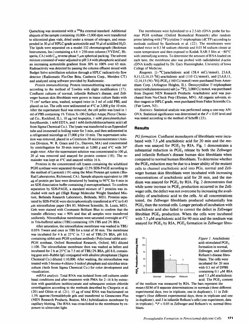
PGformation. Confluent monolayers of fibroblasts were incu-bated with 7.5 ALM arachidonic acid for 20 min and the me-dium was assayed for PGE2 by RIA. Fig. 1 demonstrates asubstantial reduction in PGE2 release by both the Zellwegerand infantile Refsum's disease human skin fibroblasts whencompared to normal human fibroblasts. To determine whetherthe PGE2reduction may be due to a lesser ability of the mutantcells to channel arachidonic acid into PGs, normal and Zell-weger human skin fibroblasts were incubated with increasingconcentrations of arachidonic acid for 20 min, and the me-dium was assayed for PGE2by RIA. Fig. 2 demonstrates thatwhile some increase in PGE2production occurred in the Zell-weger cells, the defect was not overcome by increasing the avail-ability of arachidonic acid to the cells. At all concentrationstested, the Zellweger fibroblasts produced substantially lessPGE2than the normal cells. Longer periods of incubation witharachidonic acid also failed to correct the defect in Zellwegerfibroblast PGE2 production. When the cells were incubatedwith 7.5 ,M arachidonic acid for 90 min and the medium wasassayed for PGE2by RIA, PGE2 formation in Zellweger fibro-
15- Figure 1. ArachidonicZ acid-stimulated PGE2
412- formation in normal,E°g Zellweger, and infantile
_ * Refsum's disease fibro-T6- blasts. The cells were
incubated for 20 minupX/, with 0.5 ml of DPBSUrcontaining 0.1 uM BSA
Normal Zellweger Refsum and 7.5 ALMarachidonicacid. The PGE2 content
of the medium was measured by RIA. The bars represent themean±SEMof 8 separate determinations in normals (three differentexperimental days, two in triplicate, one in duplicate), 11 in Zell-weger's (four different experimental days, three in triplicate and onein duplicate), and 3 in infantile Refsum's cells (one experiment, datain triplicate). *P < 0.05 in Zellweger and Refsum's vs. normal fibro-blasts.
Prostaglandin Formation in Peroxisomal-deficient Cells 171
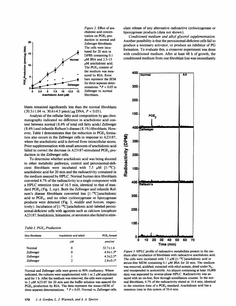
Figure 2. Effect of ara-chidonic acid concen-tration on PGE2pro-duction in normal and
20- Zellweger fibroblasts.The cells were incu-z
a bated for 20 min in15- /5 DPBScontaining 0. 1
Normal ,M BSA and 2.5-15
s10- M,uM arachidonic acid.0 1,/ The PGE2content of
the medium was mea-
2S_5 sured by RIA. Error
0.a-~ ~ ~ ~ ~ g iains ~l< .5i" , * bars represent the SEM2 t/
Zeliweer for three separate deter-O0_ minations. *P < 0.05 in
0| 2.5 5 7.5 10 12.5 15 Zellweger vs. normalArachidonic Acid (pM) fibroblasts.
blasts remained significantly less than the normal fibroblasts(20.5±1.04 vs. 30.6±4.5 pmol/,gg DNA, P < 0.05).
Analysis of the cellular fatty acid composition by gas chro-matography indicated no difference in arachidonic acid con-tent between normal (8.4% of total cell fatty acids) Zellweger(8.4%) and infantile Refsum's disease (8.1% ) fibroblasts. How-ever, Table I demonstrates that the reduction in PGE2 forma-tion also occurs in the Zellweger cells in response to A23 187,where the arachidonic acid is derived from intracellular stores.Prior supplementation with small amounts of arachidonic acidfailed to correct the decrease in A23 187-stimulated PGE2pro-duction in the Zellweger cells.
To determine whether arachidonic acid was being shuntedto other metabolic pathways, control and peroxisomal-defi-cient fibroblasts were incubated with 7.5 uM [1- 14C]-arachidonic acid for 20 min and the radioactivity contained inthe medium assayed by HPLC. Normal human skin fibroblastsconverted 4.7% of the radioactivity to a single component witha HPLCretention time of 16.5 min, identical to that of stan-dard PGE2(Fig. 3, top). Both the Zellweger and infantile Ref-sum's disease fibroblasts converted less [1-"'4C] arachidonicacid to PGE2, and no other cyclooxygenase or lipoxygenaseproducts were detected (Fig. 3, middle and bottom, respec-tively). Incubation of [1-4'4C] arachidonic acid-labeled peroxi-somal-deficient cells with agonists such as calcium ionophoreA23 187, bradykinin, histamine, or serotonin also failed to stim-
Table L PGE2Production
Skin fibroblasts Arachidonic acid added PGE2 formed
JM pmol/mI
Normal 0 32.7±1.6Zellweger 0 4.9±1.3*Zellweger 1 4.3±2.5*Zellweger 2 2.8±0. *
Normal and Zellweger cells were grown to 90% confluency. Whereindicated, the cultures were supplemented with 1 or 2 gAMarachidonicacid for 1 h. After the medium was removed, the cells were exposedto 2 ,gM A23 187 for 20 min and the buffer solution was assayed forPGE2production by RIA. The data represent the mean±SEMofthree separate determinations. * P < 0.05. Normal vs. Zellweger cells.
ulate release of any alternative radioactive cyclooxygenase orlipoxygenase products (data not shown).
Conditioned medium and alkyl glycerol supplementation.Another possibility is that the peroxisomal-deficient cells fail toproduce a necessary activator, or produce an inhibitor of PGformation. To evaluate this, a crossover experiment was donewith conditioned medium. After at least 48 h of growth, theconditioned medium from one fibroblast line was immediately
Normal AA
300.
200.
100. PGE2
400W .-.
0- 300o*0
200-
0
co8
100(:
Refsum's - _
300
200
100
0oo0 10 20 30 40 50 60 70
Time (min)
Figure 3. HPLCprofile of radioactive metabolites present in the me-
dium after incubation of fibroblasts with radioactive arachidonic acid.The cells were incubated with 7.5 AM[1- 14CIarachidonic acid inserum-free MEMcontaining 0. I MMBSA for 20 min. The mediumwas removed, acidified, extracted with ethyl acetate, dried under N2,and resuspended in acetonitrile. An aliquot containing at least 10,000dpm was separated by reverse-phase HPLC. Radioactivity was as-
sayed with an on-line, flow through scintillation counter. In the nor-
mal fibroblasts, 4.7% of the radioactivity eluted at 16.4 min, identicalto the retention time of a PGE2 standard. Arachidonic acid has a
retention time in this system of 50.4 min.
172 J. A. Gordon, L. J. Warnock, and A. A. Spector
Zellweger

transferred to the opposite cell line. Conditioned medium fromthe Zellweger fibroblasts did not inhibit normal fibroblastPGE2production. After 4 h in Zellweger-conditioned medium,PGE2 production by the normal fibroblasts increased from acontrol value of 6.36±0.06 to 9.73±0.06 pmol/,g DNA. Like-wise, conditioned medium from normal fibroblasts did notcorrect the attenuated PGE2 production in Zellweger fibro-blasts. After 4 h in conditioned medium from normal cells,PGE2production by the Zellweger cells was 3.74±0.05 pmol/,gg DNA, still significantly reduced as compared with theamount released by normal fibroblasts. Thus, the deficiency inthe Zellweger fibroblast is not due to either the production of asoluble, transferrable inhibitor, or failure to produce a trans-ferrable activator.
An alternate possibility is that by virtue of the plasmalogendeficiency that occurs in peroxisomal-deficient fibroblasts, animportant potential intracellular source of arachidonic acid forprostaglandin formation is reduced. To test this possibility,subconfluent monolayers of Zellweger fibroblasts were incu-bated for 48 h in medium supplemented with 1-0-alkyl hexa-decaglycerol. Exposure to this plasmalogen precursor did notappreciably increase arachidonic acid-stimulated PGE2 pro-duction in the Zellweger fibroblasts. PGE2production by cellsnot exposed to this compound was 1.42±0.06 pmol/,tg DNA,compared with 1.48±0.01 and 1.86±0.19 pmol/htg DNA incells exposed to 7.5 and 12.5 ,tM 1-0-alkyl hexadecaglycerol,respectively.
Alteration ofperoxide tone. Because of the peroxisomal de-ficiency, it is possible that H202 is not properly metabolized inthe Zellweger cells. For example, H202 maybuild up because ofthe aberrant localization of catalase within the cells (24), andthereby inactivate PGHsynthase. To determine if elevated per-oxide content may be a factor in inhibiting PGE2production,10-100 ,M catalase was incubated with the Zellweger cells for60-120 min, and the cells then were exposed to 7.5 ,uM arachi-donic acid for 20 min. RIA analysis revealed that this pretreat-ment with catalase did not increase Zellweger fibroblast PGE2production (data not shown). Because catalase was added tothe medium, it may have had limited access to the intracellularspace. Therefore, we attempted to more effectively increase theintracellular concentration of catalase by preincubating thecells for 24 h with 0.0 125, 0.25, and 0.5 mg/ml catalase conju-gated to polyethylene glycol. Previous work by Beckman et al.(16) suggests that this is a more efficient way of increasing the
15- Figure 4. Effect of in-creasing concentrations
a 12- of H202 on normal andZellweger fibroblast
o PGE2 production. TheE fibroblasts were incu-
Normal bated for 10 min witho 6- modified MEMcon-X \ taining 0. 1 gMBSAand
3- 5-l100 MH202. AfterW 4 removal of the medium
Zellweger and washing, the cells06 8 were incubated with 7.50 20 40 60 80 100
H202 (IJM) AMarachidonic acid for20 min and the medium
assayed for PGE2by RIA. Data points represent the mean±SEMforthree separate determinations.
20- Figure 5. Effect of in-creasing time of expo-
zZ 16 Normal sure to PMAon PGE2cm I |production by normalo and Zellweger fibro-E 12 blasts. The fibroblasts
o \ P / Zellweger were incubated for 1-12-0t 8 h with MEMcontainingS 5%heat-inactivated FBS
4- supplemented with 10o nMPMA. At the ap-IL propriate time, the me-
0 2 6 dium was removed and0 2 4 6 8 10 12 the cells were washed
Time (h)and exposed for 20 min
to DPBScontaining 0.1 MuMBSAand 7.5 MiM arachidonic acid. PGE2was measured by RIA, and each point represents the mean±SEMforthree separate determinations.
cellular catalase content and thereby lower intracellular H202concentration. However, exposure to the catalase-polyethyleneglycol preparation also did not increase PGE2production whenthe Zellweger fibroblasts were subsequently incubated with 7.5AtM arachidonic acid for 20 min (data not shown).
The alternative possibility is that because of aberrant cata-lase localization, the peroxide tone of the cell may be too low totrigger PG formation. If so, addition of H202 may overcomethis deficiency. To test this, we incubated confluent mono-layers of normal and Zellweger human skin fibroblasts with1-100 ,iM H202 for 10 min, removed this medium, and thenexposed the cells to 7.5 gM arachidonic for 20 min. Fig. 4demonstrates that PGE2formation in the Zellweger fibroblastswas not increased by prior treatment with H202. Furthermore,incubation of the normal fibroblasts with H202 resulted in adose-dependent reduction in PGE2formation. After exposureto 50 or 100 ,M H202, the amount of PGE2produced by thenormal cells was reduced to the level observed with the Zell-weger cells.
Effect ofphorbol ester. PMAhas been shown to increase PGsynthesis in many cells by increasing PGHsynthase formation(25-27) To determine if this would restore the defect in PGE2production, monolayers of normal and Zellweger fibroblastswere incubated with 10 nM PMAfor 1-12 h. After this me-dium was removed and the cells washed, they were incubatedwith DPBS/0. 1 gM BSA supplemented with 7.5 ,uM arachi-donic acid and the medium assayed by RIA. Fig. 5 demon-strates that after a 6- or 12-h incubation with PMA, PGE2for-mation in the Zellweger fibroblasts was restored to a level iden-tical to that seen in the normal human skin fibroblast treated inthe same manner.
PMA's effect to increase PGE2formation might be throughits previously described effect of increasing phospholipase A2activity and, hence, increasing the availability of arachidonicacid (28). To determine whether this might be the mechanismof the PMAeffect, monolayers of normal and Zellweger fibro-blasts were labelled with [3H] arachidonic acid for 12 h. Thismedium was removed and the cells were incubated with 5%FBS supplemented with 10 nM PMAfor an additional 12 h.A23187 at a final concentration of 2 ,uM was added to thecultures for 20 min, and the release of arachidonic acid wasdetermined. Table II indicates that treatment with PMAinboth the normal and Zellweger fibroblasts failed to augmentthe release of radioactivity from the pulse-labeled cells.
Prostaglandin Formation in Peroxisomal-deficient Cells 173
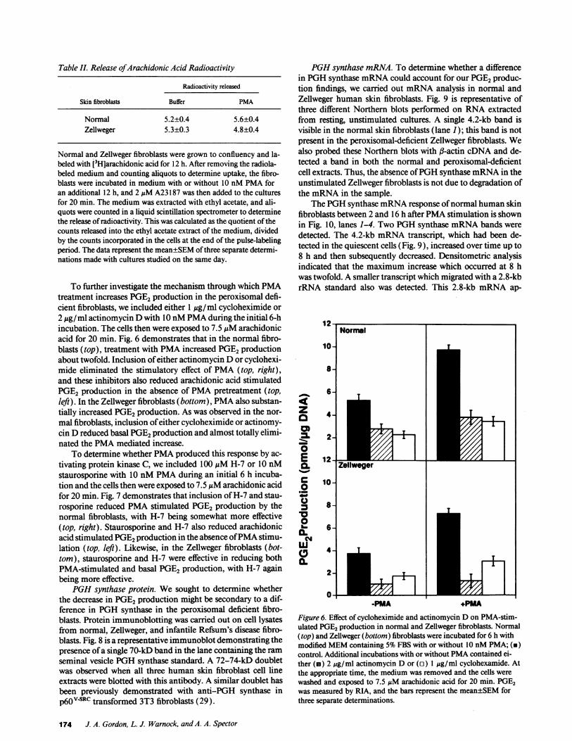
Table I. Release ofArachidonic Acid Radioactivity
Radioactivity released
Skin fibroblasts Buffer PMA
Normal 5.2±0.4 5.6±0.4Zellweger 5.3±0.3 4.8±0.4
Normal and Zellweger fibroblasts were grown to confluency and la-beled with [3Hjarachidonic acid for 12 h. After removing the radiola-beled medium and counting aliquots to determine uptake, the fibro-blasts were incubated in medium with or without 10 nMPMAforan additional 12 h, and 2 ,uM A23187 was then added to the culturesfor 20 min. The medium was extracted with ethyl acetate, and ali-quots were counted in a liquid scintillation spectrometer to determinethe release of radioactivity. This was calculated as the quotient of thecounts released into the ethyl acetate extract of the medium, dividedby the counts incorporated in the cells at the end of the pulse-labelingperiod. The data represent the mean±SEMof three separate determi-nations made with cultures studied on the same day.
To further investigate the mechanism through which PMAtreatment increases PGE2production in the peroxisomal defi-cient fibroblasts, we included either 1 Mig/ml cycloheximide or2 Mg/ml actinomycin Dwith 10 nMPMAduring the initial 6-hincubation. The cells then were exposed to 7.5 AMarachidonicacid for 20 min. Fig. 6 demonstrates that in the normal fibro-blasts (top), treatment with PMAincreased PGE2productionabout twofold. Inclusion of either actinomycin Dor cyclohexi-mide eliminated the stimulatory effect of PMA(top, right),and these inhibitors also reduced arachidonic acid stimulatedPGE2 production in the absence of PMApretreatment (top,left). In the Zellweger fibroblasts (bottom), PMAalso substan-tially increased PGE2production. As was observed in the nor-mal fibroblasts, inclusion of either cycloheximide or actinomy-cin D reduced basal PGE2production and almost totally elimi-nated the PMAmediated increase.
To determine whether PMAproduced this response by ac-tivating protein kinase C, we included 100 AMH-7 or 10 nMstaurosporine with 10 nMPMAduring an initial 6 h incuba-tion and the cells then were exposed to 7.5 MMarachidonic acidfor 20 min. Fig. 7 demonstrates that inclusion of H-7 and stau-rosporine reduced PMAstimulated PGE2 production by thenormal fibroblasts, with H-7 being somewhat more effective(top, right). Staurosporine and H-7 also reduced arachidonicacid stimulated PGE2production in the absence of PMAstimu-lation (top, left). Likewise, in the Zellweger fibroblasts (bot-tom), staurosporine and H-7 were effective in reducing bothPMA-stimulated and basal PGE2production, with H-7 againbeing more effective.
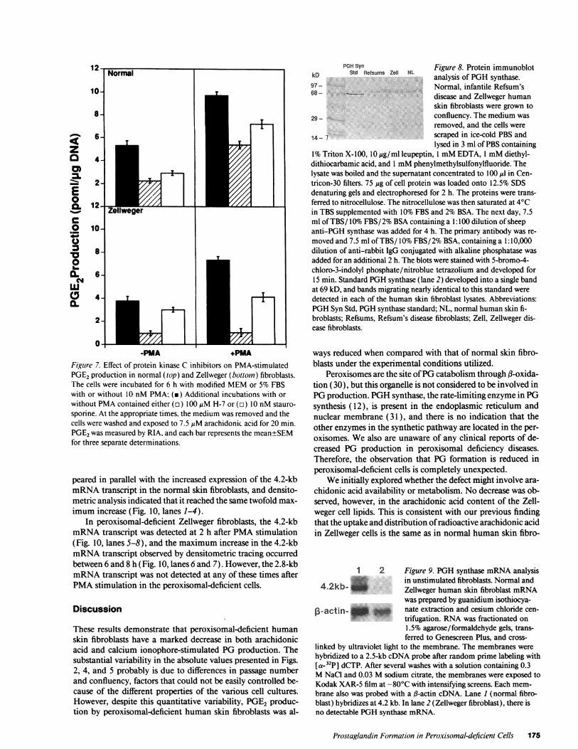
PGHsynthase protein. Wesought to determine whetherthe decrease in PGE2production might be secondary to a dif-ference in PGHsynthase in the peroxisomal deficient fibro-blasts. Protein immunoblotting was carried out on cell lysatesfrom normal, Zellweger, and infantile Refsum's disease fibro-blasts. Fig. 8 is a representative immunoblot demonstrating thepresence of a single 70-kD band in the lane containing the ramseminal vesicle PGHsynthase standard. A 72-74-kD doubletwas observed when all three human skin fibroblast cell lineextracts were blotted with this antibody. A similar doublet hasbeen previously demonstrated with anti-PGH synthase inp60v4Rc transformed 3T3 fibroblasts (29).
PGHsynthase mRNA. To determine whether a differencein PGHsynthase mRNAcould account for our PGE2produc-tion findings, we carried out mRNAanalysis in normal andZellweger human skin fibroblasts. Fig. 9 is representative ofthree different Northern blots performed on RNAextractedfrom resting, unstimulated cultures. A single 4.2-kb band isvisible in the normal skin fibroblasts (lane 1); this band is notpresent in the peroxisomal-deficient Zellweger fibroblasts. Wealso probed these Northern blots with ,B-actin cDNAand de-tected a band in both the normal and peroxisomal-deficientcell extracts. Thus, the absence of PGHsynthase mRNAin theunstimulated Zellweger fibroblasts is not due to degradation ofthe mRNAin the sample.
The PGHsynthase mRNAresponse of normal human skinfibroblasts between 2 and 16 h after PMAstimulation is shownin Fig. 10, lanes 1-4. Two PGHsynthase mRNAbands weredetected. The 4.2-kb mRNAtranscript, which had been de-tected in the quiescent cells (Fig. 9), increased over time up to8 h and then subsequently decreased. Densitometric analysisindicated that the maximum increase which occurred at 8 hwas twofold. A smaller transcript which migrated with a 2.8-kbrRNA standard also was detected. This 2.8-kb mRNAap-
z0
01-.Ea.c00v
0.NM
0.
-PMA +PMAFigure 6. Effect of cycloheximide and actinomycin D on PMA-stim-ulated PGE2production in normal and Zellweger fibroblasts. Normal(top) and Zellweger (bottom) fibroblasts were incubated for 6 h withmodified MEMcontaining 5%FBS with or without 10 nMPMA; (i)control. Additional incubations with or without PMAcontained ei-ther (i) 2 gg/ml actinomycin D or (o) 1 ug/ml cyclohexamide. Atthe appropriate time, the medium was removed and the cells werewashed and exposed to 7.5 uM arachidonic acid for 20 min. PGE2was measured by RIA, and the bars represent the mean±SEMforthree separate determinations.
174 J. A. Gordon, L. J. Warnock, and A. A. Spector
10-
8-
zza
E0a.
C0
0
NW
wa,a.
6-
4-
2-
Normal
12-E
10-
8-
6-
4- Er .1
-PMA +PMAFigure 7. Effect of protein kinase C inhibitors on PMA-stimulatedPGE2production in normal (top) and Zellweger (bottom) fibroblasts.The cells were incubated for 6 h with modified MEMor 5% FBSwith or without 10 nM PMA; (m) Additional incubations with orwithout PMAcontained either (n) 100MgM H-7 or (o) 10 nM stauro-sporine. At the appropriate times, the medium was removed and thecells were washed and exposed to 7.5 MMarachidonic acid for 20 min.PGE2was measured by RIA, and each bar represents the mean±SEMfor three separate determinations.
peared in parallel with the increased expression of the 4.2-kbmRNAtranscript in the normal skin fibroblasts, and densito-metric analysis indicated that it reached the same twofold max-imum increase (Fig. 10, lanes 1-4).
In peroxisomal-deficient Zellweger fibroblasts, the 4.2-kbmRNAtranscript was detected at 2 h after PMAstimulation(Fig. 10, lanes 5-8), and the maximum increase in the 4.2-kbmRNAtranscript observed by densitometric tracing occurredbetween 6 and 8 h (Fig. 10, lanes 6 and 7). However, the 2.8-kbmRNAtranscript was not detected at any of these times afterPMAstimulation in the peroxisomal-deficient cells.
Discussion
These results demonstrate that peroxisomal-deficient humanskin fibroblasts have a marked decrease in both arachidonicacid and calcium ionophore-stimulated PGproduction. Thesubstantial variability in the absolute values presented in Figs.2, 4, and 5 probably is due to differences in passage numberand confluency, factors that could not be easily controlled be-cause of the different properties of the various cell cultures.However, despite this quantitative variability, PGE2 produc-tion by peroxisomal-deficient human skin fibroblasts was al-
PGHSyn Figure 8. Protein immunoblotkD Std Refsums Zell NL analysis of PGHsynthase.97- Normal, infantile Refsum's68- disease and Zellweger human
skin fibroblasts were grown to
29- confluency. The medium was
removed, and the cells were
14- 7 scraped in ice-cold PBS andlysed in 3 ml of PBScontaining
1%Triton X-100, 10 Mg/ml leupeptin, 1 mMEDTA, 1 mMdiethyl-dithiocarbamic acid, and 1 mMphenylmethylsulfonylfluoride. Thelysate was boiled and the supernatant concentrated to 100 Ml in Cen-tricon-30 filters. 75 Mg of cell protein was loaded onto 12.5% SDSdenaturing gels and electrophoresed for 2 h. The proteins were trans-ferred to nitrocellulose. The nitrocellulose was then saturated at 40Cin TBS supplemented with 10% FBS and 2%BSA. The next day, 7.5ml of TBS/ 10%FBS/ 2%BSAcontaining a 1: 100 dilution of sheepanti-PGH synthase was added for 4 h. The primary antibody was re-moved and 7.5 ml of TBS/ 10%FBS/2% BSA, containing a 1:10,000dilution of anti-rabbit IgG conjugated with alkaline phosphatase wasadded for an additional 2 h. The blots were stained with 5-bromo-4-chloro-3-indolyl phosphate/nitroblue tetrazolium and developed for15 min. Standard PGHsynthase (lane 2) developed into a single bandat 69 kD, and bands migrating nearly identical to this standard weredetected in each of the human skin fibroblast lysates. Abbreviations:PGHSyn Std, PGHsynthase standard; NL, normal human skin fi-broblasts; Refsums, Refsum's disease fibroblasts; Zell, Zellweger dis-ease fibroblasts.
ways reduced when compared with that of normal skin fibro-blasts under the experimental conditions utilized.
Peroxisomes are the site of PGcatabolism through f-oxida-tion (30), but this organelle is not considered to be involved inPGproduction. PGHsynthase, the rate-limiting enzyme in PGsynthesis ( 12), is present in the endoplasmic reticulum andnuclear membrane (31 ), and there is no indication that theother enzymes in the synthetic pathway are located in the per-oxisomes. Wealso are unaware of any clinical reports of de-creased PG production in peroxisomal deficiency diseases.Therefore, the observation that PG formation is reduced inperoxisomal-deficient cells is completely unexpected.
Weinitially explored whether the defect might involve ara-chidonic acid availability or metabolism. No decrease was ob-served, however, in the arachidonic acid content of the Zell-weger cell lipids. This is consistent with our previous findingthat the uptake and distribution of radioactive arachidonic acidin Zellweger cells is the same as in normal human skin fibro-
1 2 Figure 9. PGHsynthase mRNAanalysisin unstimulated fibroblasts. Normal and
4.2kb Zellweger human skin fibroblast mRNAwas prepared by guanidium isothiocya-
13-actin- _ I_ nate extraction and cesium chloride cen-trifugation. RNAwas fractionated on1.5% agarose/formaldehyde gels, trans-ferred to Genescreen Plus, and cross-
linked by ultraviolet light to the membrane. The membranes werehybridized to a 2.5-kb cDNAprobe after random prime labeling withIa-32p] dCTP. After several washes with a solution containing 0.3MNaCl and 0.03 Msodium citrate, the membranes were exposed toKodak XAR-5 film at -80°C with intensifying screens. Each mem-brane also was probed with a (3-actin cDNA. Lane 1 (normal fibro-blast) hybridizes at 4.2 kb. In lane 2 (Zellweger fibroblast), there isno detectable PGHsynthase mRNA.
Prostaglandin Formation in Peroxisomal-deficient Cells 175
I
T
v
T00,
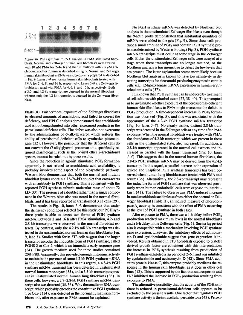
1 2 3 4 5 6 7 84.2 kb-3.0 kb-
Figure 10. PGHsynthase mRNAanalysis in PMAstimulated fibro-blasts. Normal and Zellweger human skin fibroblasts were treatedwith 10 nM PMAfor 2-16 h and then stimulated with 7.5 gM ara-chidonic acid for 20 min as described in Fig. 5. Normal and Zellwegerhuman skin fibroblast mRNAwas subsequently prepared as describedin Fig. 9. Lanes 1-4 are normal human skin fibroblasts treated withPMAfor 2, 6, 8, and 16 h, respectively. Lanes 5-8 are Zellweger fi-broblasts treated with PMAfor 4, 6, 8, and 16 h, respectively. Botha 3.0- and 4.2-kb transcript are detected in the normal fibroblastwhereas only the 4.2-kb transcript is detected in the Zellweger fibro-blast.
blasts (8). Furthermore, exposure of the Zellweger fibroblaststo elevated amounts of arachidonic acid failed to correct thedeficiency, and HPLCanalysis demonstrated that arachidonicacid is not being shunted into other eicosanoid products in theperoxisomal-deficient cells. The defect was also not overcomeby the administration of O-alkylglycerol, which restores theability of peroxisomal-deficient cells to synthesize plasmalo-gens (32). However, the possibility that the deficient cells donot convert the O-alkylglycerol precursor to a specifically re-quired plasmologen, such as the 1-vinyl ether 2-arachidonylspecies, cannot be ruled out by these results.
Since the reduction in agonist stimulated PGE2 formationapparently is not related to arachidonic acid availability, itprobably involves some aspect of the biosynthetic pathway.Western blots demonstrate that both the normal and mutantfibroblast lysates contain a 72-74-kD doublet that cross-reactswith an antibody to PGHsynthase. This is consistent with thereported PGHsynthase subunit molecular mass of about 72kD (33). The presence of a doublet rather than a single compo-nent in the Western blots also was noted in the normal fibro-blasts, and it has been reported in transformed 3T3 cells (29).
The results in Fig. 10, lanes 1-4, demonstrate that underthe stringency conditions selected for this study, our PGHsyn-thase probe is able to detect two forms of PGH synthasemRNA.Between 2 and 16 h after PMAstimulation, 4.2- and2.8-kb transcripts were observed in the normal fibroblast ex-tracts. By contrast, only the 4.2 kb mRNAtranscript was de-tected in the unstimulated normal human skin fibroblasts (Fig.9, lane 1). Studies with Swiss 3T3 cells suggest that the largertranscript encodes the inducible form of PGHsynthase, calledPGHS-2 or Cox-2, which is an immediate early response gene(34). The growth medium used for these studies contained20% FBS. Apparently, this provided enough mitogenic activityto maintain the presence of some 4.2-kb PGHsynthase mRNAin the unstimulated fibroblasts. In this regard, a 4.8-kb PGHsynthase mRNAtranscript has been detected in unstimulatednormal human monocytes (35), and a 5.5-kb transcript is pres-ent in unstimulated normal human lung fibroblasts (36). Inthese cells, however, a 2.7-2.8-kb PGHsynthase mRNAtran-script also was detected (35, 36). Whythe smaller mRNAtran-script, which probably encodes the constitutive PGHsynthase-
or Cox- 1 (34), was detected in the normal human skin fibro-blasts only after exposure to PMAcannot be explained.
No PGHsynthase mRNAwas detected by Northern blotanalysis in the unstimulated Zellweger fibroblasts even thoughthe ,3-actin probe demonstrated that substantial quantities ofmRNAwere added to the gels (Fig. 9). Since these cells pro-duce a small amount of PGE2and contain PGHsynthase pro-tein as determined by Western blotting (Fig. 8), PGHsynthasemRNAtranscripts must occur at some stage in the Zellwegercells. Either the unstimulated Zellweger cells were assayed at astage when these transcripts are no longer retained, or theNorthern analysis is too insensitive to detect the low levels thatare present. The latter explanation seems more likely becauseNorthern blot analysis is known to have low sensitivity in de-tecting transcripts for eicQsanoid-producing enzymes in certaincells, e.g., 12-lipoxygenase mRNAexpression in human eryth-roleukemia cells (37).
It is known that PGHsynthase can be induced by treatmentof cell cultures with phorbol esters (27, 38-40). This promptedus to investigate whether exposure of the peroxisomal-deficienthuman skin fibroblasts to PMAmight overcome the deficit inPGE2production. A time-dependent increase in PGE2 forma-tion was observed (Fig. 5), and this was associated with theappearance of the 4.2-kb PGH synthase mRNAtranscript(Fig. 10, lanes 5-8). No clearly visible, distinct 2.8-kb tran-script was detected in the Zellweger cells at any time after PMAexposure. Whenthe normal fibroblasts were treated with PMA,the abundance of 4.2-kb transcript, which was present in thesecells in the unstimulated state, also increased. In addition, a2.8-kb transcript appeared in the normal cell extracts and in-creased in parallel with the larger transcript (Fig. 10, lanes1-4). This suggests that in the normal human fibroblasts, the2.8-kb PGHsynthase mRNAmay be derived from the 4.2-kbtranscript. In this regard, a parallel increase in the expression ofspliced and unspliced PGHsynthase transcripts has been ob-served when human lung fibroblasts are treated with PMAandserum (36). Alternatively, the 2.8-kb transcript may representthe inducible form of PGHsynthase that was observed previ-ously when human endothelial cells were exposed to interleu-kin 1 (41 ). The failure to observe any PMA-induced increasein total arachidonic acid release from either the normal or Zell-weger fibroblast (Table II), an indirect measure of phospholi-pase A2 activity, is consistent with the effect of PMAoccurringat the level of PGHsynthase in both cases.
After exposure to PMA, there was a 4-h delay before PGE2production reached maximum levels in the normal fibroblastsand a 6-h delay in the Zellweger cells (Fig. 5). Such a lag periodalso is compatible with a mechanism involving PGHsynthasegene expression. Likewise, the inhibitory effects of actinomy-cin D and cycloheximide suggest that gene expression is in-volved. Results obtained in 3T3 fibroblasts exposed to plateletderived growth factor are consistent with this interpretation;the increase in PGE2 synthesis resulting from production ofPGHsynthase exhibited a lag period of 2-6 hand was inhibitedby cycloheximide and actinomycin D (42). Since PMAacti-vates protein kinase C, this enzyme probably mediates the re-sponse in the human skin fibroblasts, as it does in other celllines ( 12). This is supported by the fact that staurosporine andH-7 inhibited the increase in PGE2production resulting fromexposure to PMA.
The alternative possibility that the activity of the PGHsyn-thase is reduced in peroxisomal-deficient cells appears to beexcluded by the present results. One factor that regulates PGHsynthase activity is the intracellular peroxide tone (43). Peroxi-
176 J. A. Gordon, L. J. Warnock, and A. A. Spector
somes generate H202 through fatty acid /-oxidation (3, 44)and they contain catalase, the enzyme that breaks down H202( 1, 2). Depending on which of these processes predominates, aperoxisomal deficiency might either increase or decrease theperoxide tone. However, addition of neither H202 nor a poly-ethylene glycol-catalase complex stimulated PGE2 output bythe Zellweger cultures. The concentrations of H202 tested ap-parently were effective because, as in endothelial cells (45),prostaglandin production was reduced when the normal fibro-blasts were treated with these quantities of H202 (Fig. 4). Basedon these findings, it seems unlikely that abnormalities in perox-ide tone are responsible for the decreased PGE2 formation inthe peroxisomal deficient cells.
As opposed to the present findings, Tiffany et al. (46) haveobserved that PGE2 production is increased in peroxisomaldeficient fibroblasts. The culture conditions, however, werequite different in the two studies. Tiffany et al. (46) utilized agas phase containing 10% C02 and supplemented the growthmedia with 10% FBS, whereas we used 5%CO2and 20% FBS.In addition, Tiffany et al. (46) tested the fibroblasts after expo-sure to a serum-free medium supplemented with 2% TCM, aproprietary mixture containing vitamins, growth factors, andfatty acids. Someor all of these factors might have modified theresponse in the mutant cells, leading to an increase rather thanthe decrease in PGE2 formation that we observed.
In conclusion, our findings demonstrate that under com-monly used culture conditions, the capacity of peroxisomal-de-ficient human skin fibroblasts to produce PGs is impaired.Therefore, peroxisome integrity appears to be necessary fornormal PGformation, a relationship not previously recognizedin eukaryotic cells. These results also suggest the possibility thatsome of the abnormalities associated with peroxisomal defi-ciency syndromes may be related to an inability to produceadequate amounts of PGs.
Acknowledgments
The authors thank Drs. Nagender Yerram, Larry Oberley, Larry Kar-niski, and James Elwell for their help in protein immunoblotting. Thetechnical assistance of Carolyn Heath and Sharon Heller, and the secre-tarial assistance of Cay Wieland is gratefully acknowledged.
Dr. Gordon is a recipient of a Clinician-Scientist Award from theAmerican Heart Association (87-0426). This work was also supportedby grants from the Iowa Affiliate of the American Heart Association(IA-88-G12, IA-90-G-9) and The Children's Miracle Network Tele-thon of Iowa, and grant HL-49264 from the National Heart, Lung andBlood Institute, National Institutes of Health.
References
1. Lazarow, P. B., and H. W. Moser. 1989. Disorders of peroxisome biogene-sis. In The Metabolic Basis of Inherited Disease. C. R. Scriver, A. L. Beaudet,W. S. Sly, and D. Valle, editors. McGraw-Hill Information Services, Philadel-phia. 1479-1510.
2. De Duve, C., and P. Baudhin. 1966. Peroxisomes ( microbodies and relatedparticles). Phvsiol. Rev. 46:323-357.
3. Hiltunen, J. K., T. Karki, 1. E. Hassinene, and H. Osmudsen. 1986. 13-Oxi-dation of polyunsaturated fatty acids by rat liver peroxisomes. J. Biol. Chem.26 1:16484-16493.
4. Hajra, A. K., C. L. Burke, and C. L. Jones. 1979. Subcellular localization ofacyl-coenzyme A: dihydroxyacetone phosphate acyltransferase in rat liver peroxi-somes (microbodies). J. Biol. Chem. 254:10896-10900.
5. Krisans, S. K., S. L. Thompson, L. A. Pena, E. Kok, and N. B. Javitt. 1985.Bile acid synthesis in rat liver peroxisomes: metabolism of 26-hydroxycholesterolto 3 /3-hydroxy-5-cholenic acid. J. Lipid Res. 26:1324-1332.
6. Poulos, S., P. Sharp, and M. Whiting. 1984. Infantile Refsum's disease(phytanic acid storage disease): a variant of Zellweger's syndrome? Clin. Genet.26:579-586.
7. Trijbels, J. M. F., L. A. H. Monnens, J. A. J. M. Bakkeren, A. H. J. VanRaay-Selten, and J. M. B. Cortiaensen. 1979. Biochemical studies in the cerebro-heptato-renal syndrome of Zellweger: a disturbance in the metabolism of pipeco-lic acid. J. Inherited AMetab. Dis. 2:39-42.
8. Gordon, J. A., P. H. Figard, and A. A. Spector. 1990. HETEmetabolism incultured human skin fibroblasts: evidence for peroxisomal 3-oxidation. J. Clin.Invest. 85:1173-1181.
9. Kelly, R. 1. 1983. Review: the cerebro-hepato-renal syndrome of Zellweger.Morphologic and metabolic aspects. Am. J. Med. Genet. 16:503-517.
10. Goldfischer, S., C. L. Moore, A. B. Johnson, A. J. Spiro, M. P. Valsamis,H. K. Wisniewski, R. H. Ritch, W. T. Norton, 1. Rapin, and L. M. Gartner. 1973.Peroxisomal and mitochondrial defects in the cerebro-hepato-renal syndrome.Science (Was/h. DC). 182:62-64.
11. Smith, W. L., and P. Borgeat. 1985. The eicosanoids: Prostaglandins,thromboxanes, leukotrienes, and hydroxyeicosaenoic acids. In Biochemistry ofLipids and Membranes. D. Vance and J. E. Vance, editors. The Benjamin/Cum-mings Publishing Company, Menlo Park, CA. 325-360.
12. DeWitt, D. L. 1991. Prostaglandin endoperoxide synthase: regulation ofenzyme expression. Biochim. Biophv s. Acta. 1083:12 1-134.
13. Marshall, P. J.. R. J. Kulmacz. and W. E. M. Lands. 1987. Constraints ofprostaglandin biosynthesis in tissues. J. Biol. Chem. 262:3510-3517.
14. Labarca, C.. and K. Paisen. 1980. A simple, rapid, and sensitive DNAassay procedure. Anal. Biochem. 102:344-352.
15. Gordon, J. A., and A. A. Spector. 1987. Effects of 12-HETE on renaltubular epithelial cells. Aim. J. Ph.vsiol. 253:C277-C285.
16. Beckman, J. S.. R. L. Minor, C. W. White, J. E. Repine, G. M. Rosen, andB. E. Freeman. 1988. Superoxide dismutase and catalase conjugated to polyethyl-ene glycol increases endothelial enzyme activity and oxidant resistance. J. Biol.C(hem. 263:6884-6892.
17. Towbin, H., T. Staehelen. and J. Gordon. 1979. Electrophoretic transferof proteins from polyacrylamide gels to nitrocellulose sheets: procedures andsome applications. Proc. Natl. Acad. Sci. USA. 76:4350-4354.
18. Lees, M. B., and S. Paxman. 1972. Modification of the Lowry procedurefor the analysis of proteolipid protein. Anal. Biochem. 47:184-192.
19. Laemmli, U. K. 1970. Cleavage of structural proteins during the assemblyof the head of bacteriophage T4. Nature (Lond.). 227:680-685.
20. Chirgwin, J. M.. A. E. Przybyla, R. J. McDonald, and W. J. Rutter. 1979.Isolation of biologically active ribonucleic acid from sources enriched in ribonu-clease. Biochemnistrv. 18:5294-5299.
21. Glisin, V., R. Crkvenjakov. and C. Byus. 1974. Ribonucleic acid isolatedby cesium chloride centrifugation. Biochemistrv. 13:2633-2637.
22. Sambrook. J., E. F. Fritsch. and T. Maniatis. 1989. Preparation of radiola-beled DNAand RNAprobes. In Molecular Cloning: A Laboratory Manual. ColdSpring Harbor Laboratory Press. Cold Spring Harbor, NY. 10.13-10.55.
23. Winer, B. J. 1971. Statistical Principles in Experimental Design. McGrawHill Book Co., New York. 907.
24. Wanders, R. J. A., M. Kos, B. Roestg, A. J. Meijer, G. Schrakamp, H. S. A.Heymans, H. Tegelaers, H. van den Bosch, R. B. H. Schutgens, and J. M. Tager.1984. Activity of peroxisomal enzymes and intracellular distribution of catalasein Zellweger syndrome. Biochem. Biophys. Res. Commun. 123:1054-1061.
25. Duniec, Z. M., P. Nettesheim, and T. E. Eling. 1991. Stimulation ofprostaglandin H synthase mRNAlevels and prostaglandin biosynthesis by phor-bol ester: mediation by protein kinase C. Mol. Pharmacol. 39:164-170.
26. Parker, J., L. W. Daniel, and M. Waite. 1987. Evidence of protein kinase Cinvolvement in phorbol diester-stimulated arachidonic acid release and prosta-glandin synthesis. J. Biol. Chem. 262:5385-5393.
27. Beaudry, G. A., M. Waite, and L. W. Daniel. 1985. Regulation of arachi-donic acid metabolism in Madin-Darby canine kidney cells: stimulation of syn-thesis of the cyclooxygenase system by 12-0-tetradecanoyl-phorbol- 13-acetate.Arch. Biochem. Biophys. 239:242-247.
28. Daniel, L. W., L. King, and M. Waite. 1981. Source of arachidonic acidsynthesis for prostaglandin synthesis in Madin Darby canine kidney cells. J. Biol.Chem. 256:12830-12835.
29. Han, J. W., H. Sadowski, D. A. Young, and I. G. Marara. 1990. Persistentinduction of cyclooxygenase in p60"-transformed 3T3 fibroblasts. Proc. Natl.Acad. Sci. USA. 87:3373-3377.
30. Diczfalusy, U., and S. E. H. Axelson. 1990. Identification of d-oxidationof prostaglandins: metabolites from peroxisomal 13-oxidation of prostaglandins.J. Lipid Res. 31:307-314.
31. Rollins, T. E., and W. L. Smith. 1980. Subcellular localization of prosta-glandin-forming cyclooxygenase in Swiss mouse 3T3 fibroblasts by electron mi-croscopic immunocytochemistry. J. Biol. Chem. 255:4872-4875.
32. Schrakamp, G., C. G. Schalkwijk, R. B. H. Schutgens, R. J. A. Wanders,J. M. Tager, and H. van den Bosch. 1988. Plasmalogen biosynthesis in peroxiso-mal disorders: fatty alcohol versus alkylglycerol precursors. J. Lipid Res. 29:325-334.
33. Pagels, W. R., R. J. Sachs, L. J. Marnett, D. L. DeWitt, J. S. Day, andW. L. Smith. 1983. Immunochemical evidence for the involvement of prostaglan-din H synthase in hydroperoxide-dependent oxidations by ram seminal vesiclemicrosomes. J. BioL. Chem. 258:6517-6523.
Prostaglandin Formation in Peroxisomal-deficient Cells 177
34. Kujubu, D. A., and Herschman, H. R. 1992. Dexamethasone inhibitsmitogen induction of the Ti S10 prostaglandin synthase/cyclooxygenase gene. J.Biol. Chem. 267:7991-7994.
35. O'Banion, M. K., V. D. Winn, and D. A. Young. 1992. cDNAcloning andfunctional activity of a glucocorticoid-regulated inflammatory cyclooxygenase.Proc. NatL. Acad. Sci. USA. 89:4888-4892.
36. Diaz, A., A. M. Reginato, and S. A. Jimenez. 1992. Alternative splicing ofhuman prostaglandin G/H synthase mRNAand evidence of differential regula-tion of the resulting transcripts by transforming growth factor #I, interleukin 1,,and tumor necrosis factor a. J. Bio. Chem. 267:10816-10822.
37. Funk, C. D., Furci, L., and G. A. Fitzerald. 1990. Molecular cloning,primary structure, and expression of the human/platelet/erythroleukemia cell12-lipoxygenase. Proc. Nail. Acad. Sci. USA. 87:5638-5642.
38. Wu, K. K., H. Hatzakis, S. S. Lo, D. C. Seong, S. K. Sanduja, and H. H.Tai. 1988. Stimulation of de novo synthesis of prostaglandin G/H synthase inhuman endothelial cells by phorbol ester. J. Bid. Chem. 263:19043-19047.
39. Hla, T., and K. Neilson. 1992. Human cyclooxygenase-2 cDNA. Proc.Nati. Acad. Sci. USA. 89:7384-7388.
40. Kujubu, D. A., B. S. Fletcher, B. C. Varnum, R. W. Lim, and H. R.Herschman. 1991. TIS 10, a phorbol ester tumor promoter-inducible mRNA
from Swiss 3T3 cells, encodes a novel prostaglandin synthase/cyclooxygenasehomologue. J. Biol. Chem. 266:12866-12872.
41. Maier, J. A. M., T. Hla, and T. Maciag. 1990. Cyclooxygenase is anintermediate-early gene induced by interleukin- 1 in human endothelial cells. J.Bid. Chem. 265:10805-10808.
42. Habenicht, A. J. R., M. Goerig, J. Grulich, J. Rothe, D. Gronwald, U.Loth, G. Schettler, B. Kommerell, and R. Ross. 1985. Human platelet-derivedgrowth factor stimulates prostaglandin synthesis by activation and by rapid denovo synthesis of cyclooxygenase. J. Clin. Invest. 75:1381-1387.
43. Hemler, M. E., H. W. Cook, and W. E. M. Lands. 1979. Prostaglandinbiosynthesis can be triggered by lipid peroxides. Arch. Biochem. Biophys.193:340-345.
44. Lazarow, P. B. 1978. Rat liver peroxisomes catalyze the fl-oxidation offatty acids. J. Biol. Chem. 253:1522-1528.
45. Whorton, A. R., M. E. Montgomery, and R. S. Kent. 1985. Effect ofhydrogen peroxide on prostaglandin production and cellular integrity in culturedporcine aortic endothelial cells. J. Clin. Invest. 76:295-302.
46. Tiffany, C. W., S. Hoefler, H. W. Moser, and R. M. Burch. 1991. Arachi-donic acid metabolism in fibroblasts from patients with peroxisomal disorders:response to interleukin 1. Biochim. Biophys. Acta. 1096:41-46.
178 J. A. Gordon, L. J. Warnock, and A. A. Spector