
Attachment of phycobilisomes in an antenna photosystem I ...
6
Attachment of phycobilisomes in an antenna– photosystem I supercomplex of cyanobacteria Mai Watanabe a,b,1 , Dmitry A. Semchonok c,1 , Mariam T. Webber-Birungi c , Shigeki Ehira d,e , Kumiko Kondo a,f , Rei Narikawa a,e , Masayuki Ohmori d , Egbert J. Boekema c , and Masahiko Ikeuchi a,b,2 a Department of Life Sciences (Biology), Graduate School of Arts and Sciences, University of Tokyo, Komaba, Meguro, Tokyo 153-8902, Japan; b Japan Science and Technology Agency, Core Research for Evolutionary Science and Technology, Meguro, Tokyo 153-8902, Japan; c Department of Electron Microscopy, Groningen Biomolecular Sciences and Biotechnology Institute, University of Groningen, 9747 AG, Groningen, The Netherlands; d Department of Biological Science, Faculty of Science and Engineering, Chuo University, Bunkyo, Tokyo 112-8551, Japan; e Japan Science and Technology Agency, Precursory Research for Embryonic Science and Technology, Meguro, Tokyo 153-8902, Japan; and f RIKEN Plant Science Center, Tsurumi-ku, Yokohama, Kanagawa 230-0045, Japan Edited by Robert Haselkorn, The University of Chicago, Chicago, IL, and approved January 9, 2014 (received for review November 1, 2013) Oxygenic photosynthesis is driven by photosystems I and II (PSI and PSII, respectively). Both have specific antenna complexes and the phycobilisome (PBS) is the major antenna protein complex in cyanobacteria, typically consisting of a core from which several rod-like subcomplexes protrude. PBS preferentially transfers light energy to PSII, whereas a PSI-specific antenna has not been identified. The cyanobacterium Anabaena sp. PCC 7120 has rod– core linker genes (cpcG1-cpcG2-cpcG3-cpcG4). Their products, ex- cept CpcG3, have been detected in the conventional PBS. Here we report the isolation of a supercomplex that comprises a PSI tetra- mer and a second, unique type of a PBS, specific to PSI. This rod- shaped PBS includes phycocyanin (PC) and CpcG3 (hereafter renamed “CpcL”), but no allophycocyanin or CpcGs. Fluorescence excitation showed efficient energy transfer from PBS to PSI. The supercomplex was analyzed by electron microscopy and single- particle averaging. In the supercomplex, one to three rod-shaped CpcL–PBSs associate to a tetrameric PSI complex. They are mostly composed of two hexameric PC units and bind at the periphery of PSI, at the interfaces of two monomers. Structural modeling indi- cates, based on 2D projection maps, how the PsaI, PsaL, and PsaM subunits link PSI monomers into dimers and into a rhombically shaped tetramer or “pseudotetramer.” The 3D model further shows where PBSs associate with the large subunits PsaA and PsaB of PSI. It is proposed that the alternative form of CpcL–PBS is functional in harvesting energy in a wide number of cyanobacteria, partially to facilitate the involvement of PSI in nitrogen fixation. I n photosynthesis, light-harvesting antennas are essential to efficiently collect solar energy. Photosynthetic organisms have diverse antenna protein-pigment complexes, which are specifi- cally associated with photosystems I or II (PSI or PSII, re- spectively) (1). Light-harvesting chlorophyll a/b-binding proteins form the peripheral antenna of PSI or PSII in green plants (2) and light-harvesting chlorophyll a/c-binding proteins are present in brown algae and related organisms (3, 4). In cyanobacteria the phycobilisome (PBS) serves as a major antenna for PSII. No specific antenna has been isolated for PSI in cyanobacteria, al- though PBS transfers light energy to PSI under conditions of state transition (5), a temporal energy redistribution mechanism between PSII and PSI (6, 7). Generally, the PBS is a supercomplex of rod and core sub- complexes, which consist of various phycobilin-binding proteins connected by several classes of colorless linker proteins (8). Whereas phycocyanin (PC) is the major phycobiliprotein of the rod, allophycocyanin (APC) is the major phycobiliprotein of the core cylinders. The rod–core linker cyanobacterial phycocyanin protein G (CpcG), which connects the rod to the core, plays a key role in the assembly of the PBS (9). The chromosome of the filamentous, N 2 -fixing cyanobacterium Anabaena sp. PCC 7120 (hereafter “Anabaena”) bears tandem repeats of rod–core linker genes (cpcG1-cpcG2-cpcG3-cpcG4). Except for CpcG3, the products of these genes have been detected in the PBS (10– 13). Only cpcG3 encodes a hydrophobic region at the C terminus, although all four copies share the sequence for the highly con- served N-terminal linker domain (Fig. S1). Given the distinct role of the hydrophobic CpcG variant protein in docking to PSI as shown in this article, the hydrophobic CpcG is hereafter renamed “CpcL.” In this study, a supercomplex comprising PSI and its specific antenna was isolated from Anabaena and biochemically and structurally characterized. We found that within the supercomplex, PSI is organized into tetramers, which were previously suggested from biochemical data (14). Single-particle electron microscopy (EM) shows that in fact they are structurally organized as a dimer of dimers. In addition, we describe a specific association of CpcL– PBS rods with the PSI pseudotetramers and discuss the role of these unique supercomplexes. Results The thylakoid membrane from Anabaena was solubilized with n-dodecyl-β-D-maltoside (DM) and fractionated using a linear sucrose density gradient centrifugation with 0.005–0.05% DM (Fig. S2A). Three green bands (PSI/PSII monomer, PSI/PSII dimer, and PSI tetramer) were resolved at 0.03–0.05% DM, in agreement with our previous results (14). The solubilized com- plexes were aggregated into precipitation at 0.005% DM. At 0.01–0.02% DM, PSI was mostly recovered in the tetramer band, and bluish-green band 7 was newly detected below the PSI tet- ramer (band 6) (Fig. 1A and Fig. S2A). Blue native PAGE re- vealed that both fractions 6 and 7 contained the PSI tetramer Significance Light-harvesting antenna are essential for photosynthetic sys- tems, which comprise photosystems I and II (PSI and PSII, re- spectively). Phycobilisome (PBS) is a dominant and efficient antenna for PSII in cyanobacteria and some algae, whereas the attachment of PBS to PSI is a long-standing open question. We isolated a unique PBS–PSI supercomplex from a nitrogen-fixing cyanobacterium. Biochemical and spectral studies revealed that PBS is functionally connected to the PSI tetramer via a new uni- versal connecting component. A pseudoatomic model explains the configuration of the PSI tetramer and how the PBS is con- nected to PSI. Such antenna may play an important role for light harvesting in PSI-driven cyclic electron transport to facilitate ni- trogen fixation and other reactions. Author contributions: M.W. and M.I. designed research; M.W., D.A.S., and M.T.W.-B. performed research; M.W., D.A.S., M.T.W.-B., S.E., K.K., R.N., M.O., E.J.B., and M.I. ana- lyzed data; and M.W., E.J.B., and M.I. wrote the paper. The authors declare no conflict of interest. This article is a PNAS Direct Submission. 1 M.W. and D.A.S. contributed equally to this work. 2 To whom correspondence should be addressed. E-mail: [email protected]. This article contains supporting information online at www.pnas.org/lookup/suppl/doi:10. 1073/pnas.1320599111/-/DCSupplemental. 2512–2517 | PNAS | February 18, 2014 | vol. 111 | no. 7 www.pnas.org/cgi/doi/10.1073/pnas.1320599111 Downloaded by guest on January 22, 2022
Transcript of Attachment of phycobilisomes in an antenna photosystem I ...

Attachment of phycobilisomes in an antenna–photosystem I supercomplex of cyanobacteriaMai Watanabea,b,1, Dmitry A. Semchonokc,1, Mariam T. Webber-Birungic, Shigeki Ehirad,e, Kumiko Kondoa,f,Rei Narikawaa,e, Masayuki Ohmorid, Egbert J. Boekemac, and Masahiko Ikeuchia,b,2
aDepartment of Life Sciences (Biology), Graduate School of Arts and Sciences, University of Tokyo, Komaba, Meguro, Tokyo 153-8902, Japan; bJapan Scienceand Technology Agency, Core Research for Evolutionary Science and Technology, Meguro, Tokyo 153-8902, Japan; cDepartment of Electron Microscopy,Groningen Biomolecular Sciences and Biotechnology Institute, University of Groningen, 9747 AG, Groningen, The Netherlands; dDepartment of BiologicalScience, Faculty of Science and Engineering, Chuo University, Bunkyo, Tokyo 112-8551, Japan; eJapan Science and Technology Agency, Precursory Research forEmbryonic Science and Technology, Meguro, Tokyo 153-8902, Japan; and fRIKEN Plant Science Center, Tsurumi-ku, Yokohama, Kanagawa 230-0045, Japan
Edited by Robert Haselkorn, The University of Chicago, Chicago, IL, and approved January 9, 2014 (received for review November 1, 2013)
Oxygenic photosynthesis is driven by photosystems I and II (PSIand PSII, respectively). Both have specific antenna complexes andthe phycobilisome (PBS) is the major antenna protein complex incyanobacteria, typically consisting of a core from which severalrod-like subcomplexes protrude. PBS preferentially transfers lightenergy to PSII, whereas a PSI-specific antenna has not beenidentified. The cyanobacterium Anabaena sp. PCC 7120 has rod–core linker genes (cpcG1-cpcG2-cpcG3-cpcG4). Their products, ex-cept CpcG3, have been detected in the conventional PBS. Here wereport the isolation of a supercomplex that comprises a PSI tetra-mer and a second, unique type of a PBS, specific to PSI. This rod-shaped PBS includes phycocyanin (PC) and CpcG3 (hereafterrenamed “CpcL”), but no allophycocyanin or CpcGs. Fluorescenceexcitation showed efficient energy transfer from PBS to PSI. Thesupercomplex was analyzed by electron microscopy and single-particle averaging. In the supercomplex, one to three rod-shapedCpcL–PBSs associate to a tetrameric PSI complex. They are mostlycomposed of two hexameric PC units and bind at the periphery ofPSI, at the interfaces of two monomers. Structural modeling indi-cates, based on 2D projection maps, how the PsaI, PsaL, and PsaMsubunits link PSI monomers into dimers and into a rhombicallyshaped tetramer or “pseudotetramer.” The 3D model further showswhere PBSs associate with the large subunits PsaA and PsaB of PSI. Itis proposed that the alternative form of CpcL–PBS is functional inharvesting energy in a wide number of cyanobacteria, partially tofacilitate the involvement of PSI in nitrogen fixation.
In photosynthesis, light-harvesting antennas are essential toefficiently collect solar energy. Photosynthetic organisms have
diverse antenna protein-pigment complexes, which are specifi-cally associated with photosystems I or II (PSI or PSII, re-spectively) (1). Light-harvesting chlorophyll a/b-binding proteinsform the peripheral antenna of PSI or PSII in green plants (2)and light-harvesting chlorophyll a/c-binding proteins are presentin brown algae and related organisms (3, 4). In cyanobacteria thephycobilisome (PBS) serves as a major antenna for PSII. Nospecific antenna has been isolated for PSI in cyanobacteria, al-though PBS transfers light energy to PSI under conditions ofstate transition (5), a temporal energy redistribution mechanismbetween PSII and PSI (6, 7).Generally, the PBS is a supercomplex of rod and core sub-
complexes, which consist of various phycobilin-binding proteinsconnected by several classes of colorless linker proteins (8).Whereas phycocyanin (PC) is the major phycobiliprotein of therod, allophycocyanin (APC) is the major phycobiliprotein of thecore cylinders. The rod–core linker cyanobacterial phycocyaninprotein G (CpcG), which connects the rod to the core, playsa key role in the assembly of the PBS (9). The chromosome ofthe filamentous, N2-fixing cyanobacterium Anabaena sp. PCC7120 (hereafter “Anabaena”) bears tandem repeats of rod–corelinker genes (cpcG1-cpcG2-cpcG3-cpcG4). Except for CpcG3,the products of these genes have been detected in the PBS (10–13). Only cpcG3 encodes a hydrophobic region at the C terminus,
although all four copies share the sequence for the highly con-served N-terminal linker domain (Fig. S1). Given the distinctrole of the hydrophobic CpcG variant protein in docking to PSIas shown in this article, the hydrophobic CpcG is hereafterrenamed “CpcL.”In this study, a supercomplex comprising PSI and its specific
antenna was isolated from Anabaena and biochemically andstructurally characterized. We found that within the supercomplex,PSI is organized into tetramers, which were previously suggestedfrom biochemical data (14). Single-particle electron microscopy(EM) shows that in fact they are structurally organized as a dimerof dimers. In addition, we describe a specific association of CpcL–PBS rods with the PSI pseudotetramers and discuss the role ofthese unique supercomplexes.
ResultsThe thylakoid membrane from Anabaena was solubilized withn-dodecyl-β-D-maltoside (DM) and fractionated using a linearsucrose density gradient centrifugation with 0.005–0.05% DM(Fig. S2A). Three green bands (PSI/PSII monomer, PSI/PSIIdimer, and PSI tetramer) were resolved at 0.03–0.05% DM, inagreement with our previous results (14). The solubilized com-plexes were aggregated into precipitation at 0.005% DM. At0.01–0.02% DM, PSI was mostly recovered in the tetramer band,and bluish-green band 7 was newly detected below the PSI tet-ramer (band 6) (Fig. 1A and Fig. S2A). Blue native PAGE re-vealed that both fractions 6 and 7 contained the PSI tetramer
Significance
Light-harvesting antenna are essential for photosynthetic sys-tems, which comprise photosystems I and II (PSI and PSII, re-spectively). Phycobilisome (PBS) is a dominant and efficientantenna for PSII in cyanobacteria and some algae, whereas theattachment of PBS to PSI is a long-standing open question. Weisolated a unique PBS–PSI supercomplex from a nitrogen-fixingcyanobacterium. Biochemical and spectral studies revealed thatPBS is functionally connected to the PSI tetramer via a new uni-versal connecting component. A pseudoatomic model explainsthe configuration of the PSI tetramer and how the PBS is con-nected to PSI. Such antenna may play an important role for lightharvesting in PSI-driven cyclic electron transport to facilitate ni-trogen fixation and other reactions.
Author contributions: M.W. and M.I. designed research; M.W., D.A.S., and M.T.W.-B.performed research; M.W., D.A.S., M.T.W.-B., S.E., K.K., R.N., M.O., E.J.B., and M.I. ana-lyzed data; and M.W., E.J.B., and M.I. wrote the paper.
The authors declare no conflict of interest.
This article is a PNAS Direct Submission.1M.W. and D.A.S. contributed equally to this work.2To whom correspondence should be addressed. E-mail: [email protected].
This article contains supporting information online at www.pnas.org/lookup/suppl/doi:10.1073/pnas.1320599111/-/DCSupplemental.
2512–2517 | PNAS | February 18, 2014 | vol. 111 | no. 7 www.pnas.org/cgi/doi/10.1073/pnas.1320599111
Dow
nloa
ded
by g
uest
on
Janu
ary
22, 2
022

(Fig. S2B). Subsequent SDS/PAGE and N-terminal sequencingshowed that fraction 7 contained CpcL, CpcC, CpcB, and CpcAin addition to the PSI subunits (PsaA/B, PsaC, etc.) (Fig. 1B andTable S1). Neither the other rod–core linker proteins (CpcG1,CpcG2, and CpcG4) nor any of the PBS core components (suchas ApcA or ApcB) were detected in this fraction. Immunoblotanalysis showed that CpcL was predominantly associated withthylakoids (Fig. 1C). After solubilization with DM, CpcL waspredominantly recovered in fraction 7, whereas CpcG4 wasdetected in fractions 3–5, which included PBS subcomplexes.Thus, CpcL specifically interacts with PSI to form a unique PBS–CpcL–PSI supercomplex in the bluish-green fraction 7. The bandof CpcL was appreciably less intense than the band of the PSIsubunit PsaD in fraction 7, suggesting the substoichiometricabundance of CpcL in the PSI tetramer (Fig. 1B). This suggeststhat only one or two PBS rods are attached to the tetramer.The absorption spectra revealed that the bluish-green fraction
7 contained PC (Fig. 2A). The absence of the APC shoulderaround 650 nm, together with the absence of the core poly-peptides, strongly supports the idea that PC and CpcL directlyinteract with the PSI tetramer to form a supercomplex. Energytransfer from PC to the PSI tetramer was estimated throughanalysis of 77-Kelvin (K) fluorescence spectra (Fig. 2B). Fol-lowing excitation at 600 nm, which excites PC efficiently andchlorophyll less efficiently, emission at 725 nm was higher fromPSI in the PBS–CpcL–PSI supercomplex than from the PSItetramer (Fig. 2B). The higher excitation peak for fluorescenceemission from PSI corresponded well to the higher absorptionpeak of PC in the supercomplex than the PSI tetramer, con-firming the energy transfer from PC to PSI in the supercomplex(Fig. 2C).CpcL–PBS was isolated from vegetative cells of Anabaena with
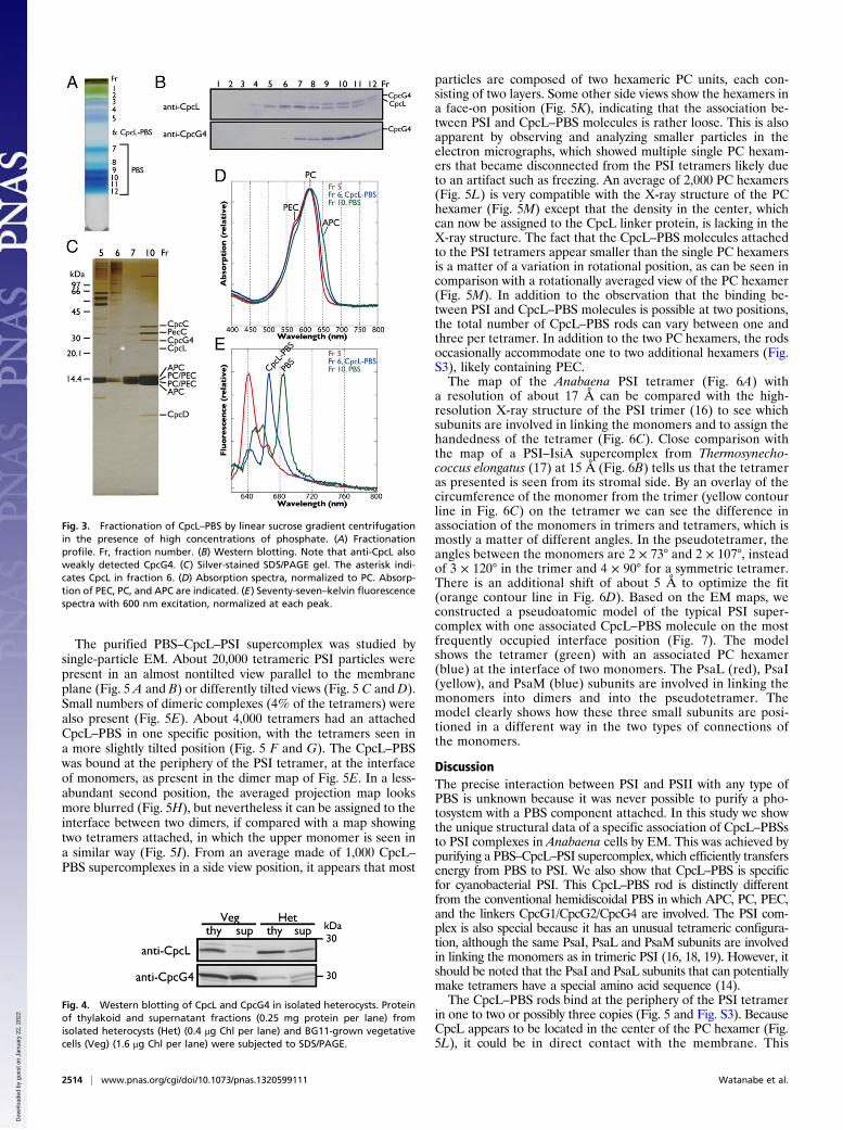
high phosphate linear sucrose gradient centrifugation withmodification (Fig. 3A). CpcL and CpcG4 were detected infractions 5–12 and 7–12, respectively (Fig. 3B). Because theantigenic peptide for CpcL was partially shared with CpcG4 (Fig.S1A), the upper band in the anti-CpcL Western blot was derived
from the weak cross-reaction with CpcG4, which correspondedto the band in the anti-CpcG4 blot.The conventional PBS that contained CpcG4 was resolved
into at least five discrete bands with a linear sucrose gradient(Fig. 3A), but only one condensed band with a typical stepwisegradient (13). Because the multiple bands showed nearly iden-tical polypeptide compositions, and band 7 corresponded to theintact PBS band of Synechocystis, the multiple bands may havebeen derived from the oligomerization of the conventional rod–core PBS complex. Although Western analysis showed thatfractions 7–12 also contained some CpcL, silver staining of SDSgels and protein sequencing detected CpcC, PecC, and CpcG4,but not CpcL, probably due to its very low abundance (Fig. 3C).Fraction 6 that mainly contained CpcL in Western analysis was
a pale blue region between the two blue bands in the gradient(Fig. 3A). The absorption spectra revealed that the pale bluefraction 6 contained PC and phycoerythrocyanin (PEC) but notAPC (Fig. 3D). After concentration, the bands of CpcL, PC, andPEC, but not CpcG4 or APC, were visible in the silver-stainedSDS gel, indicative of CpcL–PBS complex (Fig. 3C), which isconsistent with Western analysis. The CpcL–PBS emitted fluo-rescence at 665 nm at 77 K, even though it did not contain APC(Fig. 3E). This is different from the conventional CpcG–PC rodthat emits at 652 nm (15). Notably, the same fluorescence at 665nm was also detected in the PBS–CpcL–PSI supercomplex (Fig.2B). Red-shifted fluorescence of PC due to CpcL may help en-ergy transfer from PBS to PSI without APC.We further investigated the PBS–CpcL–PSI supercomplex
during N2 fixation. The N2-fixing heterocyst cells were isolated bymild disruption of the N2-fixing filaments. Immunoblot analysisindicated that heterocysts contained high levels of CpcL but verylittle CpcG4 (Fig. 4). Some of CpcL was recovered in the het-erocyst supernatant. When expressed in terms of chlorophyllcontent, total CpcL was approximately four times more abun-dant in heterocysts than in vegetative cells.
Fig. 1. Isolation of PBS–CpcL–PSI supercomplex by centrifugation througha linear sucrose gradient containing low-salt buffer and 0.01% DM. (A)Fractionation profile of thylakoid (thy) and supernatant (sup) after centri-fugation at 130,000 × g for 6 h at 4 °C. (B) Silver staining following SDS/PAGEof the conventional PBS, PSI tetramer (see 6 in A) and supercomplex (see 7 inA). N-terminal sequences of the bands are listed in Table S1. Asterisks in-dicate degradation products of PsaL. (C) Western blotting of CpcL andCpcG4. Lane numbers correspond to the numbered bands in A.
Fig. 2. Absorption spectra and 77-K fluorescence spectra of PSI tetramer(red) and supercomplex (blue) fractions. (A) Absorption spectra were nor-malized relative to the chlorophyll peak at 678 nm. (B) Emission spectraexcited at 600 nm were normalized relative to the chlorophyll concentration.(C) Excitation spectra were normalized relative to the chlorophyll peak at435 nm. PSI fluorescence was measured at 725 nm.
Watanabe et al. PNAS | February 18, 2014 | vol. 111 | no. 7 | 2513
BIOCH
EMISTR
Y
Dow
nloa
ded
by g
uest
on
Janu
ary
22, 2
022
The purified PBS–CpcL–PSI supercomplex was studied bysingle-particle EM. About 20,000 tetrameric PSI particles werepresent in an almost nontilted view parallel to the membraneplane (Fig. 5 A and B) or differently tilted views (Fig. 5 C and D).Small numbers of dimeric complexes (4% of the tetramers) werealso present (Fig. 5E). About 4,000 tetramers had an attachedCpcL–PBS in one specific position, with the tetramers seen ina more slightly tilted position (Fig. 5 F and G). The CpcL–PBSwas bound at the periphery of the PSI tetramer, at the interfaceof monomers, as present in the dimer map of Fig. 5E. In a less-abundant second position, the averaged projection map looksmore blurred (Fig. 5H), but nevertheless it can be assigned to theinterface between two dimers, if compared with a map showingtwo tetramers attached, in which the upper monomer is seen ina similar way (Fig. 5I). From an average made of 1,000 CpcL–PBS supercomplexes in a side view position, it appears that most
particles are composed of two hexameric PC units, each con-sisting of two layers. Some other side views show the hexamers ina face-on position (Fig. 5K), indicating that the association be-tween PSI and CpcL–PBS molecules is rather loose. This is alsoapparent by observing and analyzing smaller particles in theelectron micrographs, which showed multiple single PC hexam-ers that became disconnected from the PSI tetramers likely dueto an artifact such as freezing. An average of 2,000 PC hexamers(Fig. 5L) is very compatible with the X-ray structure of the PChexamer (Fig. 5M) except that the density in the center, whichcan now be assigned to the CpcL linker protein, is lacking in theX-ray structure. The fact that the CpcL–PBS molecules attachedto the PSI tetramers appear smaller than the single PC hexamersis a matter of a variation in rotational position, as can be seen incomparison with a rotationally averaged view of the PC hexamer(Fig. 5M). In addition to the observation that the binding be-tween PSI and CpcL–PBS molecules is possible at two positions,the total number of CpcL–PBS rods can vary between one andthree per tetramer. In addition to the two PC hexamers, the rodsoccasionally accommodate one to two additional hexamers (Fig.S3), likely containing PEC.The map of the Anabaena PSI tetramer (Fig. 6A) with
a resolution of about 17 Å can be compared with the high-resolution X-ray structure of the PSI trimer (16) to see whichsubunits are involved in linking the monomers and to assign thehandedness of the tetramer (Fig. 6C). Close comparison withthe map of a PSI–IsiA supercomplex from Thermosynecho-coccus elongatus (17) at 15 Å (Fig. 6B) tells us that the tetrameras presented is seen from its stromal side. By an overlay of thecircumference of the monomer from the trimer (yellow contourline in Fig. 6C) on the tetramer we can see the difference inassociation of the monomers in trimers and tetramers, which ismostly a matter of different angles. In the pseudotetramer, theangles between the monomers are 2 × 73° and 2 × 10⁷°, insteadof 3 × 120° in the trimer and 4 × 90° for a symmetric tetramer.There is an additional shift of about 5 Å to optimize the fit(orange contour line in Fig. 6D). Based on the EM maps, weconstructed a pseudoatomic model of the typical PSI super-complex with one associated CpcL–PBS molecule on the mostfrequently occupied interface position (Fig. 7). The modelshows the tetramer (green) with an associated PC hexamer(blue) at the interface of two monomers. The PsaL (red), PsaI(yellow), and PsaM (blue) subunits are involved in linking themonomers into dimers and into the pseudotetramer. Themodel clearly shows how these three small subunits are posi-tioned in a different way in the two types of connections ofthe monomers.
DiscussionThe precise interaction between PSI and PSII with any type ofPBS is unknown because it was never possible to purify a pho-tosystem with a PBS component attached. In this study we showthe unique structural data of a specific association of CpcL–PBSsto PSI complexes in Anabaena cells by EM. This was achieved bypurifying a PBS–CpcL–PSI supercomplex, which efficiently transfersenergy from PBS to PSI. We also show that CpcL–PBS is specificfor cyanobacterial PSI. This CpcL–PBS rod is distinctly differentfrom the conventional hemidiscoidal PBS in which APC, PC, PEC,and the linkers CpcG1/CpcG2/CpcG4 are involved. The PSI com-plex is also special because it has an unusual tetrameric configura-tion, although the same PsaI, PsaL and PsaM subunits are involvedin linking the monomers as in trimeric PSI (16, 18, 19). However, itshould be noted that the PsaI and PsaL subunits that can potentiallymake tetramers have a special amino acid sequence (14).The CpcL–PBS rods bind at the periphery of the PSI tetramer
in one to two or possibly three copies (Fig. 5 and Fig. S3). BecauseCpcL appears to be located in the center of the PC hexamer (Fig.5L), it could be in direct contact with the membrane. This
Fig. 3. Fractionation of CpcL–PBS by linear sucrose gradient centrifugationin the presence of high concentrations of phosphate. (A) Fractionationprofile. Fr, fraction number. (B) Western blotting. Note that anti-CpcL alsoweakly detected CpcG4. (C) Silver-stained SDS/PAGE gel. The asterisk indi-cates CpcL in fraction 6. (D) Absorption spectra, normalized to PC. Absorp-tion of PEC, PC, and APC are indicated. (E) Seventy-seven–kelvin fluorescencespectra with 600 nm excitation, normalized at each peak.
Fig. 4. Western blotting of CpcL and CpcG4 in isolated heterocysts. Proteinof thylakoid and supernatant fractions (0.25 mg protein per lane) fromisolated heterocysts (Het) (0.4 μg Chl per lane) and BG11-grown vegetativecells (Veg) (1.6 μg Chl per lane) were subjected to SDS/PAGE.
2514 | www.pnas.org/cgi/doi/10.1073/pnas.1320599111 Watanabe et al.
Dow
nloa
ded
by g
uest
on
Janu
ary
22, 2
022

makes sense if we assume that the C-terminal hydrophobic tailof CpcL anchors the PBS rods at the membrane close to or indirect contact with PSI via hydrophobic interactions. Sequence
comparison of related CpcL species reveals a conserved regionthat harbors a hydrophobic segment of 23-aa residues (Fig. S4).Most of the interaction of the CpcL–PBS can be direct, becausethe docking sites are outside the large hydrophilic ridge of thestromally located PSI subunits PsaC, -D, and -E (16).We previously reported that a CpcG variant (CpcG2), which
possesses a hydrophobic segment in the C-terminal region, bindsa rod subcomplex (CpcG2–PBS) without APC core subunits inthe unicellular cyanobacterium Synechocystis sp. PCC 6803 (20).Although a specific association of CpcG2–PBS with PSI was notshown (21), selective energy transfer from PBS to PSI is impli-cated in mutants of the conventional CpcG1 but not CpcG2 (22).Hence, CpcG2 in Synechocystis is functionally similar to theAnabaena CpcL. Synechocystis CpcG2 and related sequences,which harbor the C-terminal hydrophobic segment, are widelydistributed among cyanobacteria (Fig. S5). Although not closerelatives of Anabaena CpcL, we call CpcG2 “CpcL” and proposethat the supercomplex configuration, as characterized for Ana-baena, will be present in other cyanobacterial species as well. Afunctional role for the PBS–CpcL–PSI supercomplex may be inN2 fixation, because the level of supercomplexes is appreciablyhigher under the N2-fixing condition than the nonfixing condi-tion in Anabaena (Fig. 4).Clustering analysis illustrated the unique evolutionary features
of the CpcG/CpcL superfamily (Fig. S5), which is a member ofthe clan of linker proteins (23). First, clades of hydrophilic rod–core linker CpcG and hydrophobic rod–PSI linker CpcL coexistin the phylogeny. Second, their phylogeny can be separated intothree sets of CpcG and CpcL, depending on the grouping oforganisms: (i) group I, which consists of thermophilic species andsome heterocyst-forming species that typically carry multiplecopies that are encoded by a gene cluster [cpcG1-cpcG2-cpcG3(cpcL)-cpcG4]; (ii) group II, which consists of major cyano-bacterial species and eukaryotic algae that each carry one copy ofCpcG and CpcL, although algae and some cyanobacteria onlyhave the hydrophilic CpcG; and (iii) group III, which consists ofall of the marine Synechococcus and related species with onecopy of CpcG and one or two copies of CpcL. Third, the CpcL
Fig. 5. Single-particle EM analysis of projections of Anabaena PSI fractions. (A) Averaged projection map showing the PSI tetrameric complex in an almostnontilted view parallel to the membrane plane. (B) Twofold rotationally symmetrized projection map of A. (C and D) Projection maps of tilted tetramers. (E)Projection map of the dimer, in an equivalent position to the left half of the tetramer. (F) Projection map of tilted tetramer with a CpcL–PBS rod attached. (Gand H) Projection maps of a stronger tilted tetramer with a CpcL–PBS rod attached. (I) Map of two associated tetramers, both in a tilted position. (J) PSIcomplex in side-view position with an attached CpcL–PBS rod composed of two hexameric units seen in side view. (K) PSI complex in side-view position witha CpcL–PBS rod attached in the top-view position. (L) Projection of a PC hexamer in the top-view position. (M) Map of PC hexamer, derived from the X-raystructure of P. yezoensis. (N) Projection map of L, rotationally averaged. (Scale bar = 200 Å.)
Fig. 6. Interpretation of the Anabaena PSI tetrameric projection map. (A)Projection map of the tetramer. Red dots mark the densities of PsaL subunitslinking monomers into dimers. (B) Projection map of the PSI–IsiA super-complex from T. elongatus (from ref. 17). (C) Projection map of the high-resolution trimer structure (16), truncated to 15 Å for comparison, seen fromthe stromal side of the membrane. The circumference of one monomer isindicated (in yellow). (D) Map of the tetramer, with two monomers, as po-sitioned in the trimer, overlaid (in yellow). The trimer in C has been shiftedand rotated clockwise by 13° to optimize the fit in the tetramer (orangecontour). (E) Model of the tetramer, composed of the truncated X-raymonomers of the projection map of C. (Scale bar = 100 Å.)
Watanabe et al. PNAS | February 18, 2014 | vol. 111 | no. 7 | 2515
BIOCH
EMISTR
Y
Dow
nloa
ded
by g
uest
on
Janu
ary
22, 2
022

clade is closely related to its cognate CpcG clade in each group,suggesting convergent evolution of the hydrophobic CpcL fromthe hydrophilic CpcG. And fourth, there were some intermediaryspecies between groups I and II. The heterocyst-forming speciesNostoc punctiforme possesses single copies of CpcG and CpcLthat belong to group II (24), whereas the closely related Cylin-drospermopsis raciborskii CS-505 possesses the multicopy-typegroup I cpcG1-cpcG2-cpcG4 and group II cpcL (25). These resultssuggest that the heterocyst-forming cyanobacteria of group I ac-quired the multiple cpcG copies by lateral gene transfer from thethermophilic cyanobacteria and later created cpcL from them.Cyclic electron transport around PSI provides the ATP needed
for N2 fixation in heterocyst cells of Anabaena. In heterocysts,efficient energy transfer has been suggested to occur from PC toPSI (26), although the transfer mechanism and structural detailswere unknown. With the discovery of CpcL–PBS as a specificantenna for PSI in which CpcL is a unique and indispensablecomponent of the PBS–CpcL–PSI supercomplex, it is relevant tomention that levels of CpcL were approximately fourfold higherin heterocysts than in vegetative cells (Fig. 4). It should also bementioned that CpcL of groups I or II is present in many speciesthat fix nitrogen in the daytime (heterocystous species and Tri-chodesmium) but mostly absent in the species that fix in the dark(Crocosphaera and Cyanothece American Type Culture Collec-tion 51142; Fig. S5). This suggests that increasing levels of PBS–CpcL–PSI supercomplexes could be efficient in N2 fixation, es-pecially under light-limited conditions. Under normal light con-ditions, it has been thought that the core antenna chlorophylls ofPSI and state transition using PSII-associated PBS (5) harvestsufficient light energy for PSI in cyanobacteria. Interestingly, insome cyanobacteria, cpcL expression is induced under PSII-excitinggreen or orange light in chromatic acclimation (22, 24, 27). Thus,the spectral range of light for PSI chlorophyll may be expandedusing CpcL–PBS to improve the overall photosynthetic activity.CpcL–PBS appears to be widely distributed in many cyanobac-teria (Fig. S4). Together, these facts suggest that optimal pho-tosynthesis could be achieved by both short-term state transitionof PBS and long-term PSI-specific CpcL–PBS in cyanobacteria.This mechanism would be a general concept for efficient photo-synthesis in photosynthetic organisms.Developmental regulation of CpcL is another interesting
issue. The expression of the hydrophobic CpcL (CpcG2) is
transcriptionally regulated by the cyanobacteriochrome pho-toreceptor CcaS and its cognate response regulator CcaR inSynechocystis and N. punctiforme (24, 27), whereas a CcaS andCcaR homolog was not found in the Anabaena genome. Ac-tivation of a specific promoter for cpcL under nitrogen star-vation was suggested by RNA-sequencing analysis, althoughthe regulation mechanism is unclear (28). In cyanobacteria,phycobiliproteins are specifically degraded by the NblA systemunder nitrogen starvation. In Anabaena, they are transientlydegraded before development of nitrogen fixation (29, 30).Thus, heterocyst cells show reduced phycobiliprotein content,whereas cells in the nblA disruptant are highly fluorescent dueto the lack of their degradation (29, 31). The reduced contentof CpcG4 in heterocysts may be due to protein turnover by theNblA system. It is more likely that specific activation of cpcLand enhanced degradation of the conventional PBS may un-derlie the selective accumulation of CpcL–PBS in heterocystcells, although it remains unclear how the NblA system acts onthe conventional PBS and CpcL–PBS in heterocyst and veg-etative cells.
Materials and MethodsGrowth Conditions. Anabaena cells were grown at 31 °C in liquid BG11 orN-free medium (BG110) supplemented with 20 mM Hepes·potassium hy-droxide (pH 8.2) (32) and with bubbling of 1% CO2 under continuous illu-mination with white fluorescent lamps (ca. 20 μmol photons·m–2·s–1).
Isolation of Heterocyst Cells and Cell Fractionation. Heterocysts were purifiedas described (33) with minor modifications. Cells grown in BG110 wereharvested and washed with a buffer [8% (wt/vol) sucrose, 50 mM EDTA, and50 mM Tris·HCl (pH 8.0)]. The cells were resuspended in the same buffersupplemented with 5% (vol/vol) Triton X-100 and 1 mg·mL−1 lysozyme andvortexed for a few minutes. Cells were then broken with a French pressurecell with two passages at 210 kg·cm–2 and 350 kg·cm–2. Heterocyst cells werecollected by centrifugation at 600 × g for 3 min, washed twice with thebuffer, and the purity was confirmed with light microscopy.
Thylakoids and soluble proteins were isolated from the heterocyst andvegetative cells with a low salt buffer. Cells were harvested and resuspendedin buffer A containing 50 mM 4-Morpholineethanesulfonic acid·sodium hy-droxide (pH 6.5), 10 mM MgCl2, 5 mM CaCl2, and 25% (wt/vol) glycerol. Thecells were disrupted with zirconia beads with a bead beater (Micro SmashMS-100R; TOMY). Cells were agitated at 4 °C for 15 s and then cooled on icefor 2 min, and this cycle repeated 10 times. After removal of unbroken cells,the resulting supernatant was centrifuged at 300,000 × g for 30 min at 4 °Cto separate thylakoid membranes and soluble proteins.
Isolation of Photosystem Complexes. Thylakoids from vegetative cells [1 mgchlorophyll (Chl)·mL–1] were solubilized with 1% DM (Sigma-Aldrich) on icefor 30 min, followed by centrifugation at 300,000 × g for 30 min at 4 °C. Thesupernatant was diluted with three volumes of buffer A without glycerol,loaded onto a 10–30% linear sucrose density gradient in buffer A containingDM but not glycerol, and centrifuged at 130,000 × g at 4 °C.
Blue-Native PAGE. Blue-native PAGE was performed as described (34). Thy-lakoids (1 mg Chl·mL–1) were solubilized with 1% DM on ice for 30 min,followed by centrifugation at 30,000 × g for 30 min at 4 °C, and then sub-jected to blue-native PAGE with a gradient of 3–13% (wt/vol) acrylamide ina separation gel at 4 °C.
SDS/PAGE and Immunoblotting. Proteins were separated with SDS-urea-PAGEwith a 16–22% (wt/vol) linear gradient of polyacrylamide gel containing7.5 M urea (35), followed by silver staining (36). Immunoblotting was per-formed as described (22), except that a 16% (wt/vol) polyacrylamide gelcontaining 7.5 M urea was used. The antipeptide antibodies were producedby Takara Bio. Synthetic peptides (for CpcL, CTTDRPFSFTPRYGAD; for CpcG4,QTDWRFTLDKFYSRKSC; the underlined Cys was added for conjugation) wereconjugated to keyhole limpet hemocyanin and injected into rabbits.
N-Terminal Sequencing. SDS/PAGE gels were blotted to a Immobilon-PSQmembrane (Millipore) and subjected to N-terminal sequencing using theEdman degradation method (PPSQ-21; Shimadzu).
Fig. 7. Model of the most abundant PBS–CpcL–PSI supercomplex, witha CpcL–PBS rod (blue) associated at tetrameric PSI (green) at the interface oftwo monomers. The PsaL (red), PsaI (yellow), and PsaM (blue) subunits,linking the monomers into dimers and dimers into the pseudotetramer, havebeen highlighted. The model shows the supercomplex from the stromal sideof the membrane. Pigment molecules have been left out.
2516 | www.pnas.org/cgi/doi/10.1073/pnas.1320599111 Watanabe et al.
Dow
nloa
ded
by g
uest
on
Janu
ary
22, 2
022

Absorption and 77-K Fluorescence Spectroscopy. Absorption spectra weremeasured at room temperature using a spectrophotometer (UV-2400PC; Shi-madzu). Fluorescence was measured with a spectrofluorometer (RF-5300PC;Shimadzu). Emission spectra were recorded with excitation at 600 nm (PC).Fluorescence excitation spectra were recordedwith an emission at 725 nm (PSI).
Isolation of PBS. PBS was isolated as described (37) with modifications asnoted below. Cells grown in BG11 were harvested, washed twice with 0.9 Mpotassium phosphate buffer (pH 7.0), and broken with zirconia beads witha bead beater (Micro Smash MS-100R; TOMY). The cell extract was treatedwith 2% (vol/vol) Triton X-100 in 0.9 M phosphate buffer for 30 min andcentrifuged at 20,000 × g for 20 min at 18 °C to separate into the uppergreen Triton X-100 layer and the lower blue aqueous layer. The blue layerwas loaded onto a 10–50% (wt/vol) linear sucrose density gradient with0.9 M phosphate buffer and centrifuged at 130,000 × g for 16 h at 18 °C.
Clustering Analysis. Clustering analysis was performed by automatic sequencealignment and classification with the neighbor-joining algorithm usingClustalX (http://www.clustal.org/). Hydropathy plots were obtained with theSOSUI program (http://bp.nuap.nagoya-u.ac.jp/sosui/).
EM and Analysis. Supercomplexes were negatively stained with 2% uranylacetate for EM. Imaging was performed on a Philips CM120 equipped with
a LaB6 tip operating at 120 kV. The GRACE system for semiautomatedspecimen selection and data acquisition (38) was used to record 2048 × 2048pixel images at 130,000 × magnification using a Gatan 4000 SP 4K slow-scanCCD camera with a pixel size of 0.225 nm. Over 3,000 images were recordedand a total of 30,000 particle projections were collected and analyzed bysingle-particle averaging with Groningen image processing software in-cluding multireference and nonreference alignments, multivariate statisticalanalysis and classification, as in ref. 39. The best class averages were used asreferences to sharpen the images in the subsequent alignments. For the finalclass-sums the best ∼30% of the projections were summed. The tetramerstructure was generated from the cyanobacterial PSI monomeric structure[Protein Data Bank (PDB) ID code 1JB0]. The structure of Porphyra yezoensis(PDB code 1kn1) was used for modeling the PC hexamer. For final modeling,University of California, San Francisco Chimera 1.8, EMAN 2, Adobe Photo-shop CS4, and GIMP software were used.
ACKNOWLEDGMENTS. This work was supported by the Ministry of Educationand Science [Grants-in-Aid for Young Scientists (to R.N.), Scientific Researchand the Global Center of Excellence (GCOE) program (to M.I.)] and the CoreResearch of Evolutional Science and Technology program and Precursory Re-search for Embryonic Science and Technology from the Japan Science andTechnology Agency. This work was further supported by the HARVEST MarieCurie Research Training Network (PITN-GA-2009-238017) (to E.J.B.).
1. Neilson JAD, Durnford DG (2010) Structural and functional diversification of the light-harvesting complexes in photosynthetic eukaryotes. Photosynth Res 106(1-2):57–71.
2. Minagawa J (2011) State transitions—the molecular remodeling of photosyntheticsupercomplexes that controls energy flow in the chloroplast. Biochim Biophys Acta1807(8):897–905.
3. Ikeda Y, et al. (2008) Photosystem I complexes associated with fucoxanthin-chloro-phyll-binding proteins from a marine centric diatom, Chaetoceros gracilis. BiochimBiophys Acta 1777(4):351–361.
4. Veith T, Büchel C (2007) The monomeric photosystem I-complex of the diatom Phaeo-dactylum tricornutum binds specific fucoxanthin chlorophyll proteins (FCPs) as light-har-vesting complexes. Biochim Biophys Acta 1767(12):1428–1435.
5. Mullineaux CW (2008) Phycobilisome-reaction centre interaction in cyanobacteria.Photosynth Res 95(2-3):175–182.
6. Bellafiore S, Barneche F, Peltier G, Rochaix JD (2005) State transitions and light ad-aptation require chloroplast thylakoid protein kinase STN7. Nature 433(7028):892–895.
7. Kargul J, Barber J (2008) Photosynthetic acclimation: Structural reorganisation of lightharvesting antenna—role of redox-dependent phosphorylation of major and minorchlorophyll a/b binding proteins. FEBS J 275(6):1056–1068.
8. Liu LN, Chen XL, Zhang YZ, Zhou BC (2005) Characterization, structure and function oflinker polypeptides in phycobilisomes of cyanobacteria and red algae: An overview.Biochim Biophys Acta 1708(2):133–142.
9. Bryant DA (1991) Cell Culture and Somatic Cell Genetics of Plants, eds Bogorad L,Vasil IK (Academic, New York), Vol 7B, pp 255–298.
10. Glauser M, et al. (1992) Phycobilisome structure in the cyanobacteria Mastigocladuslaminosus and Anabaena sp. PCC 7120. Eur J Biochem 205(3):907–915.
11. Bryant DA, et al. (1991) A small multigene family encodes the rod-core linker poly-peptides of Anabaena sp. PCC7120 phycobilisomes. Gene 107(1):91–99.
12. Ducret A, Sidler W, Wehrli E, Frank G, Zuber H (1996) Isolation, characterization andelectron microscopy analysis of a hemidiscoidal phycobilisome type from the cyano-bacterium Anabaena sp. PCC 7120. Eur J Biochem 236(3):1010–1024.
13. Cai YA, Schwartz SH, Glazer AN (1997) Transposon insertion in genes coding for thebiosynthesis of structural components of the Anabaena sp. phycobilisome. Photo-synth Res 53:109–120.
14. Watanabe M, Kubota H, Wada H, Narikawa R, Ikeuchi M (2011) Novel supercomplexorganization of photosystem I in Anabaena and Cyanophora paradoxa. Plant CellPhysiol 52(1):162–168.
15. Pizarro SA, Sauer K (2001) Spectroscopic study of the light-harvesting proteinC-phycocyanin associated with colorless linker peptides. Photochem Photobiol 73(5):556–563.
16. Jordan P, et al. (2001) Three-dimensional structure of cyanobacterial photosystem I at2.5 A resolution. Nature 411(6840):909–917.
17. Chauhan D, et al. (2011) A novel photosynthetic strategy for adaptation to low-ironaquatic environments. Biochemistry 50(5):686–692.
18. Chitnis VP, Chitnis PR (1993) PsaL subunit is required for the formation of photosys-tem I trimers in the cyanobacterium Synechocystis sp. PCC 6803. FEBS Lett 336(2):330–334.
19. Naithani S, Hou JM, Chitnis PR (2000) Targeted inactivation of the psaK1, psaK2 andpsaM genes encoding subunits of Photosystem I in the cyanobacterium Synechocystissp. PCC 6803. Photosynth Res 63(3):225–236.
20. Kondo K, Geng XX, Katayama M, Ikeuchi M (2005) Distinct roles of CpcG1 and CpcG2in phycobilisome assembly in the cyanobacterium Synechocystis sp. PCC 6803. Pho-tosynth Res 84(1-3):269–273.
21. Kondo K, Mullineaux CW, Ikeuchi M (2009) Distinct roles of CpcG1-phycobilisome and
CpcG2-phycobilisome in state transitions in a cyanobacterium Synechocystis sp. PCC
6803. Photosynth Res 99(3):217–225.22. Kondo K, Ochiai Y, Katayama M, Ikeuchi M (2007) The membrane-associated CpcG2-
phycobilisome in Synechocystis: A new photosystem I antenna. Plant Physiol 144(2):
1200–1210.23. Watanabe M, Ikeuchi M (2013) Phycobilisome: Architecture of a light-harvesting su-
percomplex. Photosynth Res 116(2-3):265–276.24. Hirose Y, Narikawa R, Katayama M, Ikeuchi M (2010) Cyanobacteriochrome CcaS
regulates phycoerythrin accumulation in Nostoc punctiforme, a group II chromatic
adapter. Proc Natl Acad Sci USA 107(19):8854–8859.25. Stucken K, et al. (2010) The smallest known genomes of multicellular and toxic cya-
nobacteria: Comparison, minimal gene sets for linked traits and the evolutionary
implications. PLoS ONE 5(2):e9235.26. Peterson RB, Dolan E, Calvert HE, Ke B (1981) Energy transfer from phycobiliproteins
to photosystem I in vegetative cells and heterocysts of Anabaena variabilis. Biochim
Biophys Acta 634(2):237–248.27. Hirose Y, Shimada T, Narikawa R, Katayama M, Ikeuchi M (2008) Cyanobacter-
iochrome CcaS is the green light receptor that induces the expression of phycobili-
some linker protein. Proc Natl Acad Sci USA 105(28):9528–9533.28. Mitschke J, Vioque A, Haas F, Hess WR, Muro-Pastor AM (2011) Dynamics of tran-
scriptional start site selection during nitrogen stress-induced cell differentiation in
Anabaena sp. PCC7120. Proc Natl Acad Sci USA 108(50):20130–20135.29. Collier JL, Grossman AR (1994) A small polypeptide triggers complete degradation of
light-harvesting phycobiliproteins in nutrient-deprived cyanobacteria. EMBO J 13(5):
1039–1047.30. Baier K, Lehmann H, Stephan DP, Lockau W (2004) NblA is essential for phycobilisome
degradation in Anabaena sp. strain PCC 7120 but not for development of functional
heterocysts. Microbiology 150(Pt 8):2739–2749.31. Li H, Sherman LA (2002) Characterization of Synechocystis sp. strain PCC 6803 and
deltanbl mutants under nitrogen-deficient conditions. Arch Microbiol 178(4):256–266.32. Rippka R (1988) Isolation and purification of cyanobacteria. Methods Enzymol 167:3–27.33. Golden JW, Whorff LL, Wiest DR (1991) Independent regulation of nifHDK operon
transcription and DNA rearrangement during heterocyst differentiation in the cya-
nobacterium Anabaena sp. strain PCC 7120. J Bacteriol 173(22):7098–7105.34. Watanabe M, Iwai M, Narikawa R, Ikeuchi M (2009) Is the photosystem II complex
a monomer or a dimer? Plant Cell Physiol 50(9):1674–1680.35. Ikeuchi M, Inoue Y (1988) A new 4.8-kDa polypeptide intrinsic to the PS II reaction
center, as revealed by modified SDS-PAGE with improved resolution of low-molecu-
lar-weight proteins. Plant Cell Physiol 29:1233–1239.36. Aro EM, et al. (2005) Dynamics of photosystem II: A proteomic approach to thylakoid
protein complexes. J Exp Bot 56(411):347–356.37. Gray BH, Gantt E (1975) Spectral properties of phycobilisomes and phycobiliproteins
from the blue-green alga-nostoc sp. Photochem Photobiol 21(2):121–128.38. Oostergetel GT, Keegstra W, Brisson A (1998) Automation of specimen selection and
data acquisition for protein electron crystallography. Ultramicroscopy 74:47–59.39. Boekema EJ, Van Roon H, Van Breemen JF, Dekker JP (1999) Supramolecular orga-
nization of photosystem II and its light-harvesting antenna in partially solubilized
photosystem II membranes. Eur J Biochem 266(2):444–452.
Watanabe et al. PNAS | February 18, 2014 | vol. 111 | no. 7 | 2517
BIOCH
EMISTR
Y
Dow
nloa
ded
by g
uest
on
Janu
ary
22, 2
022


