Atopic Dermatitis, Eczema, and Noninfectious Immunodeficiency Disorders Kristy P. Gilbert, D.O....
112
Atopic Dermatitis, Atopic Dermatitis, Eczema, and Eczema, and Noninfectious Noninfectious Immunodeficiency Immunodeficiency Disorders Disorders Kristy P. Gilbert, D.O. Kristy P. Gilbert, D.O. November 29, 2005 November 29, 2005
-
date post
19-Dec-2015 -
Category
Documents
-
view
219 -
download
1
Transcript of Atopic Dermatitis, Eczema, and Noninfectious Immunodeficiency Disorders Kristy P. Gilbert, D.O....
Atopic Dermatitis, Eczema, Atopic Dermatitis, Eczema, and Noninfectious and Noninfectious Immunodeficiency Immunodeficiency
DisordersDisorders
Kristy P. Gilbert, D.O.Kristy P. Gilbert, D.O.
November 29, 2005November 29, 2005
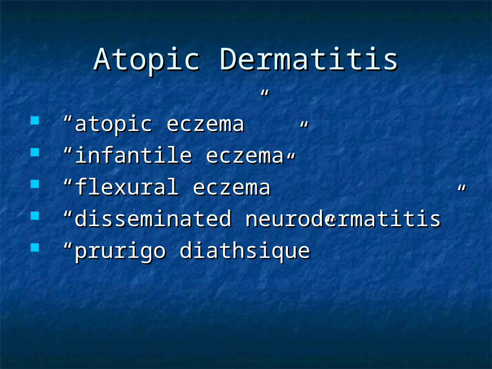
Atopic DermatitisAtopic Dermatitis
“ “atopic eczema”atopic eczema” “ “infantile eczema”infantile eczema” “ “flexural eczema”flexural eczema” “ “disseminated neurodermatitis”disseminated neurodermatitis” “ “prurigo diathsique” prurigo diathsique”
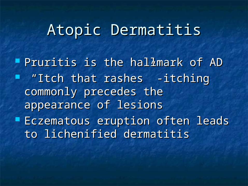
Atopic DermatitisAtopic Dermatitis
Pruritis is the hallmark of ADPruritis is the hallmark of AD “ “Itch that rashes” -itching commonly Itch that rashes” -itching commonly
precedes the appearance of lesionsprecedes the appearance of lesions Eczematous eruption often leads to Eczematous eruption often leads to
lichenified dermatitislichenified dermatitis
Atopic DermatitisAtopic Dermatitis Commonly associated with xerosis and Commonly associated with xerosis and
susceptibility to irritants and proteins, as well as an susceptibility to irritants and proteins, as well as an atopic diathesis (tendency towards asthma, allergic atopic diathesis (tendency towards asthma, allergic rhinitis, IgE mediated systemic manisfestations) rhinitis, IgE mediated systemic manisfestations)
IgE bound to Langerhans cells in atopic skinIgE bound to Langerhans cells in atopic skin Food exacerbates symptoms in some patients: soy, Food exacerbates symptoms in some patients: soy,
eggs, peanuts, cow’s milk represent up to 75% of eggs, peanuts, cow’s milk represent up to 75% of positive tests for food allergies- most commonly positive tests for food allergies- most commonly seen in infants & kids w/ mod-severe AD (up to 40% seen in infants & kids w/ mod-severe AD (up to 40% of pts.)of pts.)
Most frequent allergens leading to respiratory Most frequent allergens leading to respiratory allergies are dust mites, pollen, animal fur, & allergies are dust mites, pollen, animal fur, & molds- (seen in up to 70% of pts and associated w/ molds- (seen in up to 70% of pts and associated w/ adult AD)adult AD)
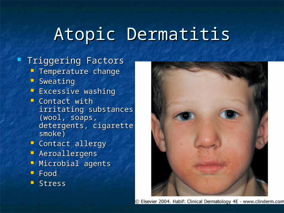
Atopic DermatitisAtopic Dermatitis Triggering FactorsTriggering Factors
Temperature changeTemperature change SweatingSweating Excessive washingExcessive washing Contact with irritating Contact with irritating
substances (wool, substances (wool, soaps, detergents, soaps, detergents, cigarette smoke)cigarette smoke)
Contact allergyContact allergy AeroallergensAeroallergens Microbial agentsMicrobial agents FoodFood StressStress
AD – 3 StagesAD – 3 Stages
InfantileInfantile
2 months to 2 years2 months to 2 years ChildhoodChildhood
2 years to 10 years2 years to 10 years AdultAdult
adolescence to adulthoodadolescence to adulthood
Infantile Atopic DermatitisInfantile Atopic Dermatitis
60% of AD present in the first year of life, 60% of AD present in the first year of life, after 2 months of ageafter 2 months of age
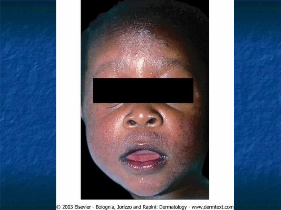
Begin as itchy erythema of the cheeksBegin as itchy erythema of the cheeks Distribution include scalp, neck, forehead, Distribution include scalp, neck, forehead,
wrist, and extensorswrist, and extensors May desquamate leading to erythrodermaMay desquamate leading to erythroderma Buttocks and diaper area frequently Buttocks and diaper area frequently
sparedspared
Infantile Atopic DermatitisInfantile Atopic Dermatitis
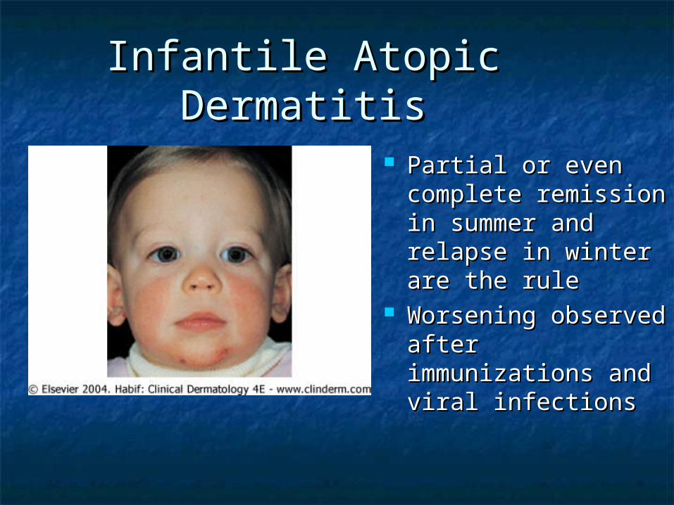
Partial or even Partial or even complete remission complete remission in summer and in summer and relapse in winter relapse in winter are the ruleare the rule
Worsening Worsening observed after observed after immunizations and immunizations and viral infectionsviral infections
Infantile Atopic DermatitisInfantile Atopic Dermatitis
In most cases the symptoms will In most cases the symptoms will disappear toward the end of the disappear toward the end of the second year.second year.
Egg, peanut, milk, wheat, fish, soy, Egg, peanut, milk, wheat, fish, soy, and chicken may exacerbate infantile and chicken may exacerbate infantile ADAD
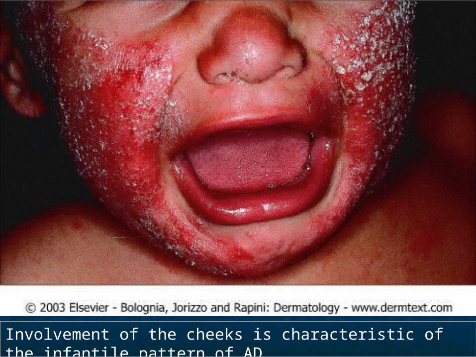
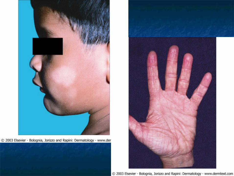
Involvement of the cheeks is characteristic of the infantile pattern of AD.
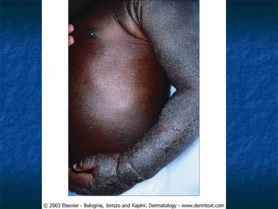
Childhood Atopic DermatitisChildhood Atopic Dermatitis
Characterized by less acute lesionsCharacterized by less acute lesions Distribution: antecubital and Distribution: antecubital and
popliteal fossae, flexor wrist, eyelids, popliteal fossae, flexor wrist, eyelids, and face.and face.
Severe atopic dermatitis involving Severe atopic dermatitis involving more than 50% of body surface area more than 50% of body surface area is associated with growth is associated with growth retardation. retardation.
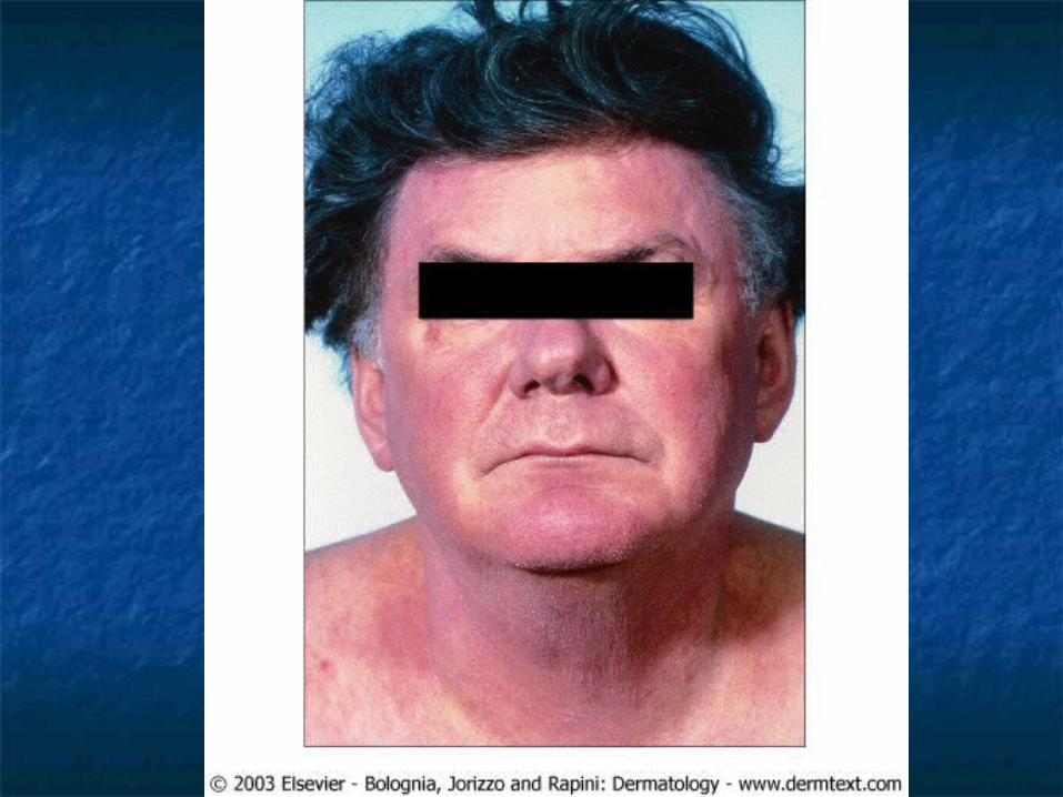
Adult Atopic DermatitisAdult Atopic Dermatitis
Distribution: antecubital and popliteal Distribution: antecubital and popliteal fossae, the front side of the neck, the fossae, the front side of the neck, the forehead, and area around the eyes.forehead, and area around the eyes.
Atopic individuals are at greater risk of Atopic individuals are at greater risk of developing hand dermatitis than are the developing hand dermatitis than are the rest of the populationrest of the population
70% develop hand dermatitis sometime in 70% develop hand dermatitis sometime in their livestheir lives Common in women after birth of their 1Common in women after birth of their 1stst child, child,
when increased exposure to soaps and water when increased exposure to soaps and water trigger diseasetrigger disease

Cutaneous stigmataCutaneous stigmata
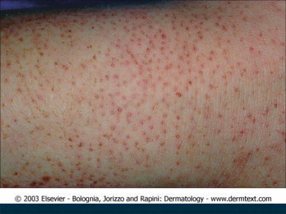
Dennie-Morgan foldsDennie-Morgan folds Pityriasis albaPityriasis alba Keratosis pilarisKeratosis pilaris Hertoghe’s sign – thinning of the lateral eyebrowsHertoghe’s sign – thinning of the lateral eyebrows Keratosis punctata palmaris et plantarisKeratosis punctata palmaris et plantaris XerosisXerosis Icthyosis vulgarisIcthyosis vulgaris Palmarplantar hyperlinearityPalmarplantar hyperlinearity CheilitisCheilitis LSCLSC Prurigo NodularisPrurigo Nodularis
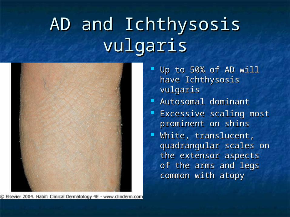
AD and Ichthysosis vulgarisAD and Ichthysosis vulgaris
Up to 50% of AD will Up to 50% of AD will have Ichthysosis have Ichthysosis vulgarisvulgaris
Autosomal dominantAutosomal dominant Excessive scaling most Excessive scaling most
prominent on shinsprominent on shins White, translucent, White, translucent,
quadrangular scales on quadrangular scales on the extensor aspects of the extensor aspects of the arms and legs the arms and legs common with atopycommon with atopy

Vascular StigmataVascular Stigmata
Headlight sign – perinasal and Headlight sign – perinasal and periorbital pallorperiorbital pallor
White dermographism – blanching of White dermographism – blanching of the skin at the site of stroking with a the skin at the site of stroking with a blunt instrument – cause edema and blunt instrument – cause edema and obscure color of underlying vessels.obscure color of underlying vessels.
InfectionInfection
Pts w/ AD more prone to certain cutaneous infx, Pts w/ AD more prone to certain cutaneous infx, may have more widespread infx, & may have may have more widespread infx, & may have exacerbations of their AD provoked by skin infxexacerbations of their AD provoked by skin infx
Staph aureus – 90% of chronic lesionsStaph aureus – 90% of chronic lesions Eczema herpeticum – generalized herpes simplex Eczema herpeticum – generalized herpes simplex
infection. Young children usually.infection. Young children usually. Secondary to reduced cell-mediated immunitySecondary to reduced cell-mediated immunity
Vaccination against smallpox is contraindicated in Vaccination against smallpox is contraindicated in person with atopic dermatitis. Even when person with atopic dermatitis. Even when condition is in remission, widespread and even condition is in remission, widespread and even fatal vaccinia can occur.fatal vaccinia can occur.
Also more prone to other cutaneous viral diseases Also more prone to other cutaneous viral diseases such as flat warts and molluscum contagiosumsuch as flat warts and molluscum contagiosum
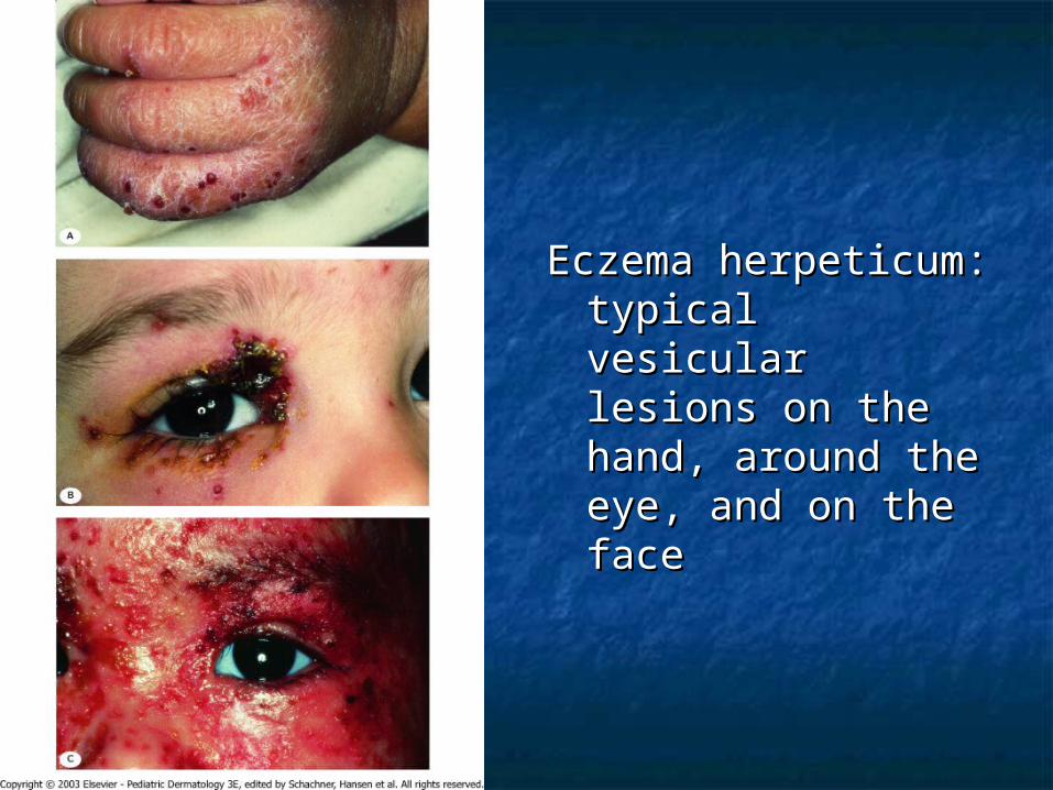
Eczema herpeticum: Eczema herpeticum: typical vesicular typical vesicular lesions on the lesions on the hand, around the hand, around the eye, and on the eye, and on the faceface
ImmunologyImmunology
T helper cell type 2 (Th2) dominanceT helper cell type 2 (Th2) dominance Th2 produces IL-4, 5, and 10Th2 produces IL-4, 5, and 10 IL-4 and IL-5 produce elevated IgE and IL-4 and IL-5 produce elevated IgE and
eosinophiliaeosinophilia IL-10 inhibits delayed type IL-10 inhibits delayed type
hypersensitivityhypersensitivity Th2 may be sensitive to house mites Th2 may be sensitive to house mites
or grass pollenor grass pollen

ImmunologyImmunology
Monocytes produces elevated Monocytes produces elevated amount of prostaglandin E2 (PGE2)amount of prostaglandin E2 (PGE2)
PGE2 reduces gamma-interferon PGE2 reduces gamma-interferon production, but not IL-4 from helper production, but not IL-4 from helper cells thereby enhancing the Th2 cells thereby enhancing the Th2 dominancedominance
PGE2 also directly enhances IgE PGE2 also directly enhances IgE production from B cellsproduction from B cells
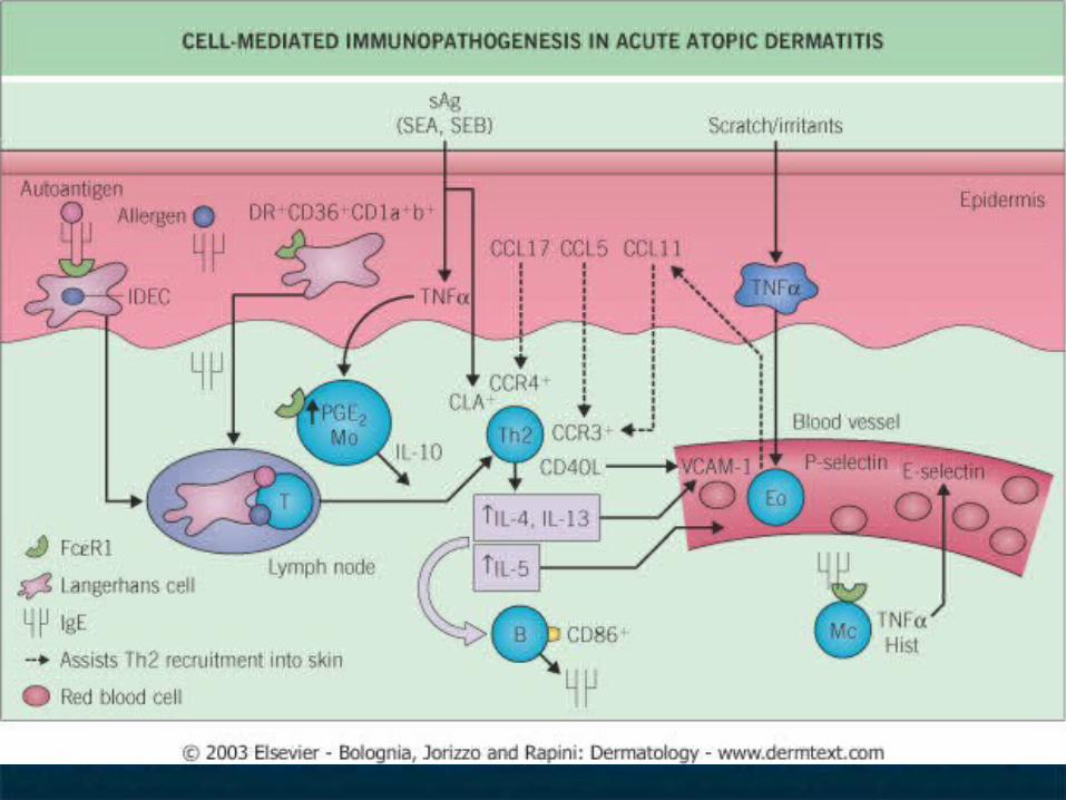
ImmunologyImmunology
Langerhans cells of AD patient Langerhans cells of AD patient stimulate helper T cells into Th2 stimulate helper T cells into Th2 phenotype without the presence of phenotype without the presence of antigenantigen
Langerhans cells have IgE bound to Langerhans cells have IgE bound to their suface receptors. These IgE are their suface receptors. These IgE are associated with atopic antigens, such associated with atopic antigens, such as house dust mitesas house dust mites
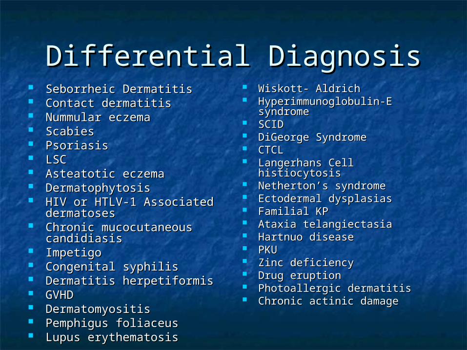
Differential DiagnosisDifferential Diagnosis Seborrheic DermatitisSeborrheic Dermatitis Contact dermatitisContact dermatitis Nummular eczemaNummular eczema ScabiesScabies Psoriasis Psoriasis LSCLSC Asteatotic eczemaAsteatotic eczema DermatophytosisDermatophytosis HIV or HTLV-1 Associated HIV or HTLV-1 Associated
dermatosesdermatoses Chronic mucocutaneous Chronic mucocutaneous
candidiasiscandidiasis ImpetigoImpetigo Congenital syphilisCongenital syphilis Dermatitis herpetiformisDermatitis herpetiformis GVHDGVHD DermatomyositisDermatomyositis Pemphigus foliaceusPemphigus foliaceus Lupus erythematosisLupus erythematosis
Wiskott- AldrichWiskott- Aldrich Hyperimmunoglobulin-E Hyperimmunoglobulin-E
syndromesyndrome SCIDSCID DiGeorge SyndromeDiGeorge Syndrome CTCLCTCL Langerhans Cell histiocytosisLangerhans Cell histiocytosis Netherton’s syndromeNetherton’s syndrome Ectodermal dysplasiasEctodermal dysplasias Familial KPFamilial KP Ataxia telangiectasiaAtaxia telangiectasia Hartnuo diseaseHartnuo disease PKUPKU Zinc deficiencyZinc deficiency Drug eruptionDrug eruption Photoallergic dermatitisPhotoallergic dermatitis Chronic actinic damageChronic actinic damage
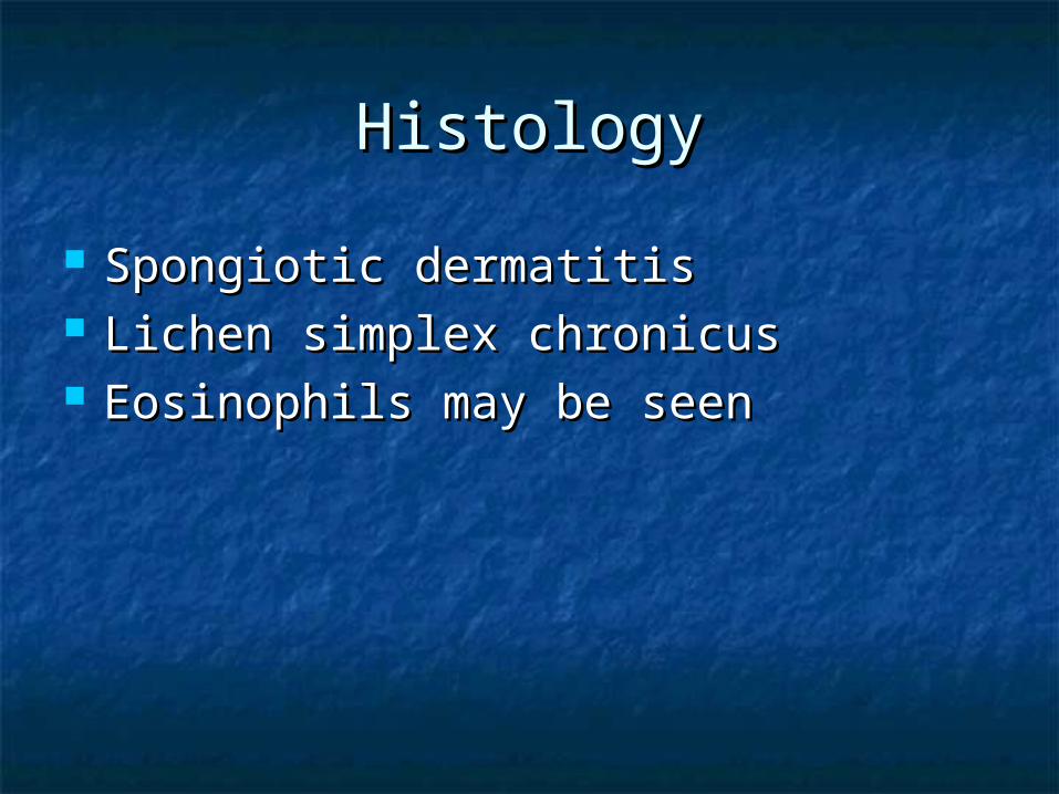
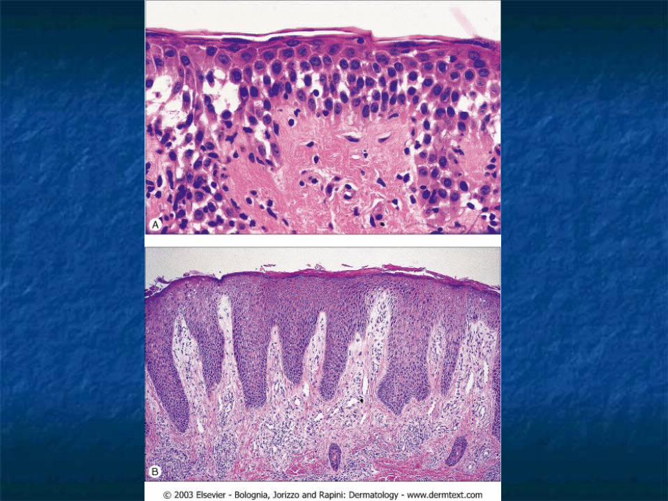
HistologyHistology
Spongiotic dermatitisSpongiotic dermatitis Lichen simplex chronicusLichen simplex chronicus Eosinophils may be seenEosinophils may be seen
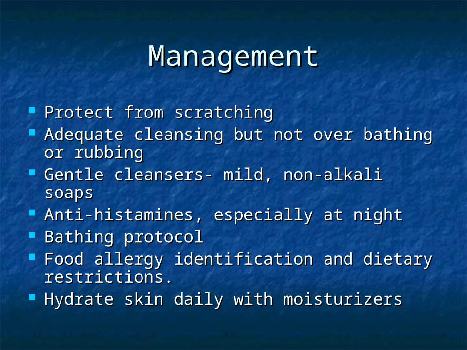
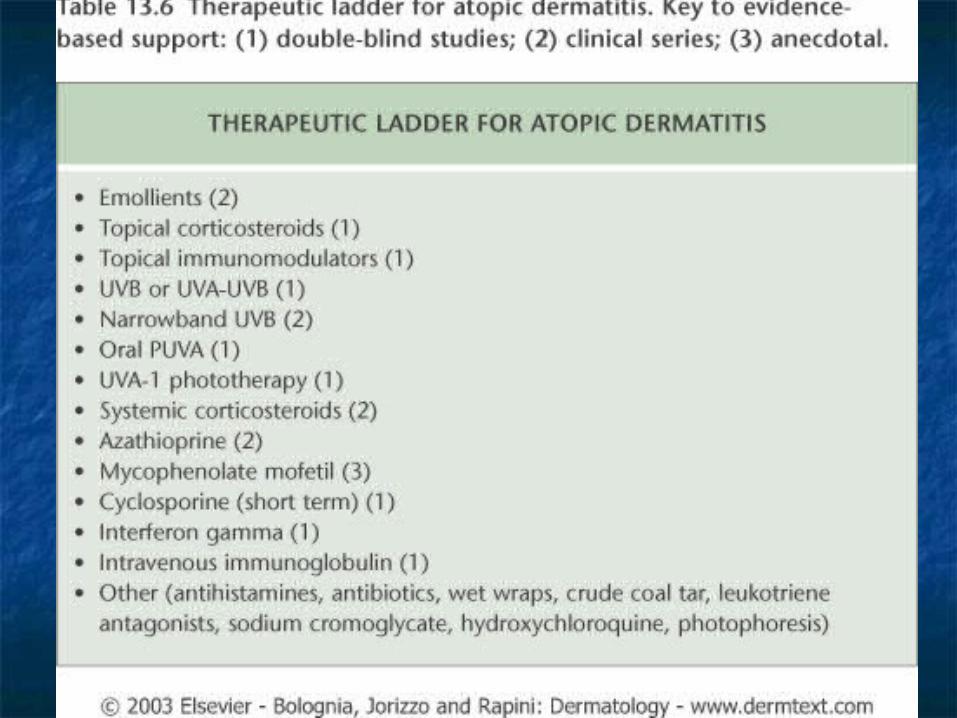
ManagementManagement
Protect from scratchingProtect from scratching Adequate cleansing but not over bathing Adequate cleansing but not over bathing
or rubbingor rubbing Gentle cleansers- mild, non-alkali soapsGentle cleansers- mild, non-alkali soaps Anti-histamines, especially at nightAnti-histamines, especially at night Bathing protocolBathing protocol Food allergy identification and dietary Food allergy identification and dietary
restrictions.restrictions. Hydrate skin daily with moisturizersHydrate skin daily with moisturizers

ManagementManagement
Topical steroidsTopical steroids Wet compress of Burow’s solution Wet compress of Burow’s solution
such as Domeboro.such as Domeboro. Crude coal tar/liquor corbonis Crude coal tar/liquor corbonis
detergens (LCD)detergens (LCD) PUVAPUVA CyclosporineCyclosporine
ManagementManagement
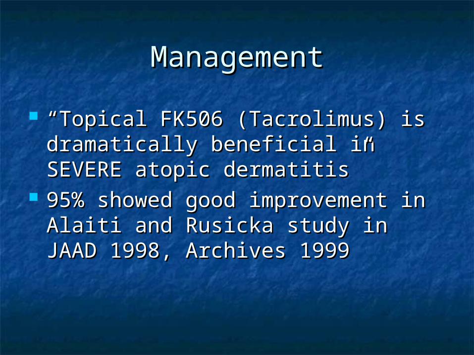
““Topical FK506 (Tacrolimus) is Topical FK506 (Tacrolimus) is dramatically beneficial in SEVERE dramatically beneficial in SEVERE atopic dermatitis”atopic dermatitis”
95% showed good improvement in 95% showed good improvement in Alaiti and Rusicka study in JAAD Alaiti and Rusicka study in JAAD 1998, Archives 19991998, Archives 1999
Regional EczemaRegional Eczema
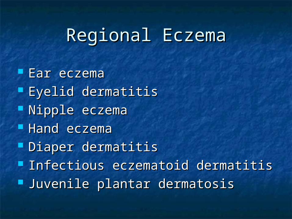
Ear eczemaEar eczema Eyelid dermatitisEyelid dermatitis Nipple eczemaNipple eczema Hand eczemaHand eczema Diaper dermatitisDiaper dermatitis Infectious eczematoid dermatitisInfectious eczematoid dermatitis Juvenile plantar dermatosisJuvenile plantar dermatosis
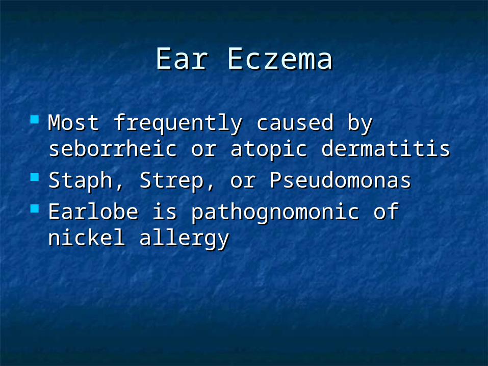
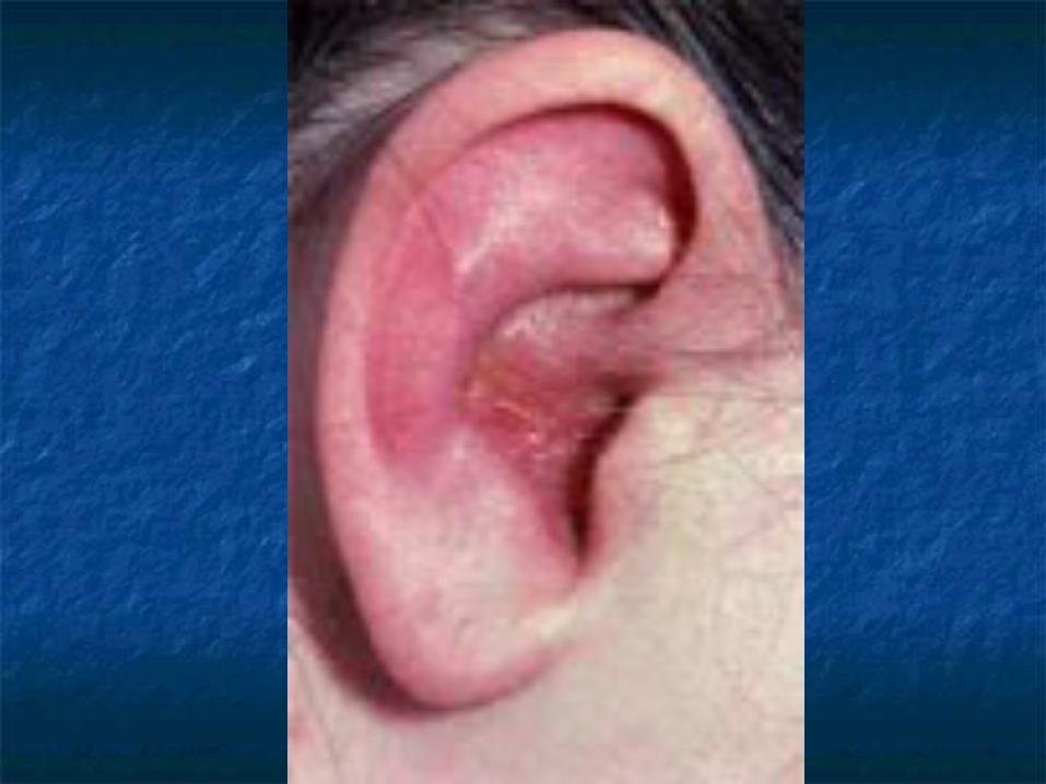
Ear EczemaEar Eczema
Most frequently caused by seborrheic Most frequently caused by seborrheic or atopic dermatitisor atopic dermatitis
Staph, Strep, or PseudomonasStaph, Strep, or Pseudomonas Earlobe is pathognomonic of nickel Earlobe is pathognomonic of nickel
allergyallergy
Eyelid dermatitisEyelid dermatitis
When on one eye only, it is most When on one eye only, it is most frequently caused by nail polish, and frequently caused by nail polish, and usually affects the upper eyelidusually affects the upper eyelid
When both eyes are involved, When both eyes are involved, consider mascara, eye shadow, consider mascara, eye shadow, eyelash cement, eyeliner, etceyelash cement, eyeliner, etc
In contrast, atopic dermatitis affects In contrast, atopic dermatitis affects both upper and lower eyelid.both upper and lower eyelid.
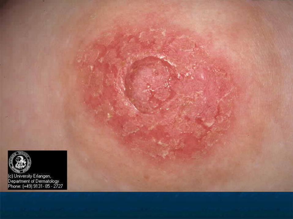
Nipple eczemaNipple eczema
Painful fissuring, seen especially in Painful fissuring, seen especially in nursing mothersnursing mothers
Maybe an isolated manifestation of Maybe an isolated manifestation of atopic dermatitisatopic dermatitis
If persist more than 3 months, and/or If persist more than 3 months, and/or unilateral, biopsy is mandatory to unilateral, biopsy is mandatory to rule out Paget’srule out Paget’s
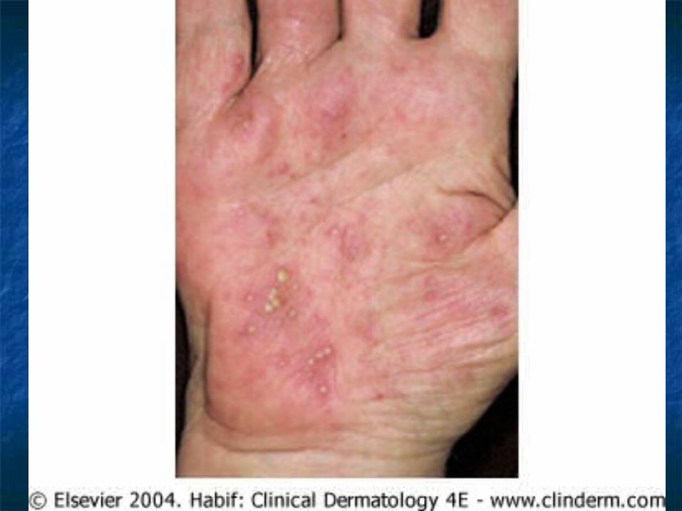
Hand eczemaHand eczema
Spongiosis histologicallySpongiosis histologically Irritant hand dermatitis- seen in Irritant hand dermatitis- seen in
homemakers, nurses. Resulting from homemakers, nurses. Resulting from excessive exposure to soapsexcessive exposure to soaps
Pompholyx- tapioca vesicles, on Pompholyx- tapioca vesicles, on sides of fingers, palms, and solessides of fingers, palms, and soles
Differentials – Bullous tinea, id, Differentials – Bullous tinea, id, allergic contact dermatitisallergic contact dermatitis
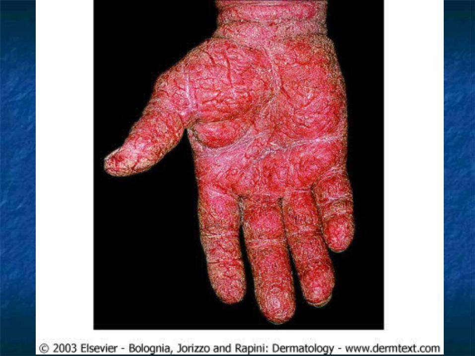

TreatmentTreatment BarrierBarrier MoisturizerMoisturizer Systemic CorticosteroidsSystemic Corticosteroids Phototherapy – UVA, PUVA, Phototherapy – UVA, PUVA,
Radiotherapy (Grenz Ray)Radiotherapy (Grenz Ray) New research suggests use of oral New research suggests use of oral
retinoids for severe recalcitrant hand retinoids for severe recalcitrant hand eczemaeczema
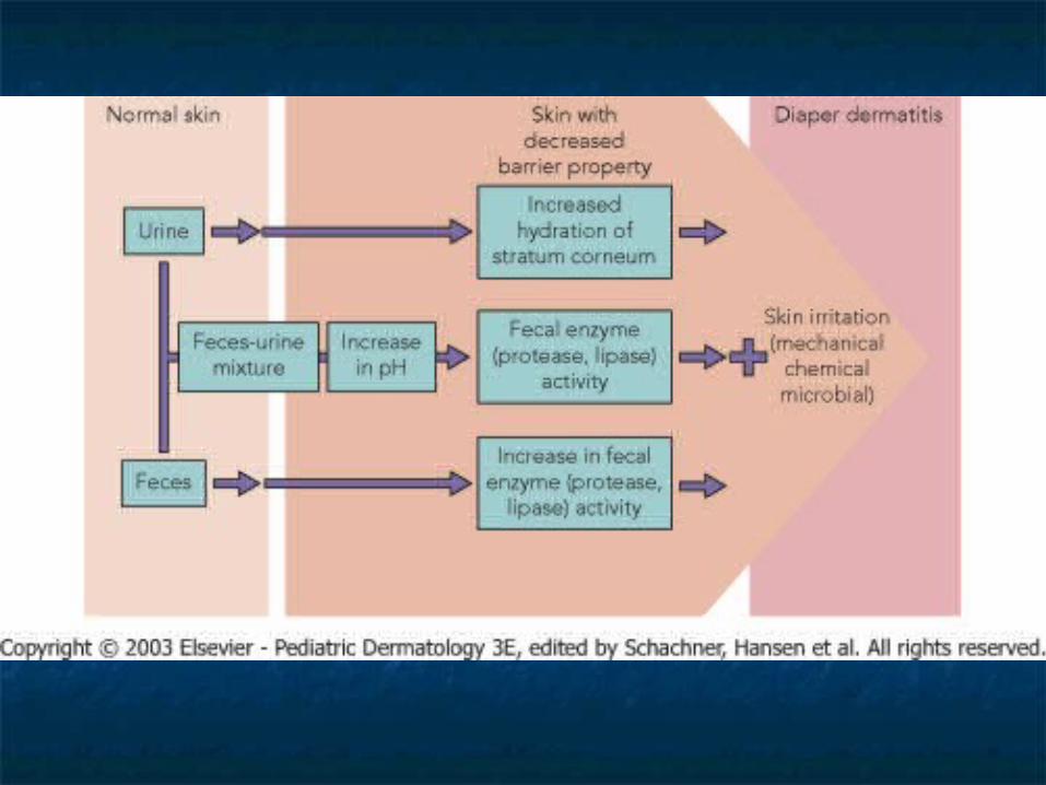
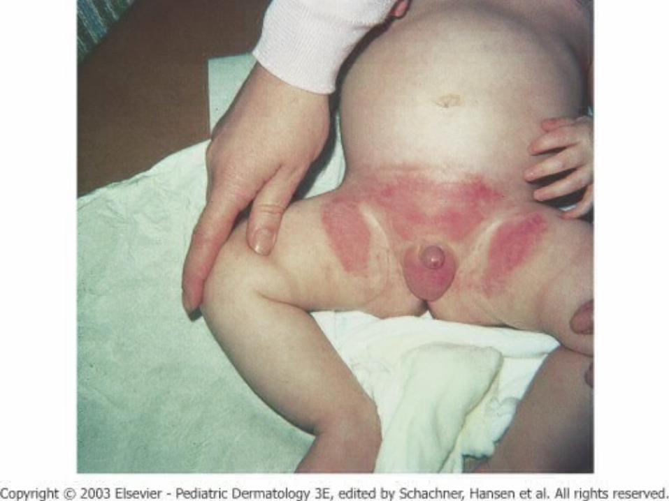
Diaper (Napkin) DermatitisDiaper (Napkin) Dermatitis
Erythematous, papulovesicular Erythematous, papulovesicular dermatitis distributed over the lower dermatitis distributed over the lower abdomen, genitals, thighs, and the abdomen, genitals, thighs, and the convex surfaces of the buttocksconvex surfaces of the buttocks
Irritation caused by bacteria, change Irritation caused by bacteria, change in the environment (moisture, lower in the environment (moisture, lower PH, feces)PH, feces)
Candida albicans secondary infection.Candida albicans secondary infection.
Diaper dermatitis - Diaper dermatitis - complicationscomplications
Jacquet’s erosive diaper dermatitisJacquet’s erosive diaper dermatitis Punched out ulcers/erosions with Punched out ulcers/erosions with
elevated borderselevated borders Pseudoverrucous papule and nodulesPseudoverrucous papule and nodules Granuloma gluteal infantumGranuloma gluteal infantum
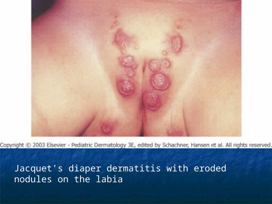
Jacquet’s diaper dermatitis with eroded nodules on the labia
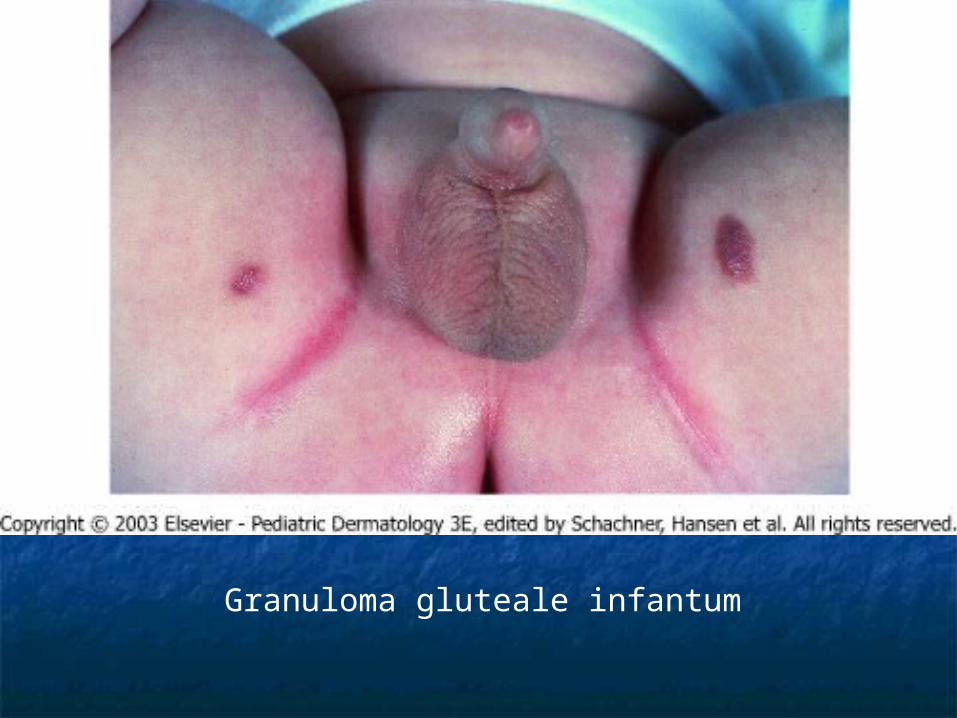
Granuloma gluteale infantum
Diaper DermatitisDiaper Dermatitis
Differential diagnosis: Napkin Differential diagnosis: Napkin psoriasis, seborrheic dermatitis, psoriasis, seborrheic dermatitis, atopic dermatitis, langerhans cell atopic dermatitis, langerhans cell histiocytosis, tinea cruris, allergic histiocytosis, tinea cruris, allergic contact dermatitis, acrodermatitis contact dermatitis, acrodermatitis enteropathica, biotin deficiency, enteropathica, biotin deficiency, congenital syphilliscongenital syphillis
Treatment: preventionTreatment: prevention
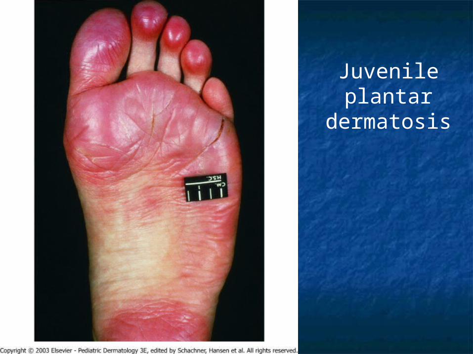
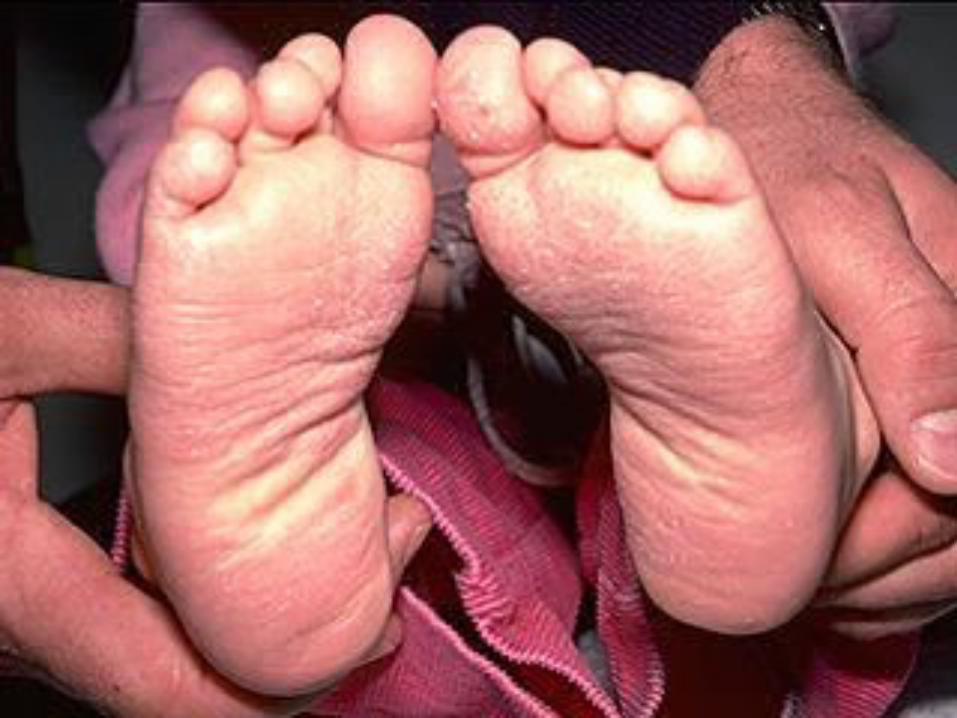
Juvenile plantar dermatosisJuvenile plantar dermatosis
Begins as a patchy symmetrical, smooth, red, Begins as a patchy symmetrical, smooth, red, glazed macules on the base of the great toesglazed macules on the base of the great toes
Affect age 3 to puberty.Affect age 3 to puberty. Symmetrical lesions on weight bearing areaSymmetrical lesions on weight bearing area ““toxic sock syndrome” – caused by repeated toxic sock syndrome” – caused by repeated
maceration of the feet by occlusive shoes and maceration of the feet by occlusive shoes and nonabsorbent synthetic socksnonabsorbent synthetic socks
Virtually always resolve after pubertyVirtually always resolve after puberty
Juvenile plantar dermatosis

Xerotic EczemaXerotic Eczema
Aka winter itch, nummular eczema, Aka winter itch, nummular eczema, eczema craquele, and asteototic eczema craquele, and asteototic eczema.eczema.
Anterior shins, extensor arms, and flankAnterior shins, extensor arms, and flank Elderly person predisposed.Elderly person predisposed. Use of bath oils in bath water is Use of bath oils in bath water is
recommended to prevent water lossrecommended to prevent water loss Moisturizers – urea or lactic acid.Moisturizers – urea or lactic acid.
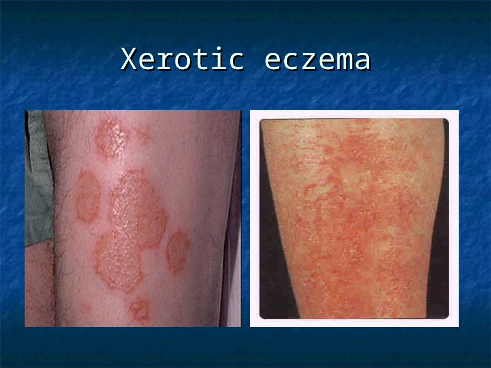
Xerotic eczemaXerotic eczema
Hormone Induced Hormone Induced DermatosesDermatoses
Autoimmune progesterone dermatitis – Appear 5-10 days Autoimmune progesterone dermatitis – Appear 5-10 days before menses. Oophorectomy, danazol, and tamoxifen are before menses. Oophorectomy, danazol, and tamoxifen are treatment modalitiestreatment modalities- Urticaria- Urticaria- Angioedema- Angioedema- Eczema- Eczema- Erythema multiforme- Erythema multiforme- Stomatitis- Stomatitis- Folliculitis- Folliculitis- Papulopustular/papulovesicular lesions- Papulopustular/papulovesicular lesions- Stephens-Johnson syndrome- Stephens-Johnson syndrome- Vesiculobullous reactions- Vesiculobullous reactions- Dermatitis herpetiformis-like rash- Dermatitis herpetiformis-like rash- Mucosal lesions- Mucosal lesions
Autoimmune estrogen dermatitis – a cyclic skin disorder Autoimmune estrogen dermatitis – a cyclic skin disorder with variable morphologies. Exacerbate premenstrually or with variable morphologies. Exacerbate premenstrually or occur only immediately before the menses. Treatment with occur only immediately before the menses. Treatment with tamoxifen maybe effective.tamoxifen maybe effective.
Immunodeficiency Immunodeficiency SyndromesSyndromes
X-Linked AgammaglobulinemiaX-Linked Agammaglobulinemia Isolated IgA DeficiencyIsolated IgA Deficiency Common Variable ImmunodificiencyCommon Variable Immunodificiency Isolated Primary IgM DeficiencyIsolated Primary IgM Deficiency Immunodificiency with Hyper-IgMImmunodificiency with Hyper-IgM Thymic HypoplasiaThymic Hypoplasia Thymic Dysplasia with Normal Thymic Dysplasia with Normal
Immunoglobulins (Nezelof Syndrome)Immunoglobulins (Nezelof Syndrome)
Immunodeficiency Immunodeficiency SyndromesSyndromes
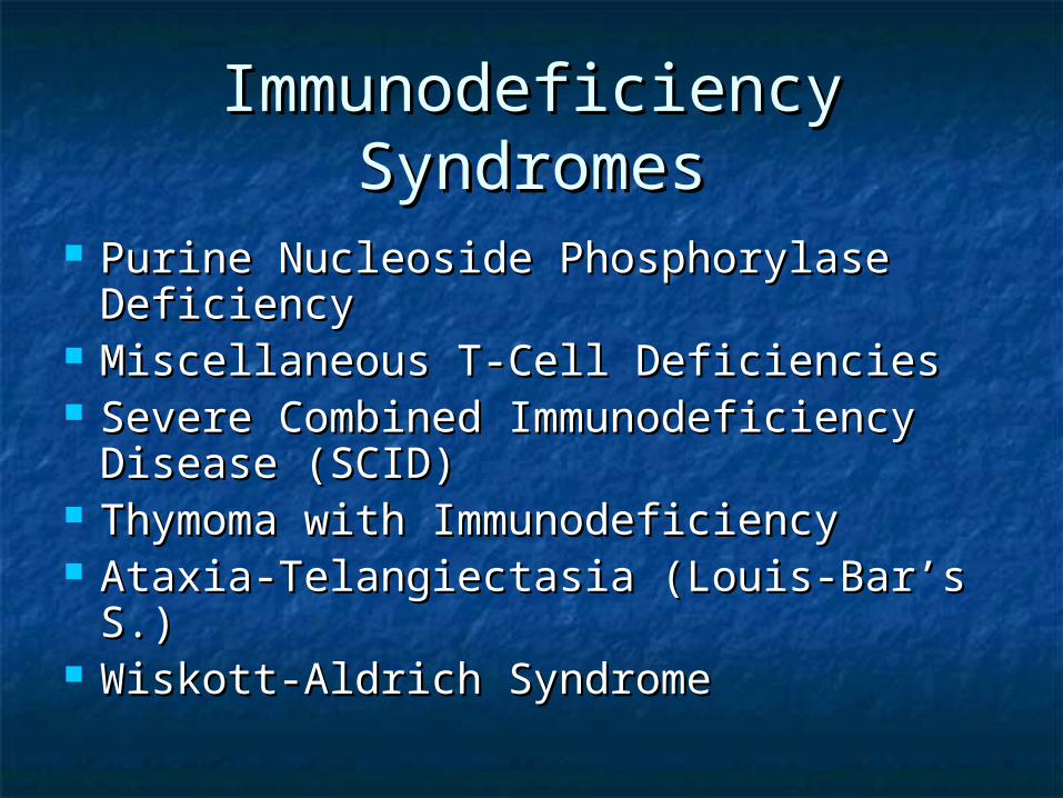
Purine Nucleoside Phosphorylase Purine Nucleoside Phosphorylase DeficiencyDeficiency
Miscellaneous T-Cell DeficienciesMiscellaneous T-Cell Deficiencies Severe Combined Immunodeficiency Severe Combined Immunodeficiency
Disease (SCID)Disease (SCID) Thymoma with ImmunodeficiencyThymoma with Immunodeficiency Ataxia-Telangiectasia (Louis-Bar’s S.)Ataxia-Telangiectasia (Louis-Bar’s S.) Wiskott-Aldrich SyndromeWiskott-Aldrich Syndrome
Immunodeficiency Immunodeficiency SyndromesSyndromes
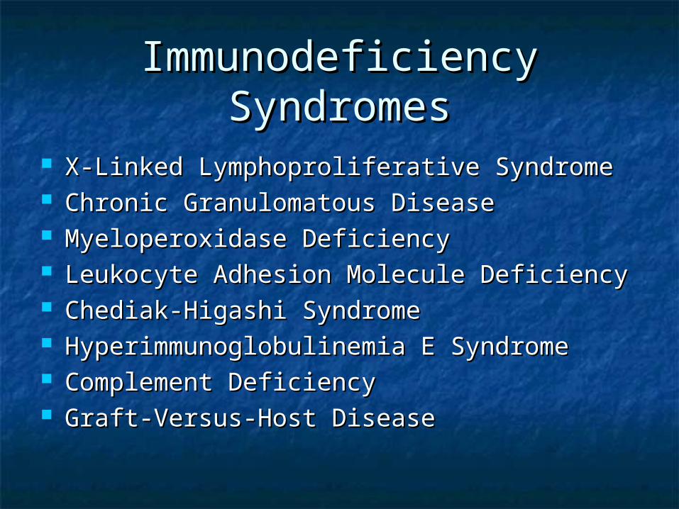
X-Linked Lymphoproliferative SyndromeX-Linked Lymphoproliferative Syndrome Chronic Granulomatous DiseaseChronic Granulomatous Disease Myeloperoxidase DeficiencyMyeloperoxidase Deficiency Leukocyte Adhesion Molecule DeficiencyLeukocyte Adhesion Molecule Deficiency Chediak-Higashi SyndromeChediak-Higashi Syndrome Hyperimmunoglobulinemia E SyndromeHyperimmunoglobulinemia E Syndrome Complement DeficiencyComplement Deficiency Graft-Versus-Host DiseaseGraft-Versus-Host Disease
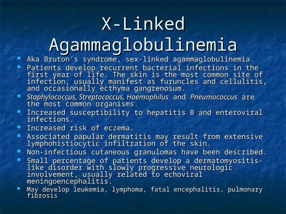
X-Linked X-Linked AgammaglobulinemiaAgammaglobulinemia
Aka Bruton’s syndrome, sex-linked agammaglobulinemia.Aka Bruton’s syndrome, sex-linked agammaglobulinemia. Patients develop recurrent bacterial infections in the first Patients develop recurrent bacterial infections in the first
year of life. The skin is the most common site of infection, year of life. The skin is the most common site of infection, usually manifest as furuncles and cellulitis, and occasionally usually manifest as furuncles and cellulitis, and occasionally ecthyma gangrenosum. ecthyma gangrenosum.
Staphylococcus, Streptococcus, HaemophilusStaphylococcus, Streptococcus, Haemophilus and and PneumococcusPneumococcus are the most common organisms. are the most common organisms.
Increased susceptibility to hepatitis B and enteroviral Increased susceptibility to hepatitis B and enteroviral infections. infections.
Increased risk of eczema. Increased risk of eczema. Associated papular dermatitis may result from extensive Associated papular dermatitis may result from extensive
lymphohistiocytic infiltration of the skin. lymphohistiocytic infiltration of the skin. Non-infectious cutaneous granulomas have been described.Non-infectious cutaneous granulomas have been described. Small percentage of patients develop a dermatomyositis-like Small percentage of patients develop a dermatomyositis-like
disorder with slowly progressive neurologic involvement, disorder with slowly progressive neurologic involvement, usually related to echoviral meningoencephalitis. usually related to echoviral meningoencephalitis.
May develop leukemia, lymphoma, fatal encephalitis, pulmonary May develop leukemia, lymphoma, fatal encephalitis, pulmonary fibrosisfibrosis
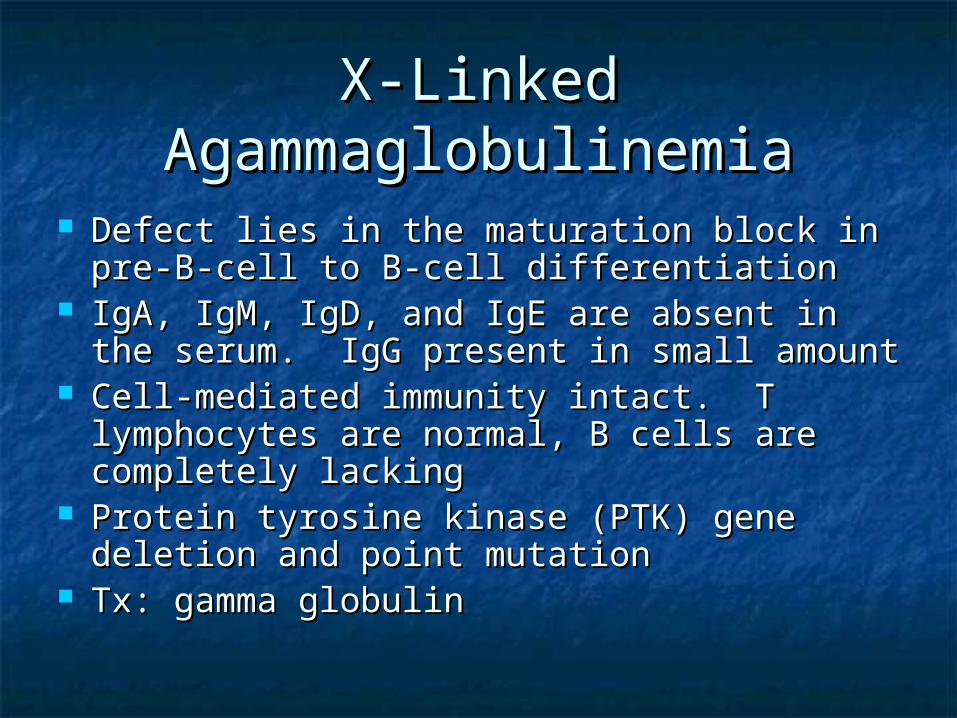
X-Linked X-Linked AgammaglobulinemiaAgammaglobulinemia
Defect lies in the maturation block in pre-Defect lies in the maturation block in pre-B-cell to B-cell differentiationB-cell to B-cell differentiation
IgA, IgM, IgD, and IgE are absent in the IgA, IgM, IgD, and IgE are absent in the serum. IgG present in small amountserum. IgG present in small amount
Cell-mediated immunity intact. T Cell-mediated immunity intact. T lymphocytes are normal, B cells are lymphocytes are normal, B cells are completely lackingcompletely lacking
Protein tyrosine kinase (PTK) gene deletion Protein tyrosine kinase (PTK) gene deletion and point mutationand point mutation
Tx: gamma globulinTx: gamma globulin

Selective IgA DeficiencySelective IgA Deficiency Most common immunoglobulin deficiencyMost common immunoglobulin deficiency Usually asymptomaticUsually asymptomatic Clinical manifestations 10-15% Clinical manifestations 10-15%
Sinopulmonary bacterial infectionsSinopulmonary bacterial infections Giardia Giardia gastroenteritisgastroenteritis
1/3 with clinical disease develop autoimmune disorders1/3 with clinical disease develop autoimmune disorders SLE, Vitiligo, chronic mucocutaneous candidiasis, SLE, Vitiligo, chronic mucocutaneous candidiasis,
lipodystrophia centrifugalis abdominalis, ITPlipodystrophia centrifugalis abdominalis, ITP No sexual predilectionNo sexual predilection A number of patients have severe atopic-like dermatitis, A number of patients have severe atopic-like dermatitis,
asthma, cow's milk allergy, and/or allergic asthma, cow's milk allergy, and/or allergic rhinoconjunctivitis.rhinoconjunctivitis.
An extremely rare selective immunoglobulin deficiency, IgM An extremely rare selective immunoglobulin deficiency, IgM deficiency, is apparently caused by an inability of T-helper deficiency, is apparently caused by an inability of T-helper cell function to stimulate IgM production. In addition to cell function to stimulate IgM production. In addition to recurrent bacterial infections, severe eczema and extensive recurrent bacterial infections, severe eczema and extensive large warts have been described. large warts have been described.
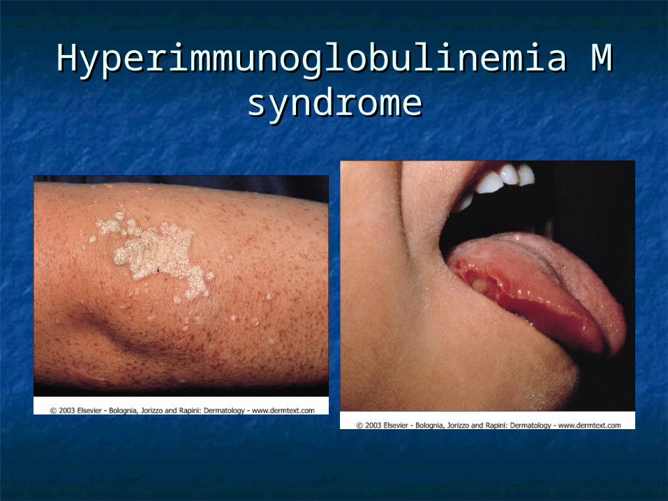
Hyperimmunoglobulinemia M Hyperimmunoglobulinemia M syndromesyndrome
Isolated IgA DeficiencyIsolated IgA Deficiency
Absence or marked reduction of Absence or marked reduction of serum IgAserum IgA
1:600 in white population, most are 1:600 in white population, most are entirely well.entirely well.
Malignancy is increased in adult with Malignancy is increased in adult with IgA deficiency.IgA deficiency.

Common Variable Common Variable ImmunodificiencyImmunodificiency
Aka acquired hypogammaglobulinemiaAka acquired hypogammaglobulinemia HLA marker B8 and DR3 are affectedHLA marker B8 and DR3 are affected B cells present but not terminally B cells present but not terminally
differentiateddifferentiated T cell dysfunction evidentT cell dysfunction evident Recurrent sinopulmonary infections- Recurrent sinopulmonary infections-
patients are especially predisposed to patients are especially predisposed to pyogenic upper and lower respiratory tract pyogenic upper and lower respiratory tract infectionsinfections
Common Variable Common Variable ImmunodeficiencyImmunodeficiency
Increased risk of autoimmune disordersIncreased risk of autoimmune disorders Vitiligo, alopecia areata, vasculitisVitiligo, alopecia areata, vasculitis
8- to 13-fold increased risk of cancer overall8- to 13-fold increased risk of cancer overall Increased incidence of lymphomaIncreased incidence of lymphoma
400 fold increase risk in female patients400 fold increase risk in female patients GiardiaGiardia infections are more common in CVID than in the X- infections are more common in CVID than in the X-
linked form. Patients frequently have cutaneous pyodermas linked form. Patients frequently have cutaneous pyodermas and eczema.and eczema.
Abnormalities of cell-mediated immunity may occur in Abnormalities of cell-mediated immunity may occur in addition to the Ig deficiency and may manifest in the skin as addition to the Ig deficiency and may manifest in the skin as widespread warts and extensive dermatophyte infections. widespread warts and extensive dermatophyte infections.
Pts may develop non-caseating granulomas of the lungs, Pts may develop non-caseating granulomas of the lungs, liver, spleen, and/or skin that are not due to liver, spleen, and/or skin that are not due to microorganisms. microorganisms.
Death in patients with CVID usually results from infection, Death in patients with CVID usually results from infection, respiratory insufficiency, or neoplasia. respiratory insufficiency, or neoplasia.
Isolated Primary IgM Isolated Primary IgM DeficiencyDeficiency
Eczematous dermatitis presents in Eczematous dermatitis presents in 1/5 of patients with this condition1/5 of patients with this condition
Predisposition to bacterial infectionPredisposition to bacterial infection Defect in maturation of IgM Defect in maturation of IgM
producing plasma cell.producing plasma cell.
Immunodificiency with Immunodificiency with Hyper-IgMHyper-IgM
Low or absent IgG, IgE, and IgA level. Low or absent IgG, IgE, and IgA level. Normal or elevated IgM and IgDNormal or elevated IgM and IgD
X-linked form caused by mutation or X-linked form caused by mutation or deletion of Xq26.3-27.1 region, which deletion of Xq26.3-27.1 region, which encodes a ligand of CD40, gp39encodes a ligand of CD40, gp39
Gp39-CD40 interaction signals for Ig Gp39-CD40 interaction signals for Ig isotype switching.isotype switching.
Tx: IVGG, and allogenic bone marrow Tx: IVGG, and allogenic bone marrow transplanttransplant
Thymic HypoplasiaThymic Hypoplasia
DiGeorge anomaly, aka III and IV pharyngeal DiGeorge anomaly, aka III and IV pharyngeal pouch syndromepouch syndrome
Facies: notched and low-set ears, micrognathia, Facies: notched and low-set ears, micrognathia, shorten philtrum, hypertelorismshorten philtrum, hypertelorism
Congenital absence of the parathyroid, thymus, Congenital absence of the parathyroid, thymus, and abnormal aortaand abnormal aorta
Hypocalcemia is the first signHypocalcemia is the first sign Aortic and cardiac defects are the most common Aortic and cardiac defects are the most common
cause of deathcause of death Deletions within proximal long arm of Deletions within proximal long arm of
chromosone 22chromosone 22
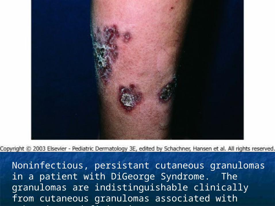
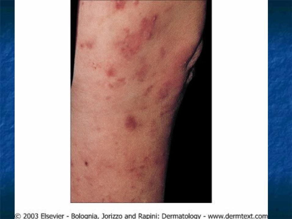
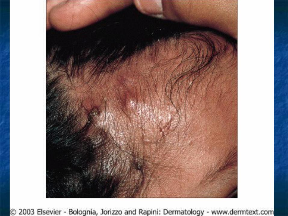
Noninfectious, persistant cutaneous granulomas in a patient with DiGeorge Syndrome. The granulomas are indistinguishable clinically from cutaneous granulomas associated with other immunodeficiencies.

Thymic Dysplasia with Thymic Dysplasia with Normal Immunoglobulins Normal Immunoglobulins
(Nezelof Syndrome)(Nezelof Syndrome) Faulty development of thymus glandFaulty development of thymus gland Autosomal recessiveAutosomal recessive Thymus is present but Thymus is present but
underdeveloped; no cardiac underdeveloped; no cardiac abnormalitiesabnormalities Contrast to DiGeorge syndromeContrast to DiGeorge syndrome

Purine Nucleoside Purine Nucleoside Phosphorylase DeficiencyPhosphorylase Deficiency
Greatly reduced T-Cell counts, Greatly reduced T-Cell counts, depressed cell mediated immunitydepressed cell mediated immunity
B cells and antibody formation intactB cells and antibody formation intact Mutation on 14q13Mutation on 14q13 Usually die of overwhelming viral Usually die of overwhelming viral
infectioninfection

Miscellaneous T-Cell Miscellaneous T-Cell DeficienciesDeficiencies
Cartilage-hair hypoplasia syndromeCartilage-hair hypoplasia syndrome AR, patient with short-limbed dwarfism, fine AR, patient with short-limbed dwarfism, fine
sparse, hypopigmented hair, defective cell sparse, hypopigmented hair, defective cell mediated immunity. Some may also have mediated immunity. Some may also have deecreased humoral immunity.deecreased humoral immunity.
Most common in Amish and FinnsMost common in Amish and Finns May have “doughy” skin secondary to May have “doughy” skin secondary to
degenerated elastic tissuedegenerated elastic tissue Increased risk of non-Hodgkin’s lymphoma and Increased risk of non-Hodgkin’s lymphoma and
basal cell carcinomasbasal cell carcinomas Patients are highly susceptible to severe Patients are highly susceptible to severe
disseminated varicella disseminated varicella
Miscellaneous T-Cell Miscellaneous T-Cell DeficienciesDeficiencies
Omenn’s syndromeOmenn’s syndrome ARAR Mimics GVHDMimics GVHD exfoliative erythroderma, eosinophilia, exfoliative erythroderma, eosinophilia,
recurrent infection, recurrent infection, hypogammaglobulinema, diarrhea, hypogammaglobulinema, diarrhea, hepatosplenomegly, alopeciahepatosplenomegly, alopecia
Early death by 6 monthsEarly death by 6 months Inefficient and abnormal generation of T-Inefficient and abnormal generation of T-
Cell receptors.Cell receptors.
SCID: Severe Combined SCID: Severe Combined Immunodeficiency DiseaseImmunodeficiency Disease
Severe impairment of humoral and cellular immunitySevere impairment of humoral and cellular immunity 75% of pts are males75% of pts are males Recurrent infections, diarrhea, and failure to thrive are Recurrent infections, diarrhea, and failure to thrive are
apparent by 3-6 months of ageapparent by 3-6 months of age Triad of Moniliasis of the oropharynx and skin, intractable Triad of Moniliasis of the oropharynx and skin, intractable
diarrhea, and pneumonia.diarrhea, and pneumonia. Overwhelming viral infection is the cause of death.Overwhelming viral infection is the cause of death. Deficiency or total absence of circulating lymphocytesDeficiency or total absence of circulating lymphocytes Infants can present with diffuse skin involvement: Infants can present with diffuse skin involvement:
seborrheic-like dermatitis or morbilliform eruptions.seborrheic-like dermatitis or morbilliform eruptions. Common early infections are mucocutaneous candidiasis, Common early infections are mucocutaneous candidiasis,
virus-induced chronic diarrhea with malabsorption, and virus-induced chronic diarrhea with malabsorption, and pneumonia due to bacteria, viruses, or pneumonia due to bacteria, viruses, or Pneumocystis Pneumocystis carinii.carinii.
Cutaneous infections are most often caused by Cutaneous infections are most often caused by C. albicans, C. albicans, S. aureusS. aureus, and , and S. pyogenesS. pyogenes. .
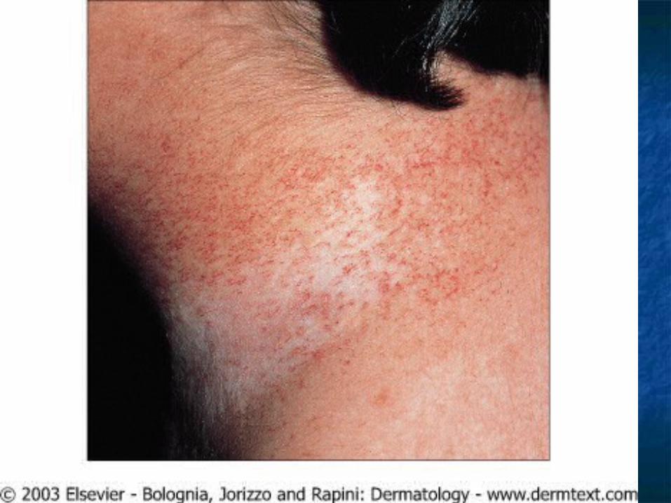
Ataxia-Telangiectasia Ataxia-Telangiectasia (Louis-Bar Syndrome)(Louis-Bar Syndrome)
Distinctive telangiectasia in bulbar conjuctiva Distinctive telangiectasia in bulbar conjuctiva and flexural suraces of the arm developing and flexural suraces of the arm developing during the 5during the 5thth year of age year of age
Telangiectasia occurs on butterfly area of the Telangiectasia occurs on butterfly area of the face, palate, ear, and exposed skin. Café au face, palate, ear, and exposed skin. Café au lait patches, and graying hair also present. lait patches, and graying hair also present.
Cerebellar ataxia is the first sign of this Cerebellar ataxia is the first sign of this syndrome, beginning in the second year of syndrome, beginning in the second year of life.life.
Choreic and athetoid movement present.Choreic and athetoid movement present.
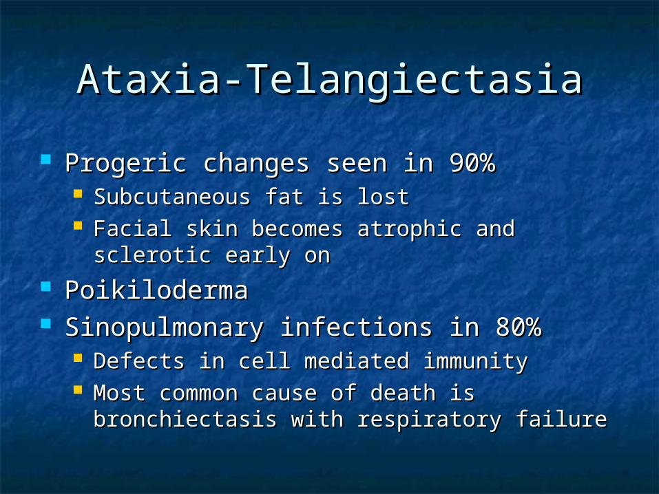
Ataxia-TelangiectasiaAtaxia-Telangiectasia
Progeric changes seen in 90%Progeric changes seen in 90% Subcutaneous fat is lostSubcutaneous fat is lost Facial skin becomes atrophic and sclerotic Facial skin becomes atrophic and sclerotic
early onearly on PoikilodermaPoikiloderma Sinopulmonary infections in 80%Sinopulmonary infections in 80%
Defects in cell mediated immunityDefects in cell mediated immunity Most common cause of death is bronchiectasis Most common cause of death is bronchiectasis
with respiratory failurewith respiratory failure
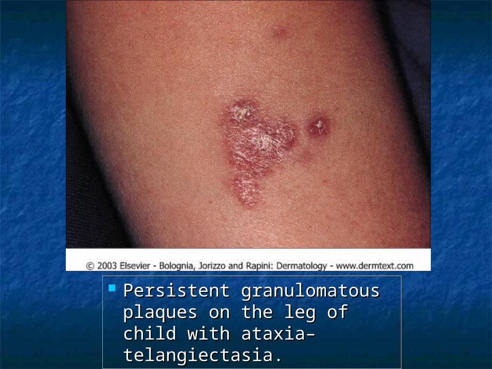
Persistent granulomatous Persistent granulomatous plaques on the leg of child plaques on the leg of child with ataxia–telangiectasia. with ataxia–telangiectasia.
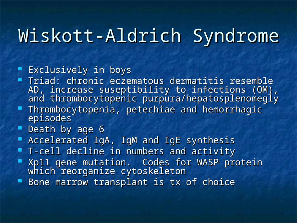
Wiskott-Aldrich SyndromeWiskott-Aldrich Syndrome
Exclusively in boysExclusively in boys Triad: chronic eczematous dermatitis resemble Triad: chronic eczematous dermatitis resemble
AD, increase suseptibility to infections (OM), and AD, increase suseptibility to infections (OM), and thrombocytopenic purpura/hepatosplenomeglythrombocytopenic purpura/hepatosplenomegly
Thrombocytopenia, petechiae and hemorrhagic Thrombocytopenia, petechiae and hemorrhagic episodes episodes
Death by age 6Death by age 6 Accelerated IgA, IgM and IgE synthesisAccelerated IgA, IgM and IgE synthesis T-cell decline in numbers and activityT-cell decline in numbers and activity Xp11 gene mutation. Codes for WASP protein Xp11 gene mutation. Codes for WASP protein
which reorganize cytoskeletonwhich reorganize cytoskeleton Bone marrow transplant is tx of choiceBone marrow transplant is tx of choice
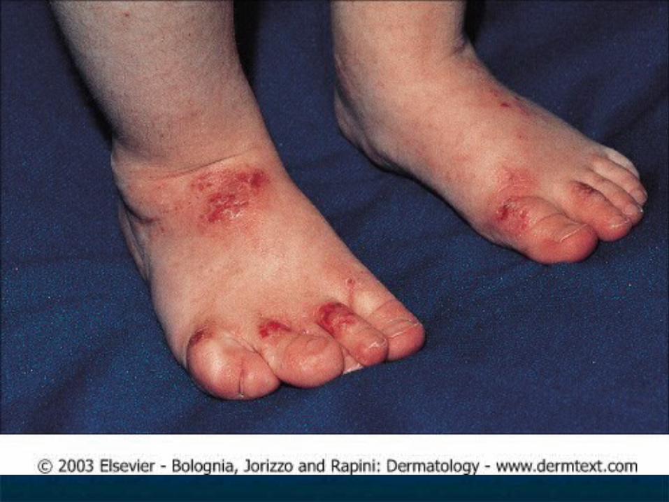
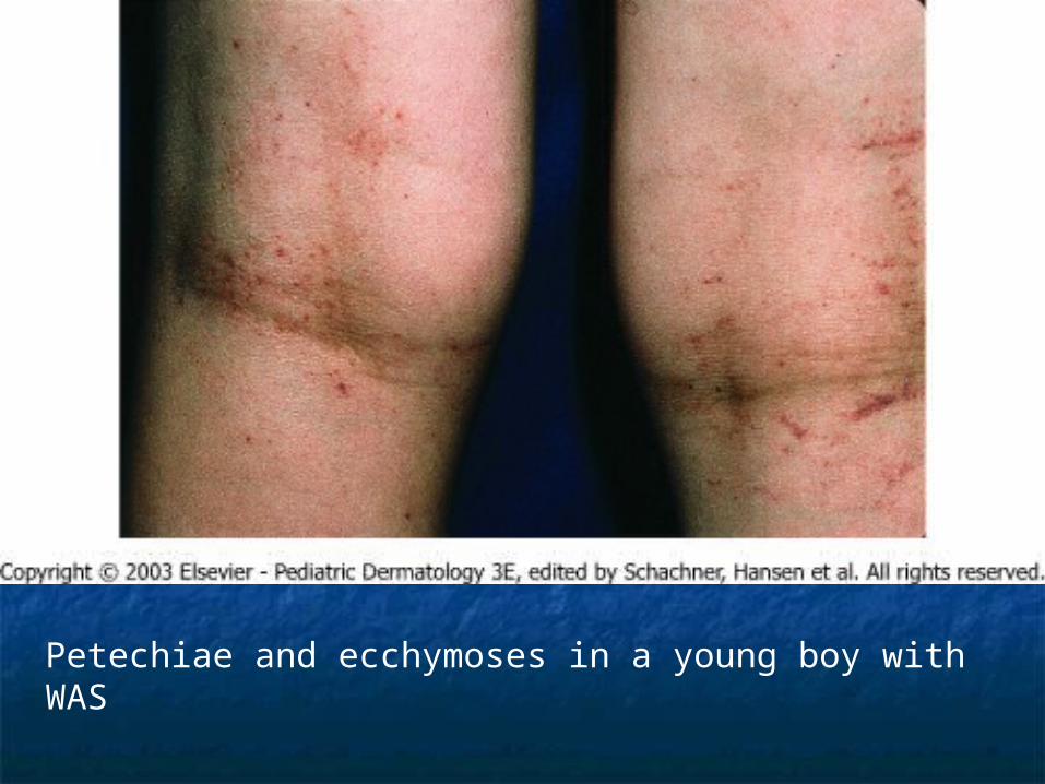
Petechiae and ecchymoses in a young boy with WAS
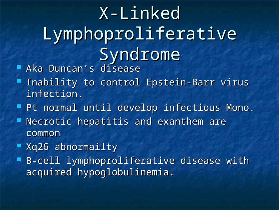
X-Linked X-Linked Lymphoproliferative Lymphoproliferative
SyndromeSyndrome Aka Duncan’s diseaseAka Duncan’s disease Inability to control Epstein-Barr virus Inability to control Epstein-Barr virus
infection.infection. Pt normal until develop infectious Mono.Pt normal until develop infectious Mono. Necrotic hepatitis and exanthem are Necrotic hepatitis and exanthem are
commoncommon Xq26 abnormailtyXq26 abnormailty B-cell lymphoproliferative disease with B-cell lymphoproliferative disease with
acquired hypoglobulinemia.acquired hypoglobulinemia.
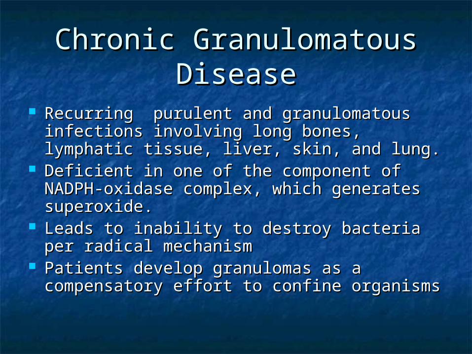
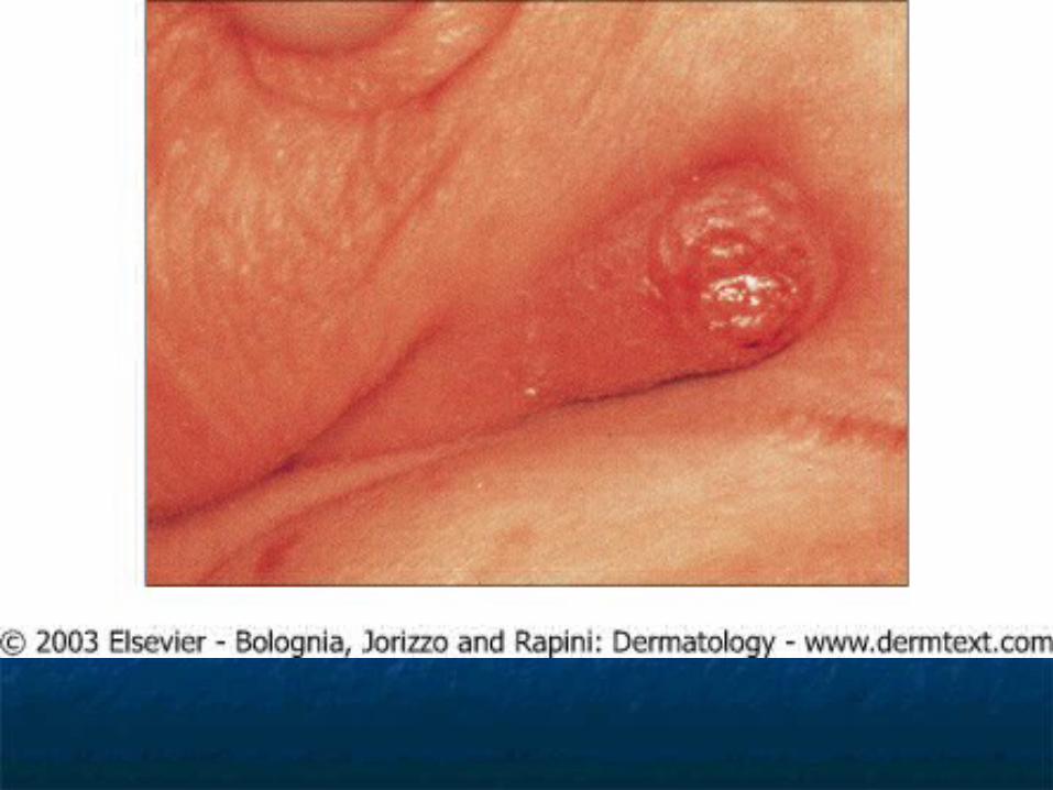
Chronic Granulomatous Chronic Granulomatous DiseaseDisease
Recurring purulent and granulomatous Recurring purulent and granulomatous infections involving long bones, lymphatic infections involving long bones, lymphatic tissue, liver, skin, and lung.tissue, liver, skin, and lung.
Deficient in one of the component of Deficient in one of the component of NADPH-oxidase complex, which generates NADPH-oxidase complex, which generates superoxide.superoxide.
Leads to inability to destroy bacteria per Leads to inability to destroy bacteria per radical mechanismradical mechanism
Patients develop granulomas as a Patients develop granulomas as a compensatory effort to confine organismscompensatory effort to confine organisms
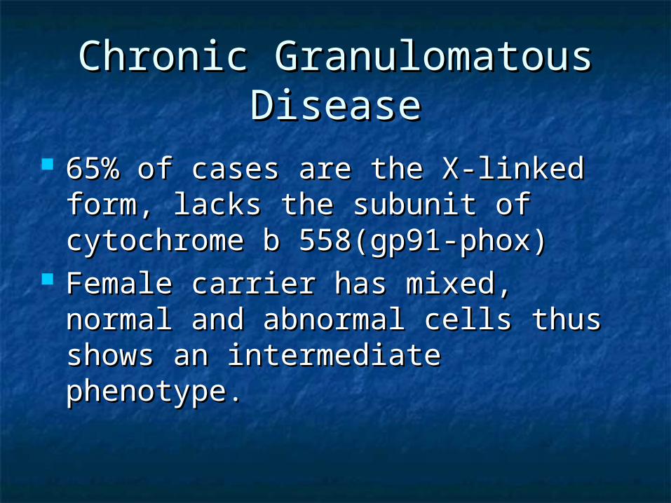
Chronic Granulomatous Chronic Granulomatous DiseaseDisease
65% of cases are the X-linked form, 65% of cases are the X-linked form, lacks the subunit of cytochrome b lacks the subunit of cytochrome b 558(gp91-phox)558(gp91-phox)
Female carrier has mixed, normal Female carrier has mixed, normal and abnormal cells thus shows an and abnormal cells thus shows an intermediate phenotype.intermediate phenotype.
Chronic Granulomatous Chronic Granulomatous DiseaseDisease
Myeloperoxidase producing bacteria Myeloperoxidase producing bacteria characteristically cause infections characteristically cause infections because their destruction requires because their destruction requires generation of oxygen free radicalsgeneration of oxygen free radicals Staph. AureusStaph. Aureus SerratiaSerratia

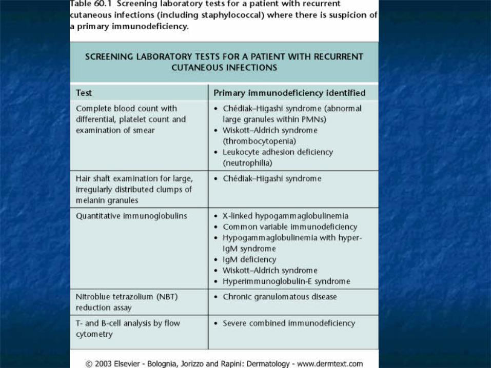
Chronic Granulomatous Chronic Granulomatous DiseaseDisease
Screening test: Nitroblue tetrazolium Screening test: Nitroblue tetrazolium (NBT) reduction assay(NBT) reduction assay NBT is normally yellowNBT is normally yellow 80-90% of normal leukocytes reduce 80-90% of normal leukocytes reduce
NBT during phagocytosis to insoluble NBT during phagocytosis to insoluble precipitate, turning it blueprecipitate, turning it blue
Only 5-10% of leukocytes from patients Only 5-10% of leukocytes from patients with CGD are able to reduce NBT during with CGD are able to reduce NBT during phagocytosisphagocytosis

Leukocyte Adhesion Leukocyte Adhesion Molecule DeficiencyMolecule Deficiency
Autosomal recessiveAutosomal recessive Affects the adherence of neutrophils, cytolytic T Affects the adherence of neutrophils, cytolytic T
lymphocytes, and monocytes lymphocytes, and monocytes Faulty complexing of the CD11 and CD18 integrinsFaulty complexing of the CD11 and CD18 integrins Necrotic ulcerations resembling pyoderma Necrotic ulcerations resembling pyoderma
gangrenosumgangrenosum Frequent skin infections, mucositis, and otitisFrequent skin infections, mucositis, and otitis Poor wound healingPoor wound healing Delayed separation of the umbilical cordDelayed separation of the umbilical cord Death usually occurs by 5 years of life unless bone Death usually occurs by 5 years of life unless bone
marrow transplant is undertaken.marrow transplant is undertaken.
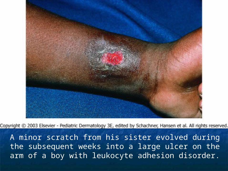
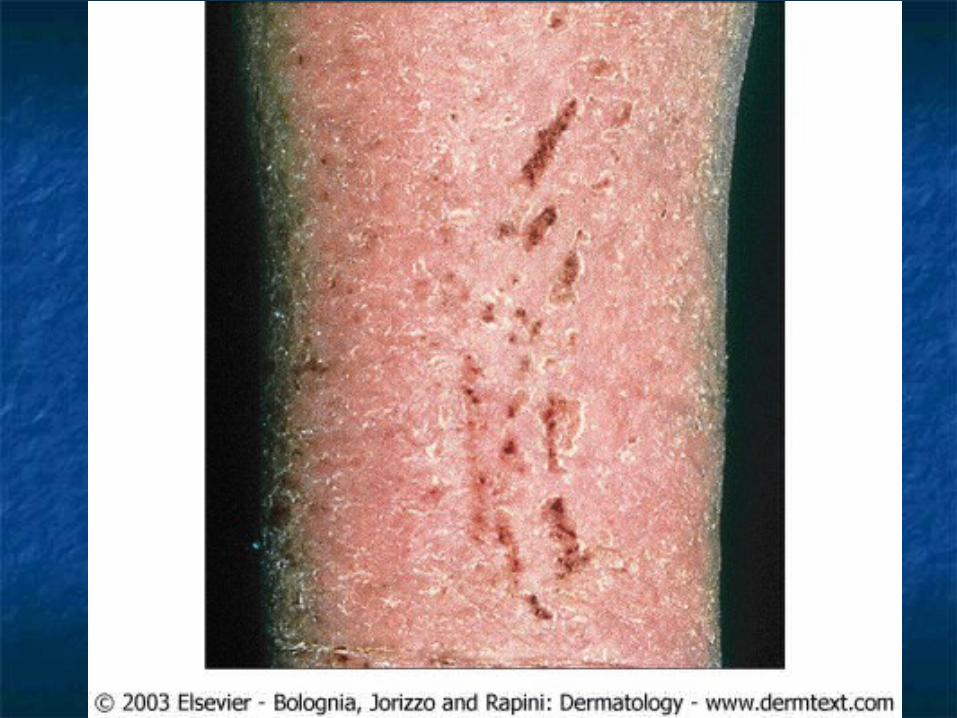
A minor scratch from his sister evolved during the subsequent weeks into a large ulcer on the arm of a boy with leukocyte adhesion disorder.
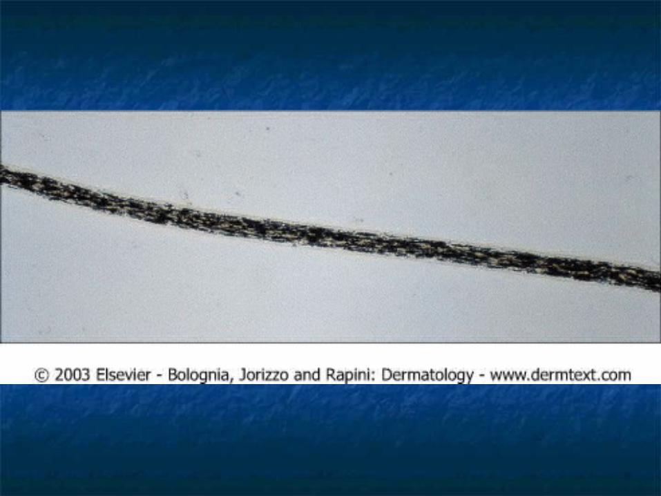
Chediak-Higashi SyndromeChediak-Higashi Syndrome
Autosomal recessiveAutosomal recessive Abnormal pigmentation with silvery hairAbnormal pigmentation with silvery hair PhotophobiaPhotophobia Partial oculocutaneous albinism, Partial oculocutaneous albinism,
cutaneous and intestinal infections early cutaneous and intestinal infections early in childhoodin childhood
Ocular albinism is accompanied by Ocular albinism is accompanied by nystagmus and photophobianystagmus and photophobia
Parental consanguinity commonParental consanguinity common
Chediak-Higashi SyndromeChediak-Higashi Syndrome
Defect in the gene LYST, resulting in Defect in the gene LYST, resulting in defective vesicular transport to and defective vesicular transport to and from the lysosome and melanosomefrom the lysosome and melanosome Causes the “giant” intracytoplasmic Causes the “giant” intracytoplasmic
granules found within leukocytes, granules found within leukocytes, melanocytes, hair shafts, renal tubular melanocytes, hair shafts, renal tubular cells, CNS neurons, and other tissuescells, CNS neurons, and other tissues
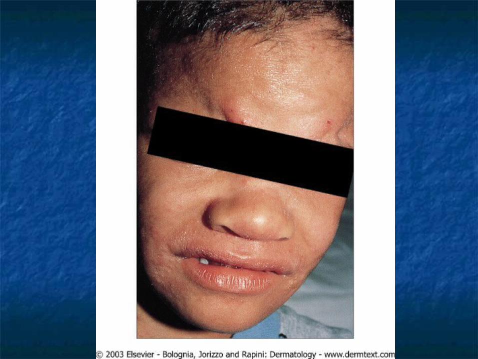
Hyperimmunoglobulinemia Hyperimmunoglobulinemia E SyndromeE Syndrome
Autosomal dominant with variable Autosomal dominant with variable expressivityexpressivity
Consists of atopic-like eczematous Consists of atopic-like eczematous dermatitis, recurrent pyogenic infection, dermatitis, recurrent pyogenic infection, high level of IgE, elevated IgD, IgE anti-high level of IgE, elevated IgD, IgE anti-staphlococcal antibodies, and eosinophilia.staphlococcal antibodies, and eosinophilia.
Face is consistently involved. Begin early Face is consistently involved. Begin early in life (2 month to 2 years)in life (2 month to 2 years)
Lesions resemble prurigoLesions resemble prurigo Keratoderma of the palms and solesKeratoderma of the palms and soles

Job’s syndromeJob’s syndrome
AKA Buckley SyndromeAKA Buckley Syndrome Subset of HIE.Subset of HIE. Mainly affect girls with red hair, Mainly affect girls with red hair,
freckles, and blue eyes.freckles, and blue eyes. Hyperextensible joints Hyperextensible joints Cold abscesses occur.Cold abscesses occur.
Graft-Versus-Host DiseaseGraft-Versus-Host Disease
Immunocompetent cells are introduced Immunocompetent cells are introduced as graft or blood transfusion to host as graft or blood transfusion to host who is unable to reject the graft cell.who is unable to reject the graft cell.
Most commonly after bone marrow Most commonly after bone marrow transplant.transplant.
Begins between 4-5Begins between 4-5thth weeks after weeks after transplant.transplant.
Result in exfoliative erythroderma.Result in exfoliative erythroderma.
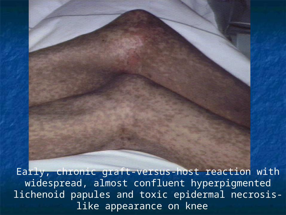
Early, chronic graft-versus-host reaction with widespread, almost confluent hyperpigmented lichenoid papules and toxic epidermal
necrosis-like appearance on knee
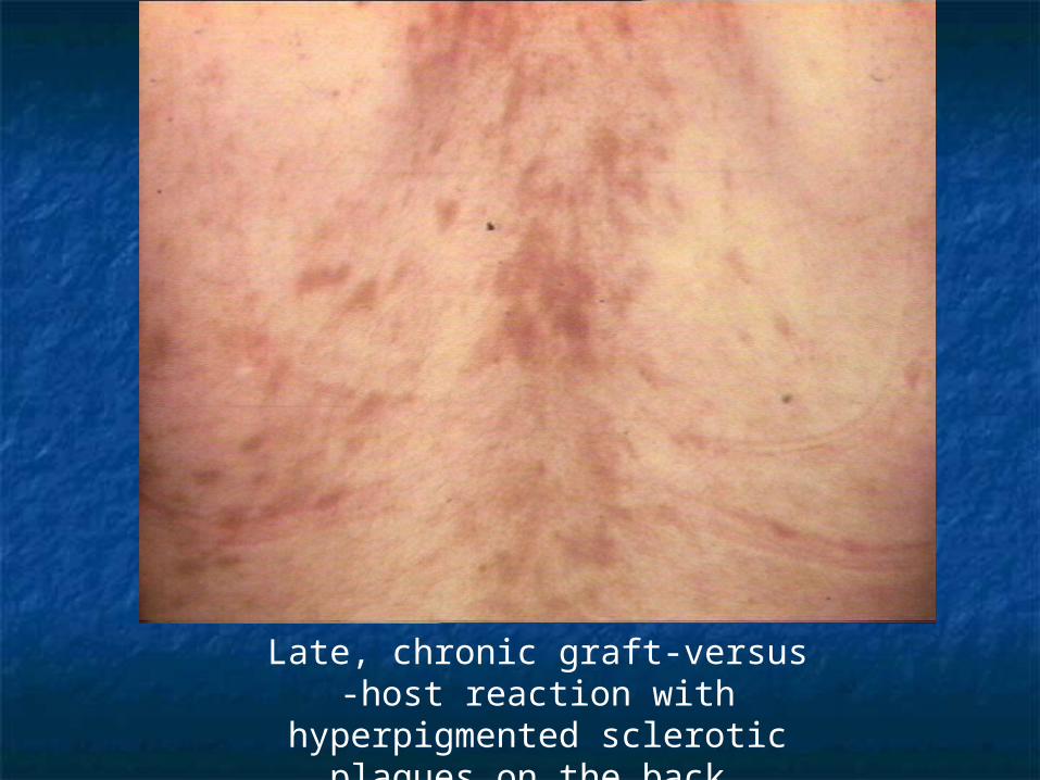
Late, chronic graft-versus -host reaction with hyperpigmented sclerotic plaques
on the back
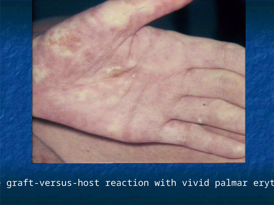
Acute graft-versus-host reaction with vivid palmar erythema
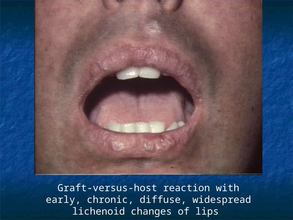
Graft-versus-host reaction with early, chronic, diffuse, widespread lichenoid changes of lips
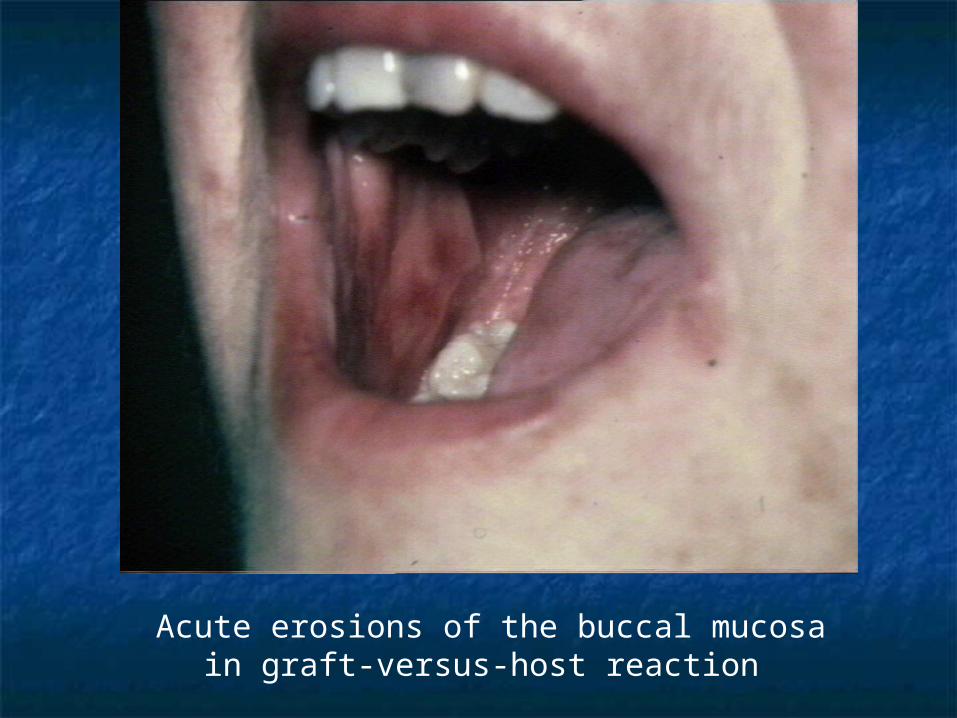
Acute erosions of the buccal mucosa in graft-versus-host reaction
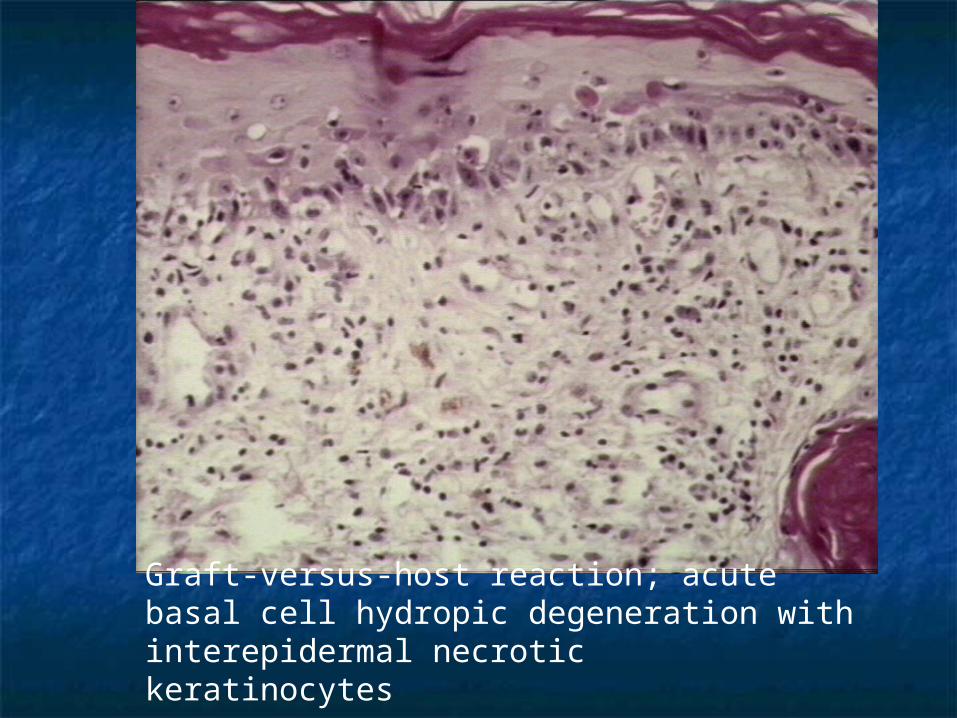
Graft-versus-host reaction; acute basal cell hydropic degeneration with interepidermal necrotic keratinocytes
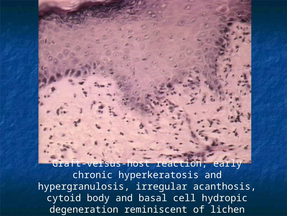
Graft-versus-host reaction; early chronic hyperkeratosis and hypergranulosis, irregular acanthosis, cytoid body
and basal cell hydropic degeneration reminiscent of lichen planus
THE ENDTHE END