Atopic dermatitis
25
ATOPIC DERMATITIS Manal Bosseila Prof of Dermatology Faculty of Medicine, Cairo University
-
Upload
manal-bosseila -
Category
Health & Medicine
-
view
418 -
download
2
Transcript of Atopic dermatitis
ATOPIC DERMATITIS
Manal Bosseila
Prof of Dermatology
Faculty of Medicine, Cairo University

Reference
DERMATOLOGY Text Book. 3rd Edition 2012. Eds: Jean L Bolognia, Joseph L Jorizzo, Julie V
Schaffer. ElSevier Publishing
Atopic Dermatitis

DEFINITION OF ATOPY
The term “atopy” is tightly linked to the presence
of allergen-specific IgE antibodies in the serum,
as documented by positive fluorescence enzyme
immunoassays (previously radioallergosorbent
[RAST] tests) or skin prick tests.
Atopic Dermatitis
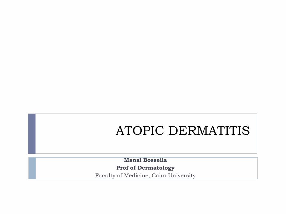
SPECTRUM OF AD Sp
ect
rum
of A
D
An IgE associated or allergic form of dermatitis corresponds to AD in the strict sense (formerly known as extrinsic AD
The remaining 20–30% of patients with the clinical phenotype of AD who have no evidence of IgE-sensitization are categorized as having a non-IgE-associated or non-allergic form of dermatitis (formerly known as intrinsic AD).
Atopic Dermatitis

THE ATOPIC MARCH
Atopic Dermatitis

PATHOGENESIS of Atopic Dermatitis
AD
Genetic
Environ-mental
Immunologic Mechanisms
Epidermal Barrier
Dysfunction
Atopic Dermatitis
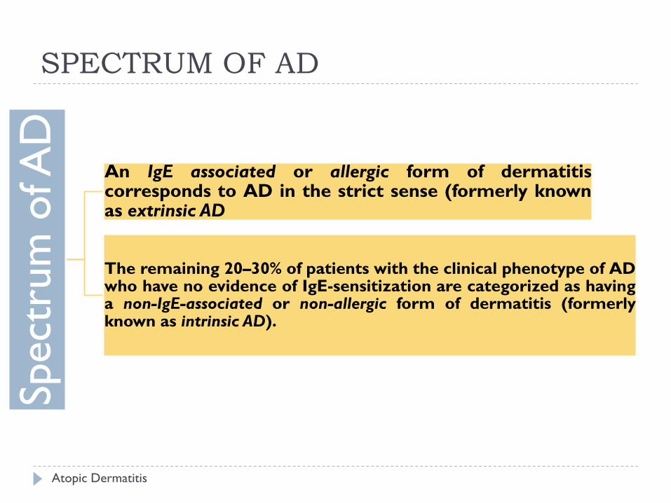
GENETICS
The entities in the atopic triad cluster together in
families.
AD is a complex genetic disease, and both gene–
gene and gene–environment interactions have
pathogenic roles. Existence of
genes specific to ADermtitis
Genes encoding proteins with immunologic
functions
Genes encoding epidermal proteins
Atopic Dermatitis
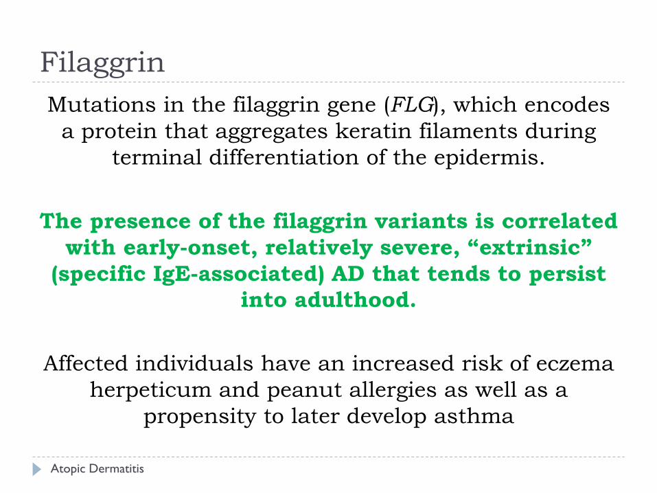
Filaggrin
Mutations in the filaggrin gene (FLG), which encodes
a protein that aggregates keratin filaments during
terminal differentiation of the epidermis.
The presence of the filaggrin variants is correlated
with early-onset, relatively severe, “extrinsic”
(specific IgE-associated) AD that tends to persist
into adulthood.
Affected individuals have an increased risk of eczema
herpeticum and peanut allergies as well as a
propensity to later develop asthma
Atopic Dermatitis
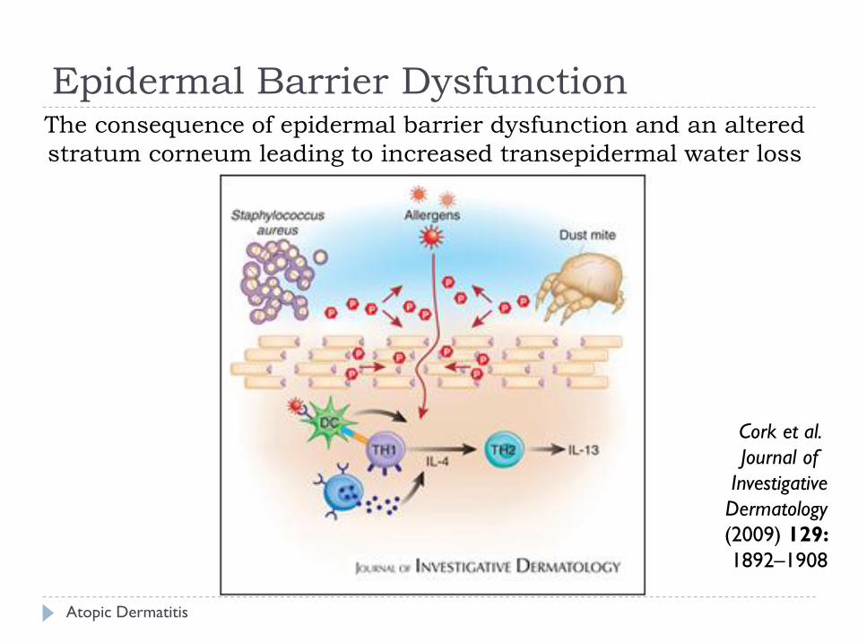
Epidermal Barrier Dysfunction The consequence of epidermal barrier dysfunction and an altered
stratum corneum leading to increased transepidermal water loss
Cork et al.
Journal of
Investigative
Dermatology
(2009) 129:
1892–1908
Atopic Dermatitis
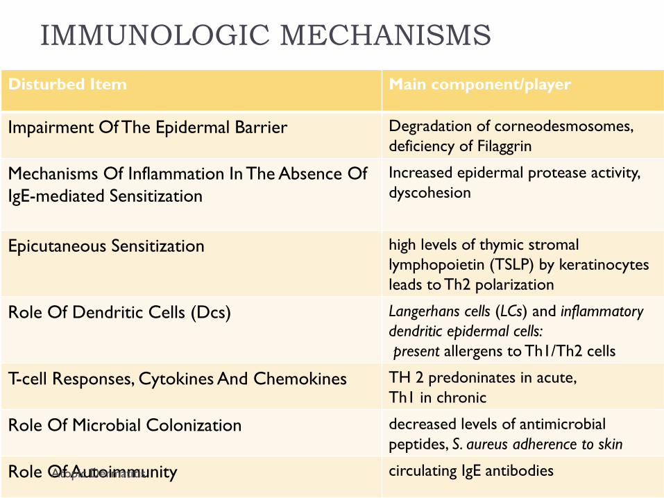
IMMUNOLOGIC MECHANISMS
Main component/player Disturbed Item
Degradation of corneodesmosomes,
deficiency of Filaggrin Impairment Of The Epidermal Barrier
Increased epidermal protease activity,
dyscohesion Mechanisms Of Inflammation In The Absence Of
IgE-mediated Sensitization
high levels of thymic stromal
lymphopoietin (TSLP) by keratinocytes
leads to Th2 polarization
Epicutaneous Sensitization
Langerhans cells (LCs) and inflammatory
dendritic epidermal cells:
present allergens to Th1/Th2 cells
Role Of Dendritic Cells (Dcs)
TH 2 predoninates in acute,
Th1 in chronic T-cell Responses, Cytokines And Chemokines
decreased levels of antimicrobial
peptides, S. aureus adherence to skin Role Of Microbial Colonization
circulating IgE antibodies Role Of Autoimmunity Atopic Dermatitis

PRURITUS & IL-31
Classic antihistamines are ineffective in AD
neuropeptides, proteases, kinins, and cytokines
such as interleukin (IL)-31 are known to induce itch.
IL-31 is strongly pruritogenic and exerts its biologic
activity through a heterodimeric receptor composed
of the IL-31 receptor A and oncostatin M receptor β
protein, both of which are overexpressed in lesional
skin of AD.
Atopic Dermatitis
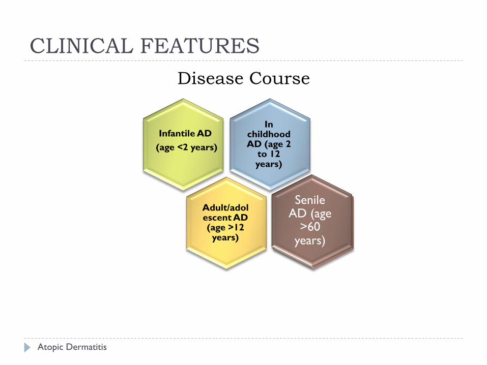
CLINICAL FEATURES
Disease Course
In
childhood AD (age 2
to 12 years)
Adult/adolescent AD (age >12
years)
Infantile AD
(age <2 years)
Senile AD (age
>60 years)
Atopic Dermatitis

Clinical diagnosis of AD Atopic stigmata
1. Xerosis
2. Keratosis Pilaris
3. Ichthyosis
Vulgaris
4. Dennie Morgan
lines
5. Periorbital
darkening
6. White
dermoographism
Pruritus
eczematous skin lesions in
typical age-specific
distribution patterns,
a chronic or chronically
relapsing course,
Early age at onset,
A personal and/or family
history of atopy.
13

Associated Features Associated Complications
Pityriasis Alba
Pruritus
Atopic Stigmata
Infections, esp
eczema herpeticum
Occular
complications
14
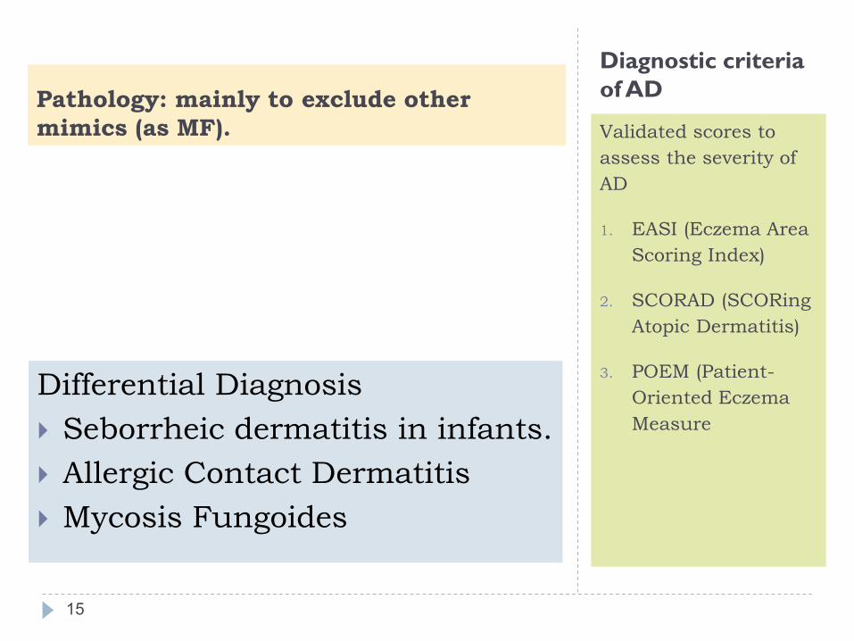
Diagnostic criteria
of AD
Validated scores to
assess the severity of
AD
1. EASI (Eczema Area
Scoring Index)
2. SCORAD (SCORing
Atopic Dermatitis)
3. POEM (Patient-
Oriented Eczema
Measure
Differential Diagnosis
Seborrheic dermatitis in infants.
Allergic Contact Dermatitis
Mycosis Fungoides
Pathology: mainly to exclude other
mimics (as MF).
15
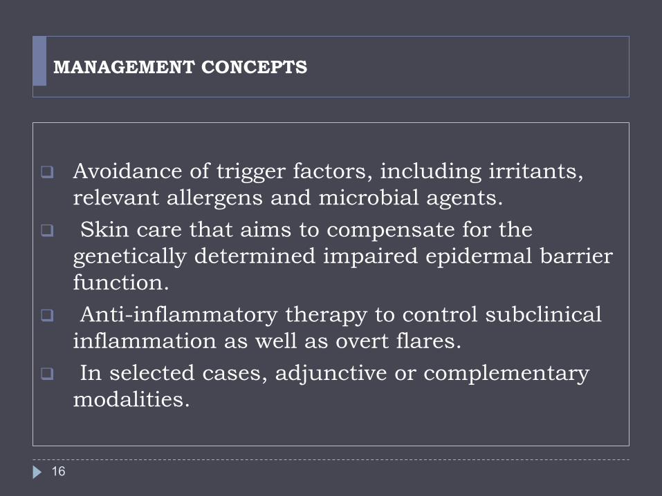
MANAGEMENT CONCEPTS
Avoidance of trigger factors, including irritants,
relevant allergens and microbial agents.
Skin care that aims to compensate for the
genetically determined impaired epidermal barrier
function.
Anti-inflammatory therapy to control subclinical
inflammation as well as overt flares.
In selected cases, adjunctive or complementary
modalities.
16
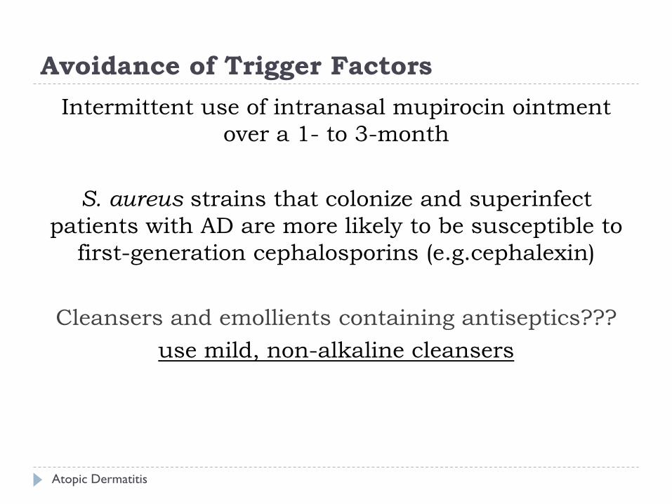
Avoidance of Trigger Factors
Intermittent use of intranasal mupirocin ointment
over a 1- to 3-month
S. aureus strains that colonize and superinfect
patients with AD are more likely to be susceptible to
first-generation cephalosporins (e.g.cephalexin)
Cleansers and emollients containing antiseptics???
use mild, non-alkaline cleansers
Atopic Dermatitis

Avoidance of Trigger Factors
According to Patch test/RAST IgE test
Avoid House mites
Avoid Food Allergens
A continuous basic therapy with emollients, even in periods and sites in which the AD is not active.
Atopic Dermatitis

Topical Anti-inflammatory Therapy
The corticosteroid with appropriate potency to
quickly gain control of the flare, continuation of
daily therapy until active dermatitis minimized.
In moderate to severe AD, risk of relapse can be
significantly reduced by proactive maintenance
with twice-weekly application of a mid-potency
topical corticosteroid.
Topical calcineurin inhibitors (TCIs)
Atopic Dermatitis
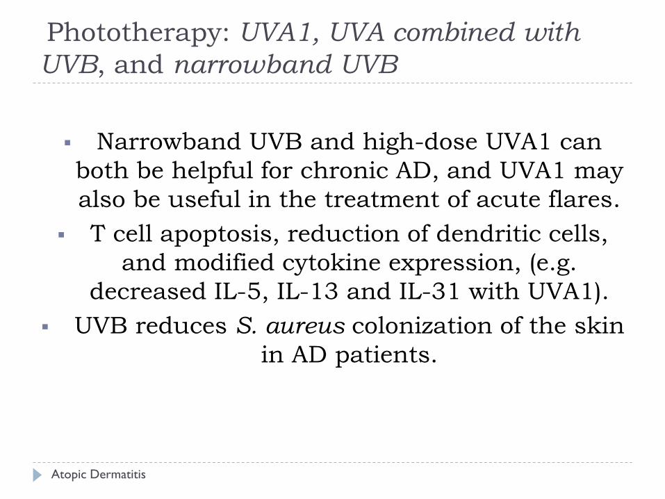
Phototherapy: UVA1, UVA combined with
UVB, and narrowband UVB
Narrowband UVB and high-dose UVA1 can
both be helpful for chronic AD, and UVA1 may
also be useful in the treatment of acute flares.
T cell apoptosis, reduction of dendritic cells,
and modified cytokine expression, (e.g.
decreased IL-5, IL-13 and IL-31 with UVA1).
UVB reduces S. aureus colonization of the skin
in AD patients.
Atopic Dermatitis
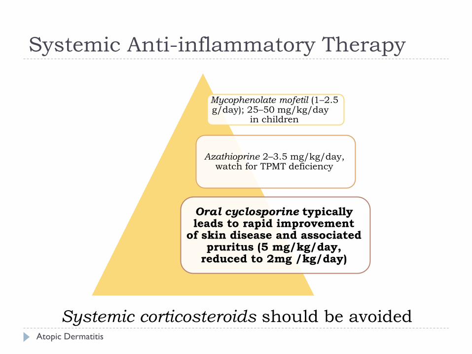
Systemic Anti-inflammatory Therapy
Systemic corticosteroids should be avoided
Mycophenolate mofetil (1–2.5 g/day); 25–50 mg/kg/day
in children
Azathioprine 2–3.5 mg/kg/day, watch for TPMT deficiency
Oral cyclosporine typically leads to rapid improvement
of skin disease and associated pruritus (5 mg/kg/day,
reduced to 2mg /kg/day)
Atopic Dermatitis
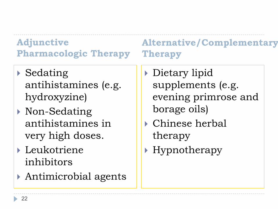
Adjunctive
Pharmacologic Therapy Alternative/Complementary
Therapy
Sedating
antihistamines (e.g.
hydroxyzine)
Non-Sedating
antihistamines in
very high doses.
Leukotriene
inhibitors
Antimicrobial agents
Dietary lipid
supplements (e.g.
evening primrose and
borage oils)
Chinese herbal
therapy
Hypnotherapy
22

Targeted Molecular
Therapy (“Biologics”) Emerging Therapies
Anti-IgE monoclonal antibody omalizumab, which inhibits the binding of IgE to its high-affinity receptor (FcεRI), is FDA-approved for the treatment of asthma in patients ≥12 years.
The anti-CD20 monoclonal antibody rituximab (administered via 2 IV infusions separated by 2 weeks), which inhibits mature B cells
mepolizumab inhibits IL-5, a crucial factor for growth and differentiation of eosinophils. Although mepolizumab can decrease the eosinophil count in patients with AD, it failed to lead to a significant clinical improvement
Goals of blocking factors
such as cytokines involved
in the regulation of IgE
synthesis (e.g. IL-4) or
chemoattractant receptor-
homologous molecule
expressed
on Th2 cells (CRTH2).
23
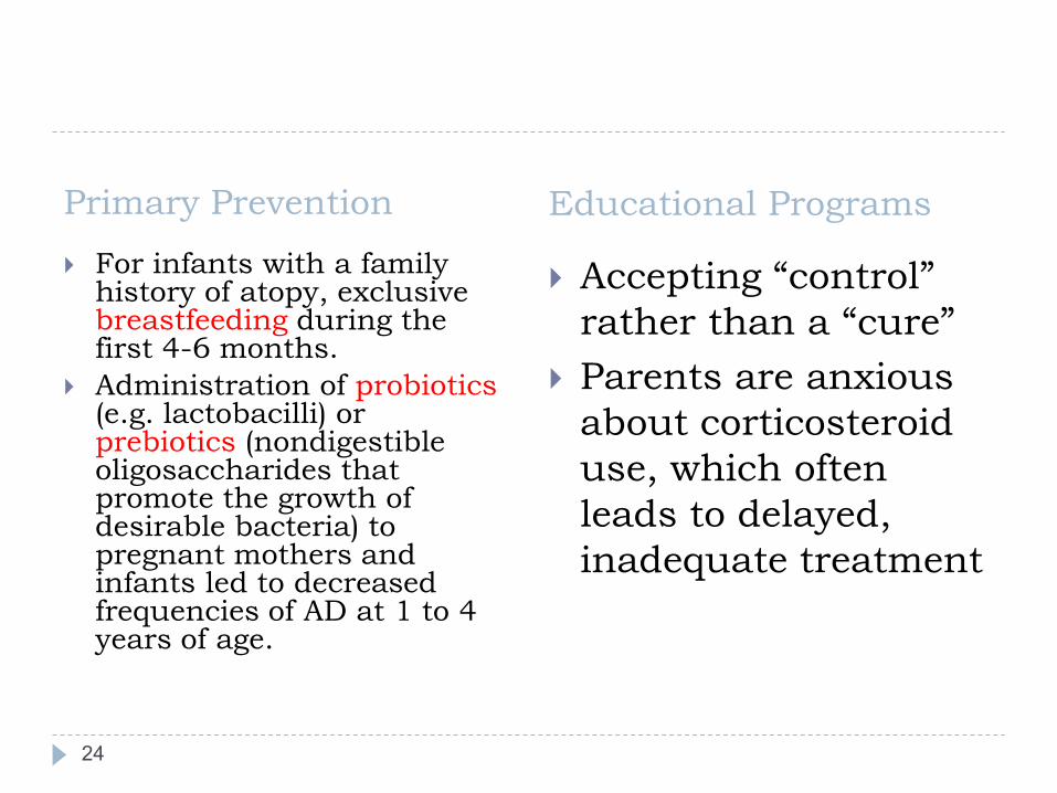
Primary Prevention Educational Programs
For infants with a family history of atopy, exclusive breastfeeding during the first 4-6 months.
Administration of probiotics (e.g. lactobacilli) or prebiotics (nondigestible oligosaccharides that promote the growth of desirable bacteria) to pregnant mothers and infants led to decreased frequencies of AD at 1 to 4 years of age.
Accepting “control”
rather than a “cure”
Parents are anxious
about corticosteroid
use, which often
leads to delayed,
inadequate treatment
24

Resumé: The APPROACH TO AD
Management should not be concentrated solely on the treatment of acute flares, but also be directed
towards improving the underlying genetically determined epidermal barrier dysfunction and
preventing active dermatitis (e.g. via maintenance therapy).
Such an approach could potentially block the sensitizations and ongoing inflammation that drive
the atopic march forward
Atopic Dermatitis