Atomic force microscopy on polymers and polymer...
8
Polymer Bulletin 26, 215-222 (1991) Polymer Bullelin 9 Springer-Verlag 1991 Atomic force microscopy on polymers and polymer related compounds 2. Monocrystals of normal and cyclic alkanes Ca3Hss, Ca6Hr4, (CH2)~, (CH2)72" W. Stocker 1'2, G. Bar 1, M. Kunz 2, M. M611er 3, S. N. Magonov TM, and H.-J. Cantow ~'*** 1Freiburger MateriaI-Forschungszentrum FMF and 21nstitut fL~r Makromolekulare Chemie, Universit&t, Stefan-Meier-Strasse 31, W-7800 Freiburg, Federal Republic of Germany 3Department of Chemical Technology of the University of Twente, NL-7500 Enschede, The Netherlands ABSTRACT Atomic Force Microscopy (AFM) images have been recorded from the surfaces of platelet type monocrystals of linear alkanes, n-tritriacontane, C33H68, and n-hexatrlacontane, C36H74. Structural details have been revealed in the range from several hundreds of nano- meters down to the atomic scale. High resolution AFM micrographs of the linear alkanes show a regular pattern of elevations characterized by two main periodicities: aAF M = .70 + .02 nm, DAF M = .52 + ,05 nm, "~AFM = 88 4- 3 ~ and aAF M = .79+ .05 nm, bAF M = .61 + .05 nm, "~AFM = 87 + 3 ~ , for 033H68 and 036H74, respectively. This structure is in fair agreement with the or- thorhombic subcell of the bulk structure as confirmed by electron diffraction. Further, AFM results are in accordance with an orthorhombic unit cell for C33H68, and a monoclinic one for C36H74 under investigation. Consequently, the elevations in the AFM images can be as- signed to individual methyl groups, which form the surface layer of a paraffine crystal. A regular surface structure has also been observed in AFM images of platelet crystals of cyclic alkanes, cyclooctatetracontane (CH2)48, and cyclodoheptacontane (CH2)72. In these cases, the periodicities are characterized by aAF M = ] .10 4- .01 nm, b~,FM= .85 4- .01 nm, "~AFM --" 8 9 4- 1~ and aAF M = 1,1 ] 4- .05 nm, DAF M = .91 + ,05 nm, and ~'AFM = 90 + 3~ for (CH2)48 and (CH2)72, respectively. The images are consistent with the arrangement of the adja- cent reentry folds obtained from crystallographic data. INTRODUCTION Scanning probe techniques, i. e. scanning tunneling microscopy, STM [1], and atomic force microscopy, AFM [2], are novel tools for the analysis of surface structures, showing details with atomic resolution. While STM has been developed first and is widely used for conductive and semiconductive materials [3], AFM studies with a sub-nanometer re- solution have been reported only recently. STM studies on monocrystals of organic con- ductors and superconductors show good agreement between STM pictures and the sur- face structure as derived from crystallographic data [4-6], and no evidence for surface reconstruction has been detected. This was also demonstrated for AFM by recent coope- rative STM/AFM studies of the same monocrystals [7]. AFM images with atomic resolution were generally consistent with the corresponding STM data. Unlike other probe tech- niques, AFM does not require special interactions between the probe and the analyzed surface such as a tunneling current, magnetic force, etc. As based on probing interatomic van der Waals forces, AFM shows great promise to become a valuable tool for direct examination of surfaces of all kinds of materials. Thus, also AFM studies of nonconducting macromolecular materials can give detailed and important information on molecular sur- face structures. This has been demonstrated recently by AFM studies on surfaces of COid- *Herrn Prof. Dr. Harald Cherdron zu seinem 60. Geburtstag herzlich gewidmet ** Permanent address: Institute of Chemical Physics of the USSR Academy of Sciences, Kosygin u I. 4, SU-117977 Moscow, USSR ***To whom offprint requests should be sent
Transcript of Atomic force microscopy on polymers and polymer...
Polymer Bulletin 26, 215-222 (1991) Polymer Bullelin �9 Springer-Verlag 1991
Atomic force microscopy on polymers and polymer related compounds 2. Monocrysta ls of normal and cyclic a lkanes Ca3Hss, Ca6Hr4, (CH2)~, (CH2)72"
W. Stocker 1'2, G. Bar 1, M. Kunz 2, M. M611er 3, S. N. Magonov TM, and H.-J. Cantow ~'***
1Freiburger MateriaI-Forschungszentrum FMF and 21nstitut fL~r Makromolekulare Chemie, Universit&t, Stefan-Meier-Strasse 31, W-7800 Freiburg, Federal Republic of Germany 3Department of Chemical Technology of the University of Twente, NL-7500 Enschede, The Netherlands
ABSTRACT
Atomic Force Microscopy (AFM) images have been recorded from the surfaces of platelet type monocrystals of linear alkanes, n-tritriacontane, C33H68, and n-hexatrlacontane, C36H74. Structural details have been revealed in the range from several hundreds of nano- meters down to the atomic scale. High resolution AFM micrographs of the linear alkanes show a regular pattern of elevations characterized by two main periodicities: aAF M = .70 +
.02 nm, DAF M = .52 + ,05 nm, "~AFM = 88 4- 3 ~ and aAF M = .79+ .05 nm, bAF M = .61 + .05 nm, "~AFM = 87 + 3 ~ , for 033H68 and 036H74, respectively. This structure is in fair agreement with the or- thorhombic subcell of the bulk structure as confirmed by electron diffraction. Further, AFM results are in accordance with an orthorhombic unit cell for C33H68, and a monoclinic one for C36H74 under investigation. Consequently, the elevations in the AFM images can be as- signed to individual methyl groups, which form the surface layer of a paraff ine crystal.
A regular surface structure has also been observed in AFM images of platelet crystals of cyclic alkanes, cyc looctatet racontane (CH2)48, and cyc lodoheptacontane (CH2)72. In these cases, the periodicities are characterized by aAF M = ] .10 4- .01 nm, b~,FM = .85 4- .01 nm, "~AFM --" 89 4- 1~ and aAF M = 1,1 ] 4- .05 nm, DAF M = .91 + ,05 nm, and ~'AFM = 90 + 3 ~ f o r (CH2)48
and (CH2)72, respectively. The images are consistent with the arrangement of the adja- cent reentry folds obta ined from crystallographic data.
INTRODUCTION
Scanning probe techniques, i. e. scanning tunneling microscopy, STM [1], and atomic force microscopy, AFM [2], are novel tools for the analysis of surface structures, showing details with atomic resolution. While STM has been deve loped first and is widely used for conduct ive and semiconductive materials [3], AFM studies with a sub-nanometer re- solution have been reported only recently. STM studies on monocrystals of organic con- ductors and superconductors show good agreement between STM pictures and the sur- face structure as derived from crystallographic data [4-6], and no evidence for surface reconstruction has been detected. This was also demonstrated for AFM by recent coope- rative STM/AFM studies of the same monocrystals [7]. AFM images with atomic resolution were generally consistent with the corresponding STM data. Unlike other probe tech- niques, AFM does not require special interactions between the probe and the analyzed surface such as a tunneling current, magnet ic force, etc. As based on probing interatomic van der Waals forces, AFM shows great promise to become a valuable tool for direct examination of surfaces of all kinds of materials. Thus, also AFM studies of nonconduct ing macromolecular materials can give detai led and important information on molecular sur- face structures. This has been demonstrated recently by AFM studies on surfaces of COid-
*Herrn Prof. Dr. Harald Cherdron zu seinem 60. Geburtstag herzlich gewidmet ** Permanent address: Institute of Chemical Physics of the USSR Academy of Sciences, Kosygin u I. 4, SU-117977 Moscow,
USSR ***To whom offprint requests should be sent
216
extruded polyethylene [8], epitaxially crystallized isotactic polypropylene [9], and of monocrystals of diacetylene and polydiacetylene derivatives [10]. This paper is directed towards the study of low molecular weight oligomer crystals with well established and regular lamellar surfaces consisting of chain ends and tight folds as model compounds to approach AFM research on the less defined polymeric homologues. Surfaces of mono- crystals of linear and cyclic alkanes, n-tritriacontane, n-hexatriacontane, cyclooctatetra- contane, and cyc lodoheptacontane have been examined.
EXPERIMENTAL
AFM experiments were carried out with a Nanoscope II microscope (Digital Instruments, Inc., Santa Barbara, Cal., USA) at ambient conditions. Sample surfaces were scanned by a tiny pyramidal SigN 4 tip which is a t tached to a microfabricated canti lever (200 Fm tri- angular base). Deflections of the cantilever were registered via deflection of a laser beam. The largest scan area was 700 x 700 nm (head type A). Cantilevers had a force constant of .06 and .12 N/m. Scanning line frequencies were 1Hz for large scale scans and up to 39 Hz for smaller areas. Thermal drift of images was observed even by scanning at highest fre- quencies. This resulted in minor neglectable deviations in the structure parameters, i. e. repeat distances and angles. In most experiments, images were taken repeatedly where- by samples were scanned in different directions. This provides an important opportunity to verify observed images.
An important requirement for samples to be studied by AFM is flatness of the surface. Mo- nocrystais of linear and cyclic alkanes have a platelet shape, and large lamellar surfaces are suitable for AFM studies. Monocrystals of orthorhombic n-tritriacontane, C33H68, of monoclinic n-hexatriacontane, 036H74, of cyclooctatetracontane, (CH2)48, and of cyclo- doheptacontane, (0H2)72, were prepared as described elsewhere [11-12]. Electron dif- fraction experiments on C33H68 and C36H74 monocrystals were done with a 100 kV Philips 400 electron microscope. SCHAKAL, a molecular graphics computer software [13], was used for construction of the atomic picture of the surfaces based on X-ray analysis da ta of
033H68 [14] and (CH2)36 [15].
RESULTS AND DISCUSSION
n-Alkanes
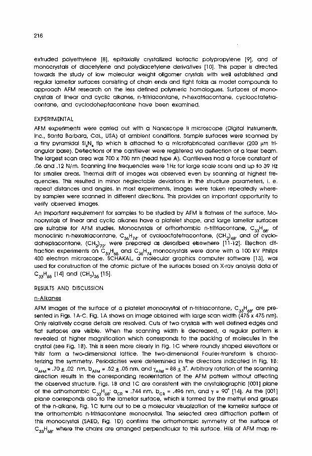
AFM images of the surface of a platelet monocrystal of n-tritriacontane, C33H68 , are pre- sented in Figs. 1A-C. Fig. 1A shows an image obtained with large scan width (475 x 475 nm). Only relatively coarse details are resolved. Cuts of two crystals with well def ined edges and flat surfaces are visible. When the scanning width is decreased, a regular pattern is revealed at higher magnif ication which corresponds to the packing of molecules in the crystal (see Fig. 1B). This is seen more clearly in Fig. lC where roundly shaped elevations or 'hills" form a two-dimensional lattice. The two-dimensional Fourier-transform is charac- terizing the symmetry. Periodicities were determined in the directions indicated in Fig. 1B: QAF M = .70 _+ .02 nm, bAF M = .52 -I- .05 rim, and ~tAF M = B8 -I- 3 ~. Arbitrary rotation of the scanning direction results in the corresponding reorientation of the AFM pattern without affecting the observed structure. Figs. 1B and lC are consistent with the crystallographic [001] plane of the orthorhombic C33H68: QcR = ,744 nm, bcR = .496 nm, and y = 90" [14]. As the [001] plane corresponds also to the lamellar surface, which is formed by the methyl end groups of the n-alkane, Fig. 1C turns out to be a molecular visualization of the lamellar surface of the orthorhombic n-tritriacontane monocrystal. The selected area diffraction pattern of this monocrystal (SAED, Fig. 1D) confirms the orthorhombic symmetry of the surface of C33H68, where the chains are arranged perpendicular to this surface. Hills of AFM map re-
2 1 7
a~
i �9
::'24 ~~.ii:i!i
.... ~...~. .,,,,..:..
<.~: . . . . . ~2-:</ <.~. . . . . . . .~.
i,~...~., i~,..,..-" b ~
�9 ~" . . . . . . i'"~"
:}!i; Figure 1: AFM images of or thorhombic n-tr i t r iacontane A - Top-view, 475 x 475 nm
B - Top-view, 14,2 x 14,2 nm C - Top-view, 5.7 x 5.7 nm, with 2D-Fourier transform
D - Se lec ted a r e a e lect ron dif fraction pat tern (SAED) of n- t r i t r iacontane
E l = Crystal structure of monocl in ic n -hexa t r i acon tane E - [001] p l a n e F - [100] p l a n e
218
219
A �9 ~ ~ . ; ~ D,.J,j=-'~,~
i (~... ",,._.
~...4)
�9 , . . 4 i
i
L-.. ~ '---'.4
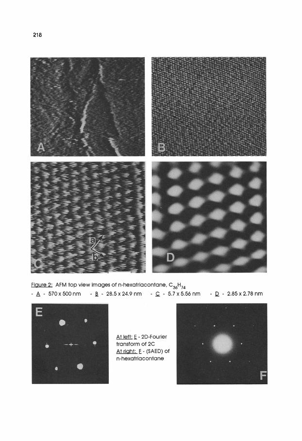
F igure 3: Crys ta l s t ruc tu re o f c v c l o h e x a t r i a c o n t a n e
t o D l e f t A [001] p l a n e - t o p r iqht B [ 0 0 i ] p l a n e
AFM o f c y c l o o c t a t e t r a c o n t a n e - C - t o p - v i e w 49
x 57 n m D - 3 D - v i e w a t 60 ~ r o t a t i o n - E - t o p - v i e w
8.1 x 9.5 n m - a t 30" r o t a t i o n - p a r t o f i m a g e a f t e r f i l t ra t ion F - 2D FT o f D _~G - 2D FT o f E
220
present individual methyl groups whereby atomic details, i. e. hydrogen and carbon atoms, cannot be distinguished. A corresponding assignment was proposed for the AFM images of DL-leucine monocrystals [16]. Also in this case, individual carbon and hydrogen atoms have not been resolved. However, at ambient temperature, fast rotation of the methyl group around the s-carbon-carbon bond is well established [11] and gives man- datory explanat ion for the diffusive pattern. Variations in the height have been estimated to be approximately .3 ~,. An interpretation of the surface profile is not possible definitely at present due the missing theoretical understanding of AFM imaging.
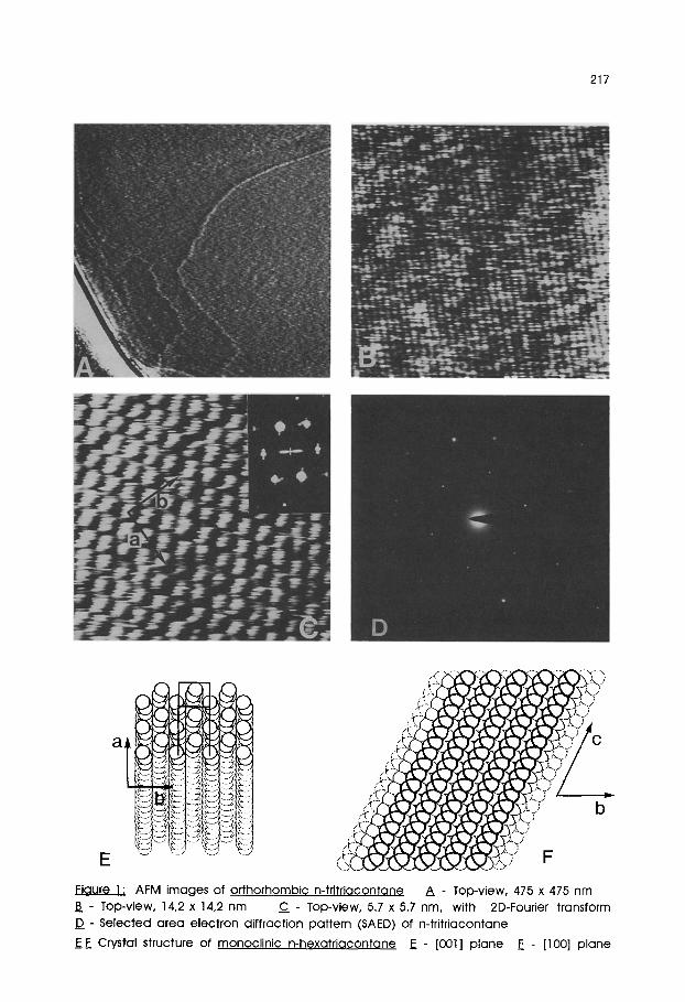
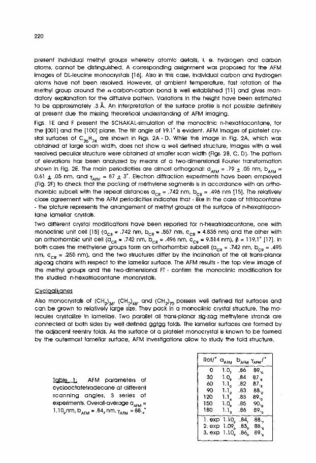
Figs. 1E and F present the $CHAKAL-simulation of the monoclinic n-hexatriacontane, for the [001] and the [100] plane. The tilt angle of 19.1 ~ is evident. AFM images of platelet cry- stal surfaces of C36H74 are shown in Figs. 2A- D. While the image in Fig. 2A, which was obtained at large scan width, does not show a well def ined structure, images with a well resolved peculiar structure were obtained at smaller scan width (Figs. 2B, C, D). The pattern of elevations has been analyzed by means of a two-dimensional Fourier transformation shown in Fig, 2E. The main periodicities are almost orthogonal: aAF M = .79 + .05 nm, bAF M = 0.61 + .05 nm, and ~/AFM = 87 + 3 ~ Electron diffraction experiments have been employed (Fig. 2F) to check that the packing of methylene segments is in accordance with an ortho- rhombio subcell with the repeat distances acR = .742 rim, bcR --- .496 nm [15]. The relatively close agreement with the AFM periodicities indicates that - like in the case of tritriacontane - the picture represents the arrangement of methyl groups at the surface of n-hexatriacon- tane lamellar crystals.
Two different crystal modifications have been reported for n-hexatr iaacontane, one with monoclinlc unit cell [15] (acR = .742 nm, bcR = .557 nm, CcR = 4.835 nm) and the other with an orthorhomblc unit cell (acR = .742 nm, bcR = .496 nm, Cc~ = 9.514 nm), j3 = 119,1" [17]. In both cases the methylene groups form an orthorhombic subcell (acR = .742 nm, ben = .495 nm, CcR = .255 nm), and the two structures differ by the inclination of the all trans-planar zig-zag chains with respect to the lamellar surface. The AFM results - the top view image of the methyl groups and the two-dimensional FT- confirm the monoclinic modif ication for the studied n-hexatr iacontane moncrystals.
Cycloalkanes
Also monocrystals of (0H2)36, (0H2)48, and (CH2)72 possess well defined flat surfaces and can be grown to relatively large size. They pack in a monoclinic crystal structure. The mo- lecules crystallize in lamellae. Two parallel all trans-planar zig-zag methylene strands are connected at both sides by well def ined ggtgg folds. The lamellar surfaces are formed by the ad jacent reentry folds. As the surface of a platelet monocrystal is known to be formed by the outermost lamellar surface, AFM investigations allow to study the fold structure.
Table 1: AFM parameters of cyc loocta te t radecane at different scanning angles, 3 series of experiments. Overall-overage aAF M = 1.1 02nm, bAF M = .847 rim, "~AFM = 88'9"
Rot/" aAF M bAF M ~AFM / ~
0 1.07 .86 89. 0 30 1.0~ .84 87. 8 60 I . I o .82 87. 4 90 1.12 .83 88.2
120 1.14 .83 89. 0 150 1.0,;, .85 90. 0 180 1.12 .86 89. 0
I. exp 1.102 .84, 88. 7 2. exp 1.09~ .839 88.~ 3. exp 1.109 .86 o 89.~
221
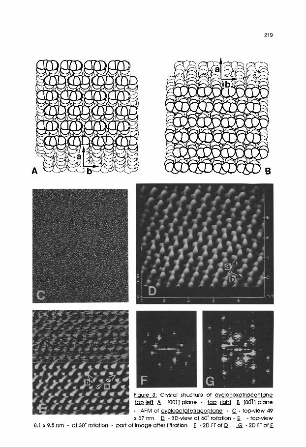
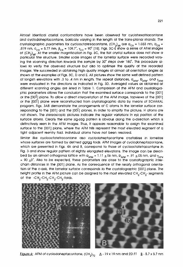
Almost identical crystal conformations have been observed for cyclohexatr iacontane and cyc lodoheptacontane, basically varying in the length of the trans-planar strands. The crystallographic parameters for cyclooctatetracontane, (0H2)48 are ac~ = 1.033 nm, bcR = .819 nm, CcR = 5.77 nm, ~CR = 109'7~ YCR = 90~ [18]. Figs. 3C-E ShOW a series of AFM images of (CH2)48. At the smaller magnif ication in Fig. 3C, the flat crystal surface does not show a particular fine structure. Smaller scale images of the lamellar surface were recorded vary- ing the scanning direction towards the sample by 30 ~ steps over 180 ~ . This procedure al- lows to verify the observed structure but also to optimize the quality of the recorded images. We succeeded in obtaining high quality images at almost all orientation angles as shown at the examples of Figs. 3C, D and E. All pictures show the same well def ined pattern of Iongish elevations with .3 to .4 nm in length. The repeat distances, aAF M, bAFM, and ~AFM were evaluated in the directions as indicated in Fig. 3D. Averaged values as obta ined at different scanning angles are listed in Table 1. Comparison of the AFM and crystallogra- phic parameters allows the conclusion that the examined surface corresponds to the [001] or the [00T] plane. To al low a direct interpretation of the AFM image, topviews of the [001] or the [00T] plane were reconstructed from crystallographic data by means of SCHAKAL program. Figs. 3AB demonstrate the_arrangements of C atoms in the lamellar surface cor- responding to the [001] and the [001] planes. In order to simplify the picture, H atoms are not shown. The stereoscopic pictures indicate the regular variations in xyz position of the surface atoms. Clearly the same zig-zag pattern is obvious along the a-direction which is distinctively seen in the AFM images. Thus, it appears reasonable to assign the examined surface to the [001] plane, where the AFM hills represent the most e levated segment of a tight ad jacent reentry fold. Individual atoms have not been resolved.
Similar like cyc looctatet racontane also cyc lodoheptacontane crystallizes in lamellae whose surfaces are formed by def ined ggtgg folds. AFM images of cyc lodoheptacontane, which are presented in Figs. 4A and B, correspond to those of cyc looctatet racontane in Fig. 3 and show regular pattern of slightly e longated elevations. The image can be descri- bed by an almost orthogonal lattice with aAF M = 1.11 +.06 nm, bAF M = .91 +.05 rim, and "~AFM = 90 +3 ~ Also to be expected, these parameters are close to the crystallographic inter- chain distances in the [001] plane. As the consequence of the nearly orthogonal orienta- tion of the c-axis, the lamellar surface corresponds to the crystallographic [001] plane. The height profile in the AFM picture can be assigned to the most e levated CH2-CH 2- segments of the -CH2-CH2-CH2-CH2-folds.
Figure 4~ AFM of cyc lodoheptacontane, (CH2)72 A - 1 9 x 19 nm and 2D FT B - 5.7 x 5.7 nm
222
CONCLUSIONS
The experimental results presented above demonstrate the potential of atomic force mi- croscopy for quantitative surface structure analysis of organic monocrystals. For both types of alkanes - linear and cyclic - well resolved AFM images have been obtained and assigned. Even such structural details like differences in periodicities of orthorhombic and monoclinic n-alkanes and variations in molecular arrangement of the [001] and [001] planes of cyclic alkanes were detected.
The examined normal and cyclic paraffines can be regarded as well defined models for polyethylene, demonstrating the packing of strands and adjacent reentry of folded chains. Thus, AFM may become a powerful tool for the examination of important structural details of crystalline polymers, i.e. stems, folds, and end groups, and consequently also structural defects can be visualized by AFM.
ACKNOWLEDGEMENTS
We thank cordially to Dr. Egbert Keller for modifying the SCHAKAL program for the visuali- zation of surfaces and to Dr. Markus Drechsler for measuring the SAED of C33H68. To Prof. Dr. Gerd Strobl we are grateful for the C33H68 sample and for helpful discussions. Cooperation with Dr. Vergil Elings from Digital Instruments is gratefully acknowledged. We are indebted to Eva SchilI-Wendt for her photographic work.
REFERENCES
1. Binnig G, Rohrer H, Gerber Ch, Weibel E (1982) Phys Rev Lett 49:52
2. Binnig G, Quate C F, Gerber Ch (1986) Phys Rev Lett 56:930
3. "Scanning Tunneling Microscopy and Related Methods" B6hm R, Garcia N, Rohrer H (eds) (:1990) Kluwer Acad Publ Dordrecht
4. Magonov S N, Schuchhardt J, Kempf S, Keller E, Cantow H-J (1991) Synth Met 40:59
5. Magonov S N, Kempf S, Rotter H, Cantow H-J (1991) Synth Met 40:73
6. Magonov S N, Bar G, Keller E, Yagubskii E B, Cantow H-J (1991) Synth Met 40:247
7. Magonov S N, Bar G, Laukhina E E, Yagubskii E B, Cantow H-J (1991) in prep
8. Magonov S N, QvarnstrOm K, Elings V, Cantow H-J (1991) Polym Bull in press
9. Lotz B, Stacker W, Wittmann J C, Magonov S N, Cantow H-J (1991) Polym Bull in press
10. Magonov S N, Bar G, Cantow H-J, Bauer H-D, M[~ller I, Schwoerer M (1991) Polym Bull in press
11. M@ller M, Cantow H-J, Drotloff H, Emeis D, Lee K-S, Wegner G (1987) Makrom Chem 187:1237
12. Schill G, ZEircher C, Fritz H (1978) Chem Ber 111:2901
13. Keller E J (1989) J Appl Crystallogr 22:19
14. Strobl G, Ewen B, Fischer E W, Piesczek (1974) J Chem Phys 61:5257
15. Shearer H M M, Vand V [1956) Acta Cryst 9:379
16. Gould S, Marti O, Drake B, Hellemans L, Bracker C E, Hansma P K, Keder N L, Eddy M M, Stucky G D (1988) Nature 332:332
17. Teare P W (1959) Acta Cryst 12:19
18. Trzebiatowski T, Drc3ger M, Strobl G R (1982) Macromol Chem 183:731
Accepted March 21, 1991 C