ATM Promotes the Obligate XY Crossover and both Crossover ...ATM Promotes the Obligate XY Crossover...
17
ATM Promotes the Obligate XY Crossover and both Crossover Control and Chromosome Axis Integrity on Autosomes Marco Barchi 1.¤a , Ignasi Roig 2. , Monica Di Giacomo 2¤b , Dirk G. de Rooij 3,4 , Scott Keeney 2,5 *, Maria Jasin 1,5 * 1 Developmental Biology Program, Memorial Sloan-Kettering Cancer Center, New York, New York, United States of America, 2 Molecular Biology Program, Memorial Sloan-Kettering Cancer Center, New York, New York, United States of America, 3 Department of Endocrinology and Metabolism, Faculty of Science, Utrecht University, The Netherlands, 4 Center for Reproductive Medicine, Academic Medical Center, Amsterdam, The Netherlands, 5 Weill Graduate School of Medical Sciences of Cornell University, New York, New York, United States of America Abstract During meiosis in most sexually reproducing organisms, recombination forms crossovers between homologous maternal and paternal chromosomes and thereby promotes proper chromosome segregation at the first meiotic division. The number and distribution of crossovers are tightly controlled, but the factors that contribute to this control are poorly understood in most organisms, including mammals. Here we provide evidence that the ATM kinase or protein is essential for proper crossover formation in mouse spermatocytes. ATM deficiency causes multiple phenotypes in humans and mice, including gonadal atrophy. Mouse Atm 2/2 spermatocytes undergo apoptosis at mid-prophase of meiosis I, but Atm 2/2 meiotic phenotypes are partially rescued by Spo11 heterozygosity, such that ATM-deficient spermatocytes progress to meiotic metaphase I. Strikingly, Spo11 +/2 Atm 2/2 spermatocytes are defective in forming the obligate crossover on the sex chromosomes, even though the XY pair is usually incorporated in a sex body and is transcriptionally inactivated as in normal spermatocytes. The XY crossover defect correlates with the appearance of lagging chromosomes at metaphase I, which may trigger the extensive metaphase apoptosis that is observed in these cells. In addition, control of the number and distribution of crossovers on autosomes appears to be defective in the absence of ATM because there is an increase in the total number of MLH1 foci, which mark the sites of eventual crossover formation, and because interference between MLH1 foci is perturbed. The axes of autosomes exhibit structural defects that correlate with the positions of ongoing recombination. Together, these findings indicate that ATM plays a role in both crossover control and chromosome axis integrity and further suggests that ATM is important for coordinating these features of meiotic chromosome dynamics. Citation: Barchi M, Roig I, Di Giacomo M, de Rooij DG, Keeney S, et al. (2008) ATM Promotes the Obligate XY Crossover and both Crossover Control and Chromosome Axis Integrity on Autosomes. PLoS Genet 4(5): e1000076. doi:10.1371/journal.pgen.1000076 Editor: R. Scott Hawley, Stowers Institute for Medical Research, United States of America Received February 14, 2008; Accepted April 17, 2008; Published May 23, 2008 Copyright: ß 2008 Barchi et al. This is an open-access article distributed under the terms of the Creative Commons Attribution License, which permits unrestricted use, distribution, and reproduction in any medium, provided the original author and source are credited. Funding: This work was supported in part by the Lalor Foundation (MB and MDG), an American-Italian Cancer Foundation Fellowship (MB), an MFAG-2007 grant from Associazione Italiana per la Ricerca sul Cancro (AIRC) (MB), a Scholarship from the Leukemia and Lymphoma Society (SK), and NIH grant R01 HD40916 (MJ and SK). Sponsors played no role in design or conduct of the study, in the collection, analysis, and interpretation of the data, or in the preparation, review, or approval of the manuscript. Competing Interests: The authors have declared that no competing interests exist. * E-mail: [email protected] (SK); [email protected] (MJ) ¤a Current address: Department of Public Health and Cell Biology, Section of Anatomy, University of Rome, Rome, Italy ¤b Current address: EMBL Monterotondo, Adriano Buzzati-Traverso Campus, Monterotondo, Italy . These authors contributed equally to this work. Introduction Crossing-over between homologous chromosomes in conjunc- tion with sister chromatid cohesion provides physical connections necessary for accurate chromosome segregation during the first meiotic division [1]. Due to their central role in meiosis, crossovers are tightly controlled in most organisms such that each chromosome pair gets at least one crossover, and multiple crossovers on the same chromosome tend to be evenly and widely spaced [2,3]. One example of this control is the fact that non- exchange chromosomes are very rare even though the average number of crossovers per chromosome pair is low (often only 1–2 per pair). This observed tendency for at least one crossover to form per pair of homologous chromosomes is often referred to as the ‘‘obligate’’ crossover [3]. (The obligate crossover is viewed as one of the outcomes of the process(es) through which most crossovers form, not as a special type of crossover.) An especially striking example of this phenomenon is the sex chromosomes in males of many mammalian species, for which recombination between the X and Y is restricted to a relatively short region of homology, the pseudoautosomal region or PAR, which is ,700 kb in some mouse strains [4]. Because a crossover must be formed to ensure segregation of the X and Y, the crossover rate per Mb of DNA is orders of magnitude higher in the PAR than in other regions of the genome. A second manifestation of the regulation of crossing-over is interference, in which crossing-over in one genomic region makes it less likely that another crossover will be found nearby [2,3,5,6]. A third manifestation is crossover homeostasis, documented in PLoS Genetics | www.plosgenetics.org 1 May 2008 | Volume 4 | Issue 5 | e1000076
Transcript of ATM Promotes the Obligate XY Crossover and both Crossover ...ATM Promotes the Obligate XY Crossover...

ATM Promotes the Obligate XY Crossover and bothCrossover Control and Chromosome Axis Integrity onAutosomesMarco Barchi1.¤a, Ignasi Roig2., Monica Di Giacomo2¤b, Dirk G. de Rooij3,4, Scott Keeney2,5*, Maria
Jasin1,5*
1 Developmental Biology Program, Memorial Sloan-Kettering Cancer Center, New York, New York, United States of America, 2 Molecular Biology Program, Memorial
Sloan-Kettering Cancer Center, New York, New York, United States of America, 3 Department of Endocrinology and Metabolism, Faculty of Science, Utrecht University, The
Netherlands, 4 Center for Reproductive Medicine, Academic Medical Center, Amsterdam, The Netherlands, 5 Weill Graduate School of Medical Sciences of Cornell
University, New York, New York, United States of America
Abstract
During meiosis in most sexually reproducing organisms, recombination forms crossovers between homologous maternaland paternal chromosomes and thereby promotes proper chromosome segregation at the first meiotic division. Thenumber and distribution of crossovers are tightly controlled, but the factors that contribute to this control are poorlyunderstood in most organisms, including mammals. Here we provide evidence that the ATM kinase or protein is essentialfor proper crossover formation in mouse spermatocytes. ATM deficiency causes multiple phenotypes in humans and mice,including gonadal atrophy. Mouse Atm2/2 spermatocytes undergo apoptosis at mid-prophase of meiosis I, but Atm2/2
meiotic phenotypes are partially rescued by Spo11 heterozygosity, such that ATM-deficient spermatocytes progress tomeiotic metaphase I. Strikingly, Spo11+/2Atm2/2 spermatocytes are defective in forming the obligate crossover on the sexchromosomes, even though the XY pair is usually incorporated in a sex body and is transcriptionally inactivated as in normalspermatocytes. The XY crossover defect correlates with the appearance of lagging chromosomes at metaphase I, which maytrigger the extensive metaphase apoptosis that is observed in these cells. In addition, control of the number anddistribution of crossovers on autosomes appears to be defective in the absence of ATM because there is an increase in thetotal number of MLH1 foci, which mark the sites of eventual crossover formation, and because interference between MLH1foci is perturbed. The axes of autosomes exhibit structural defects that correlate with the positions of ongoingrecombination. Together, these findings indicate that ATM plays a role in both crossover control and chromosome axisintegrity and further suggests that ATM is important for coordinating these features of meiotic chromosome dynamics.
Citation: Barchi M, Roig I, Di Giacomo M, de Rooij DG, Keeney S, et al. (2008) ATM Promotes the Obligate XY Crossover and both Crossover Control andChromosome Axis Integrity on Autosomes. PLoS Genet 4(5): e1000076. doi:10.1371/journal.pgen.1000076
Editor: R. Scott Hawley, Stowers Institute for Medical Research, United States of America
Received February 14, 2008; Accepted April 17, 2008; Published May 23, 2008
Copyright: � 2008 Barchi et al. This is an open-access article distributed under the terms of the Creative Commons Attribution License, which permitsunrestricted use, distribution, and reproduction in any medium, provided the original author and source are credited.
Funding: This work was supported in part by the Lalor Foundation (MB and MDG), an American-Italian Cancer Foundation Fellowship (MB), an MFAG-2007 grantfrom Associazione Italiana per la Ricerca sul Cancro (AIRC) (MB), a Scholarship from the Leukemia and Lymphoma Society (SK), and NIH grant R01 HD40916 (MJand SK). Sponsors played no role in design or conduct of the study, in the collection, analysis, and interpretation of the data, or in the preparation, review, orapproval of the manuscript.
Competing Interests: The authors have declared that no competing interests exist.
* E-mail: [email protected] (SK); [email protected] (MJ)
¤a Current address: Department of Public Health and Cell Biology, Section of Anatomy, University of Rome, Rome, Italy¤b Current address: EMBL Monterotondo, Adriano Buzzati-Traverso Campus, Monterotondo, Italy
. These authors contributed equally to this work.
IntroductionCrossing-over between homologous chromosomes in conjunc-
tion with sister chromatid cohesion provides physical connections
necessary for accurate chromosome segregation during the first
meiotic division [1]. Due to their central role in meiosis, crossovers
are tightly controlled in most organisms such that each
chromosome pair gets at least one crossover, and multiple
crossovers on the same chromosome tend to be evenly and widely
spaced [2,3]. One example of this control is the fact that non-
exchange chromosomes are very rare even though the average
number of crossovers per chromosome pair is low (often only 1–2
per pair). This observed tendency for at least one crossover to form
per pair of homologous chromosomes is often referred to as the
‘‘obligate’’ crossover [3]. (The obligate crossover is viewed as one
of the outcomes of the process(es) through which most crossovers
form, not as a special type of crossover.) An especially striking
example of this phenomenon is the sex chromosomes in males of
many mammalian species, for which recombination between the
X and Y is restricted to a relatively short region of homology, the
pseudoautosomal region or PAR, which is ,700 kb in some
mouse strains [4]. Because a crossover must be formed to ensure
segregation of the X and Y, the crossover rate per Mb of DNA is
orders of magnitude higher in the PAR than in other regions of the
genome.
A second manifestation of the regulation of crossing-over is
interference, in which crossing-over in one genomic region makes
it less likely that another crossover will be found nearby [2,3,5,6].
A third manifestation is crossover homeostasis, documented in
PLoS Genetics | www.plosgenetics.org 1 May 2008 | Volume 4 | Issue 5 | e1000076

budding yeast as a tendency for crossover numbers to be
maintained despite reduction in the number of recombination
initiation events [7].
The number and distribution of crossovers are thus subject to
multiple layers of regulation, which include both crossover-
promoting (e.g., the obligate crossover and crossover homeostasis)
and crossover-suppressing (e.g., interference) aspects. The term
‘‘crossover control’’ is often used as a catchall phrase to encompass
these distinct aspects [3]. The various manifestations of crossover
control may reflect a single underlying mechanism or closely
interrelated set of mechanisms, although this remains to be
experimentally verified ([2,3,5,8] but see also [9]). The biochem-
ical and genetic factors that govern crossover number and
distribution are not well understood in most organisms, including
in mammals.
Although key for chromosome segregation, crossing-over
between homologous chromosomes is just one outcome of meiotic
recombination, since noncrossovers also occur. Meiotic recombi-
nation initiates with DNA double-strand breaks (DSBs) introduced
by the SPO11 transesterase [10]. DSBs are nucleolytically
processed and the strand exchange proteins RAD51 and its
meiotic homolog DMC1 act on the resulting single-stranded DNA
ends to promote strand invasion into intact homologous DNA.
Evidence from Saccharomyces cerevisiae, for which the mechanisms of
meiotic recombination are best understood, suggests that the
crossover versus noncrossover decision is made at or about this
step during meiotic prophase [11]. In mouse spermatocytes,
noncrossovers are estimated to outnumber crossovers approxi-
mately 10 to 1, as inferred from the ratio of RAD51 foci to MLH1
foci, which apparently mark sites of crossing-over [12,13].
In male mice, several different molecular defects cause apoptosis
of spermatocytes at the same point in meiotic prophase, equivalent
to mid-pachynema in normal males [14,15]. These defects include
failure to initiate meiotic recombination (Spo112/2) [16,17] and
failure to repair SPO11-generated DSBs (Dmc12/2) [18,19].
Despite the similar timing of apoptosis, spermatocytes from these
mutants appear to arrest at different stages of meiotic progression,
such that Spo112/2 spermatocytes express markers of early to mid-
pachynema, whereas Dmc12/2 spermatocytes primarily express
earlier markers (mid to late zygonema) [20]. Epistasis analysis with
Spo112/2 revealed that the apparently earlier arrest in Dmc12/2
spermatocytes is a response to unrepaired DSBs [20]. Although
the timing of apoptosis is quite different in females, oocytes also
display distinct DNA damage- dependent and independent
responses, such that Spo112/2 oocytes progress further than
Dmc12/2 oocytes [21].
Loss of the serine/threonine kinase ATM also causes defects in
meiotic progression during prophase I [22–24]. ATM activates cell
cycle checkpoints in response to DSBs in somatic cells [25], and
orthologs of ATM and the related kinase ATR also serve
checkpoint monitoring functions for defects in meiotic interho-
molog recombination in several organisms, including budding
yeast and Drosophila (reviewed in [26]). However, phenotypes of
Atm2/2 spermatocytes and oocytes in mice are similar in many
ways to those of Dmc12/2 meiocytes, and epistasis analysis with
Spo11 mutation further reinforces this similarity [20,21]. These
findings strongly indicate that the loss of ATM impairs the repair
of meiotic DSBs, suggesting that ATM plays a role in promoting
meiotic recombination rather than only serving a monitoring
function. This interpretation is consistent with other studies that
demonstrate that ATM and/or ATR orthologs promote normal
recombination patterns in unperturbed yeast and Drosophila
meiosis [26–29], and also promote repair of DNA damage
[25,30] as well as basic chromosomal events [31] in non-meiotic
mammalian and yeast cells. Precisely what meiotic processes are
influenced by ATM in mammalian cells has been difficult to
uncover, however, in part because progression through meiotic
prophase I fails so catastrophically in Atm2/2 mutants.
During our investigation of the epistatic relationship between
Spo11 and Atm, we found that the testis cellularity of ATM-
deficient mice was markedly increased by Spo11 heterozygosity,
accompanied by significantly improved chromosome synapsis. A
similar finding with a different Spo11 mutation was recently
reported [32]. Spo11+/2Atm2/2 spermatocytes can progress to
meiotic metaphase I, although most cells undergo apoptosis at this
stage. The rescue of meiotic progression to this stage allowed us to
further explore the role of ATM in meiosis. Our analysis provides
evidence for involvement of ATM in several aspects of crossover
control and chromosome axis integrity.
Results
Spermatocyte Apoptosis at Metaphase I inSpo11+/2Atm2/2 Mice
Testis cellularity of ATM-deficient mice is markedly increased
by Spo11 heterozygosity [32]. To characterize the increase, we
performed a histological analysis of testis sections. Seminiferous
tubules contain germ cells at various stages of spermatogenesis,
with mitotic and early meiotic cells at the base of the tubule and
later meiotic and post-meiotic stages displaced toward the lumen.
Tubule cross sections can be classified into stages, referred to as I–
XII, based on the particular set of germ cells present [33].
Spo11+/2 testes show the normal pattern of these various stages
(Figure 1A and data not shown), whereas tubules in Atm2/2 mice
are severely depleted of cells as a result of apoptosis of pachytene
spermatocytes at stage IV [20,23] (Figure 1B). In contrast,
Spo11+/2Atm2/2 mice presented morphologically normal pachy-
tene cells in tubules at stage IV and beyond (Figure 1C and Figures
S1A and S1B). Although some apoptosis at stage IV was still
observed (data not shown), most Spo11+/2Atm2/2 spermatocytes
appeared to reach metaphase (stage XII tubules, Figure 1C).
Author Summary
Meiosis is the specialized cell division that gives rise toreproductive cells such as sperm and eggs. During meiosisin most organisms, genetic information is exchangedbetween homologous maternal and paternal chromo-somes through the process of homologous recombination.This recombination forms connections between homolo-gous chromosomes that allow them to segregate accu-rately when the meiotic cell divides. Recombinationdefects can result in reproductive cells with abnormalchromosome numbers, which are a major cause ofdevelopmental disorders and spontaneous abortions inhumans. Meiotic recombination is tightly controlled suchthat each pair of chromosomes undergoes at least onecrossover recombination event despite a low averagenumber of crossovers per chromosome. Moreover, multi-ple crossovers on the same chromosome tend to be evenlyand widely spaced. Mechanisms of this control are not wellunderstood, but here we provide evidence that ATMprotein is required for normal operation of this process(es)in male mice. ATM has long been known to be involved incellular responses to DNA damage. Our studies reveal anew function for this protein and also provide new insightinto the mechanisms by which meiotic cells ensureaccurate transmission of genetic material from onegeneration to the next.
Role of ATM in Mouse Meiosis
PLoS Genetics | www.plosgenetics.org 2 May 2008 | Volume 4 | Issue 5 | e1000076
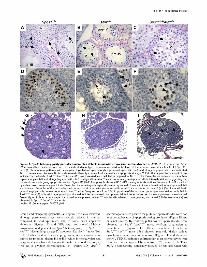
Round and elongating spermatids and sperm were also observed,
although post-meiotic stages were severely reduced in number
compared to wild-type mice, and in some cases appeared
abnormal (Figures 1C and S1B; data not shown). Meiotic
progression is dependent on Spo11 heterozygosity, as Spo112/2
Atm2/2 mice undergo a stage IV apoptosis, like Atm2/2 mice [20].
To further evaluate meiotic progression, testis sections were
stained for phospho-histone H3 (p-H3), which is normally detected
in spermatocytes from diplonema through the second division, as
well as in dividing spermatogonia [34] (Figure 1D). Atm2/2
spermatogonia were positive for p-H3 but spermatocytes were not,
as expected because of apoptosis during prophase I (Figure 1E and
data not shown). By contrast, p-H3-positive spermatocytes were
observed in Spo11+/2Atm2/2 mice, verifying progression to
metaphase I (Figure 1F). These metaphase I cells of
Spo11+/2Atm2/2 mice often showed relatively darkly stained
cytoplasm characteristic of apoptosis (Figure 1F and data not
shown). TUNEL staining confirmed that most spermatocytes were
eliminated at metaphase I by apoptosis [32] (Figure S1C). Thus,
Spo11 heterozygosity sufficiently rescued defects associated with
Figure 1. Spo11 heterozygosity partially ameliorates defects in meiotic progression in the absence of ATM. (A–C) Periodic acid Schiff(PAS)-stained testis sections from mice of the indicated genotypes. Roman numerals denote stages of the seminiferous epithelial cycle [33]. Spo11+/2
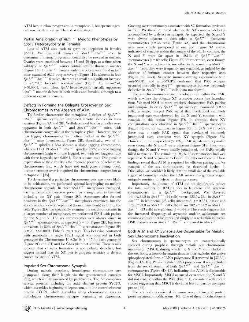
mice (A) show normal patterns, with examples of pachytene spermatocytes (p), round spermatids (rs), and elongating spermatids (es) indicated.Atm2/2 seminiferous tubules (B) show decreased cellularity as a result of spermatocyte apoptosis at stage IV. Cells that appear to be apoptotic areindicated (arrowheads). Spo11+/2Atm2/2 tubules (C) have increased testis cellularity compared to Atm2/2 mice. Examples are indicated of metaphaseI spermatocytes (MI) and elongating spermatids (es) in stage XII tubules. The cytosol of many metaphase cells is intensely stained, suggesting thatthese cells are undergoing apoptosis (see also Figure S1). (D–F) Anti-phospho-histone H3 (p-H3) staining of testis sections. Presence of p-H3 is markedby a dark brown enzymatic precipitate. Examples of spermatogonia (sg) and spermatocytes in diplonema (di), metaphase I (MI), or metaphase II (MII)are indicated. Examples of the most advanced non-apoptotic spermatocytes observed in Atm2/2 are indictated in panel E (sc). (G–I) Reduced Spo11gene dosage partially rescues oogenesis in Atm2/2 mice. Ovary sections from 17–18 dpp mice of the indicated genotypes were stained with PAS. InSpo11+/2 mice (G), as in wild type, growing and antral follicles (arrowheads) and primordial follicles at the cortex of the ovary (arrows) are observed.Essentially no follicles at any stage of maturation are present in Atm2/2 ovaries (H), whereas some growing and antral follicles (arrowheads) areobserved in Spo11+/2Atm2/2 ovaries (I).doi:10.1371/journal.pgen.1000076.g001
Role of ATM in Mouse Meiosis
PLoS Genetics | www.plosgenetics.org 3 May 2008 | Volume 4 | Issue 5 | e1000076
ATM loss to allow progression to metaphase I, but spermatogen-
esis was for the most part halted at this stage.
Partial Amelioration of Atm2/2 Meiotic Phenotypes bySpo11 Heterozygosity in Females
Loss of ATM also leads to germ cell depletion in females
[22,24]. We examined ovaries of Spo11+/2Atm2/2 mice to
determine if meiotic progression could also be rescued in oocytes.
Ovaries were examined between 17 and 29 dpp, at a time when
wild-type or Spo11+/2 ovaries contain several thousand oocytes
(Figure 1G). In Atm2/2 females, only one oocyte was found in four
mice examined (0.13 oocytes/ovary) (Figure 1H), whereas in four
Spo11+/2Atm2/2 females, there was a small but significant increase
to 7.963.7 follicular oocytes/ovary (Figure 1I; mean6sd,
p,0.0001, t test). Thus, Spo11 heterozygosity partially suppresses
Atm2/2 meiotic defects in both males and females, although to a
different extent in females.
Defects in Forming the Obligate Crossover on SexChromosomes in the Absence of ATM
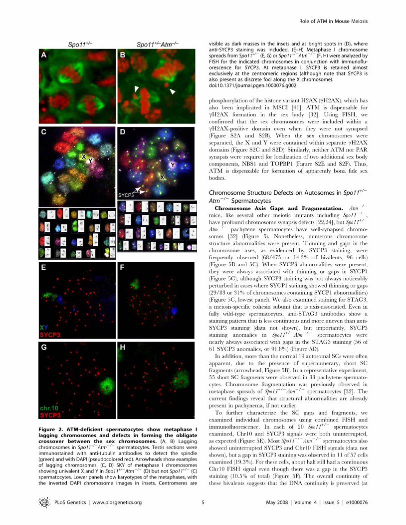
To further characterize the metaphase I defect of Spo11+/2
Atm2/2 spermatocytes, we examined meiotic spindles in testis
sections (Figure 2A and 2B). Well-developed bipolar spindles were
apparent in both Spo11+/2 and Spo11+/2Atm2/2 mice, with
chromosome congression at the metaphase plate. However, one or
two lagging chromosomes were often evident in the Spo11+/2
Atm2/2 mice (arrowheads, Figure 2B). Specifically, two of 11
Spo11+/2 spindles (18%) showed a single lagging chromosome,
whereas 11 of 13 Spo11+/2Atm2/2 spindles (85%) showed lagging
chromosomes (eight with one laggard, two with two laggards, one
with three laggards) (p = 0.0031, Fisher’s exact test). One possible
explanation of these results is the frequent presence of achiasmate
chromosomes (i.e., which have not undergone crossing over),
because crossing-over is required for chromosome congression at
metaphase I [35].
To determine if a particular chromosome pair was more likely
to be achiasmate, we performed spectral karyotyping on meiotic
chromosome spreads. In three Spo11+/2 metaphases examined,
each chromosome pair was present as a single unit (a bivalent)
including the XY pair (Figure 2C). Autosomes also formed
bivalents in five Spo11+/2Atm2/2 metaphases examined, but the
sex chromosomes were separated (formed univalents) in four of the
cells (Figure 2D). To specifically examine the sex chromosomes in
a larger number of metaphases, we performed FISH with probes
for the X and Y. The sex chromosomes were always joined in
Spo11+/2 spermatocytes, as expected (n = 10; Figure 2E), but were
univalents in 80% of Spo11+/2Atm2/2 spermatocytes (Figure 2F)
(n = 20; p,0.0001, Fisher’s exact test). This behavior contrasted
with autosomes: a single FISH signal was observed in both
genotypes for Chromosome 10 (Chr10) (n = 15 for each genotype)
(Figure 2G and 2H) and for Chr3 (data not shown). These results
indicate that chiasma formation is not globally defective, but
suggest instead that the XY pair is uniquely sensitive to defects
caused by lack of ATM.
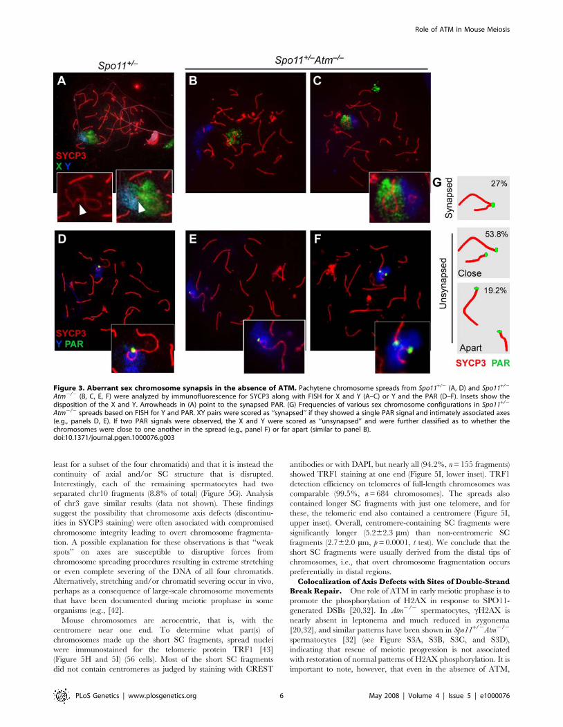
Impaired Sex Chromosome SynapsisDuring meiotic prophase, homologous chromosomes are
juxtaposed along their length via the synaptonemal complex
(SC), which is fully assembled by pachynema. The SC comprises
several proteins, including the axial element protein SYCP3,
which assembles beginning in leptonema, and the central element
protein SYCP1, which assembles along chromosome axes as
homologous chromosomes synapse beginning in zygonema.
Crossing-over is intimately associated with SC formation (reviewed
in [36]). We therefore tested whether the XY crossover defect is
accompanied by a defect in synapsis. As expected, the X and Y
were always adjacent to each other in Spo11+/2 pachytene
spermatocytes (n = 38 cells) (Figure 3A), and the chromosome
axes were closely juxtaposed at one end (Figure 3A insets),
indicative of synapsis within the context of the SC. In contrast, the
X and Y were far apart in 10.1% of Spo11+/2Atm2/2
spermatocytes (n = 89 cells; Figure 3B). Furthermore, even though
the X and Y were adjacent to one other in the remaining Spo11+/2
Atm2/2 cells, they were frequently not synapsed, as judged by the
absence of intimate contact between their respective axes
(Figure 3C inset). Separate immunostaining experiments with
anti-SYCP1 and anti-SYCP3 confirmed that PAR synapsis
occurred normally in Spo11+/2 spermatocytes but was frequently
defective in Spo11+/2Atm2/2 cells (data not shown).
The sex chromosomes share homology only within the PAR,
which is where the obligate XY crossover occurs (see Introduc-
tion). We used FISH to more precisely characterize PAR pairing
and synapsis. In every Spo11+/2 spermatocyte examined (n = 50
cells), a single, merged PAR signal that overlapped intimately
juxtaposed axes was observed for the X and Y, consistent with
synapsis in this region (Figure 3D). In contrast, three XY
configurations were observed in Spo11+/2Atm2/2 spermatocytes
(Figure 3E and 3F; summary in Figure 3G). In 27% (n = 78 cells),
there was a single PAR signal that overlapped intimately
juxtaposed axes, consistent with XY synapsis (Figure 3E).
However, in the major class (53.8%), PAR signals were separated
even though the X and Y were adjacent (Figure 3F). Thus, even
though the X and Y were usually juxtaposed, the PARs usually
failed to synapse. The remaining 19.2% of spermatocytes had well
separated X and Y (similar to Figure 3B; data not shown). These
findings reveal that ATM is required for efficient pairing and/or
synapsis of the sex chromosomes. As described further in
Discussion, we consider it likely that the small size of the available
region of homology within the PAR makes this genomic region
uniquely sensitive to defects in these processes.
Importantly, the absence of ATM did not significantly reduce
the total number of RAD51 foci in leptotene and zygotene
spermatocytes in a Spo11+/2 background. We observed
144.0631.0 in Spo11+/2 (14 cells) versus 123.5678.1 in Spo11+/2
Atm2/2 in leptonema (25 cells) (mean6sd, p = 0.354, t test); and
173.8623.8 in Spo11+/2 (20 cells) versus 202.7657.2 in Spo11+/2
Atm2/2 (23 cells) in zygonema (p = 0.041). This result suggests that
the increased frequency of asynaptic and/or achiasmate sex
chromosomes cannot be attributed simply to a reduction in overall
DSB frequencies in Spo11+/2Atm2/2 compared to Spo11+/2.
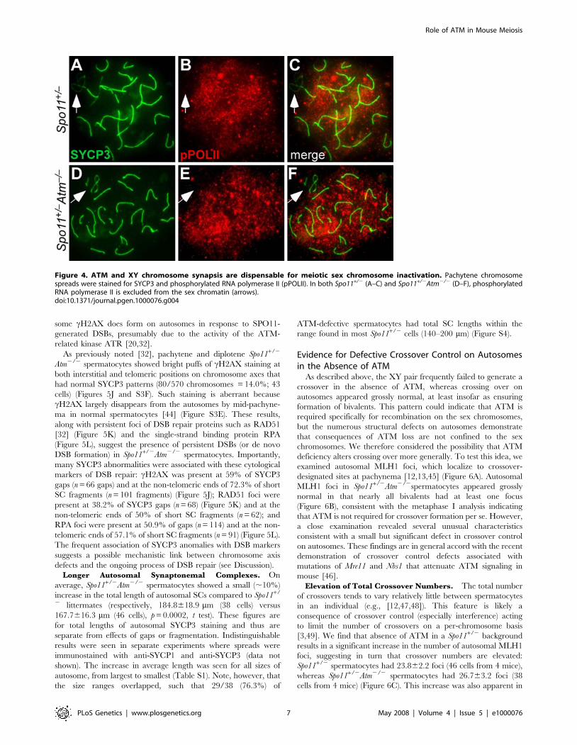
Both ATM and XY Synapsis Are Dispensable for MeioticSex Chromosome Inactivation
Sex chromosomes in spermatocytes are transcriptionally
silenced during prophase through meiotic sex chromosome
inactivation (MSCI), during which the X and Y are included in
the sex body, a heterochromatin domain that excludes the active
(phosphorylated) form of RNA polymerase II (reviewed in [37,38])
(Figure 4A–4C). Phosphorylated RNA polymerase II was excluded
from the sex chromatin of both Spo11+/2 and Spo11+/2Atm2/2
spermatocytes (Figure 4D–4F), indicating that ATM is dispensable
for MSCI. Importantly, MSCI occurred even when the X and Y
did not synapse within the PAR (Figure 4), consistent with recent
studies suggesting that MSCI is driven at least in part by asynapsis
per se [39].
The sex body is enriched for numerous proteins and protein
posttranslational modifications [40]. One of these modifications is
Role of ATM in Mouse Meiosis
PLoS Genetics | www.plosgenetics.org 4 May 2008 | Volume 4 | Issue 5 | e1000076
phosphorylation of the histone variant H2AX (cH2AX), which has
also been implicated in MSCI [41]. ATM is dispensable for
cH2AX formation in the sex body [32]. Using FISH, we
confirmed that the sex chromosomes were included within a
cH2AX-positive domain even when they were not synapsed
(Figure S2A and S2B). When the sex chromosomes were
separated, the X and Y were contained within separate cH2AX
domains (Figure S2C and S2D). Similarly, neither ATM nor PAR
synapsis were required for localization of two additional sex body
components, NBS1 and TOPBP1 (Figure S2E and S2F). Thus,
ATM is dispensable for formation of apparently bona fide sex
bodies.
Chromosome Structure Defects on Autosomes in Spo11+/2
Atm2/2 SpermatocytesChromosome Axis Gaps and Fragmentation. Atm2/2
mice, like several other meiotic mutants including Spo112/2,
have profound chromosome synapsis defects [22,24], but Spo11+/2
Atm2/2 pachytene spermatocytes have well-synapsed chromo-
somes [32] (Figure 5). Nonetheless, numerous chromosome
structure abnormalities were present. Thinning and gaps in the
chromosome axes, as evidenced by SYCP3 staining, were
frequently observed (68/475 or 14.3% of bivalents, 96 cells)
(Figure 5B and 5C). When SYCP3 abnormalities were present,
they were always associated with thinning or gaps in SYCP1
(Figure 5C), although SYCP3 staining was not always noticeably
perturbed in cases where SYCP1 staining showed thinning or gaps
(29/83 or 31% of chromosomes containing SYCP1 abnormalities)
(Figure 5C, lowest panel). We also examined staining for STAG3,
a meiosis-specific cohesin subunit that is axis-associated. Even in
fully wild-type spermatocytes, anti-STAG3 antibodies show a
staining pattern that is less continuous and more uneven than anti-
SYCP3 staining (data not shown), but importantly, SYCP3
staining anomalies in Spo11+/2Atm2/2 spermatocytes were
nearly always associated with gaps in the STAG3 staining (56 of
61 SYCP3 anomalies, or 91.8%) (Figure 5D).
In addition, more than the normal 19 autosomal SCs were often
apparent, due to the presence of supernumerary, short SC
fragments (arrowhead, Figure 5B). In a representative experiment,
55 short SC fragments were observed in 33 pachytene spermato-
cytes. Chromosome fragmentation was previously observed in
metaphase spreads of Spo11+/2Atm2/2 spermatocytes [32]. The
current findings reveal that structural abnormalities are already
present in pachynema, if not earlier.
To further characterize the SC gaps and fragments, we
examined individual chromosomes using combined FISH and
immunofluorescence. In each of 20 Spo11+/2 spermatocytes
examined, Chr10 and SYCP3 signals were both uninterrupted,
as expected (Figure 5E). Most Spo11+/2Atm2/2 spermatocytes also
showed uninterrupted SYCP3 and Chr10 FISH signals (data not
shown), but a gap in SYCP3 staining was observed in 11 of 57 cells
examined (19.3%). For these cells, about half still had a continuous
Chr10 FISH signal even though there was a gap in the SYCP3
staining (10.5% of total) (Figure 5F). The overall continuity of
these bivalents suggests that the DNA continuity is preserved (at
Figure 2. ATM-deficient spermatocytes show metaphase Ilagging chromosomes and defects in forming the obligatecrossover between the sex chromosomes. (A, B) Laggingchromosomes in Spo11+/2Atm2/2 spermatocytes. Testis sections wereimmunostained with anti-tubulin antibodies to detect the spindle(green) and with DAPI (pseudocolored red). Arrowheads show examplesof lagging chromosomes. (C, D) SKY of metaphase I chromosomesshowing univalent X and Y in Spo11+/2Atm2/2 (D) but not Spo11+/2 (C)spermatocytes. Lower panels show karyotypes of the metaphases, withthe inverted DAPI chromosome images in insets. Centromeres are
visible as dark masses in the insets and as bright spots in (D), whereanti-SYCP3 staining was included. (E–H) Metaphase I chromosomespreads from Spo11+/2 (E, G) or Spo11+/2Atm2/2 (F, H) were analyzed byFISH for the indicated chromosomes in conjunction with immunoflu-orescence for SYCP3. At metaphase I, SYCP3 is retained almostexclusively at the centromeric regions (although note that SYCP3 isalso present as discrete foci along the X chromosome).doi:10.1371/journal.pgen.1000076.g002
Role of ATM in Mouse Meiosis
PLoS Genetics | www.plosgenetics.org 5 May 2008 | Volume 4 | Issue 5 | e1000076
least for a subset of the four chromatids) and that it is instead the
continuity of axial and/or SC structure that is disrupted.
Interestingly, each of the remaining spermatocytes had two
separated chr10 fragments (8.8% of total) (Figure 5G). Analysis
of chr3 gave similar results (data not shown). These findings
suggest the possibility that chromosome axis defects (discontinu-
ities in SYCP3 staining) were often associated with compromised
chromosome integrity leading to overt chromosome fragmenta-
tion. A possible explanation for these observations is that ‘‘weak
spots’’ on axes are susceptible to disruptive forces from
chromosome spreading procedures resulting in extreme stretching
or even complete severing of the DNA of all four chromatids.
Alternatively, stretching and/or chromatid severing occur in vivo,
perhaps as a consequence of large-scale chromosome movements
that have been documented during meiotic prophase in some
organisms (e.g., [42].
Mouse chromosomes are acrocentric, that is, with the
centromere near one end. To determine what part(s) of
chromosomes made up the short SC fragments, spread nuclei
were immunostained for the telomeric protein TRF1 [43]
(Figure 5H and 5I) (56 cells). Most of the short SC fragments
did not contain centromeres as judged by staining with CREST
antibodies or with DAPI, but nearly all (94.2%, n = 155 fragments)
showed TRF1 staining at one end (Figure 5I, lower inset). TRF1
detection efficiency on telomeres of full-length chromosomes was
comparable (99.5%, n = 684 chromosomes). The spreads also
contained longer SC fragments with just one telomere, and for
these, the telomeric end also contained a centromere (Figure 5I,
upper inset). Overall, centromere-containing SC fragments were
significantly longer (5.262.3 mm) than non-centromeric SC
fragments (2.762.0 mm, p = 0.0001, t test). We conclude that the
short SC fragments were usually derived from the distal tips of
chromosomes, i.e., that overt chromosome fragmentation occurs
preferentially in distal regions.
Colocalization of Axis Defects with Sites of Double-Strand
Break Repair. One role of ATM in early meiotic prophase is to
promote the phosphorylation of H2AX in response to SPO11-
generated DSBs [20,32]. In Atm2/2 spermatocytes, cH2AX is
nearly absent in leptonema and much reduced in zygonema
[20,32], and similar patterns have been shown in Spo11+/2Atm2/2
spermatocytes [32] (see Figure S3A, S3B, S3C, and S3D),
indicating that rescue of meiotic progression is not associated
with restoration of normal patterns of H2AX phosphorylation. It is
important to note, however, that even in the absence of ATM,
Figure 3. Aberrant sex chromosome synapsis in the absence of ATM. Pachytene chromosome spreads from Spo11+/2 (A, D) and Spo11+/2
Atm2/2 (B, C, E, F) were analyzed by immunofluorescence for SYCP3 along with FISH for X and Y (A–C) or Y and the PAR (D–F). Insets show thedisposition of the X and Y. Arrowheads in (A) point to the synapsed PAR. (G) Frequencies of various sex chromosome configurations in Spo11+/2
Atm2/2 spreads based on FISH for Y and PAR. XY pairs were scored as ‘‘synapsed’’ if they showed a single PAR signal and intimately associated axes(e.g., panels D, E). If two PAR signals were observed, the X and Y were scored as ‘‘unsynapsed’’ and were further classified as to whether thechromosomes were close to one another in the spread (e.g., panel F) or far apart (similar to panel B).doi:10.1371/journal.pgen.1000076.g003
Role of ATM in Mouse Meiosis
PLoS Genetics | www.plosgenetics.org 6 May 2008 | Volume 4 | Issue 5 | e1000076
some cH2AX does form on autosomes in response to SPO11-
generated DSBs, presumably due to the activity of the ATM-
related kinase ATR [20,32].
As previously noted [32], pachytene and diplotene Spo11+/2
Atm2/2 spermatocytes showed bright puffs of cH2AX staining at
both interstitial and telomeric positions on chromosome axes that
had normal SYCP3 patterns (80/570 chromosomes = 14.0%; 43
cells) (Figures 5J and S3F). Such staining is aberrant because
cH2AX largely disappears from the autosomes by mid-pachyne-
ma in normal spermatocytes [44] (Figure S3E). These results,
along with persistent foci of DSB repair proteins such as RAD51
[32] (Figure 5K) and the single-strand binding protein RPA
(Figure 5L), suggest the presence of persistent DSBs (or de novo
DSB formation) in Spo11+/2Atm2/2 spermatocytes. Importantly,
many SYCP3 abnormalities were associated with these cytological
markers of DSB repair: cH2AX was present at 59% of SYCP3
gaps (n = 66 gaps) and at the non-telomeric ends of 72.3% of short
SC fragments (n = 101 fragments) (Figure 5J); RAD51 foci were
present at 38.2% of SYCP3 gaps (n = 68) (Figure 5K) and at the
non-telomeric ends of 50% of short SC fragments (n = 62); and
RPA foci were present at 50.9% of gaps (n = 114) and at the non-
telomeric ends of 57.1% of short SC fragments (n = 91) (Figure 5L).
The frequent association of SYCP3 anomalies with DSB markers
suggests a possible mechanistic link between chromosome axis
defects and the ongoing process of DSB repair (see Discussion).
Longer Autosomal Synaptonemal Complexes. On
average, Spo11+/2Atm2/2 spermatocytes showed a small (,10%)
increase in the total length of autosomal SCs compared to Spo11+/
2 littermates (respectively, 184.8618.9 mm (38 cells) versus
167.7616.3 mm (46 cells), p = 0.0002, t test). These figures are
for total lengths of autosomal SYCP3 staining and thus are
separate from effects of gaps or fragmentation. Indistinguishable
results were seen in separate experiments where spreads were
immunostained with anti-SYCP1 and anti-SYCP3 (data not
shown). The increase in average length was seen for all sizes of
autosome, from largest to smallest (Table S1). Note, however, that
the size ranges overlapped, such that 29/38 (76.3%) of
ATM-defective spermatocytes had total SC lengths within the
range found in most Spo11+/2 cells (140–200 mm) (Figure S4).
Evidence for Defective Crossover Control on Autosomesin the Absence of ATM
As described above, the XY pair frequently failed to generate a
crossover in the absence of ATM, whereas crossing over on
autosomes appeared grossly normal, at least insofar as ensuring
formation of bivalents. This pattern could indicate that ATM is
required specifically for recombination on the sex chromosomes,
but the numerous structural defects on autosomes demonstrate
that consequences of ATM loss are not confined to the sex
chromosomes. We therefore considered the possibility that ATM
deficiency alters crossing over more generally. To test this idea, we
examined autosomal MLH1 foci, which localize to crossover-
designated sites at pachynema [12,13,45] (Figure 6A). Autosomal
MLH1 foci in Spo11+/2Atm2/2spermatocytes appeared grossly
normal in that nearly all bivalents had at least one focus
(Figure 6B), consistent with the metaphase I analysis indicating
that ATM is not required for crossover formation per se. However,
a close examination revealed several unusual characteristics
consistent with a small but significant defect in crossover control
on autosomes. These findings are in general accord with the recent
demonstration of crossover control defects associated with
mutations of Mre11 and Nbs1 that attenuate ATM signaling in
mouse [46].
Elevation of Total Crossover Numbers. The total number
of crossovers tends to vary relatively little between spermatocytes
in an individual (e.g., [12,47,48]). This feature is likely a
consequence of crossover control (especially interference) acting
to limit the number of crossovers on a per-chromosome basis
[3,49]. We find that absence of ATM in a Spo11+/2 background
results in a significant increase in the number of autosomal MLH1
foci, suggesting in turn that crossover numbers are elevated:
Spo11+/2 spermatocytes had 23.862.2 foci (46 cells from 4 mice),
whereas Spo11+/2Atm2/2 spermatocytes had 26.763.2 foci (38
cells from 4 mice) (Figure 6C). This increase was also apparent in
Figure 4. ATM and XY chromosome synapsis are dispensable for meiotic sex chromosome inactivation. Pachytene chromosomespreads were stained for SYCP3 and phosphorylated RNA polymerase II (pPOLII). In both Spo11+/2 (A–C) and Spo11+/2Atm2/2 (D–F), phosphorylatedRNA polymerase II is excluded from the sex chromatin (arrows).doi:10.1371/journal.pgen.1000076.g004
Role of ATM in Mouse Meiosis
PLoS Genetics | www.plosgenetics.org 7 May 2008 | Volume 4 | Issue 5 | e1000076
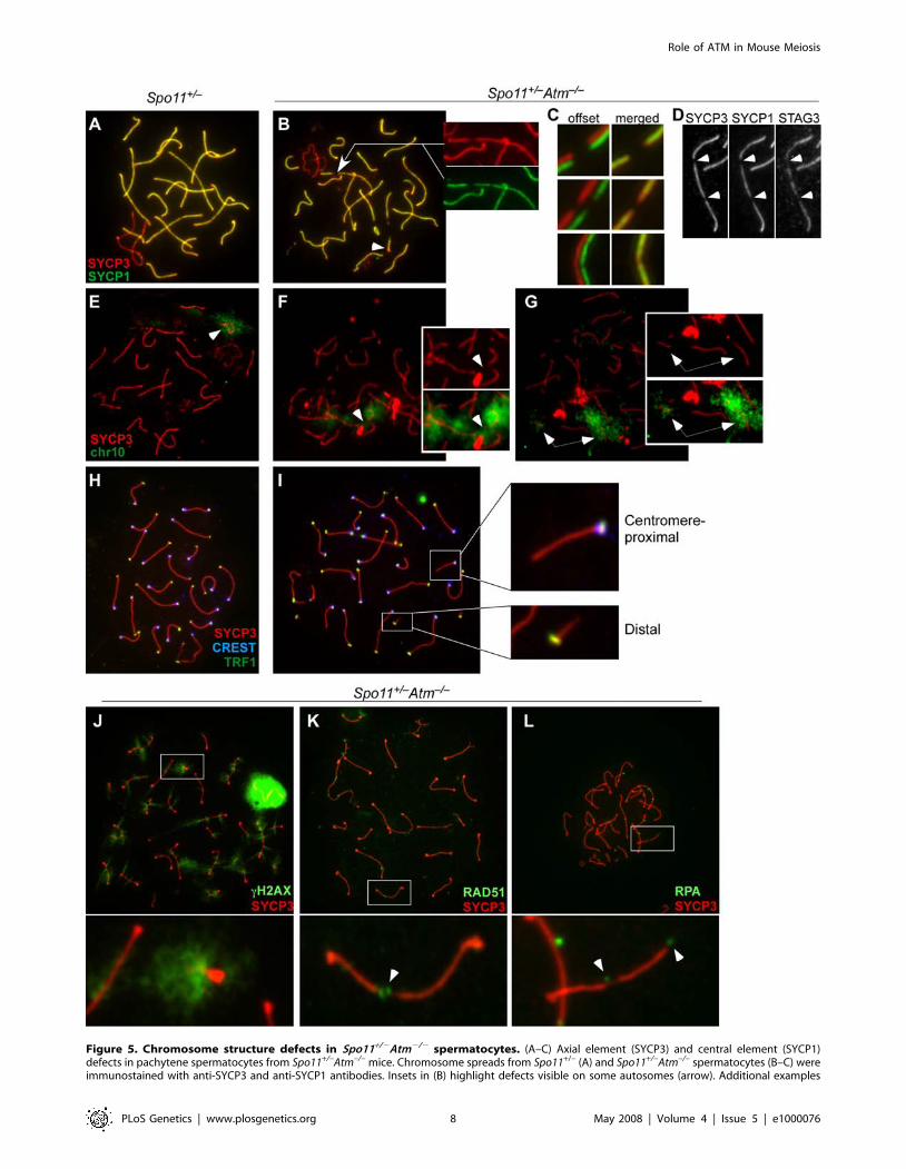
Figure 5. Chromosome structure defects in Spo11+/2Atm2/2 spermatocytes. (A–C) Axial element (SYCP3) and central element (SYCP1)defects in pachytene spermatocytes from Spo11+/–Atm–/– mice. Chromosome spreads from Spo11+/– (A) and Spo11+/–Atm–/– spermatocytes (B–C) wereimmunostained with anti-SYCP3 and anti-SYCP1 antibodies. Insets in (B) highlight defects visible on some autosomes (arrow). Additional examples
Role of ATM in Mouse Meiosis
PLoS Genetics | www.plosgenetics.org 8 May 2008 | Volume 4 | Issue 5 | e1000076
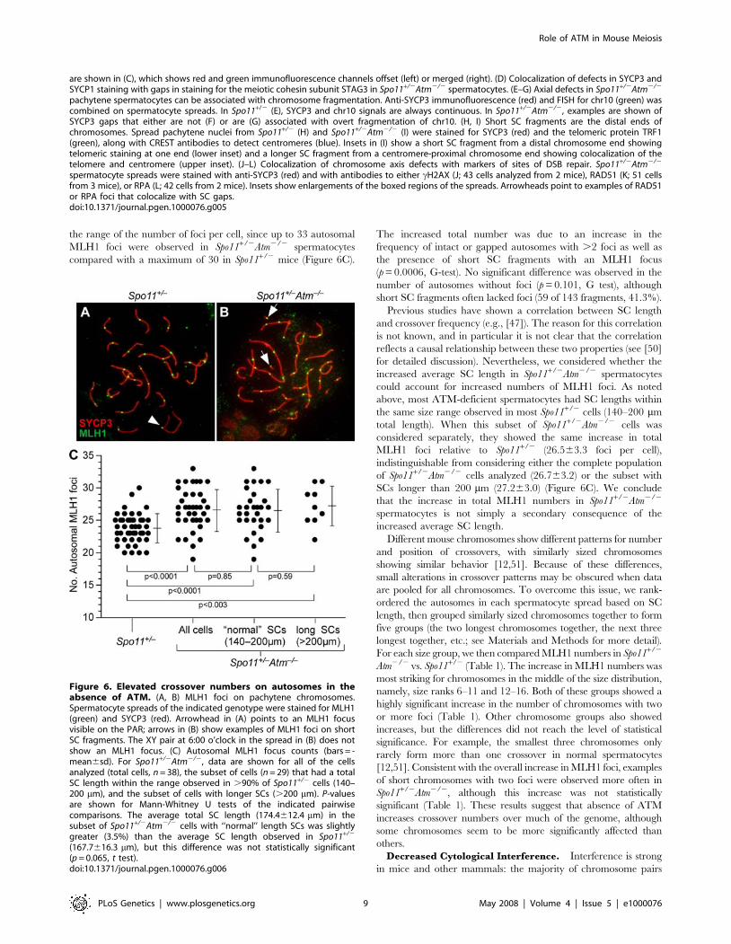
the range of the number of foci per cell, since up to 33 autosomal
MLH1 foci were observed in Spo11+/2Atm2/2 spermatocytes
compared with a maximum of 30 in Spo11+/2 mice (Figure 6C).
The increased total number was due to an increase in the
frequency of intact or gapped autosomes with .2 foci as well as
the presence of short SC fragments with an MLH1 focus
(p = 0.0006, G-test). No significant difference was observed in the
number of autosomes without foci (p = 0.101, G test), although
short SC fragments often lacked foci (59 of 143 fragments, 41.3%).
Previous studies have shown a correlation between SC length
and crossover frequency (e.g., [47]). The reason for this correlation
is not known, and in particular it is not clear that the correlation
reflects a causal relationship between these two properties (see [50]
for detailed discussion). Nevertheless, we considered whether the
increased average SC length in Spo11+/2Atm2/2 spermatocytes
could account for increased numbers of MLH1 foci. As noted
above, most ATM-deficient spermatocytes had SC lengths within
the same size range observed in most Spo11+/2 cells (140–200 mm
total length). When this subset of Spo11+/2Atm2/2 cells was
considered separately, they showed the same increase in total
MLH1 foci relative to Spo11+/2 (26.563.3 foci per cell),
indistinguishable from considering either the complete population
of Spo11+/2Atm2/2 cells analyzed (26.763.2) or the subset with
SCs longer than 200 mm (27.263.0) (Figure 6C). We conclude
that the increase in total MLH1 numbers in Spo11+/2Atm2/2
spermatocytes is not simply a secondary consequence of the
increased average SC length.
Different mouse chromosomes show different patterns for number
and position of crossovers, with similarly sized chromosomes
showing similar behavior [12,51]. Because of these differences,
small alterations in crossover patterns may be obscured when data
are pooled for all chromosomes. To overcome this issue, we rank-
ordered the autosomes in each spermatocyte spread based on SC
length, then grouped similarly sized chromosomes together to form
five groups (the two longest chromosomes together, the next three
longest together, etc.; see Materials and Methods for more detail).
For each size group, we then compared MLH1 numbers in Spo11+/2
Atm2/2 vs. Spo11+/2 (Table 1). The increase in MLH1 numbers was
most striking for chromosomes in the middle of the size distribution,
namely, size ranks 6–11 and 12–16. Both of these groups showed a
highly significant increase in the number of chromosomes with two
or more foci (Table 1). Other chromosome groups also showed
increases, but the differences did not reach the level of statistical
significance. For example, the smallest three chromosomes only
rarely form more than one crossover in normal spermatocytes
[12,51]. Consistent with the overall increase in MLH1 foci, examples
of short chromosomes with two foci were observed more often in
Spo11+/2Atm2/2, although this increase was not statistically
significant (Table 1). These results suggest that absence of ATM
increases crossover numbers over much of the genome, although
some chromosomes seem to be more significantly affected than
others.
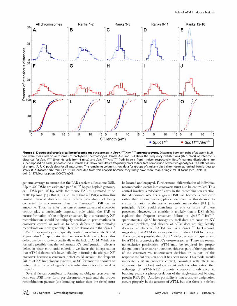
Decreased Cytological Interference. Interference is strong
in mice and other mammals: the majority of chromosome pairs
Figure 6. Elevated crossover numbers on autosomes in theabsence of ATM. (A, B) MLH1 foci on pachytene chromosomes.Spermatocyte spreads of the indicated genotype were stained for MLH1(green) and SYCP3 (red). Arrowhead in (A) points to an MLH1 focusvisible on the PAR; arrows in (B) show examples of MLH1 foci on shortSC fragments. The XY pair at 6:00 o’clock in the spread in (B) does notshow an MLH1 focus. (C) Autosomal MLH1 focus counts (bars = -mean6sd). For Spo11+/2Atm2/2, data are shown for all of the cellsanalyzed (total cells, n = 38), the subset of cells (n = 29) that had a totalSC length within the range observed in .90% of Spo11+/2 cells (140–200 mm), and the subset of cells with longer SCs (.200 mm). P-valuesare shown for Mann-Whitney U tests of the indicated pairwisecomparisons. The average total SC length (174.4612.4 mm) in thesubset of Spo11+/2Atm2/2 cells with ‘‘normal’’ length SCs was slightlygreater (3.5%) than the average SC length observed in Spo11+/2
(167.7616.3 mm), but this difference was not statistically significant(p = 0.065, t test).doi:10.1371/journal.pgen.1000076.g006
are shown in (C), which shows red and green immunofluorescence channels offset (left) or merged (right). (D) Colocalization of defects in SYCP3 andSYCP1 staining with gaps in staining for the meiotic cohesin subunit STAG3 in Spo11+/2Atm2/2 spermatocytes. (E–G) Axial defects in Spo11+/2Atm2/2
pachytene spermatocytes can be associated with chromosome fragmentation. Anti-SYCP3 immunofluorescence (red) and FISH for chr10 (green) wascombined on spermatocyte spreads. In Spo11+/2 (E), SYCP3 and chr10 signals are always continuous. In Spo11+/2Atm2/2, examples are shown ofSYCP3 gaps that either are not (F) or are (G) associated with overt fragmentation of chr10. (H, I) Short SC fragments are the distal ends ofchromosomes. Spread pachytene nuclei from Spo11+/2 (H) and Spo11+/2Atm2/2 (I) were stained for SYCP3 (red) and the telomeric protein TRF1(green), along with CREST antibodies to detect centromeres (blue). Insets in (I) show a short SC fragment from a distal chromosome end showingtelomeric staining at one end (lower inset) and a longer SC fragment from a centromere-proximal chromosome end showing colocalization of thetelomere and centromere (upper inset). (J–L) Colocalization of chromosome axis defects with markers of sites of DSB repair. Spo11+/2Atm2/2
spermatocyte spreads were stained with anti-SYCP3 (red) and with antibodies to either cH2AX (J; 43 cells analyzed from 2 mice), RAD51 (K; 51 cellsfrom 3 mice), or RPA (L; 42 cells from 2 mice). Insets show enlargements of the boxed regions of the spreads. Arrowheads point to examples of RAD51or RPA foci that colocalize with SC gaps.doi:10.1371/journal.pgen.1000076.g005
Role of ATM in Mouse Meiosis
PLoS Genetics | www.plosgenetics.org 9 May 2008 | Volume 4 | Issue 5 | e1000076
form only a single crossover, and multiple crossovers on the same
bivalent tend to be both widely and evenly spaced [51,52].
Directly measuring crossover interference in mice requires analysis
of viable progeny, precluding studies in sterile mutants. However,
alternative methods have been developed for measuring
cytological manifestations of interference, by measuring the
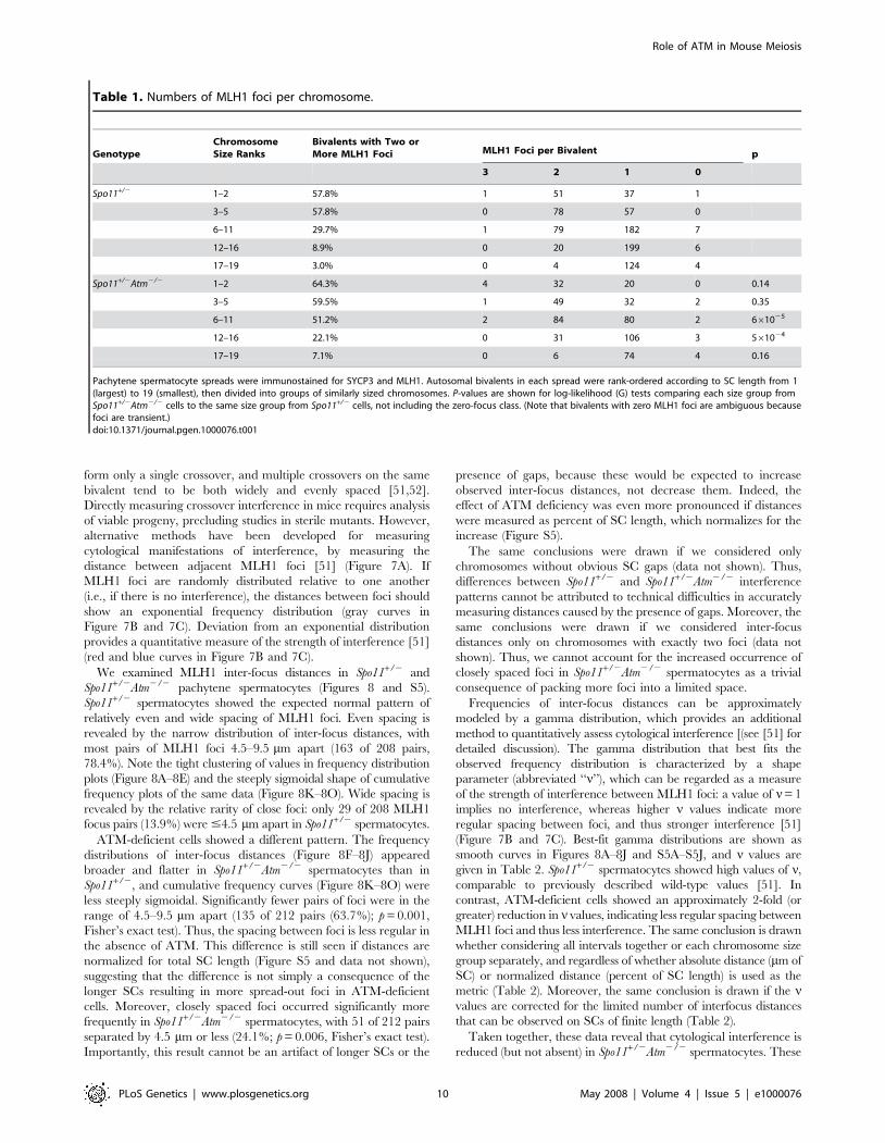
distance between adjacent MLH1 foci [51] (Figure 7A). If
MLH1 foci are randomly distributed relative to one another
(i.e., if there is no interference), the distances between foci should
show an exponential frequency distribution (gray curves in
Figure 7B and 7C). Deviation from an exponential distribution
provides a quantitative measure of the strength of interference [51]
(red and blue curves in Figure 7B and 7C).
We examined MLH1 inter-focus distances in Spo11+/2 and
Spo11+/2Atm2/2 pachytene spermatocytes (Figures 8 and S5).
Spo11+/2 spermatocytes showed the expected normal pattern of
relatively even and wide spacing of MLH1 foci. Even spacing is
revealed by the narrow distribution of inter-focus distances, with
most pairs of MLH1 foci 4.5–9.5 mm apart (163 of 208 pairs,
78.4%). Note the tight clustering of values in frequency distribution
plots (Figure 8A–8E) and the steeply sigmoidal shape of cumulative
frequency plots of the same data (Figure 8K–8O). Wide spacing is
revealed by the relative rarity of close foci: only 29 of 208 MLH1
focus pairs (13.9%) were #4.5 mm apart in Spo11+/2 spermatocytes.
ATM-deficient cells showed a different pattern. The frequency
distributions of inter-focus distances (Figure 8F–8J) appeared
broader and flatter in Spo11+/2Atm2/2 spermatocytes than in
Spo11+/2, and cumulative frequency curves (Figure 8K–8O) were
less steeply sigmoidal. Significantly fewer pairs of foci were in the
range of 4.5–9.5 mm apart (135 of 212 pairs (63.7%); p = 0.001,
Fisher’s exact test). Thus, the spacing between foci is less regular in
the absence of ATM. This difference is still seen if distances are
normalized for total SC length (Figure S5 and data not shown),
suggesting that the difference is not simply a consequence of the
longer SCs resulting in more spread-out foci in ATM-deficient
cells. Moreover, closely spaced foci occurred significantly more
frequently in Spo11+/2Atm2/2 spermatocytes, with 51 of 212 pairs
separated by 4.5 mm or less (24.1%; p = 0.006, Fisher’s exact test).
Importantly, this result cannot be an artifact of longer SCs or the
presence of gaps, because these would be expected to increase
observed inter-focus distances, not decrease them. Indeed, the
effect of ATM deficiency was even more pronounced if distances
were measured as percent of SC length, which normalizes for the
increase (Figure S5).
The same conclusions were drawn if we considered only
chromosomes without obvious SC gaps (data not shown). Thus,
differences between Spo11+/2 and Spo11+/2Atm2/2 interference
patterns cannot be attributed to technical difficulties in accurately
measuring distances caused by the presence of gaps. Moreover, the
same conclusions were drawn if we considered inter-focus
distances only on chromosomes with exactly two foci (data not
shown). Thus, we cannot account for the increased occurrence of
closely spaced foci in Spo11+/2Atm2/2 spermatocytes as a trivial
consequence of packing more foci into a limited space.
Frequencies of inter-focus distances can be approximately
modeled by a gamma distribution, which provides an additional
method to quantitatively assess cytological interference [(see [51] for
detailed discussion). The gamma distribution that best fits the
observed frequency distribution is characterized by a shape
parameter (abbreviated ‘‘n’’), which can be regarded as a measure
of the strength of interference between MLH1 foci: a value of n= 1
implies no interference, whereas higher n values indicate more
regular spacing between foci, and thus stronger interference [51]
(Figure 7B and 7C). Best-fit gamma distributions are shown as
smooth curves in Figures 8A–8J and S5A–S5J, and n values are
given in Table 2. Spo11+/2 spermatocytes showed high values of n,
comparable to previously described wild-type values [51]. In
contrast, ATM-deficient cells showed an approximately 2-fold (or
greater) reduction in n values, indicating less regular spacing between
MLH1 foci and thus less interference. The same conclusion is drawn
whether considering all intervals together or each chromosome size
group separately, and regardless of whether absolute distance (mm of
SC) or normalized distance (percent of SC length) is used as the
metric (Table 2). Moreover, the same conclusion is drawn if the nvalues are corrected for the limited number of interfocus distances
that can be observed on SCs of finite length (Table 2).
Taken together, these data reveal that cytological interference is
reduced (but not absent) in Spo11+/2Atm2/2 spermatocytes. These
Table 1. Numbers of MLH1 foci per chromosome.
GenotypeChromosomeSize Ranks
Bivalents with Two orMore MLH1 Foci MLH1 Foci per Bivalent p
3 2 1 0
Spo11+/2 1–2 57.8% 1 51 37 1
3–5 57.8% 0 78 57 0
6–11 29.7% 1 79 182 7
12–16 8.9% 0 20 199 6
17–19 3.0% 0 4 124 4
Spo11+/2Atm2/2 1–2 64.3% 4 32 20 0 0.14
3–5 59.5% 1 49 32 2 0.35
6–11 51.2% 2 84 80 2 661025
12–16 22.1% 0 31 106 3 561024
17–19 7.1% 0 6 74 4 0.16
Pachytene spermatocyte spreads were immunostained for SYCP3 and MLH1. Autosomal bivalents in each spread were rank-ordered according to SC length from 1(largest) to 19 (smallest), then divided into groups of similarly sized chromosomes. P-values are shown for log-likelihood (G) tests comparing each size group fromSpo11+/2Atm2/2 cells to the same size group from Spo11+/2 cells, not including the zero-focus class. (Note that bivalents with zero MLH1 foci are ambiguous becausefoci are transient.)doi:10.1371/journal.pgen.1000076.t001
Role of ATM in Mouse Meiosis
PLoS Genetics | www.plosgenetics.org 10 May 2008 | Volume 4 | Issue 5 | e1000076
findings suggest that crossover interference is partially defective
when ATM is not present.
Discussion
In the absence of ATM, mouse spermatocytes and oocytes die by
apoptosis during prophase of meiosis I, exhibiting profound defects
in meiotic chromosome behavior [22,24]. Remarkably, most of the
spermatocyte defects are eliminated simply by halving Spo11 gene
dosage: instead of dying by apoptosis at pachynema (like Atm2/2 and
Spo112/2 single mutants), most Spo11+/2Atm2/2 spermatocytes
progress to metaphase I, and sometimes beyond [32]. Homologous
synapsis, sex body formation, and crossing over are substantially,
albeit incompletely, rescued. In this study, we took advantage of this
intriguing phenotype to analyze the role of ATM in meiotic
recombination. As discussed further below, our findings suggest
previously undefined roles of ATM in crossover control and in
promoting integrity of higher order chromosome structures.
How does Spo11 heterozygosity rescue Atm2/2 meiotic
progression? Cytological and other evidence suggest that Spo11+/2
spermatocytes form fewer DSBs than wild type (F. Cole, S.
Keeney, and M. Jasin, unpublished observations). Given that
ATM has an established role in meiotic DSB repair [20,21,32], a
straightforward interpretation is that a reduced number of SPO11-
generated DSBs is responsible for suppression of the meiotic DSB
repair defects arising from ATM deficiency. Perhaps there is a
threshold amount of DSBs below which another kinase (e.g., ATR)
can partially substitute for ATM; above this threshold, the number
of DSBs may exceed the capacity for this kinase to substitute. It is
also possible that DSBs are formed in wild-type numbers in
Spo11+/2 mice but are delayed such that induction of ATR later in
prophase is able to substitute for ATM [32]. Current findings do
not allow us to distinguish between these and other possibilities.
Atm2/2 spermatocytes, similar to many other mutants including
Spo112/2, Dmc12/2 and Msh52/2, show pronounced defects in
forming a bona fide sex body and transcriptionally silencing the X
and Y chromosomes [20,37]. Studies of these and other mouse
mutants defective for MSCI strongly support the hypothesis that
failure to silence the sex chromosomes is sufficient to trigger
apoptosis of pachytene spermatocytes in stage IV tubules
(reviewed in [37,38]). Thus, the substantial restoration of sex
body formation and MSCI in Spo11+/2Atm2/2 spermatocytes may
account for the suppression of Atm2/2 pachytene apoptosis.
Although Spo11+/2Atm2/2 spermatocytes progress further than
Atm2/2 single mutants, they are substantially eliminated at or
prior to the first meiotic division. It is possible that apoptosis is
triggered by a spindle checkpoint responding to the lagging
chromosomes observed at metaphase I, which are likely to be the
frequently achiasmate X and Y. Indeed, metaphase I spermatocyte
apoptosis has been observed in several instances where one or a
few achiasmate chromosomes are present because of chromosomal
abnormalities [53–55], as well as in Mlh12/2 mice in which most
chromosomes lack chiasmata [56]. Alternatively, or in addition,
metaphase I apoptosis of Spo11+/2Atm2/2 spermatocytes may be a
response to unrepaired DSBs, whose presence is indicated by
persistent cH2AX, RAD51, and RPA (data not shown).
The rescue of meiotic progression in Spo11+/2Atm2/2 females
was much less pronounced than in males. We have shown that
ovarian follicle formation is particularly sensitive to the presence of
unrepaired DNA damage [21]. Thus, even if meiotic prophase
events were rescued to the same extent as in spermatocytes, it is
possible that persistent DNA damage would preclude rescue of
oocytes at this stage.
ATM and Control of the Normal Number and Distributionof Crossovers
Forming an Obligate Crossover. In most organisms,
nonexchange chromosomes occur rarely, giving rise to the
concept of the obligate crossover [57] (see Introduction). The
mammalian XY pair shares homology only within the very small
PAR, and as a result, the crossover frequency in males is
elevated .100-fold in the PAR over the genome average. DSB
frequency in the PAR must be elevated at least 10-fold over the
Figure 7. Measuring cytological interference between MLH1foci. (A) Pachytene spermatocyte spreads were immunostained forSYCP3 (shown in red) and MLH1 (shown in green), then the distancesbetween foci were measured on autosomal bivalents that contain twoor more MLH1 foci. (B, C) Examples of relative (B) and correspondingcumulative (C) frequency plots of gamma distributions. If there is nointerference between MLH1 foci, an exponential frequency distributionis expected (gray lines). Deviation from exponential behavior indicatesthe existence of interference (red and blue lines): short and longdistances become more rare and the spacing becomes more even (i.e.,distances are tightly clustered). Curves were calculated using anaverage interfocus distance of 10 and the indicated increasing valuesfor the shape parameter, n. See text and [51] for further discussion.doi:10.1371/journal.pgen.1000076.g007
Role of ATM in Mouse Meiosis
PLoS Genetics | www.plosgenetics.org 11 May 2008 | Volume 4 | Issue 5 | e1000076
genome average to ensure that the PAR receives at least one DSB.
(Up to 300 DSBs are estimated per 36109 bp per haploid genome,
or 1 DSB per 107 bp, while the mouse PAR is estimated to be
,106 bp long [4].) But it is also likely that a DSB(s) within this
limited physical distance has a greater probability of being
converted to a crossover than the ‘‘average’’ DSB on an
autosome. Thus, we infer that one or more aspects of crossover
control play a particularly important role within the PAR to
ensure formation of the obligate crossover. By this reasoning, XY
recombination should be uniquely sensitive to perturbations in
crossover control as well as to other defects in interhomolog
recombination more generally. Here, we demonstrate that Spo11+/
2Atm2/2 spermatocytes frequently contain an achiasmate X and
Y pair. Spo11+/2 spermatocytes have no such difficulty, hence this
defect can be attributed specifically to the lack of ATM. While it is
formally possible that the achiasmate XY configuration reflects a
defect in sister chromatid cohesion, we favor the interpretation
that ATM-deficient cells have difficulty in forming the obligate XY
crossover because a crossover defect could account for frequent
failure of XY homologous synapsis, as SC formation is thought to
initiate at crossover-designated recombination sites (reviewed in
[36,49]).
Several factors contribute to forming an obligate crossover. At
least one DSB must form per chromosome pair and the proper
recombination partner (the homolog rather than the sister) must
be located and engaged. Furthermore, differentiation of individual
recombination events into crossovers must also be controlled. This
control involves a ‘‘decision’’ early in the recombination reaction
that determines whether a given DSB will become a crossover
rather than a noncrossover, plus enforcement of this decision to
ensure formation of the correct recombinant product [8,11]. In
principle, ATM could contribute to one or more of these
processes. However, we consider it unlikely that a DSB deficit
explains the frequent crossover failure in Spo11+/2Atm2/2
spermatocytes: Spo11 heterozygosity itself does not cause an XY
crossover problem, and absence of ATM does not significantly
decrease numbers of RAD51 foci in a Spo11+/2 background,
suggesting that ATM deficiency does not reduce DSB frequency.
Therefore, it is possible that the XY defect reflects a requirement
for ATM in promoting the XY crossover per se. There are several
nonexclusive possibilities. ATM may be required for proper
designation of a crossover outcome, either as part of the regulation
of the crossover vs. noncrossover decision or as part of the
response to that decision once it has been made. This model would
implicate ATM in crossover control, consistent with effects on
autosomes (see below) and consistent with the observation that
orthologs of ATM/ATR promote crossover interference in
budding yeast via phosphorylation of the single-stranded binding
protein RPA [58]. Another possibility is that crossover designation
occurs properly in the absence of ATM, but that there is a defect
Figure 8. Decreased cytological interference on autosomes in Spo11+/2Atm2/2 spermatocytes. Distances between pairs of adjacent MLH1foci were measured on autosomes of pachytene spermatocytes. Panels A–E and F–J show the frequency distributions (step plots) of inter-focusdistances for Spo11+/2 (blue; 46 cells from 4 mice) and Spo11+/2Atm2/2 (red; 38 cells from 4 mice), respectively. Best-fit gamma distributions aresuperimposed on each (smooth curves). Panels K–O show cumulative frequency plots to facilitate comparison of the two genotypes. The left columnof graphs (A, F, K) pools data for all autosomes. The remaining columns show data for groups of similarly sized chromosomes, ranked from largest tosmallest. Autosome size ranks 17–19 are excluded from this analysis because they rarely have more than a single MLH1 focus (see Table 1).doi:10.1371/journal.pgen.1000076.g008
Role of ATM in Mouse Meiosis
PLoS Genetics | www.plosgenetics.org 12 May 2008 | Volume 4 | Issue 5 | e1000076

in later steps of recombination such that crossover or chiasma
formation fails. Notably, however, there was no detectable
increase in achiasmate autosomes, suggesting that ATM is not
strictly required for maturing chiasmata.
A third possibility is that ATM is required for efficient homologous
pairing and/or choice of partner for recombination (i.e., homolog
versus sister). This hypothesis would place ATM function at or prior
to the step when the homologous chromosome is located and
engaged, i.e., before a decision is made about a crossover vs.
noncrossover outcome. Further studies of the kinetics and efficiency
of homologous pairing in normal and ATM-deficient mouse meiosis
are necessary to distinguish between these possibilities. However, it is
interesting to note that budding yeast ATM/ATR orthologs are
required to suppress recombination between ectopic repeated
sequences and to promote the normal bias toward interhomolog
rather than intersister recombination [27,28].
Why is there an obligate crossover defect on the XY but not on
autosomes? One reasonable explanation is that the small size of
the PAR makes this region uniquely sensitive to defects in any or
all of the processes listed above. At ,55–58 Mbp, even the
smallest autosome (Chr19) is some 80 times as long as the PAR
and forms on average 4–5 RPA and MSH4 foci, of which typically
only one will give rise to a crossover [51]. The formation of
multiple DSBs along each autosomal bivalent means that there are
multiple opportunities to promote pairing and to successfully
execute chiasma formation, such that a partial defect in pairing, in
formation of crossovers, or in regulation of crossover number and
distribution might have little effect on the frequency of non-
exchange bivalents. In contrast, the XY pair may have as little as a
single DSB within the PAR in any one cell, which would place a
much greater premium on efficient execution of all of the steps
that lead to chiasma formation.
Crossover Interference. Meiotic recombination is also
subject to one or more activities that depress the total number of
crossovers and ensure that crossovers are widely and evenly
spaced. Spo11+/2Atm2/2 spermatocytes had a small but significant
defect in cytological interference, suggesting that ATM is required
for normal crossover interference. We cannot exclude the
possibility that the increased heterogeneity in inter-MLH1 focus
distances is simply a consequence of altered axis structure instead
of altered crossover interference (e.g., if ATM deficiency causes the
SC to appear stretched in some places and compressed in others,
such that distances along the axis do not share the same
relationship with DNA distances as in wild type). However, such
a scenario would not explain the accompanying increase in the
number of autosomal MLH1 foci in Spo11+/2Atm2/2
spermatocytes. We therefore favor the interpretation that ATM
is partially required for both of these suppressive aspects of
crossover control. One possibility is that ATM-deficient cells form
a small number of ‘‘extra’’ autosomal crossovers that do not
display the normal constraints on number and position. Such an
outcome could occur if ATM is involved in making or enforcing
the crossover vs. noncrossover decision locally at recombination
sites, and/or if it is involved in propagation or response to
crossover-inhibiting signals between adjacent recombination sites.
Lack of ATM causes defects in both crossover-promoting (the
obligate XY crossover) and crossover-suppressing (interference)
aspects of meiotic recombination. This feature of the results is
consistent with the hypothesis that both positive and negative
aspects of crossover control are closely related processes [3,5,7,49].
Connecting Crossover Control and ChromosomeStructures
Spo11+/2Atm2/2 spermatocytes exhibited numerous defects in
chromosome axes. It is possible that the structural flaws reflect
defects in axis morphogenesis, but as discussed below there is also
reason to consider that the lack of ATM causes defects in axis
stability. Previous studies noted chromosome fragmentation in
Atm2/2 spermatocytes but were unable to distinguish whether this
defect was an indirect effect of arrest and apoptosis in early to mid
Table 2. Evidence for reduced cytological interference in Spo11+/2Atm2/2 spermatocytes.
mm of SC % of SC Length
Genotype Chromosome size rank N a n (SE) b p c n (corr.) d n (SE) b p c n (corr.) d
Spo11+/2 Total 206 9.3 (0.9) 0.25 6.3 17.9 (1.7) 0.00 12.2
1–2 47 15.0 (3.1) 0.55 11.8 17.4 (3.5) 0.36 14.7
3–5 67 12.3 (2.1) 0.015 10.2 22.9 (3.9) 0.00 20.5
6–11 74 17.3 (2.8) 0.39 11.8 21.1 (3.4) 0.50 14.6
12–16 15 34.7 (12.6) 0.27 18.2 47.2 (17.2) 0.11 25.1
Spo11+/2Atm2/2 Total 211 5.3 (0.5) 0.69 3.8 7.7 (0.7) 0.001 5.5
1–2 38 5.9 (1.3) 0.73 4.9 8.5 (1.9) 0.22 7.2
3–5 51 6.3 (1.2) 0.68 4.9 9.5 (1.8) 0.73 7.4
6–11 88 5.9 (0.9) 0.33 4.4 6.3 (0.9) 0.15 4.6
12–16 30 6.9 (1.7) 0.66 4.8 8.6 (2.2) 0.06 5.9
Pachytene spermatocyte spreads were immunostained for SYCP3 and MLH1. For autosomal bivalents with two or more MLH1 foci, distances between the foci weremeasured and expressed as absolute distance (mm of SC) or relative distance (% of SC length). Gamma distribution parameters that best fit the observed frequencydistributions of inter-focus distances were calculated. Where indicated, bivalents were further subdivided into groups of similarly sized chromosomes as described in thetext and the legend to Table 1.aNumber of interfocus distances analyzed.bShape parameter (n) and standard error (SE). A larger value of n indicates a more even spacing of MLH1 foci and thus greater cytological interference.cGoodness-of-fit of the experimental data to the gamma distribution (high p value indicates better fit).dCorrected shape parameter. The gamma distribution assumes theoretical limits of infinitely small and infinitely large interfocus distances, but the actual range of inter-
focus distances that can be detected is limited by the resolution of light microscopy and by the finite length of each SC. Corrections for these limits were estimated asdescribed in [51].
doi:10.1371/journal.pgen.1000076.t002
Role of ATM in Mouse Meiosis
PLoS Genetics | www.plosgenetics.org 13 May 2008 | Volume 4 | Issue 5 | e1000076
prophase [22]. Since progression through meiotic prophase I is
substantially rescued in Spo11+/2Atm2/2 spermatocytes, our
results indicate that axis defects are more directly tied to the lack
of ATM.
The crossover and chromosome axis defects in Spo11+/2Atm2/2
spermatocytes may be separate. However, considerations about
the relationship between meiotic recombination and higher order
chromosome structures lead us to speculate instead that these
defects may be manifestations of a single underlying problem. In
many organisms, mutations affecting chromosome structure
proteins perturb meiotic recombination and, conversely, muta-
tions affecting recombination factors perturb chromosome struc-
tures [reviewed in 36,49]. Moreover, cytological and molecular
studies reveal that meiotic recombination occurs in close spatial
coordination with chromosome axes (reviewed in [49]). Taken
together, these observations reveal functional connections between
recombination and axes. It has been argued that these connections
are important for establishing a functional chiasma, because a
chiasma is more than just a crossover at the DNA level—a
chiasma also involves higher order chromosome structure changes,
including exchange of the chromosome axes and local separation
of sister chromatids [5,49,59].
In order for chromosome structures and recombination events to
develop in parallel, signals coordinating these processes must be
transduced in both directions between the axes and the recombi-
nation machinery. Moreover, chromosome axes are likely to
participate directly in crossover control by providing a conduit for
an interference signal that governs distribution of crossovers [5,60].
We propose that ATM kinase activity generates or transduces one
or more of these signals. Consistent with this interpretation,
mutations of Mre11 and Nbs1 that attenuate ATM signaling also
cause crossover control defects in mouse spermatocytes [46].
Relevant phosphorylation targets remain to be identified, but might
include histones, structural components of the axes, and/or
recombination proteins (see also [27,58]). Non-catalytic (i.e.,
kinase-independent) functions of ATM are also possible [61].
This model suggests how axis and recombination perturbations
could both arise from absence of ATM. Sites of ongoing
recombination are also places where axes are locally destabilized,
for example showing buckling or twisting of the axes (reviewed in
[49,62]). If Atm2/2 mutants are defective for interactions between
recombinosomes and the axes (e.g., if ATR is only partly effective
as a substitute), then correlated defects would be expected in all of
the processes that depend on these interactions. If correct, this
model predicts that axial interruptions in Spo11+/2Atm2/2
spermatocytes occur specifically at sites where DSBs have
occurred. The observed correlation between chromosomal
anomalies and persistent cH2AX, RAD51, and RPA foci at
pachynema in these mice is consistent with this prediction.
Moreover, we found that axis defects that result in overt
chromosome fragmentation in the absence of ATM are spatially
correlated with chromosomal regions where crossover control is
known to play an important role—the short SC fragments in
Spo11+/2Atm2/2 spermatocytes were usually derived from the
distal tips of chromosomes, and there is a known preference in
spermatocytes for one (or the only) crossover on a bivalent to be
located distally [12,51]. This nonrandom positioning is thought to
be another manifestation of crossover control [51,63]. Thus, the
position of fragmentation is consistent with our hypothesis that
axis and crossover control defects are functionally connected.
The meiotic cell’s ability to coordinate multiple molecular
processes spanning size scales that differ by orders of magnitude is
truly remarkable. The unexpected rescue by Spo11 hemizygosity of
meiotic prophase progression in Atm2/2 spermatocytes has
allowed us to identify ATM as a prime candidate to be directly
involved in this unique feature of meiotic chromosome dynamics.
Materials and Methods
MiceSpo112/2 and Atm2/2 mice were as previously described
[16,22] on a C57Bl/66129/Sv mixed background. To minimize
variability from strain background, experimental animals were
compared to controls from the same litter or from the same
matings involving closely related parents. Each analysis was done
with 2–4 Spo11+/2Atm2/2 experimental animals (except for
TOPBP1 staining, Figure S2). In each case, experimental animals
were matched with 2–4 Spo11+/2 controls, except for RAD51
focus counts (text); phospho-Pol II staining (Figure 4A), STAG3
staining (Figure 5D), evaluation of chromosome continuity by
combined FISH/immunofluorescence (Figure 5E), and TOPBP1
staining (Figure S2). Importantly, all of the patterns described
above for XY synapsis/chiasma defects, autosomal MLH1
numbers and distributions, and chromosome axis defects were
reproducibly observed in multiple sib-pair comparisons. No
significant variations were observed in between-individual or
between-litter comparisons of animals with the same Spo11/Atm
genotype. Genotyping was performed by PCR of tail tip DNA as
previously described [21]. Experiments conformed to relevant
regulatory standards and were approved by the MSKCC
Institutional Animal Care and Use Committee.
Cytology, Histology, and FISHTestis cell preparations were prepared for surface spreading and
sectioning as described [20] from 2–4 month-old mice unless
otherwise stated. Indirect immunofluorescence analysis of spread
chromosomes was performed using described methods and
antibodies [20]. Additional primary antibodies were rabbit anti-
MLH1 (Calbiochem PC56T), 1:75 dilution; rabbit anti-RAD51
(Oncogene), 1:250; rabbit anti-TRF1 (generously provided by T.
de Lange, Rockefeller Univ.), 1:200; guinea pig anti-STAG3
(generous gift of C. Hoog, Karolinska Institute), 1:30; and CREST
serum to detect centromeres (generous gift of P. Moens, York
University), 1:500. For RAD51 and MLH1 focus counts, nuclei
were staged according to the extent of SYCP3 staining and
synapsis. Leptonema was defined as having short, unsynapsed
SYCP3 fragments. Early zygonema was defined as ,50% synapsis
and late zygonema was defined as .50% but less than 100%
synapsis. Only nuclei with at least 19 autosomal MLH1 foci were
considered for MLH1 counts, and only RAD51 and MLH1 foci
that co-localized with SYCP3 staining were counted.
Detailed methods for testis sectioning and immunohistochemistry
are described elsewhere [20]. Briefly, for histological analysis, sections
were stained by periodic acid-Schiff (PAS) and hematoxylin.
Spermatogenic staging of PAS-stained seminiferous tubule sections
was as described [33]. For immunohistochemistry, anti-phospho-
histone H3 antibody (Upstate Cell Signaling) was used at 5 mg/ml
and detected with HRP-conjugated secondary antibodies using DAB
as a substrate; slides were counter-stained with hematoxylin.
For analysis of meiotic spindles, 30-mm testis sections were placed
on poly-L-lysine (Sigma) coated slides, and dried 2 hr, followed by a 2
hr incubation at 37uC. Slides were post-fixed 10 min in cold
methanol, washed twice with PBS, then blocked for 20 min with
antibody dilution buffer [20], and washed three times with PBS
before incubation overnight at 4uC with anti-b-tubulin antibody
(Sigma T4026) at 1:200 dilution. Coverslips were mounted using
ProLong Gold (Molecular Probes) containing DAPI. Images were
analyzed using a confocal imaging system (Zeiss).
Role of ATM in Mouse Meiosis
PLoS Genetics | www.plosgenetics.org 14 May 2008 | Volume 4 | Issue 5 | e1000076
Combined immunofluorescence/FISH was performed as de-
scribed [20] using FITC-conjugated X chromosome paint
(Cambio, UK) and coumarin-conjugated (ENZO) Y-specific
repetitive BAC probe Ct7-590p11 (Invitrogen). SpectrumGreen-
conjugated (Vysis, Abbott Labs) PAR-specific probe was prepared
from mouse BAC RP24-500I4 (CHORI). Paints for chromosomes
10 and 3 were from Cambio. SKY was performed as described
[64]. Metaphase cells were documented with a Nikon E800, and
images were analyzed using the SKYview 2.1.1 software.
Interference and Statistical AnalysesAutosomal SC lengths and MLH1 focus positions were
recorded using MicroMeasure, version 3.01 (http://www.
colostate.edu/Depts/Biology/MicroMeasure). MLH1 position
was measured from the centromeric end of the chromosome as
revealed by the brighter DAPI staining of pericentromeric
heterochromatin. Once SC length and the position of each
MLH1 focus were obtained, the SCs in each spread were rank-
ordered based on their absolute length, from rank 1 (longest) to
rank 19 (shortest). Similarly ranked SCs were grouped to allow
comparison of similar chromosomes between cells and geno-
types. Based in part on published analyses of autosomal SC sizes
using combined FISH and immunofluorescence [52], we chose
the following groupings: ranks 1–2 (i.e., the two longest SCs in
each spread), ranks 3–5, ranks 6–11, ranks 12–16, and ranks 17–
19. For SCs containing gaps, the length of the gap was
subtracted from the total length of the bivalent. Whenever
possible for SC fragments, the lengths of fragments that
appeared to originate from the same bivalent were combined
to estimate the complete SC length for ranking purposes. Best fits
of frequency distributions of MLH1 inter-focus distances to the
gamma distribution were calculated using the GenStat software
package (VSN International Ltd, Hemel Hempstead, UK), as
described [51]. Correction was applied as described [51] to
adjust n values for the limited number of interfocus distances that
can be measured (see Table 2). Other statistical tests were as
specified in the text. We applied the non-parametric Mann-
Whitney U Test to total numbers of MLH1 foci per cell to avoid
the need to assume that the data were normally distributed.
However, similar conclusions as to statistical significance were
drawn if a t test was used instead (data not shown). Two-by-two
contingency tables were subjected to two-tailed Fisher’s exact
tests. For larger contingency tables, a log-likelihood test for
heterogeneity (G test) was applied.
Supporting Information
Figure S1 Additional examples of seminiferous tubule
sections from Spo11+/2Atm2/2 mice. (A) Morphologically
normal pachytene spermatocytes (p) are seen at stage IV and
later stages. (B) Round spermatids (rs) are also observed, as are
abnormal spermatids which may be diploid (ds) (inset). (C)
Apoptosis of metaphase cells in stage XII tubules was frequent
in SSpo11+/2Atm2/2 testis, as revealed by TUNEL assay
(brown stain).
Found at: doi:10.1371/journal.pgen.1000076.s001 (3.37 MB TIF)
Figure S2 Sex body formation does not require ATM or XY
synapsis. (A–D) Pachytene chromosome spreads of Spo11+/2
Atm2/2 spermatocytes (n = 51) were analyzed by immunofluo-
rescence for SYCP3 in conjunction with FISH for the XY pair
and cH2AX immunofluorescence. In all cases where the sex
chromosomes are adjacent to one another, they are included
within a common cH2AX domain whether they are synapsed
(not shown) or unsynapsed (A,B). When the sex chromosomes
are widely separated, two cH2AX signals (arrowheads) are
generally observed (7/8 spermatocytes) (C,D). (E, F) Pachytene
chromosome spreads of Spo11+/2Atm2/2 spermatocytes were
analyzed by immunofluorescence for additional sex body
components. Even when the XY pair is not synapsed, they
are included within a common NBS1 (E) or TOPBP1 (F)
domain.
Found at: doi:10.1371/journal.pgen.1000076.s002 (2.06 MB TIF)
Figure S3 cH2AX staining at leptonema and zygonema is not
rescued in Spo11+/2Atm2/2 spermatocytes, while at diplonema
puffs of cH2AX are observed. Chromosome spreads from wild
type and Spo11+/2Atm2/2 testis were stained for SYCP3 and
cH2AX. Leptotene spermatocytes from Spo11+/2Atm2/2 mice
show little or no cH2AX staining (compare A and B), while
zygotene spermatocytes have reduced levels of cH2AX relative to
wild type (compare C and D), similar to Atm2/2 spermatocytes
[20]. Diplotene Spo11+/2Atm2/2 spermatocytes have persistent
puffs of cH2AX on some autosomes (compare E and F).
Found at: doi:10.1371/journal.pgen.1000076.s003 (2.09 MB TIF)
Figure S4 SCs are longer on average in Spo11+/2Atm2/2
spermatocytes, but most cells show SC lengths within the range
found in normal cells. SC lengths for autosomal bivalents in
pachytene cells were summed to obtain a total SC length per cell.
Bars show means6sd.
Found at: doi:10.1371/journal.pgen.1000076.s004 (0.03 MB TIF)
Figure S5 Decreased cytological interference on autosomes in
Spo11+/2Atm2/2 spermatocytes. Distances between pairs of MLH1
foci are plotted as in Figure 8, but normalized to SC length. Panels
A–E and F–J show the frequency distributions (step plots) of inter-
focus distances for Spo11+/2 (blue) and Spo11+/2Atm2/2 (red),
respectively. Best-fit gamma distributions are superimposed on each
(smooth curves). Panels K–O show cumulative frequency plots to
facilitate comparison of the two genotypes. The left column of graphs
(A, F, K) pools data for all autosomes. The remaining columns show
data for groups of similarly-sized chromosomes, ranked from largest
to smallest. Autosome size ranks 17–19 are excluded from this
analysis because they rarely have more than a single MLH1 focus
(see Table 1).
Found at: doi:10.1371/journal.pgen.1000076.s005 (0.11 MB TIF)
Table S1 Autosomal SCs are longer on average in ATM-
defective spermatocytes.
Found at: doi:10.1371/journal.pgen.1000076.s006 (0.04 MB
DOC)
Acknowledgments
We thank Margaret Leversha and Lei Zhang (MSKCC, Molecular
Cytogenetics Core Facility) for FISH; Katia Manova (MSKCC, Molecular
Cytology Core Facility) for help with histology; Qingwen Wang (MSKCC)
for providing some of the mice used in this study; and Jing Pan, Esther de
Boer, Francesca Cole, and other members of the Jasin and Keeney labs for
comments, suggestions, and technical advice. We are grateful to the many
colleagues who generously provided antibodies.
Author Contributions
Conceived and designed the experiments: MB IR MD SK MJ. Performed
the experiments: MB IR MD. Analyzed the data: MB IR MD DD SK MJ.
Contributed reagents/materials/analysis tools: DD. Wrote the paper: MB
IR SK MJ.
Role of ATM in Mouse Meiosis
PLoS Genetics | www.plosgenetics.org 15 May 2008 | Volume 4 | Issue 5 | e1000076
References
1. Page SL, Hawley RS (2003) Chromosome choreography: the meiotic ballet.
Science 301: 785–789.
2. Hillers KJ (2004) Crossover interference. Curr Biol 14: R1036–R1037.
3. Jones GH, Franklin FC (2006) Meiotic crossing-over: obligation and interfer-ence. Cell 126: 246–248.
4. Perry J, Palmer S, Gabriel A, Ashworth A (2001) A short pseudoautosomal
region in laboratory mice. Genome Res 11: 1826–1832.5. Kleckner N, Zickler D, Jones GH, Dekker J, Padmore R, et al. (2004) A
mechanical basis for chromosome function. Proc Natl Acad Sci U S A 101:
12592–12597.
6. Broman KW, Weber JL (2000) Characterization of human crossoverinterference. Am J Hum Genet 66: 1911–1926.
7. Martini E, Diaz RL, Hunter N, Keeney S (2006) Crossover homeostasis in yeast
meiosis. Cell 126: 285–295.8. Borner GV, Kleckner N, Hunter N (2004) Crossover/noncrossover differenti-
ation, synaptonemal complex formation, and regulatory surveillance at the
leptotene/zygotene transition of meiosis. Cell 117: 29–45.9. Shinohara M, Oh SD, Hunter N, Shinohara A (2008) Crossover assurance and
crossover interference are distinctly regulated by the ZMM proteins during yeast
meiosis. Nat Genet 40: 299–309.
10. Keeney S (2007) Spo11 and the formation of DNA double-strand breaks inmeiosis. In: Lankenau DH, ed (2007) Recombination and meiosis. Heidelberg:
Springer-Verlag. pp 81–123.
11. Bishop DK, Zickler D (2004) Early decision; meiotic crossover interference priorto stable strand exchange and synapsis. Cell 117: 9–15.
12. Anderson LK, Reeves A, Webb LM, Ashley T (1999) Distribution of crossing
over on mouse synaptonemal complexes using immunofluorescent localization ofMLH1 protein. Genetics 151: 1569–1579.
13. Moens PB, Kolas NK, Tarsounas M, Marcon E, Cohen PE, et al. (2002) The
time course and chromosomal localization of recombination-related proteins atmeiosis in the mouse are compatible with models that can resolve the early
DNA-DNA interactions without reciprocal recombination. J Cell Sci 115:1611–1622.
14. Cohen PE, Pollard JW (2001) Regulation of meiotic recombination and
prophase I progression in mammals. Bioessays 23: 996–1009.
15. Hunt PA, Hassold TJ (2002) Sex matters in meiosis. Science 296: 2181–2183.
16. Baudat F, Manova K, Yuen JP, Jasin M, Keeney S (2000) Chromosome synapsisdefects and sexually dimorphic meiotic progression in mice lacking Spo11. Mol
Cell 6: 989–998.
17. Romanienko PJ, Camerini-Otero RD (2000) The mouse Spo11 gene is requiredfor meiotic chromosome synapsis. Mol Cell 6: 975–987.
18. Pittman DL, Cobb J, Schimenti KJ, Wilson LA, Cooper DM, et al. (1998)
Meiotic prophase arrest with failure of chromosome synapsis in mice deficientfor Dmc1, a germline-specific RecA homolog. Mol Cell 1: 697–705.
19. Yoshida K, Kondoh G, Matsuda Y, Habu T, Nishimune Y, et al. (1998) The
mouse RecA-like gene Dmc1 is required for homologous chromosome synapsisduring meiosis. Mol Cell 1: 707–718.
20. Barchi M, Mahadevaiah S, Di Giacomo M, Baudat F, de Rooij DG, et al. (2005)
Surveillance of different recombination defects in mouse spermatocytes yieldsdistinct responses despite elimination at an identical developmental stage. Mol
Cell Biol 25: 7203–7215.
21. Di Giacomo M, Barchi M, Baudat F, Edelmann W, Keeney S, et al. (2005)Distinct DNA damage-dependent and independent responses drive the loss of
oocytes in recombination-defective mouse mutants. Proc Natl Acad Sci U S A
102: 737–742.22. Barlow C, Liyanage M, Moens PB, Tarsounas M, Nagashima K, et al. (1998)
Atm deficiency results in severe meiotic disruption as early as leptonema of
prophase I. Development 125: 4007–4017.
23. Hamer G, Kal HB, Westphal CH, Ashley T, de Rooij DG (2004) Ataxiatelangiectasia mutated expression and activation in the testis. Biol Reprod 70:
1206–1212.
24. Xu Y, Ashley T, Brainerd EE, Bronson RT, Meyn MS, et al. (1996) Targeteddisruption of ATM leads to growth retardation, chromosomal fragmentation
during meiosis, immune defects, and thymic lymphoma. Genes Dev 10:2411–2422.
25. Shiloh Y (2003) ATM and related protein kinases: safeguarding genome
integrity. Nat Rev Cancer 3: 155–168.
26. Carballo JA, Cha RS (2007) Meiotic roles of Mec1, a budding yeast homolog ofmammalian ATR/ATM. Chromosome Res 15: 539–550.
27. Carballo JA, Johnson AL, Sedgwick SG, Cha RS (2008) Phosphorylation of the
axial element protein Hop1 by Mec1/Tel1 ensures meiotic interhomologrecombination. Cell 132: 758–770.
28. Grushcow JM, Holzen TM, Park KJ, Weinert T, Lichten M, et al. (1999)
Saccharomyces cerevisiae checkpoint genes MEC1, RAD17, and RAD24 are requiredfor normal meiotic recombination partner choice. Genetics 153: 607–620.
29. Carpenter AT (1979) Recombination nodules and synaptonemal complex in
recombination-defective females of Drosophila melanogaster. Chromosoma 75:259–292.
30. Riballo E, Kuhne M, Rief N, Doherty A, Smith GC, et al. (2004) A pathway of
double-strand break rejoining dependent upon ATM, Artemis, and proteinslocating to gamma-H2AX foci. Mol Cell 16: 715–724.
31. Cha RS, Kleckner N (2002) ATR homolog Mec1 promotes fork progression,
thus averting breaks in replication slow zones. Science 297: 602–606.
32. Bellani MA, Romanienko PJ, Cairatti DA, Camerini-Otero RD (2005) SPO11 isrequired for sex-body formation, and Spo11 heterozygosity rescues the prophase
arrest of Atm2/2 spermatocytes. J Cell Sci 118: 3233–3245.
33. Russell LD (1990) Histological and histopathological evaluation of the testis.Clearwater: Cache River Press. 286p.
34. Cobb J, Cargile B, Handel MA (1999) Acquisition of competence to condense
metaphase I chromosomes during spermatogenesis. Dev Biol 205: 49–64.
35. Woods LM, Hodges CA, Baart E, Baker SM, Liskay M, et al. (1999)Chromosomal influence on meiotic spindle assembly: abnormal meiosis I in
female Mlh1 mutant mice. J Cell Biol 145: 1395–1406.
36. Zickler D (2006) From early homologue recognition to synaptonemal complexformation. Chromosoma 115: 158–174.
37. Turner JMA, Burgoyne PS (2006) Male meiotic sex chromosome inactivation
and meiotic silencing. In: Lau Y-FC, Chan WY eds. Y chromosome and malegerm cell biology. Hackensack, New Jersey: World Scientific Publishers.
38. Burgoyne PS, Mahadevaiah SK, Turner JM (2007) The management of DNA
double-strand breaks in mitotic G2, and in mammalian meiosis viewed from amitotic G2 perspective. Bioessays 29: 974–986.
39. Turner JM, Mahadevaiah SK, Ellis PJ, Mitchell MJ, Burgoyne PS (2006)
Pachytene asynapsis drives meiotic sex chromosome inactivation and leads tosubstantial postmeiotic repression in spermatids. Dev Cell 10: 521–529.
40. Handel MA (2004) The XY body: a specialized meiotic chromatin domain. Exp
Cell Res 296: 57–63.
41. Turner JM, Aprelikova O, Xu X, Wang R, Kim S, et al. (2004) BRCA1, histoneH2AX phosphorylation, and male meiotic sex chromosome inactivation. Curr
Biol 14: 2135–2142.
42. Scherthan H, Wang H, Adelfalk C, White EJ, Cowan C, et al. (2007)Chromosome mobility during meiotic prophase in Saccharomyces cerevisiae. Proc
Natl Acad Sci U S A 104: 16934–16939.
43. de Lange T (2005) Shelterin: the protein complex that shapes and safeguardshuman telomeres. Genes Dev 19: 2100–2110.
44. Mahadevaiah SK, Turner JMA, Baudat F, Rogakou EP, de Boer P, et al. (2001)
Recombinational DNA double strand breaks in mice precede synapsis. NatGenet 27: 271–276.
45. Baker SM, Plug AW, Prolla TA, Bronner CE, Harris AC, et al. (1996)
Involvement of mouse Mlh1 in DNA mismatch repair and meiotic crossing over.
Nat Genet 13: 336–342.46. Cherry SM, Adelman CA, Theunissen JW, Hassold TJ, Hunt PA, et al. (2007)
The Mre11 complex influences DNA repair, synapsis, and crossing over in
murine meiosis. Curr Biol 17: 373–378.47. Lynn A, Koehler KE, Judis L, Chan ER, Cherry JP, et al. (2002) Covariation of
synaptonemal complex length and mammalian meiotic exchange rates. Science
296: 2222–2225.48. Lynn A, Schrump S, Cherry J, Hassold T, Hunt P (2005) Sex, not genotype,
determines recombination levels in mice. Am J Hum Genet 77: 670–675.
49. Kleckner N (2006) Chiasma formation: chromatin/axis interplay and the role(s)
of the synaptonemal complex. Chromosoma 115: 175–194.50. Kleckner N, Storlazzi A, Zickler D (2003) Coordinate variation in meiotic
pachytene SC length and total crossover/chiasma frequency under conditions of
constant DNA length. Trends Genet 19: 623–628.51. de Boer E, Stam P, Dietrich AJ, Pastink A, Heyting C (2006) Two levels of
interference in mouse meiotic recombination. Proc Natl Acad Sci U S A 103:
9607–9612.52. Froenicke L, Anderson LK, Wienberg J, Ashley T (2002) Male mouse
recombination maps for each autosome identified by chromosome painting.
Am J Hum Genet 71: 1353–1368.
53. Eaker S, Pyle A, Cobb J, Handel MA (2001) Evidence for melotic spindlecheckpoint from analysis of spermatocytes from Robertsonian-chromosome
heterozygous mice. J Cell Sci 114: 2953–2965.
54. Odorisio T, Rodriguez TA, Evans EP, Clarke AR, Burgoyne PS (1998) Themeiotic checkpoint monitoring synapsis eliminates spermatocytes via p53-
independent apoptosis. Nat Genet 18: 257–261.
55. Sutcliffe MJ, Darling SM, Burgoyne PS (1991) Spermatogenesis in XY, XYSxra
and XOSxra mice: a quantitative analysis of spermatogenesis throughout
puberty. Mol Reprod Dev 30: 81–89.
56. Eaker S, Cobb J, Pyle A, Handel MA (2002) Meiotic prophase abnormalities andmetaphase cell death in MLH1-deficient mouse spermatocytes: insights into
regulation of spermatogenic progress. Dev Biol 249: 85–95.
57. Jones GH (1984) The control of chiasma distribution. Symp Soc Exp Biol 38:293–320.
58. Bartrand AJ, Iyasu D, Marinco SM, Brush GS (2006) Evidence of meiotic
crossover control in Saccharomyces cerevisiae through Mec1-mediated phosphory-lation of replication protein A. Genetics 172: 27–39.
59. Blat Y, Protacio RU, Hunter N, Kleckner N (2002) Physical and functional
interactions among basic chromosome organizational features govern early stepsof meiotic chiasma formation. Cell 111: 791–802.
60. Nabeshima K, Villeneuve AM, Hillers KJ (2004) Chromosome-wide regulation
of meiotic crossover formation in Caenorhabditis elegans requires properlyassembled chromosome axes. Genetics 168: 1275–1292.
Role of ATM in Mouse Meiosis
PLoS Genetics | www.plosgenetics.org 16 May 2008 | Volume 4 | Issue 5 | e1000076
61. McSherry TD, Kitazono AA, Javaheri A, Kron SJ, Mueller PR (2007) Non-
catalytic function for ATR in the checkpoint response. Cell Cycle 6: 2019–2030.62. Storlazzi A, Tesse S, Ruprich-Robert G, Gargano S, Poggeler S, et al. (2008)
Coupling meiotic chromosome axis integrity to recombination. Genes Dev 22:
796–809.
63. Kauppi L, Jeffreys AJ, Keeney S (2004) Where the crossovers are: recombination
distributions in mammals. Nat Rev Genet 5: 413–424.
64. Heng HH, Liu G, Lu W, Bremer S, Ye CJ, et al. (2001) Spectral karyotyping
(SKY) of mouse meiotic chromosomes. Genome 44: 293–298.
Role of ATM in Mouse Meiosis
PLoS Genetics | www.plosgenetics.org 17 May 2008 | Volume 4 | Issue 5 | e1000076